This is a repository copy of
Impact of treatment with iron chelation therapy in patients with
lower-risk myelodysplastic syndromes participating in the European MDS registry
.
White Rose Research Online URL for this paper:
http://eprints.whiterose.ac.uk/149547/
Version: Published Version
Article:
Hoeks, Marlijn, Yu, Ge orcid.org/0000-0002-0891-2501, Langemeijer, Saskia et al. (29
more authors) (2019) Impact of treatment with iron chelation therapy in patients with
lower-risk myelodysplastic syndromes participating in the European MDS registry.
Haematologica-The hematology journal. ISSN 0390-6078
https://doi.org/10.3324/haematol.2018.212332
eprints@whiterose.ac.uk https://eprints.whiterose.ac.uk/
Reuse
This article is distributed under the terms of the Creative Commons Attribution (CC BY) licence. This licence allows you to distribute, remix, tweak, and build upon the work, even commercially, as long as you credit the authors for the original work. More information and the full terms of the licence here:
https://creativecommons.org/licenses/
Takedown
If you consider content in White Rose Research Online to be in breach of UK law, please notify us by
Impact of treatment with iron chelation therapy in
patients with lower-risk myelodysplastic syndromes
participating in the European MDS registry
by Marlijn Hoeks, Ge Yu, Saskia Langemeijer, Simon Crouch, Louise de Swart, Pierre Fenaux,
Argiris Symeonidis, Jaroslav
Č
ermák, Eva Hellström-Lindberg, Guillermo Sanz, Reinhard
Stau-der, Mette Skov Holm, Moshe Mittelman, Krzysztof M
ą
dry, Luca Malcovati, Aurelia Tatic,
Antonio Medina Almeida, Ulrich Germing, Aleksandar Savic, Njeto
č
ka Gredelj Šimec,
Domi-nic Culligan, Raphael Itzykson, Agnes Guerci-Bresler, Borhane Slama, Arjan van de
Loo-sdrecht, Corine van Marrewijk, Jackie Droste, Nicole Blijlevens, Marian van Kraaij, David
Bowen, Theo de Witte, and Alex Smith
Collaborative Groups: EUMDS Registry Participants)
Haematologica 2019 [Epub ahead of print]
Citation: Marlijn Hoeks, Ge Yu, Saskia Langemeijer, Simon Crouch, Louise de Swart, Pierre Fenaux,
Argiris Symeonidis, Jaroslav ermák, Eva Hellström-Lindberg, Guillermo Sanz, Reinhard Stauder,
Mette Skov Holm, Moshe Mittelman, Krzysztof M dry, Luca Malcovati, Aurelia Tatic, Antonio Medina
Almeida, Ulrich Germing, Aleksandar Savic, Njeto ka Gredelj Šimec, Dominic Culligan, Raphael
Itzykson, Agnes Guerci-Bresler, Borhane Slama, Arjan van de Loosdrecht, Corine van Marrewijk,
Jackie Droste, Nicole Blijlevens, Marian van Kraaij, David Bowen, Theo de Witte, and Alex Smith.
Collaborative Groups: EUMDS Registry Participants). Impact of treatment with iron chelation therapy
in patients with lower-risk myelodysplastic syndromes participating in the European MDS registry.
Haematologica. 2019; 104:xxx
doi:10.3324/haematol.2018.212332
Publisher's Disclaimer.
E-publishing ahead of print is increasingly important for the rapid dissemination of science.
Haematologica is, therefore, E-publishing PDF files of an early version of manuscripts that
have completed a regular peer review and have been accepted for publication. E-publishing
of this PDF file has been approved by the authors. After having E-published Ahead of Print,
manuscripts will then undergo technical and English editing, typesetting, proof correction and
be presented for the authors' final approval; the final version of the manuscript will then
appear in print on a regular issue of the journal. All legal disclaimers that apply to the
Copyright 2019 Ferrata Storti Foundation.
Impact of treatment with iron chelation therapy in patients with lower-risk
myelodysplastic syndromes participating in the European MDS registry
Authors and Affiliations:
Marlijn Hoeks1,2, Ge Yu3, Saskia Langemeijer4, Simon Crouch3, Louise de Swart4, Pierre Fenaux5, Argiris
Symeonidis6, Jaroslav Čermák7, Eva Hellström-Lindberg8, Guillermo Sanz9, Reinhard Stauder10, Mette
Skov Holm11, Moshe Mittelman12, Krzysztof Mądry13, Luca Malcovati14, Aurelia Tatic15, Antonio
Medina Almeida16, Ulrich Germing17, Aleksandar Savic18, Njetočka Gredelj Šimec19, Dominic Culligan20,
Raphael Itzykson5, Agnes Guerci-Bresler21, Borhane Slama22, Arjan van de Loosdrecht23, Corine van Marrewijk4, Jackie Droste4, Nicole Blijlevens4, Marian van Kraaij24, David Bowen25, Theo de Witte26,
and Alex Smith3, on behalf of the EUMDS Registry Participants
1Centre for Clinical Transfusion Research, Sanquin Research, Leiden, The Netherlands; 2Department of Clinical
Epidemiology, Leiden University Medical Center, Leiden, The Netherlands; 3Epidemiology and Cancer Statistics
Group, Department of Health Sciences, University of York, York, United Kingdom; 4Department of Hematology,
Radboud university medical center, Nijmegen, The Netherlands; 5Service d'Hématologie, Hôpital Saint-Louis,
Assistance Publique des Hôpitaux de Paris and Université Paris 7, Paris, France; 6Department of Medicine,
Div ision of Hematology, Univ ersity of Patras Medical School, Patras, Greece; 7Department of Clinical
Hematology, Inst. of Hematology & Blood Transfusion, Praha, Czech Republic; 8Department of Medicine,
Division of Hematology, Karolinska Institutet, Stockholm, Sweden; 9Department of Haematology, Hospital
Universitario y Politécnico La Fe, Valencia, Spain; 10Department of Internal Medicine V (Haematology and Oncology), Innsbruck Medical University, Innsbruck, Austria; 11Department of Haematology, Aarhus University
Hospital, Aarhus, Denmark; 12Department of Medicine A, Tel Aviv Sourasky (Ichilov) Medical Center and Sackler
Medical Faculty, Tel Aviv University, Tel Aviv, Israel; 13Department of Haematology, Oncology and Internal Medicine, Warszawa Medical University, Warszawa, Poland; 14Department of Hematology Oncology,
Fondazione Istituto Di Ricovero e Cura a Carettere Scientifico, Policlinico San Matteo, University of Pavia, Pavia, Italy; 15Center of Hematology and Bone Marrow Transplantation, Fundeni Clinical Institute, Bucharest,
Romania; 16Department of Hematology, Hospital da Luz, Lisbon, Portugal; 17Department of Haematology,
Oncology and Clinical Immunology, Universitätsklinik Düsseldorf, Düsseldorf, Germany; 18Clinic of Hematology -
Clinical Center of Vojvodina, Faculty of Medicine, University of Nov i Sad, Novi Sad, Serbia; 19Department of
Internal Medicine, Div ision of Hematology, Merkur Univ ersity Hospital, Zagreb, Croatia; 20Department of
Haematology, Aberdeen Royal Infirmary, Aberdeen, United Kingdom; 21Service d'Hématologie, Centre
Hospitalier Universitaire Brabois Vandoeuvre, Nancy, France; 22Service d'Hématologie, Centre Hospitalier
d'Avignon, Avignon, France; 23Department of Hematology – Cancer Center Amsterdam VU University Medical
Center, Amsterdam, The Netherlands; 24Unit Transfusion Medicine, Sanquin Blood Bank, Amsterdam, the
Netherlands; 25St. James's Institute of Oncology, Leeds Teaching Hospitals, Leeds, United Kingdom; 26Department of Tumor Immunology - Nijmegen Center for Molecular Life Sciences, Radboud univ ersity
medical center, Nijmegen, The Netherlands
Correspondence: Theo de Witte. Dept of Tumorimmunology, Radboud Institute for Molecular Life Sciences. P.O.Box 9101, 6500HB Nijmegen, Netherlands. Email: theodewitte@radboudumc.nl
Telephone: +31 (0)651691226
Running head: Iron chelation in lower risk MDS patients Word count: Abstract: 250, Text: 3959
Acknowledgments: The authors and members of the steering committee of the EUMDS registry would like to thank all local investigators and operational team members for their contribution to the registry, and W. Thomas Johnston for his contribution to the analyses.
Authorship
-Contributions: Design: MH, GY, SL, SC, DB, TDW, and AS; provision of patients, assembly of data: GY, SL, LdS, PF, ASY, JC, EHL, GS, RS, MSH, MM, KM, LM, AT, AMA, UG, ASA, NGS, DC, RI, AGB, BS, AvdL, CvM, JD, DB, TdW, and AS; statistical analysis: MH, GY, SL, SC, TDW and AS; manuscript writing: all authors; final approval: all authors.
-Disclosure of conflicts-of-interests: PF: Research funding and honoraria of Celgene, Janssen, Novartis, Roche, Amgen, and Astex; ASY: honoraria and consultancy for Amgen, Celgene/GenesisPharma, Janssen-Cilag, Gilead, Pfizer, MSD, Novartis, and Genzyme/Sanofi; EHL: research funding Celgene; GS: honoraria and research funding from Celgene, Novartis, Amgen, advisory committee for Amgen, Boehringer-Ingelheim, Celgene, MSD, and Novartis; MSH: research funding Celgene; AMA: consultancy for Novartis, Celgene, and Servier, honoraria from Bristol Meyer Squibb and Alexion; UG: research funding and honorarium from Novartis; RI: research funding from Oncoethix (now Merck MSD), honoraria from Celgene, Sanofi, Novartis, Amgen, and BMS; CvM: funded by the EUMDS project budget; NB: research funding from Novartis, Bristol Meyer Squibb, Pfizer, Ariad, MSD, Astellas, Xenikos, and Celgene, educational grant from Novartis, Celgene, and Janssen Cilag; TdW: honorarium Novartis as project coordinator EUMDS. The remaining authors (MH, GY, SL, SC, LdS, JC, RS, MM, KM, LM, AT, ASA, NGS, DC, AGB, BS, AvdL, JD, MvK, DB, and AS) declare no competing financial interests.
Financial support: The EUMDS Registry is supported by an educational grant from Novartis Pharmacy B.V. Oncology Europe, and Amgen Limited. This work is part of the MDS-RIGHT activities, which has received funding from the European Union’s Horizon 2020 research and innovation programme under grant agreement No 634789 - “Providing the right care to the right patient with MyeloDysplastic Syndrome at the right time”.
Key points
• Secondary iron overload is common in lower-risk MDS patients and is associated with morbidity and mortality
Abstract
Iron overload due to red blood cell transfusions is associated with morbidity and mortality in lower-risk myelodysplastic syndrome patients. Many studies suggested improved survival after iron chelation therapy, but valid data are limited. The aim of this study was to assess the effect of iron chelation on overall survival and hematological improvement in lower-risk myelodysplastic syndrome patients in the European MDS registry. We compared chelated patients with a contemporary, non-chelated control group within the European MDS registry, that met the eligibility criteria for starting iron chelation. A Cox proportional hazards model was used to assess overall survival, treating receipt of chelation as a time-varying variable. Additionally, chelated and non-chelated patients were compared using a propensity-score matched model. Of 2200 patients, 224 received iron chelation. The hazard ratio and 95% confidence interval for overall survival for chelated patients, adjusted for age, sex, comorbidity, performance status, cumulative red blood cell transfusions, IPSS-R, and presence of ringed sideroblasts was 0.50 (0.34-0.74). The propensity-score analysis, matched for age, sex, country, red blood cell transfusion intensity, ferritin level, comorbidity, performance status, and IPSS-R and additionally corrected for cumulative red blood cell transfusions and presence of ringed sideroblasts, demonstrated a significantly improved overall survival for chelated patients with a hazard ratio of 0.42 (0.27-0.63) compared to non-chelated patients. Up to 39% of chelated patients reached an erythroid response. In conclusion, our results suggest that iron chelation may improve overall survival and hematopoiesis in transfused lower-risk myelodysplastic syndrome patients.
Introduction
Myelodysplastic syndromes (MDS) comprise a heterogeneous group of clonal hematopoietic stem cell disorders characterized by abnormal differentiation and maturation of hematopoietic cells, bone marrow failure and genetic instability, with an enhanced risk of progressing to acute myeloid leukemia.1 Iron overload, as a consequence of frequently administered red blood cell transfusions
(RBCT) and/or ineffective erythropoiesis, is a common finding in MDS. The effects of toxic iron species in other iron loading diseases, such as primary hemochromatosis, thalassemia and sickle cell anemia are well known, but the consequences in MDS are less clear.2-4 With an expected median
survival of 2.4 to 11.8 years in lower risk MDS (LR-MDS) patients5 these patients are prone to
long-term accumulation of iron due to RBCT as well as direct iron toxicity due to the formation of reactive oxygen species (ROS).6
Several studies have reported beneficial effects of iron chelation therapy (ICT) on overall survival (OS) and other clinical outcomes in MDS patients with iron overload.7-10 However, valid data on the effect
of ICT are limited as most studies are executed in small or highly selected patient groups or suffer from serious methodological problems such as confounding by indication. Performing a randomized, controlled trial for this research question is cumbersome due to a widespread belief of patients in the beneficial effects of ICT and additionally, the personal opinion on ICT of many treating physicians, which may negatively affect enrollment. Likewise, patients included in a randomized, controlled trial do not generally reflect the actual LR-MDS patient group, which are usually elderly patients with multiple comorbidities.
In addition to the possible beneficial effects of iron chelation therapy on overall survival, increasing evidence indicates hematological improvement in patients during treatment with iron chelators.11-16
Next to improvement in hemoglobin, platelet, and neutrophil levels, transfusion independency is achieved in a minority of chelated patients.11,12,14 The underlying mechanisms are still unclear.17
Methods
The EUMDS registry prospectively collects observational data on LR-MDS patients from 142 centers in 16 countries in Europe and Israel. Patients were included within 100 days of MDS diagnosis according to the World Health Organization 2001 classification, restricted to patients with a low or intermediate-1 score according to the international prognostic scoring system (IPSS).18 IPSS was the
current prognostic indicator at the start of the registry, in accordance with the currently used prognostic score, the revised IPSS (IPSS-R) was reconstructed afterwards. The ethics committees of all participating centers approved the protocol and all patients provided written informed consent. Data were collected at baseline and at each 6-monthly outpatient routine follow-up visit. Data were collected on: comorbidity, transfusion history, use of iron chelators (agent, time frame; no drug doses or schedules were collected), peripheral blood values, conventional iron parameters (e.g. serum ferritin), bone marrow pathology, and progression to higher-risk MDS or acute myeloid leukemia. Subjects were prospectively followed until death, loss to follow-up, or withdrawal of informed consent.
In Europe, three iron chelators are available for treatment of secondary iron overload, but availability varies between countries. We analyzed all patients, chelated or non-chelated, who are eligible for receiving ICT based on at least one criterion for starting ICT (cumulative ≥15 RBC units, RBCT intensity of ≥1 unit/month during a six-month period, or serum ferritin level >1000 µg/L), thereby preventing immortal time bias. As chelated and non-chelated patients may differ in characteristics that affect outcome, two different approaches were performed in order to control for potential bias: 1) Analysis of all eligible chelated and non-chelated patients using receipt of ICT as a time-varying covariate; adjusting for covariates related to both ICT receipt and OS: sex, age, comorbidity, performance status, RBCT intensity, number of units transfused, IPSS-R, and presence of ringed sideroblasts; 2) Propensity score (PS), i.e. conditional probability for being treated with ICT on basis of patient characteristics, matching of the same group. Variables included in the PS were: age, sex, country, RBCT intensity, ferritin level, MDS comorbidity index, performance status, and IPSS-R. A 3-to-1 nearest neighbor matching method with replacement and caliper (0.2) was applied.19 Additionally,
we used a robust sandwich estimator to correct for intra-individual correlation of multiply used controls. Further details on the PS matching are provided in the supplementary methods.20-22 OS was
Erythroid responses were defined as a reduction in RBCT density (number of RBCT over time, see supplementary methods for definition and details) or as transfusion independency at least once as the transfusion density was reduced to zero, platelet responses were assessed according to the modified international working group (IWG) criteria.24 Ferritin responses were defined as a decrease
of ≥1000 µg/L or a drop of the serum ferritin value below 1000 µg/L.
All analyses were undertaken in Stata 15 (StataCorp, College Station, TX).
Results
Patient population
Data were extracted from the EUMDS registry on July 5, 2017, 2,200 patients, diagnosed between December 3, 2007 and April 25, 2017, had been registered, of which 1,161 patients received at least one RBCT and 224 patients received iron chelation therapy (ICT) (figure 1). A small proportion of patients had received ICT without being transfused or prior to starting RBCT, these subjects generally had a high ferritin level and were excluded from subsequent analyses. Of the 1,161 transfused patients, 850 patients had been transfused for a duration of ≥2 months. Out of these 850 patients, 689 met the eligibility criteria. Supplementary figure 1 summarizes the number of patients who reached each criterion. At the time of analysis, 236 patients were deceased (154 non-chelated, 82 chelated) and nine patients progressed to high-risk MDS or AML (4 non-chelated, 5 chelated).
Comparing outcome of chelated versus non-chelated patients using ICT as a time-dependent variable
subjects had similar IPPS-R scores, chelated patients had fewer co-morbidities as measured by the MDS-CI score and a better performance status as measured by Karnofsky performance status. Overall survival was estimated using receipt of ICT as a time-dependent variable – hence the number of patients reported in the risk table in Figure 2 reflects the time when a subject commences ICT. The hazard ratio for overall survival in the univariate analysis was 0.57 (95% CI: 0.45-0.73) (Figure 2, Table 1). This benefit increased when adjusted for the factors in Table 1 and the following variables: sex, RBCT intensity, and the presence of ringed sideroblasts (HR: 0.50, 95% CI: 0.34-0.74). No statistically significant interactions were detected by using a sophisticated prediction-type model. When we restricted the analysis to patients who were treated with deferasirox (the largest group), thereby excluding possible differences between patients using different chelators, the crude HR for OS was 0.53 (95% CI: 0.40-0.69) and the adjusted HR for OS was 0.38 (95% CI: 0.24-0.60). Out of the 199 chelated patients, 150 received deferasirox as the initial chelator, 36 deferoxamine, and 13 deferiprone, and differences were seen in the baseline characteristics by type of chelator with deferasirox treated patients being younger and fitter. Twenty-two patients switched from one chelator to another, or were treated with all three chelators consecutively (Supplementary Table 1), but usually the treatment period of the second chelator was shorter than the treatment period of the first chelator. The median time on chelation for all 199 patients was 13 months (range 3-41 months) and patients who were initially treated with deferoxamine had inferior overall survival compared to deferasirox treated patients (Table 1: adjusted HR: 2.46, 95% CI: 1.12-5.41). The overall survival of deferoxamine-treated patients was similar to non-chelated patients (adjusted HR: 0.98, 95% CI: 0.52-1.86).
Matching of chelated and non-chelated patients by propensity scores
The variables used in the propensity score matching are described in supplementary table 2 for all eligible patients by chelation status; initially excluding any missing variables and then after multiple imputation (MI). Along with factors already shown in table 1, there was a difference by country as to whether a patient was treated with ICT; patients in the UK were less likely to be treated.
of ICT as a time-dependent variable for the matched patients. A multivariate Cox proportional hazard model was used to adjust for potential confounders (age, sex, comorbidity, performance status, monthly RBCT intensity, number of RBC units transfused, IPSS-R, and presence of ringed sideroblasts). The estimated crude and adjusted hazard ratios were 0.70 (95% CI: 0.51-0.95) and 0.42 (0.27-0.63), respectively (Table 2) and the adjusted survival curve is shown in Figure 4. When we again restricted the analysis to the deferasirox treated patients, the crude HR for OS was 0.63 (95% CI: 0.45-0.88) and the adjusted HR was 0.34 (95% CI: 0.22-0.53).
The distribution of ESA and lenalidomide-treated patients among chelated and non-chelated patients at time of eligibility were similar in the unmatched and matched sample. A sensitivity analysis excluding the treatment of ESA and lenalidomide showed similar results.
Impact of iron chelation therapy on hematopoiesis and ferritin levels
Figure 5 shows the changes in transfusion density over eight visits in chelated and non-chelated patients. Forty-eight (62.3%) of the 77 responding patients were treated with ESA and 16 (20.8%) were treated lenalidomide during chelation therapy. Compared to visit 1, 61 of the 197 chelated patients (31.0%) had a reduction in transfusion density, i.e. an absolute decrease, during at least one visit interval, 2 patients (1.0%) maintained the same density throughout, and 134 (68.0%) never had a reduction in transfusion density. For those patients who showed a reduction, the average value in the monthly rate was -1.63 units per month (SD: 2.12, median: -0.96) compared to visit 1. Figure 6A shows the monthly red blood cell transfusion density for chelated patients with and without an erythroid response and non-chelated patients. In terms of becoming transfusion independent, 35 (17.8%) of the 197 treated patients had at least one visit interval during, approximately six months, which they had not received any further transfusions and 19 (9.6%) of the 197 patients were transfusion independent during more than one visit interval after starting chelation therapy. In total, 54 patients (27.4%) became (temporarily) transfusion independent.
In total, 77 chelated patients had an erythroid response: 61 patients had a reduction in transfusion density, and 16 patients who did not have a reduction in transfusion density became transfusion independent during at least one visit interval. We observed hematological responses with all chelating agents.
Figure 6B demonstrates ferritin levels of chelated patients with and without a ferritin response and non-chelated patients. Fifteen (51.7%) of the 29 responding patients were treated with ESA and 5 (17.2%) were treated with lenalidomide. A subgroup of patients had a ferritin response (5.6-23.5%) over time. Responding patients showed ongoing mean serum ferritin levels around 1000 µg/L, whereas non-responding chelated patients had mean ferritin values around 2100 µ/L.
Follow-up chelated patients
On average, chelated patients did not start therapy until 17 months after diagnosis (Table 1). Of the 199 chelated patients, at the time of the analysis, follow-up was ongoing for 148 patients, for seven patients their disease had progressed to higher risk MDS/AML, 29 patients had died, and 4 have missing values of treatment dates (those four patients are still ongoing), 9 patients had withdrawn from the study (four of these because of disease progression and five after starting intensive treatment like an allogeneic stem cell transplantation), and 6 were lost to follow-up. Most patients (101 of the 148 ongoing patients) were receiving chelation at the time of the last report. Twenty of the 199 chelated patients switched from deferasirox to another chelating agent.
Reasons for cessation of iron chelation therapy
Information on reasons of cessation of ICT was not routinely recorded in the study, however, information about the deferasirox-treated patients was available for 7 patients: fatigue and diarrhoea (1 patient), physician’s choice (1 patient), economic reasons (1 patient), renal failure (1 patient), no effect (1 patient), dyspepsia (1 patient) and lower limb cramps and dosage change (1 patient).
Renal function
Non-chelated patients had slightly higher median creatinine values compared to chelated patients at time of eligibility (non-chelated: median 86 µmol/L [p10-p90: 61-135]; chelated: median 79 µmol/L [p10-p90: 59-107]). Forty-four chelated patients had higher serum creatinine levels at the first visit after discontinuing chelation compared with creatinine levels at time of eligibility (p=0.02 for all chelating agents and p=0.03 for deferasiox-treated patients),
Discussion
The results of this study indicate that iron chelation therapy (ICT) may improve OS in transfusion dependent lower risk MDS patients (LR-MDS). Our results are in line with several previously reported studies.7-10,12,25-28 Some of these studies attempted to correct for confounding factors, but still
suffered from confounding by indication. This generally results in an overestimation of the beneficial effect of ICT on OS in LR-MDS patients. Currently, one randomized controlled trial has been reported on this subject. The randomized, placebo-controlled, TELESTO trial29 evaluated the event-free
survival (EFS) (a composite outcome, including non-fatal events related to cardiac and liver function, and transformation to acute myeloid leukemia or death) and safety of deferasirox versus placebo in low and intermediate-1-risk MDS patients. This study demonstrated an EFS risk reduction of 36.4% in the deferasirox arm (p=0.015). However, the median overall survival in the deferasirox-treated arm was not different (HR 0.83, 95% CI 0.54-1.28, P=0.200) when compared with placebo, but more than 50% of the placebo-treated patients switched to ICT after study treatment discontinuation (the placebo drug). The results of the TELESTO study are in line with our results, however, the included patients may not represent ‘real life’ elderly MDS patients with multiple comorbidities, as reflected by the mean age of 61 years old of the patients included in TELESTO study compared to the mean age of 70 years in the EUMDS Registry study. Furthermore, low accrual rates and the cross-over to ICT after cessation of the placebo, affected the statistical power of the TELESTO study.
Meanwhile, well-designed prospective observational data, reflecting, ‘real life’ data, contribute to the better understanding of the effect of ICT on OS in LR-MDS patients. Recently, a study from the Canadian MDS registry demonstrated a superior OS for 83 chelated patients compared to non-chelated patients (5.2 vs. 2.1 years, p<0.001).30 The patients in this study were selected at the onset
of transfusion-dependency. Chelated patients became transfusion-dependent at a much longer interval from diagnosis than non-chelated patients (median 18 versus 6 months) and OS was calculated from the time of becoming transfusion-dependent. Even after matching, some incomparability between the two groups remained in factors like concurrent treatment, presence of ringed sideroblasts, and ferritin levels. Therefore, confounding cannot be excluded in this study. Nevertheless, their conclusions are in accordance with our findings, supporting the probable beneficial effect of iron chelation therapy on OS in LR-MDS patients.
patients.31,32 The risk of dying prematurely in patients with detectable LPI levels occurred too early in
this study to explain this risk by classical iron overload due to organ toxicity (e.g. liver and heart) after long-term transfusions, but this indicates a direct toxic effect associated with elevated LPI levels.31
Likewise, increasing evidence supports that increased LPI levels may be a general predictor of an increased non-relapse mortality during and after hematopoietic stem cell transplantion.33
Toxic iron species are known to catalyze the cellular generation of reactive oxygen species (ROS), which play a key role in cellular damage.34,35 ROS damage (mitochondrial) DNA, with potential consequent genomic instability, mutagenesis, and cell death. ROS are associated with leukemic transformation of the MDS clone.6 Moreover, iron chelation therapy is associated with a decrease of
LPI and ROS.6,16 Overall, the present study indicates that iron chelation therapy may partly counteract
the unfavorable consequences of secondary iron overload.
In up to 31.0% of chelated patients a reduction in transfusion density was observed during at least one visit interval. Likewise, 27.4% of the responding patients became, at least temporarily, transfusion independent. Platelet responses were less frequently observed. However, platelet count in this context was less relevant because the platelet counts in both groups were within the normal range, and will not likely lead to severe bleeding complications. Contemporary treatment with ESA and/or lenalidomide may have enhanced these responses.
Several previous studies recorded hematological responses to ICT.11-16 While the percentage of
patients with hematological responses in these studies are in line with the present study, none of the former studies included a control group in their analyses. One of the factors playing a role is the relatively short period of ICT (median 13 months) in this study. The duration of ICT may improve by the introduction of a better tolerated formulation of deferasirox.36 Usually, ICT is prescribed
relatively late after detection of signs of iron overload. Earlier initiation of ICT may prevent or decrease the occurrence of transfusional iron toxicity on hematopoiesis. Moreover, we recorded data only at six monthly intervals. Short duration hematological responses in between visits may be missed by this approach. But on the other hand, short-lasting responses may not be clinically relevant.
Preclinical studies have shown a beneficial effect of ICT on hematopoiesis.35,37 Inhibition of the
transcription factor NF-ĸB, involved in many cellular processes, and modulation of mammalian target of rapamycin (M-TOR) signaling, a major regulator of cell death and proliferation, have been proposed to play a role.17 Future studies should address this issue appropriately.
ferritin is an imprecise surrogate marker for secondary iron overload and toxicity.38,39 This is reflected
by the observation that a relatively small proportion of chelated patients have a considerable decrease in serum ferritin levels, while these patients show a significant survival benefit. Serum ferritin levels are influenced by the stage of MDS and by concurrent infection and inflammation, which is common in LR-MDS patients.38 Additionally, no convincing evidence exists regarding its use
for monitoring secondary IO in MDS patients.38,40 Currently, tissue biopsy and MRI T2* are regarded
as most specific and sensitive diagnostic tests for detecting IO.38 However, the clinical utility of these assays remains unclear in MDS and invasiveness (biopsy), unavailability, and expense (MRI T2*) hampers their general use in clinical practice. LPI, as discussed above, is associated with inferior survival in LR-MDS patients.31,32 Future studies are warranted to evaluate the effect of ICT on LPI
levels as a measure of iron toxicity. Also measures of oxidative stress, including malondialdehyde, a long-lasting lipid peroxidation product, formed as a consequence of oxidative stress from iron overload, are possible future markers for detecting and monitoring of the biological consequences of secondary IO in LR-MDS patients, should they be proven to correlate with clinical outcomes.41,42
Analysis of renal function demonstrated that iron chelation therapy is associated with an increase of creatinine levels. In some patients this will be clinically relevant and/or a reason to stop or lower the dose of iron chelation therapy. In other patients an increase in creatinine levels will not affect cessation of iron chelation therapy.
Strengths and limitations
This large cohort, with prospectively collected ‘real life’ data from diagnosis, provides an unique opportunity to study the effect of iron chelation therapy in a large number of lower-risk MDS patients in daily practice. An important strength is that the results are widely generalizable to this, mostly elderly, patient population with multiple comorbidities, who are typically excluded from clinical trials. The variation in iron chelation practice across the different countries, due to variable interpretation of the poor quality outcome data for ICT in MDS, made it possible to compare the effects of ICT on OS to a non-chelated control group. In Europe, unlike in the United States, socio-economic status does not influence the prescription of ICT (either deferoxamine or deferasirox) because the costs are covered by the health insurance systems.
therefore a major strength of this study. To our knowledge, we are the first to apply this method in order to adequately deal with confounding in this setting.
Limitations of our study include the moderate sample size of the deferoxamine and deferiprone groups. Additionally, differences in dosing schemes and therapy compliance could not be considered in our analysis. This prevented us from drawing definitive conclusions of the effect of the separate iron chelators on OS. Data were collected at the scheduled 6-monthly intervals. Subtle changes in patient-related factors in the intervening 6-month period may have been missed. Not all patients can be matched by the propensity score approach. This might introduce selection bias. However, the same magnitude and direction of the results were seen in the analysis of the unmatched samples. Therefore, in this case, propensity-score matching will probably not have led to significant selection bias. Finally, despite using a large control group, eligible for using iron chelation therapy, and a propensity-score matched analysis corrected for many known and measured confounders, we cannot exclude residual confounding. Considering the size of the effect, it is unlikely that residual confounding would explain the difference found between chelated and non-chelated patients.
References
1. Nimer SD. Myelodysplastic syndromes. Blood. 2008;111(10):4841-4851.
2. Porter J, Garbowski M. Consequences and management of iron overload in sickle cell disease. Hematology Am Soc Hematol Educ Program. 2013;2013:447-456.
3. Olivieri NF, Brittenham GM. Iron-chelating therapy and the treatment of thalassemia. Blood. 1997;89(3):739-761.
4. Powell LW, Seckington RC, Deugnier Y. Haemochromatosis. Lancet. 2016;388(10045):706-716. 5. Greenberg P, Cox C, LeBeau MM, et al. International scoring system for evaluating prognosis in
myelodysplastic syndromes. Blood. 1997;89(6):2079-2088.
6. Porter JB, de Witte T, Cappellini MD, et al. New insights into transfusion-related iron toxicity: Implications for the oncologist. Crit Rev Oncol Hematol. 2016;99:261-271.
7. Rose C, Brechignac S, Vassilief D, et al. Does iron chelation therapy improve survival in regularly transfused lower risk MDS patients? A multicenter study by the GFM (Groupe Francophone des Myélodysplasies). Leuk Res. 2010;34(7):864-870.
8. Raptis A, Duh MS, Wang ST, et al. Treatment of transfusional iron overload in patients with myelodysplastic syndrome or severe anemia: data from multicenter clinical practices. Transfusion. 2010;50(1):190-199.
9. Neukirchen J, Fox F, Kündgen A, et al. Improved survival in MDS patients receiving iron chelation therapy - a matched pair analysis of 188 patients from the Düsseldorf MDS registry. Leuk Res. 2012;36(8):1067-1070.
10. Delforge M, Selleslag D, Beguin Y, et al. Adequate iron chelation therapy for at least six months improves survival in transfusion-dependent patients with lower risk myelodysplastic syndromes. Leuk Res. 2014;38(5):557-563.
11. Messa E, Biale L, Castiglione A, et al. Erythroid response during iron chelation therapy in a cohort of patients affected by hematologic malignancies and aplastic anemia with transfusion requirement and iron overload: a FISM Italian multicenter retrospective study. Leuk Lymphoma. 2017;58(11):2752-2754.
12. Angelucci E, Santini V, Di Tucci AA, et al. Deferasirox for transfusion-dependent patients with myelodysplastic syndromes: safety, efficacy, and beyond (GIMEMA MDS0306 Trial). Eur J Haematol. 2014;92(6):527-536.
13. Nolte F, Höchsmann B, Giagounidis A, et al. Results from a 1-year, open-label, single arm, multi-center trial evaluating the efficacy and safety of oral Deferasirox in patients diagnosed with low and int-1 risk myelodysplastic syndrome (MDS) and transfusion-dependent iron overload. Ann Hematol. 2013;92(2):191-198.
14. Jensen PD, Heickendorff L, Pedersen B, et al. The effect of iron chelation on haemopoiesis in MDS patients with transfusional iron overload. Br J Haematol. 1996;94(2):288-299.
15. List AF, Baer MR, Steensma DP, et al. Deferasirox reduces serum ferritin and labile plasma iron in RBC transfusion-dependent patients with myelodysplastic syndrome. J Clin Oncol. 2012;30(17):2134-2139.
16. Gattermann N, Finelli C, Della Porta M, et al. Hematologic responses to deferasirox therapy in transfusion-dependent patients with myelodysplastic syndromes. Haematologica. 2012;9(9):1364-1371.
17. Breccia M, Voso MT, Aloe Spiriti MA, et al. An increase in hemoglobin, platelets and white blood cells levels by iron chelation as single treatment in multitransfused patients with myelodysplastic syndromes: clinical evidences and possible biological mechanisms. Ann Hematol. 2015;94(5):771-777.
18. Bennett JM. World Health Organization classification of the acute leukemias and myelodysplastic syndrome. Int J Hematol. 2000;72(2):131-133.
20. Rosenbaum PR, Rubin DB. Constructing a Control Group Using Multivariate Matched Sampling Methods That Incorporate the Propensity Score. The American Statistician. 1985;39(1):33-38. 21. Stürmer T, Joshi M, Glynn RJ, et al. A review of the application of propensity score methods
yielded increasing use, advantages in specific settings, but not substantially different estimates compared with conventional multivariable methods. J Clin Epidemiol. 2006;59(5):437-447. 22. Schafer JL. Analysis of Incomplete Multivariate Data: Taylor & Francis, 1997.
23. Schultz LR, Peterson EL, Breslau N. Graphing survival curve estimates for time-dependent covariates. Int J Methods Psychiatr Res. 2002;11(2):68-74.
24. Cheson BD, Greenberg PL, Bennett JM, et al. Clinical application and proposal for modification of the International Working Group (IWG) response criteria in myelodysplasia. Blood. 2006;108(2):419-425.
25. Zeidan AM, Hendrick F, Friedmann E, et al. Deferasirox therapy is associated with reduced mortality risk in a medicare population with myelodysplastic syndromes. J Comp Eff Res. 2015;4(4):327-340.
26. Mainous AG, Tanner RJ, Hulihan MM, et al. The impact of chelation therapy on survival in transfusional iron overload: a meta-analysis of myelodysplastic syndrome. Br J Haematol. 2014;167(5):720-723.
27. Remacha Á, Arrizabalaga B, Villegas A, et al. Evolution of iron overload in patients with low-risk myelodysplastic syndrome: iron chelation therapy and organ complications. Ann Hematol. 2015;94(5):779-787.
28. Lyons RM, Marek BJ, Paley C, et al. Relation between chelation and clinical outcomes in lower-risk patients with myelodysplastic syndromes: Registry analysis at 5 years. Leuk Res. 2017;56:88-95.
29. Angelucci E, Li J, Greenberg PL, et al. Safety and efficacy, including event-free survival, of deferasirox versus placebo in iron-overloaded patients with low- and intermediate-1-risk myelodysplastic syndromes (MDS): outcomes from the randomized, double blind Telesto study. Blood, annual meeting abstracts. 2018;132(S1):abstract 234.
30. Leitch HA, Parmar A, Wells RA, et al. Overall survival in lower IPSS risk MDS by receipt of iron chelation therapy, adjusting for patient-related factors and measuring from time of first red blood cell transfusion dependence: an MDS-CAN analysis. Br J Haematol. 2017;179(1):83-97. 31. de Swart L, Reiniers C, Bagguley T, et al. Labile plasma iron levels predict survival in patients with
lower-risk Myelodysplastic syndromes. Haematologica. 2018;103(1):69-79.
32. Hoeks M, Bagguley T, Roelofs R, et al. Elevated labile plasma iron (LPI) levels in patients with lower-risk myelodysplastic syndromes (MDS) are associated with decreased quality of life and reduced overall survival. Blood, annual meeting abstracts. 2018;132(S1):abstract 4392.
33. Wermke M, Eckholdt J, Götze KS, et al.
Enhanced labile plasma iron and outcome in acute
myeloid leukaemia and myelodysplastic syndrome after allogeneic haemopoietic cell
transplantation (ALLIVE): a prospective, multicentre, observational trial
. Lancet Haematol. 2018;5:e201-210.34. Hershko C, Link G, Cabantchik I. Pathophysiology of iron overload. Ann N Y Acad Sci. 1998;850:191-201.
35. Pilo F, Angelucci E. A storm in the niche: Iron, oxidative stress and haemopoiesis. Blood Rev. 2018;32(1):29-35.
36. Taher AT, Saliba AN, Kuo KH, et al. Safety and Pharmacokinetics of the Oral Iron Chelator SP-420 in β-thalassemia. Am J Hematol. 2017;92(12):1356-1361.
37. Hartmann J, Braulke F, Sinziq U, et al.
Iron overload impairs proliferation of erythroid
progenitors cells (BFU-E) from patients with myelodysplastic syndromes. Leuk Res.
2013;37(3):327-332.
39. Malcovati L, Porta MG, Pascutto C, et al. Prognostic factors and life expectancy in myelodysplastic syndromes classified according to WHO criteria: a basis for clinical decision making. J Clin Oncol. 2005;23(30):7594-7603.
40. Greenberg PL, Attar E, Bennett JM, et al. NCCN Clinical Practice Guidelines in Oncology: myelodysplastic syndromes. J Natl Compr Canc Netw. 2011;9(1):30-56.
41. Pimková K, ChrastinováL, Suttnar J, et al. Plasma levels of aminothiols, nitrite, nitrate, and malondialdehyde in myelodysplastic syndromes in the context of clinical outcomes and as a consequence of iron overload. Oxid Med Cell Longev. 2014 Jan; DOI 10.1155/2014/416028. 42. de Souza GF, Barbosa MC, Santos TE, et al. Increased parameters of oxidative stress and its
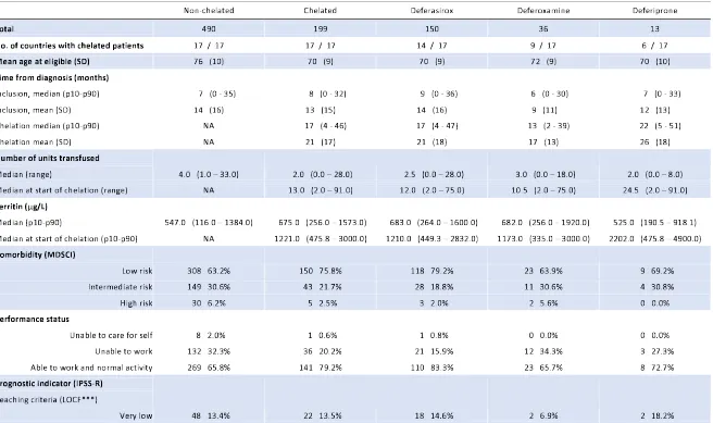
Table 1: Baseline characteristics of non-chelated and chelated patients at the visit prior to reaching the eligibility criteria and estimates of overall survival.
Non-chelated Chelated Deferasirox Deferoxamine Deferiprone
Total 490 199 150 36 13
No. of countries with chelated patients 17 / 17 17 / 17 14 / 17 9 / 17 6 / 17 Mean age at eligible (SD) 76 (10) 70 (9) 70 (9) 72 (9) 70 (10) Time from diagnosis (months)
Inclusion, median (p10-p90) 7 (0 - 35) 8 (0 - 32) 9 (0 - 36) 6 (0 - 30) 7 (0 - 33) Inclusion, mean (SD) 14 (16) 13 (15) 14 (16) 9 (11) 12 (13) Chelation median (p10-p90) NA 17 (4 - 46) 17 (4 - 47) 13 (2 - 39) 22 (5 - 51)
Chelation mean (SD) NA 21 (17) 21 (18) 17 (13) 26 (18)
Number of units transfused
Median (range) 4.0 (1.0 – 33.0) 2.0 (0.0 – 28.0) 2.5 (0.0 – 28.0) 3.0 (0.0 – 18.0) 2.0 (0.0 – 8.0) Median at start of chelation (range) NA 13.0 (2.0 – 91.0) 12.0 (2.0 – 75.0) 10.5 (2.0 – 75.0) 24.5 (2.0 – 91.0) Ferritin (µg/L)
Median (p10-p90) 547.0 (116.0 – 1384.0) 675.0 (256.0 – 1573.0) 683.0 (264.0 – 1600.0) 682.0 (256.0 – 1920.0) 525.0 (190.5 – 918.1) Median at start of chelation (p10-p90) NA 1221.0 (475.8 – 3000.0) 1210.0 (449.3 – 2832.0) 1173.0 (335.0 – 3000.0) 2202.0 (475.8 – 4900.0) Comorbidity (MDSCI)
Low risk 308 63.2% 150 75.8% 118 79.2% 23 63.9% 9 69.2% Intermediate risk 149 30.6% 43 21.7% 28 18.8% 11 30.6% 4 30.8% High risk 30 6.2% 5 2.5% 3 2.0% 2 5.6% 0 0.0% Performance status
Unable to care for self 8 2.0% 1 0.6% 1 0.8% 0 0.0% 0 0.0% Unable to work 132 32.3% 36 20.2% 21 15.9% 12 34.3% 3 27.3% Able to work and normal activity 269 65.8% 141 79.2% 110 83.3% 23 65.7% 8 72.7% Prognostic indicator (IPSS-R)
Reaching criteria (LOCF***)
Low 199 55.6% 95 58.3% 66 53.7% 21 72.4% 8 72.7% Intermediate 111 31.0% 46 28.2% 39 31.7% 6 20.7% 1 9.1%
High 38 10.6% 9 5.5% 6 4.9% 3 10.3% 0 0.0% Very high 3 0.8% 1 0.6% 1 0.8% 0 0.0% 0 0.0% Duration of treatment with chelation (months)
Median (p10-p90) NA 13 (3 - 41) 14 (3 - 41) 9 (1 - 34) 13 (2 - 30) Ever received ESAs
No 312 63.7% 115 57.8% 85 56.7% 22 61.1% 8 61.5% Yes 178 36.3% 84 42.2% 65 43.3% 14 38.9% 5 38.5% Ever received hypomethylating
No 460 93.9% 184 92.5% 136 90.7% 36 100.0% 12 92.3% Yes 30 6.1% 15 7.5% 14 9.3% 0 0.0% 1 7.7% Ever received lenalidomide
No 467 95.3% 179 89.9% 136 90.7% 33 91.7% 10 76.9% Yes 23 4.7% 20 10.1% 14 9.3% 3 8.3% 3 23.1% Overall Survival (OS)*
Unadjusted 1 0.57 (0.45 – 0.73) 1 1.99 (1.18 – 3.35) 0.42 (0.10 – 1.71) Adjusted** 1 0.50 (0.34 – 0.74) 1 2.46 (1.12 – 5.41) 0.30 (0.02 – 3.58) * HRs and 95% CI were estimated using receipt of chelation as a time-varying covariate
** adjusted by age at eligibility criteria, sex, comorbidity, performance status, number of units transfused, IPSS-R, and ringed sideroblasts present *** LOCF: last observation carried forward (only for cytogenetics and bone marrow blasts)
Table 2 Baseline characteristics for all matched subjects included in the propensity analyses
Covariates
Matched* data with imputations**
Non-chelated Chelated P-Value differences*** Standardized
N = 591 N = 197
Age (years) 71 (11) 70 (9) 0.364 -0.077
Sex 0.797 -0.021
Female 210 35.5% 72 36.5% Male 381 64.5% 125 63.5%
RBCT Intensity (per month) 0.7 (1.0) 0.6 (1.0) 0.484 -0.058 Cumulative RBCT units 4.5 (4.9) 4.3 (4.7) 0.570 -0.047 Ferritin level (µg/L, median, p25-p75) 730.6 (494.6-977.3) 683.6 (504-915.5) 0.328 -0.086
Comorbidity (MDSCI) 0.965 -0.004
Low risk 440 74.5% 150 76.1% Intermediate risk 145 24.5% 42 21.3% High risk 6 1.0% 5 2.5%
Performance status 0.279 0.090
Unable to care for self 4 0.7% 1 0.5% Unable to work 135 22.8% 38 19.3% Able to work and normal activity 452 76.5% 158 80.2%
Prognostic indicator (IPSS-R) 0.914 0.009
Very low 83 14.0% 22 11.2% Low 337 57.0% 120 60.9% Intermediate 134 22.7% 45 22.8% High 34 5.8% 9 4.6% Very high 3 0.5% 1 0.5%
Ring-sideroblast present 0.445 0.062
Yes 419 70.9% 134 68.0% No 172 29.1% 63 32.0%
Platelet level (10^9/L, median, p25-p75) 162.5 (99.2-294) 224.0 (121-324) 0.086 0.148 Haemoglobin level (g/dL, median, p25-p75) 8.8 (8.2-9.8) 8.4 (7.7-9.5) 0.021 -0.194 Quality of Life (EQ-5D)
Index (mean, SD) 0.7 (0.2) 0.7 (0.2) 0.186 0.125 VAS (mean, SD) 64.8 (21.0) 68.1 (19.9) 0.083 0.165
Country 0.140 -0.122
Austria 25 4.2% 10 5.1% Croatia 9 1.5% 1 0.5% Czech Republic 58 9.8% 25 12.7%
Denmark 15 2.5% 8 4.1% France 113 19.1% 40 20.3% Germany 23 3.9% 8 4.1%
Greece 80 13.5% 23 11.7% Israel 11 1.9% 5 2.5%
Netherlands 17 2.9% 7 3.6% Poland 22 3.7% 8 4.1% Portugal 2 0.3% 1 0.5% Romania 34 5.8% 11 5.6% Republic of Serbia 8 1.4% 2 1.0% Spain 11 1.9% 5 2.5% Sweden 97 16.4% 20 10.2%
UK 55 9.3% 18 9.1% Overall Survival (OS)
Unadjusted 1.0 0.70 (0.51 – 0.95) Adjusted**** 1.0 0.42 (0.27 – 0.63)
Note: Continuous variables are reported as mean (standard deviation), while categorical variables are reported as number (percent)
* Matched by age, gender, country, RBCT intensity, ferritin level, comorbidity, performance status, and IPSS-R at eligibility ** Multiple imputations in RBCT intensity, ferritin level, comorbidity, performance status, and IPSS-R at eligibility for non-chelated patients
*** The standardized difference in percent is the mean difference as a percentage of the average standard deviation **** Adjusted by age, sex, comorbidity, performance status, RBCT intensity, number of units transfused, IPSS-R, and RS present
Figure 1 Number of registry patients by transfusion and chelation status.
Number of registry patients by transfusion and chelation status. RBCT = Red Blood Cell Transfusion
*Cumulative RBCT units >=15 or RBCT intensity of ≥1 RBC unit/month or Serum Ferritin >1000 µg/L.
Figure 2 Overall Survival by Iron Chelation Therapy (ICT) as a Time-Dependent Variable in unmatched patients.
Figure 3 Overall Survival by Iron Chelation Therapy (ICT)as a Time-Dependent Variable in matched patients.
Figure 4 Adjusted Overall Survival by Iron Chelation Therapy (ICT)as a Time-Dependent Variable in matched patients.
Figure 5 Changes in transfusion density over time in chelated and non-chelated patients.
Legend 5:
time: eight 6-monthly visits
Figure 6 Trajectory analysis in chelated patients with and without response and for non-chelated patients.
Legend figure 6:
6A: Monthly red blood cell transfusion density for chelated patients with and without an erythroid response and for non-chelated patients. 6B: Ferritin levels of patients with and without a ferritin response, defined as a decrease of ≥1000 µg/L or a drop of the serum ferritin value below 1000 µg/L, and for non-chelated patients.
Supplementary Material
Supplementary Method Section
General
In the EUMDS registry, clinical information was collected via a bespoke web-based database on:
concomitant diseases, transfusion history, use of iron chelators (chelating agent, start date and end
date; no drug doses or schedules were collected), peripheral blood values, conventional iron
parameters (serum ferritin, transferrin saturation), concomitant treatments (lenalidomide, erythroid
stimulating agents [ESA], and hypomethylating therapy), and bone marrow pathology.
As information is recorded at 6-monthly time-points and the patients may have reached the criteria
for using iron chelation therapy between visits, the visit prior to reaching the criteria was selected.
Propensity score matched method
The main purpose of PSM was to balance the distribution of observed covariates at the time of
meeting the eligibility criteria in both the chelated and non-chelated groups, so there should be no
systematic differences in the distribution and overlap of covariates between the two groups after
matching.20 The causal effect of ICT on outcome was estimated in two stages. In the first stage, the
propensity score (PS) or the conditional probability of receiving ICT among eligible subjects were
estimated using multivariate logistic regression using the characteristics below, identified a priori to
be involved in the decision to treat a patient with ICT; A PS graph was used to check visually if the
common support condition was satisfied, i.e. if there was sufficient overlap.21 To examine the
balance in this study, we computed standardized differences that were defined as the difference
between chelated and non-chelated means of each factor, divided by the pooled standard deviation.
Absolute values of standardized differences <0.1 indicated sufficient balance.20 A p-value of 0.01 or
lower was considered to be statistically significant.
Missing data in PS estimations could result in biased estimates, and it may also shrink the pool of
potential matches. The following methods were used to impute missing values: 1) last observation
carried forward (LOCF) approach: For many patients bone marrow assessments were not repeated
after initial diagnosis, accordingly karyotype and bone marrow blast count, required for the
calculation of the IPSS-R at each visit, may be missing. A LOCF approach for only these two
components of the IPSS-R was applied; 2) Multiple imputation (MI) approach: For missing values of
IPSS-R, a MI approach was applied to create 20 multiple complete data sets consisting of all non-chelated
patients and all visits since the last visit prior to meeting the eligibility criteria.22 The imputation
model also included age, sex, and cumulative RBCT units.
Transfusion dose density
We used the beginning of the time interval in which the first transfusion started after diagnosis as
the starting point of time to calculate the cumulative number of transfusion units received and time
interval by the end of each subsequent visit. Transfusion dose density was calculated by dividing the
cumulative number of units by the time since the starting time point and standardised to monthly
Supplementary tables
Supplementary Table 1 Description of iron chelator use
Unmatched Sample Matched Sample
N
%
N
%
No iron chelation 490 71.12 591 75.00
Deferasirox only 135 19.59 134 17.01
Deferoxamine only 30 4.35 29 3.68
Deferiprone only 12 1.74 12 1.52
Deferasirox and deferoxamine 13 1.89 13 1.65
Deferasirox and deferiprone 4 0.58 4 0.51
Deferoxamine and deferiprone 2 0.29 2 0.25
All of the three 3 0.44 3 0.38
Supplementary table 2 Baseline characteristics for all unmatched transfused non-chelated and chelated patients with missing values and imputed values
N=490 N=199 N=490 N=199
Age (years) 76 (10) 70 (9) 76 (10) 70 (9)
Sex
Female 194 39,6% 72 36,2% 194 39,6% 72 36,2%
Male 296 60,4% 127 63,8% 296 60,4% 127 63,8%
RBCT Intensity (per month) 0,5 (0,8) 0,6 (1,0) 0,5 (0,8) 0,6 (1,0)
Ferritin level (ug/L, median, p25-p75) 547,0 (251.2-878.8) 675,0 (434.9-992) 693,5 (382-884) 685,8 (504-921)
Comorbidity (MDSCI)
Low risk 308 63,2% 150 75,8% 309 63,1% 151 75,9%
Intermediate risk 149 30,6% 43 21,7% 151 30,8% 43 21,6%
High risk 30 6,2% 5 2,5% 30 6,1% 5 2,5%
Performance status
Unable to care for self 8 2,0% 1 0,6% 8 1,6% 1 0,5%
Unable to work 132 32,3% 36 20,2% 151 30,8% 39 19,6%
Able to work and normal activity 269 65,8% 141 79,2% 331 67,6% 159 79,9%
Prognostic indicator (IPSS-R)
Very low 48 12,0% 22 12,7% 49 10,0% 22 11,1%
Low 199 49,9% 95 54,9% 276 56,3% 121 61,1%
Intermediate 111 27,8% 46 26,6% 124 25,3% 45 22,7%
High 38 9,5% 9 5,2% 38 7,8% 9 4,5%
Very high 3 0,8% 1 0,6% 3 0,6% 1 0,5%
Country
Austria 22 4,5% 10 5,0% 22 4,5% 10 5,0%
Croatia 3 0,6% 1 0,5% 3 0,6% 1 0,5%
Czech Republic 39 8,0% 25 12,6% 39 8,0% 25 12,6%
Denmark 24 4,9% 8 4,0% 24 4,9% 8 4,0%
France 88 18,0% 40 20,1% 88 18,0% 40 20,1%
Germany 7 1,4% 8 4,0% 7 1,4% 8 4,0%
Greece 34 6,9% 23 11,6% 34 6,9% 23 11,6%
Israel 20 4,1% 5 2,5% 20 4,1% 5 2,5%
Italy 19 3,9% 5 2,5% 19 3,9% 5 2,5%
Netherlands 10 2,0% 8 4,0% 10 2,0% 8 4,0%
Poland 15 3,1% 9 4,5% 15 3,1% 9 4,5%
Portugal 15 3,1% 1 0,5% 15 3,1% 1 0,5%
Romania 11 2,2% 11 5,5% 11 2,2% 11 5,5%
Republic of Serbia 7 1,4% 2 1,0% 7 1,4% 2 1,0%
Spain 32 6,5% 5 2,5% 32 6,5% 5 2,5%
Sweden 34 6,9% 20 10,1% 34 6,9% 20 10,1%
UK 110 22,4% 18 9,0% 110 22,4% 18 9,0%
Covariates
Unmatched data with missing values Unmatched data with imputations* Non-chelated Chelated
P-Value Standardise
d
Non-chelated Chelated
P-ValueStandardised
differences**
0,038 0,169 0,046 0,161
-0,554 0,000 -0,554
0,405 0,070 0,405 0,070
0,000
0,046 0,204 0,152 0,124
0,001 -0,291 0,001 -0,296
0,001 0,313 0,001 0,290
0,001 -0,283 0,001 -0,283
0,138 -0,137 0,106 -0,139
RBCT: red blood cell transfusion; MDSCI: myelodysplastic syndrome specific comorbidity index; IPSS-R: revised international prognostic scoring system; EQ-5D: European Quality of Life - 5 dimensions
Note: Continuous variables are reported as mean (standard deviation), while categorical variables are reported as number(percent) * Multiple imputations in RBCT intensity, ferritin level, comorbidity, performance status, and IPSS-R at eligibility criteria for unchelated patients ** The standardised difference in percent is the the mean difference as a percentage of the average standard deviation
Supplementary figures
Supplementary figure 1 Proportion of subjects meeting the eligibility criteria (n=689)