approach to reconstruct the osteobiography of an Italian worker community
.
White Rose Research Online URL for this paper:
http://eprints.whiterose.ac.uk/137825/
Version: Published Version
Article:
Baldoni, Marica, Scorrano, Gabriele, Gismondi, Angelo et al. (7 more authors) (2018) Who
were the miners of Allumiere? A multidisciplinary approach to reconstruct the
osteobiography of an Italian worker community. PLOS one. e0205362. ISSN 1932-6203
https://doi.org/10.1371/journal.pone.0205362
eprints@whiterose.ac.uk https://eprints.whiterose.ac.uk/
Reuse
Items deposited in White Rose Research Online are protected by copyright, with all rights reserved unless indicated otherwise. They may be downloaded and/or printed for private study, or other acts as permitted by national copyright laws. The publisher or other rights holders may allow further reproduction and re-use of the full text version. This is indicated by the licence information on the White Rose Research Online record for the item.
Takedown
If you consider content in White Rose Research Online to be in breach of UK law, please notify us by
Who were the miners of Allumiere? A
multidisciplinary approach to reconstruct the
osteobiography of an Italian worker
community
Marica Baldoni1,2 , Gabriele Scorrano3 , Angelo Gismondi4 , Alessia D’Agostino4,
Michelle Alexander5, Luca Gaspari3, Fabrizio Vallelonga6, Antonella Canini4, Olga Rickards3, Cristina Martı´nez-Labarga
ID1,3*
1Laboratorio di Antropologia Forense e Biologia dello Scheletro, Dipartimento di Biologia Universitàdegli Studi di Roma “Tor Vergata”, Roma, Italia,2Laboratorio di Medicina Legale, Dipartimento di Biomedicina e Prevenzione, Universitàdegli Studi di Roma “Tor Vergata”, Roma, Italia,3Centro di Antropologia Molecolare per lo Studio del DNA antico, Dipartimento di Biologia Universitàdegli Studi di Roma “Tor Vergata”, Roma, Italia,4Laboratorio di Botanica, Dipartimento di Biologia Universitàdegli Studi di Roma “Tor Vergata”, Roma, Italia,5Department of Archaeology, BioArCh, University of York, York, United Kingdom,6Sapienza Universitàdi Roma, Dipartimento di Scienza dell’Antichità, Cattedra di Archeologia Cristiana e Medievale, Roma, Italia
These authors contributed equally to this work.
Current address: Natural History Museum of Denmark; University of Copenhagen, Copenhagen, Denmark.
*martine@uniroma2.it
Abstract
This research presents an in-depth study of the skeletal remains collected from the archaeo-logical site of Allumiere (15th-16thcenturies CE; Rome, Italy). A multidisciplinary approach was used, combining skeletal biology, molecular anthropology and archaeobotany with the aim of reconstructing the osteobiography of the alum miners buried at the site. Since 1460, the area of the Tolfa Mountains was significant for the exploitation of alum which was used for a wide range of purposes in the Middle Ages, ranging from woven production to medical practice. A total of 70 individuals (63 adults and 7 juveniles) were studied. The sex ratio of the community indicated a higher prevalence of males with respect to females. Morphologi-cal examination indicated occupational musculoskeletal stress markers, which might reflect the specific phase of alum production that each individual was occupied in. Dietary recon-struction was primarily performed through carbon and nitrogen stable isotope analysis with integration of the results obtained by microscopic, genetic and GC-MS investigations on dental calculus. The diet was omnivorous, indicating a reliance on C3-terrestrial protein and evidence for limited C4consumption by some individuals. Herbivores, such as sheep and cattle, appear to have contributed to the diet more than pigs and chickens. Consumption of Fagaceae and Poaceae species was predominant; moreover, indicators of Brassicaceae and milk and its derivatives were abundantly recurrent in the population, followed by plant oils and theophylline. Furthermore, the detection of pharmacological alkaloids indicated the knowledge and application of medicinal plants by the community. The novel use of multiple techniques based on cutting-edge technologies has provided a unique window on the life-styles of individuals from one of the first Italian settlements of alum workers.
a1111111111 a1111111111 a1111111111 a1111111111 a1111111111 OPEN ACCESS
Citation:Baldoni M, Scorrano G, Gismondi A, D’Agostino A, Alexander M, Gaspari L, et al. (2018) Who were the miners of Allumiere? A
multidisciplinary approach to reconstruct the osteobiography of an Italian worker community. PLoS ONE 13(10): e0205362.https://doi.org/ 10.1371/journal.pone.0205362
Editor:David Caramelli, University of Florence, ITALY
Received:April 13, 2018
Accepted:September 23, 2018
Published:October 11, 2018
Copyright:©2018 Baldoni et al. This is an open access article distributed under the terms of the
Creative Commons Attribution License, which permits unrestricted use, distribution, and reproduction in any medium, provided the original author and source are credited.
Data Availability Statement:All relevant data are within the paper and its Supporting Information files.
Introduction
The exploitation of alum in Italy started in 1460, when Giovanni di Castro, a commissioner of the Pontifical State, identified the presence of alunite, a mineral from which alum could be extracted, in the territory of the Tolfa Mountains (Rome, Italy) [1–3]. Alum is a salt made up of ammonium sulfate and potassium associated with twenty-four molecules of crystallization water, whose applications ranged from textile production to medicine. Because of its water solubility, alum is not directly available in nature, but it is obtained through transformation of the less solu-ble aluminum minerals, such as alunite. The discovery of alunite on the Tolfa Mountains was of vital importance for the papal coffers and influenced the rise of the Western European textile industry. This source became particularly important after the conquest of Constantinople by the Turks in 1453 meaning that alunite deposits located in the Eastern Mediterranean area became difficult to access [2]. The extraction of the alum was entrusted to companies of contractors while the Apostolic Chamber handled its marketing. From the beginning, the Medici family secured the control of commercialization of the product in collaboration with Genoese mer-chants. In 1499, the banker Agostino Chigi was responsible for the organization of the mining enterprise and settlement of the area and the birth of the village that would later become Allu-miere, is likely down to his actions [2].
Extraction and processing of the alum required the involvement of specialized manpower. The production cycle of alum, described in detail by historical sources [1,3], began with the excavation of the mineral from the rocks through the use of picks. Then, alunite stones were heated at high temperature (i.e. 600–700˚C) in special furnaces for 12–14 hours. The "roasted" mineral was finally treated with water, in order to obtain a doughy solution, which was then heated again and concentrated until alum crystals began to separate.
In the area ofLa Bianca, several excavation campaigns have unearthed a Medieval cemetery and a church namedCappella dei Minatori(Fig 1). It has a single nave, is East-West oriented and is 19 m long and 8 m wide. The structure and dating of the church suggest that the archae-ological site was related to one of the first human settlements in the area associated with alum production. A total of 70 burials were found, most of them located outside the church (Fig 1). Archaeologically, the stratigraphic sequence of the site is challenging to define, as is com-mon in Medieval cemeteries. In the majority, of the taphonomic observations, the burials were made up of simple earthen graves with individuals wrapped in shrouds, with the exception of a wooden coffin identified through the presence ofin situnails (SU 296). A chronological seq-uence of burials was indicated by earlier burials were being laid in a N-S orientation and subse-quent later individuals buried on an E-W axis.
Items of jewelry and coins were recovered with a few individuals. In SU 110, three silver coins were found close to the left hand; SU 135 was buried with a religious medal, portraying the Immaculate Conception and SU 245 had eleven gold and two silver coins close to the left hand. All these artifacts confirmed the dating of the cemetery; in particular, the silver coins of SU 110 were dated to 1476–1483 [4]. Further archaeological analyses revealed various origins for these coins. Some coins possessed a “fleur-de-lis”, symbol of the city of Florence (Italy). Others were also identified as three ducats of Ferdinando I from Naples, one coin from the Pontifical State and three from Venice. Others were of Iberian origin, specifically, adoble
exce-lente de la Granadaand anexcelenteof the kings Fernando de Aragon and Isabel de Castilla
and a Portuguesecruzadoof King Giovanni II d’Aviz. There was also a duchy from Rhodes. The dating of the coins ranged between 1464–1523. The variety of coins and their origins are an important proof of the wealth of commercial traffic linked to alum.
The present research aims to reconstruct the osteobiography of these alum miners of the Tolfa Mountains through a multi-proxy approach, combining skeletal biology, molecular
design, data collection and analysis, decision to publish, or preparation of the manuscript.
anthropology and archaeobotany. Osteological analysis allows the reconstruction of the bio-logical features of the Medieval workers, while molecular and archeobotanical analyses are used to shed light on the dietary patterns and medicinal habits of the community of Allumiere.
There is a general lack of information about the dietary habits of the lower social classes in his-torical texts from the Medieval period. Therefore, to better understand the dietary pattern of the studied community, we applied carbon ( 13C) and nitrogen ( 15N) stable isotope analysis to human and animal bone collagen. Stable isotope analysis of bone collagen represents an established technique to identify the main protein sources in the diet of archaeological populations. This allows us to investigate marine and terrestrial and C3and C4sources of protein in addition to the trophic
position an individual is feeding at [5–7]. Analysis of dental calculus was also carried out to further investigate which vegetal and animal species were present in miners’ diet. Indeed, the analysis of dental calculus is a very informative archaeobiological approach to reconstruct past food habits [8– 10]. Dental calculus is a dense mineral matrix made up of inorganic salts and organic molecules derived from ingested foods, crystallized remains of oral microbiota and accidentally inhaled microremains [11–12]. Light microscopy, genetic and gas-chromatographic mass-spectrometry approaches were carried out on dental calculus to provide direct evidence of the main categories of foods and plant drugs introduced, at least once, in the lifetime of our ancient exemplars [13–23]. The present work provides new information on an important period of the Italian Medieval history, shedding light on the lives of those who lived in one of the first Italian settlement of alum miners.
Materials and methods
The present research deals with the skeletal remains recovered in the area of La Bianca in Allu-miere (Rome, Italy) (Fig 1A) in a graveyard close to the church named asCappella dei Minatori (Fig 1B). The archaeological excavation started in 2010 directed by Dr. Fabrizio Vallelonga authorized by the “Comune di Allumiere” (Municipality of Allumiere). The research was car-ried out at the Department of Biology of the University of Rome “Tor Vergata” and directed by Dr. Cristina Martı´nez-Labarga who received the authorization for the analysis of the skeletal remains from La Bianca (Allumiere, Rome, Italy) in 2015. The complete list of the specimens is provided inS1 Table.
Osteological examination
Seventy individuals were analysed, both adults and non-adults collected from the cemetery area ofLa Bianca(Allumiere, Rome, Italy). The preservation index for the individuals was cal-culated following the method proposed by Walker et al. [24]. The age estimation for adult indi-viduals (from ca. 18 year old) followed methods based on morphological changes in the pubic symphysis [25–27], in the auricular surface of the ilium [28] and in the sternal end of the fourth rib [29–30]. Secondarily, dental wear [31–32] and obliteration of the cranial sutures [33] were also observed. Age estimation for infant and juvenile skeletal remains (until ca. 18 years old) was carried out through the diaphyseal length of bones [34–36], and tooth eruption [37]. Moreover, secondary ossification centers were taken into account, following the methods proposed by different authors and summarized in Minozzi and Canci [38].
Sex diagnosis was performed only on adult samples, as proposed by Acsàdi and Nemeskèri [39] and revised by Ferembach et al. [40] and Phenice’s [41]. Metric analysis complemented the morphological data, as the state of preservation of the sample allowed the measurement of sexually dimorphic bones, utilizing univariate and multivariate techniques [42–46].
average value obtained using multiple methods [52–56]. When skeletal remains were frag-mented, Steele’s formulae [57] were applied to estimate long bone length. Moreover, working activities were determined analyzing enthesal changes (EC) as proposed by Mariotti et al. [58– 59] and by Borgognini Tarli and Reale [60]. The paleopathological survey was performed through morphological observation of the skeletal remains. In one pathological case, X-ray monitoring was carried out at the “Dipartimento di Diagnostica per Immagini, Imaging mole-colare, Radiologia, Interventistica e Radioterapia, Azienda Ospedaliera Universitaria Policli-nico Tor Vergata”, using GMM Opera Swing, in order to achieve a differential diagnosis.
Particular attention was paid to evidence of osteoarthritis, which was scored according to the standards proposed by Buikstra and Ubelaker [61]. These authors evaluated intensity and distri-bution of lipping, porosity and eburnation of the osteoarthritis. A scale system from 0 -absence of injury on bone tissue- to 3 -marked modification of the joint surface- was used. In order to better evaluate the position in which the lesion occurred, Prieto’s joint division scheme was adopted [62]. This method distinguishes proximal and distal epiphyses, dividing them in four areas: two superiors (anterior and posterior) and two inferiors (anterior and posterior).
Statistical analyses were carried out using the statistical software R (v. 3.4.1) for the Stu-dent’s t-test and chi square test with Yates correction.
Isotope analyses
Collagen extraction was carried out on 68 human and 12 faunal skeletal remains from the archaeological cistern (1Hystrix cristata, 1Cervus elaphus, 2Equus asinus, 2Felix catus, 1
Canis lupus2Sus domesticus, 1Equus caballusand 2 unidentified carnivores) in order to
con-duct carbon and nitrogen stable isotope analysis. Human samples comprised of rib bones, whereas animal samples were taken from the various skeletal elements that were available.
Collagen extraction followed a modified Longin method [63]. Initially, to remove potential contaminants a sterile surgical blade was used on the outer surface of the bone samples and c. 500 mg of bone was subsequently pulverized using a mortar and pestle. To demineralize the bone, 8 mL of HCl 0.6 M at 4˚C was added to the powder and left at 4˚C on a horizontal mixer for two days, changing the acid after 24 hours.
Once the mineral component of the bone removed, samples were rinsed three times with ddH2O, until the pH level became neutral. The resultant pellet was gelatinized at 75˚C, for 24–
48 hours, with HCl pH 3.0 (0.001 M). The solution was then frozen at -80˚C for four hours and then freeze-dried for two days. A simultaneous extraction on modern bovine bone was performed and used as reference control. Approximately 0.8–1.2 mg of collagen was weighed and analysed in duplicate by EA-IRMS on a Sercon GSL analyser coupled to a Sercon 20–22 Mass Spectrometer at the University of York. The analytical error, calculated from repeated measurements of each sample, an internal laboratory control (fish gelatine), and international standards, was<0.2‰ (1σ) for both 13C and 15N. International Atomic Energy Agency
(IAEA) standards were N-2, and 600 for nitrogen and International Atomic Energy Agency IAEA-600 and Iso-Analytical R006 for carbon. Isotope data are reported as delta ( ) values rel-ative to V-PDB (Vienna Pee Dee Belemnite) for carbon and AIR (Atmospheric air) for nitro-gen. Carbon content (%C), nitrogen content (%N), protein yield, and C/N ratios were checked to monitor the diagenesis of bone [64] and determine bone protein quality for paleodietary reconstruction, according to DeNiro [65] and van Klinken [66].
Molecular and archaeobotanical analyses on dental calculus
Each analysis was carried out on different aliquots of calculus. Sodium hypochlorite 5% and UV exposure were employed to treat tools and working surfaces to restrict contamination dur-ing the analysis. In addition, different laboratories were used to carry out the various phases of this work [67]. Deposits of dental calculus were generally slight, according to Brothwell [31], on all dentition. Initially, to eliminate any contamination before sampling, a scraping action was applied by a sterile surgical blade on the outer surface of the dental calculus. All collecting procedures were carried out under a sterile vertical laminar flow hood (Heraeus HERAsafe HS12 Type). In order to remove any environmental contaminants from the calculus surface, samples were UV-treated for 10 minutes on each side and soaked in 5% sodium hypochlorite for 15 minutes. Lastly, the calculus was washed in sterilized bidistilled water and rinsed in 100% ethanol to eliminate the aqueous components before being left to dry [22,68]. To vali-date sterilization protocols, five dental calculus samples previously subjected to decontamina-tion procedures, were randomly selected and washed with 200μL of water. These last washing solutions were subjected to light microscopy, genetic and GC-MS analysis. No microremains, nucleic acid or chromatographic signals were detected, confirming the efficacy of the cleaning methods.
DNA extraction, amplification and sequencing. All criteria and precautions for the study and analysis of ancient DNA (aDNA) were applied [69–71]. For aDNA extraction a modified protocol suggested by Warinner and collaborators [10] was used. For each sample (50 mg of pulverized dental calculus), 600μL of extraction buffer (100 mM Tris-HCl pH 8, 100 mM NaCl, 10 mM EDTA and 2% SDS) and 50μL of proteinase K (20 mg/mL) were added. The samples were incubated in a shaking water bath at 56˚C, for 6 hours, adding 20μL of fresh proteinase K (20 mg/mL) every 2 hours. The sample was then incubated overnight at 37˚C. Following this, the sample was centrifuged for 5 minutes at 13.000 rpm and the supernatant was transferred to a new 2 mL Eppendorf tube. The supernatant was mixed with 500μL of phenol/chloroform/isoamyl alcohol (25:24:1) and, after a centrifugation of 5 minutes at the 13.000 rpm, transferred into a new 15 mL falcon tube. DNA was purified by QIAquick PCR purification kit following the manufacturer’s procedure, and eluted into 50μL of the elution buffer. Ancient DNA extracts were stored at 4˚C.
For each sample, different polymorphic regions of mitochondrial genome of several animal species were amplified (35 PCR cycles), using pairs of primers that only amplified for the target species (S2 Table). For ovine and pig, the COX 1 gene was amplified, while 12S rRNA and 16S rRNA were analysed for bovine and chicken, respectively. These primers were proposed by Natonek-Wiśniewska and colleagues [72]. For fish investigations, the DNA Mini-Barcoding System reported in Shokralla and collaborators [73] was applied. Amplifications were also per-formed for negative and positive controls. Amplifications of modern and ancient DNA were performed separately in two different laboratories. In particular aDNA analysis was carried out in the aDNA Laboratory in the Departmental Center of Molecular Anthropology for Ancient DNA Studies, University of Rome, Tor Vergata, in Villa Mondragone, Monte Porzio Catone, Rome (http://www.bio.uniroma2.it/biologia/laboratori/lab-antropologia/DNAantico/ DNA_antico/Facilities.htm) which has all the facilities to minimize potential contamination with extant DNA [71]. In order to detect the correct activity of the different reagents, the posi-tive controls that contained modern faunal DNA were prepared in the laboratory for modern DNA processing in the department of Biology, of the University of Rome “Tor Vergata”.
ncbi.nlm.nih.gov/Blast.cgi?PROGRAM=blastn&PAGE_TYPE=BlastSearch&LINK_LOC= blasthome) was used to confirm the identity of the sequences found in the dental calculus. The presence of fish DNA was detected by polyacrylamide gel, using a molecular weight marker (GelPilot 50bp Ladder Qiagen).
Light microscopy analysis (LM). A protocol based on Hardy et al. [75] method was used to extract starch granules and other microremains from dental calculus. Twenty mg of each sample were resuspended in 500μL of 1 M HCl and sonicated for 10 minutes (Falc Instru-ments MOD: LBS1 34) before being left under agitation for 24 h. After centrifugation at maxi-mum speed for 10 minutes, the pellet was subjected to 3 consecutive washings with bidistilled water. The last pellet was resuspended in 100μL of bidistilled water, sonicated for 5 minutes and observed at OM (Nikon Eclipse E100), under white and polarized light. The whole volume of the sample was examined and microfossils were photographed at 100X magnification using software for capturing images (ProgRes CapturePro 2.9.0.1) and measured (i.e. for starch gran-ule, maximum length through thehilumand maximum width anywhere along the
perpendicu-laraxis) by the SuperAmpelo 2.0 program. Taxonomic identification of pollens, phytoliths and
starch granules was carried out by direct comparison with a modern experimental collection (hosted in Laboratory of Botany and Botanical Gardens of the Department of Biology at Uni-versity of Rome “Tor Vergata”) or data from literature, including a pollen Atlas [76].
Gas-chromatography mass-spectrometry (GC-MS) analysis. This analysis was carried out in qualitative and non-quantitative terms, according to Gismondi et al. [77]. Ten mg of cal-culus for each individual were dissolved in 1 mL of 6% HCl. Once left in agitation for three days, 1 mL of hexane was added and shaken for two hours. After centrifugation for 10 minutes at 10.000 rpm, the supernatant hexane fraction was recovered and dried by a speed-vac system (Eppendorf AG 22331 Hamburg, Concentration Plus). The dried pellet was then derivatized by resuspension with 50μL of hexane and 50μL of the Methyl-8-Reagent (Thermo Scientific), in thermostated bath, at 60˚C for 20 minutes. At least 3 analyses for each sample, by injecting
2μL of extract in GC-MS (QP2010, Shimadzu, Japan; column DB-5 Phenomenex; helium as
gas carrier; splitless modality), were carried out. The run was conducted by a temperature gra-dient: initial oven temperature was set at 60˚C for 5 min; then, increasing temperature at a fixed rate of 6˚C per minute, the column was heated up at 150˚C for 5 minutes, 250˚C for 5 minutes and 330˚C for 25 minutes. Parameters and conditions relating to mass spectrometry were: ion source temperature 230˚C; interface temperature 320˚C; solvent cut time 6 minutes; ionization mode: EI; ionization voltage: 0.70 eV. The identification of each molecule (similarity values were considered acceptable only if higher than 85%) was performed comparing their mass spectrum with those registered in the NIST Library 14 loaded on detection software. No significant differences among replicates were detected.
Results
Skeletal biology
For adult individuals, the highest mortality falls between the young adult and adult classes. Data on the juveniles’ mortality rate data should be considered cautiously due to the small sample size.
Living stature was estimated on 43 individuals (37 males and 6 females). The mean stature for males was 169.22±6.26 cm, while females exhibited an average stature of 156.70±5.96 cm. A Student’s t-test indicated a statistically significant difference between the sexes (p-value = 3.17x10⁻⁷). Post-cranial indices revealed high skeletal robusticity and skeletal asymme-try in males. In particular, the right side seemed to have been subjected to a higher biomechan-ical stress than the left. The results obtained for all the indices are shown inS3 Table.
Pathological assessment was carried out on 57 adult individuals (S4 Table). Six individuals were excluded due to poor preservation.
The highest frequencies were recorded for degenerative diseases (86%), Schmo¨rl’s nodules (75%), periostitis (32%), and fractures (27%). Degenerative diseases were considered, jointly with enthesal changes, able to associate each individual to the most plausible phase in alum production. Fractures were relatively common within the community. For example, SU 245 demonstrated anante-mortemcompound fracture which affected both tibia and fibula. This trauma (Fig 2) caused the loss of ca. 5 cm in limb length.
[image:9.612.38.578.88.313.2]The analysis of musculoskeletal stress markers was conducted on male individuals as histor-ical evidence indicates [1,3] that they would have been involved in alum extraction and pro-duction. No detection attempt was made on juveniles because the high plasticity of their skeletal remains could provide unreliable results. Only 20 male specimens were considered suitable for all further analyses on enthesal changes. The remainder were excluded either due to their state of preservation, which prevented macroscopic observation of muscular insertion sites, or because, as was the case for SU 245, a trauma or skeletal disease was present that could alter interpretation. Indeed, the musculoskeletal stress markers of SU 245 seemed to be Table 1. Sex and age at death determination in the analyzed series from Allumiere (Rome. Italy).
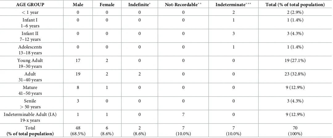
AGE GROUP Male Female Indefinite Not-Recordable Indeterminate Total (% of total population)
<1 year 0 0 0 0 2 2 (2.9%)
Infant I 1–6 years
0 0 0 0 1 1 (1.4%)
Infant II 7–12 years
0 0 0 0 3 3 (4.3%)
Adolescents 13–18 years
0 0 0 0 1 1 (1.4%)
Young Adult 19–30 years
17 2 0 0 0 19 (27.1%)
Adult 31–40 years
19 2 2 0 0 23 (32.8%)
Mature 41–50 years
8 1 0 0 0 9 (12.9%)
Senile
>50 years
3 0 0 0 0 3 (4.3%)
Indeterminable Adult (IA) 19-x years
1 1 0 7 0 9 (12.9%)
Total
(% of total population)
48 (68.5%) 6 (8.6%) 2 (8.6%) 7 (10.0%) 7 (10.0%) 70 (100%)
Indefinite indicates a mixture of male and female traits;
Not-recordable indicates the impossibility to perform sex and age at death diagnosis;
Indeterminate indicates non-adults for whom no assumption of sex was performed.
influenced by skeletal lesions. The enthesal changes observed in the upper limbs of this indi-vidual (i.e. a greater muscle development on the right humerus than on the left) led us to hypothesize that they would have used a crutch to partially solve the walking difficulty caused by the loss of the original length of the limb as consequence of theante-mortemfracture. Fig 2.Ante-mortemcompound fracture affecting SU 245.A) compound fracture on both tibia and fibula; B) X-rays analysis (X-ray exposure: 78 kV).
[image:10.612.74.576.79.591.2]On the basis of the development of occupational stress markers, the individuals seemed to cluster in four groups, corresponding to the four different phases of alum production (excavation, calcination, moistening and lixiviation) [78]. These data were also corroborated by the results of the post-cranial indices, which revealed different patterns of skeletal strength as well as a func-tional asymmetry. Musculoskeletal stress data were finally integrated with the analysis of degener-ative pattern and joint diseases. Osteoarthritis could be observed on 45 samples (S5 Table).
Osteoarthritis confirmed all previous suppositions about musculoskeletal stresses and alum working activity of the individuals. The excavation phase caused a marked enthesal develop-ment of shoulders, elbows, hips and feet; in the calcination the most involved functional groups are shoulder, elbow, hip and knee; the individuals involved in moistening exhibit a high degree of biomechanical stress that affected shoulders, hips, knee and feet, while those employed in lix-iviation show a great muscle development in the shoulders, forearms, hip and foot.
All the working phases are characterized by well-developed musculoskeletal markers on the shoulders and the hip however, the combination of other functional groups significantly diverges amongst the other clusters. For example, the skeletons that show high robusticity of
m.pectoralis major(horizontal flexion of the upper limb),m.deltoideus(abduction, extension
and flexion of the limb) andm.biceps brachii(flexion of the forearm on the arm) could be related to the excavation phase according to the upper limb’s movements involved in the use of the picks. Even if, as mentioned, all phases of alum production cause a high biomechanical stress on muscles, the movement related to each one as well as the functional groups involved are different, and the excavation was observed to be the most strenuous phase involving the strain on both the shoulder and the elbow [1,3].
Furthermore, a high degree of biomechanical stress on the knee could be observed in both moistening and calcination phases. However, moistening should involve a higher robusticity of thequadriceps tendon, whereas in the calcinationm.vastus medialisshould appear more tinctly stressed. The combined results of musculoskeletal stress markers and degenerative dis-eases are provided inTable 2.
Unfortunately, a clear relationship between musculoskeletal markers, osteoarthritis and performed working activity could be identified only for twenty-one individuals because the others exhibited musculoskeletal stress markers or osteoarthritis patterns that could made them suitable for more than one working phase.
Stable isotope analysis from bone collagen and molecular analysis from
dental calculus
Isotopic data and quality indicators are presented inTable 3.
Ten humans (SU 213, 330, 198, 325, 320, 307, 179, 385.3, 385.5, 416) and one animal (SU 101 AEquus caballus), were excluded from the analysis due to their C/N ratio being outside the satisfactory range. The remaining samples were acceptable according to criteria proposed by Ambrose and Norr [64] and van Klinken [66].Fig 3shows the plot of 13C versus 15N iso-tope values for both faunal and human samples.
The 13C values of the three herbivore samples (Cervus elaphusandEquus asinuus) ranged from -21.4‰ to -21.1‰ (mean -21.3±0.1‰), while 15N values ranged between 3.5‰ and 5.9‰ (mean 4.5±1.3‰). Omnivores (Sus domesticusandHystrix cristata,) possessed values from 21.8‰ to -20.6‰ (mean -21.0±0.7‰) for 13C and from 3.6‰ to 4.7‰ (mean 4.1±0.6‰) for 15N. The carnivore (Felix catus,Canis familiarisand unidentified carnivores) data ranged from -20.8‰ to -19.5‰ (mean -20.1±0.46‰) and from 7.5‰ to 8.5‰ (mean 7.8±0.4‰) for 15N. Isotope data for all animals are in a range typically associated with the consumption of C3plants. In terms
Mediterranean area [79]. Omnivore 15N values are similar to those of the herbivores, which indi-cates that at Allumiere they consumed similar dietary resources, i.e. mainly plants with little/no ani-mal protein. TheHystrix cristata(porcupine) show isotope values that are of an essentially
herbivorous and it seems that any occasional consumption of insects and small vertebrates [80] did not play a key role in the stable isotope values for this animal. The analysis of the pig diet is useful to understand the variation in husbandry practices [81–88] among the different communities. Indeed, pigs raised in a home-based system are expected to have a more controlled diet than free-range animals. For Medieval northwestern Europe communities, pigs had a diet based on terres-trial plant and human refuse [87], whereas our data show a comparable 15N values between the
Sus domesticusand the other herbivores analyzed. During the Middle Ages, in some areas
(includ-ing Lazio) pigs were free to roam in the fallow land close to the city [82], where they would typically consume acorns (pannaga) [89]. Our results seem to suggest that the Allumiere pigs were predomi-nantly fed with plant products, which might indicate that they wandered about in forested environ-ments. Therefore, the difference between Allumiere and data from published northwestern European populations [87] is probably related to the different social status of the communities: the Allumiere population was made up of workers of low social status, while the other data derives from predominantly coastal and elite/urban communities with different husbandry practices.
[image:12.612.36.585.81.359.2]The human remains fromLa Bianca(Allumiere) possessed 13C values between -20.6‰ and -17.1‰ (mean -19.2±1‰) and 15N values between 5.9‰ and 12.1‰ (mean 8.6±1.4‰). Both nitrogen and carbon stable isotope values for humans showed a high variability, in particu-lar two individuals (SU 245 and 317) have elevated 15N values of 12.1‰ and 11.9‰ and one (SU 388) has a particularly low 15N value of 5.9‰. Despite the variability in isotopic values, no statistically significant difference was observed, between different sexes or age at death. The majority of humans demonstrate an enrichment of15N (about 3–5‰, with a nitrogen offset equal 3.5‰) in comparison to the animals (Fig 3), reflecting a typical trophic level shift Table 2. Combined results of musculoskeletal stress markers and osteoarthritic pattern (nd: not determinable).
INDIVIDUAL SEX AGE AT DEATH ENTHESAL DEVELOPMENT DEGENERATIVE PATTERN HYPOTHESIZED ACTIVITY
139 M 41–50 shoulder/knee shoulder/ankle/foot moistening
144 M 31–40 shoulder/hip/knee shoulder moistening
147 Aa M 19–30 shoulder/hip/foot shoulder/elbow/hip lixiviation
159 M 31–40 shoulder/elbow/hip shoulder lixiviation
169 M 19–30 shoulder/foot shoulder/wrist moistening
176 M 19–30 shoulder/forearm/hip/foot shoulder/elbow/hip lixiviation
185 M 51–60 hip/foot shoulder/wrist/hip/knee lixiviation
189 M 19–30 hip/knee/foot shoulder/wrist/hip/knee moistening
198 M 31–40 shoulder/elbow/hip/foot elbow/wrist/hip/ankle excavation
221 M 31–40 nd shoulder/elbow/wrist/hip/knee calcination
231 M 41–50 nd shoulder/elbow/wrist/hip excavation
277 M 41–50 nd shoulder/elbow/hip/knee calcination
280 M 41–50 shoulder/elbow/hip/knee elbow/hip calcination
296 M 51–60 high biomechanical stress shoulder/elbow/wrist/hip excavation
303 M 19–30 shoulder/elbow/hip/foot shoulder/elbow/hip/ankle excavation
307 M 31–40 shoulder/hip/knee/foot shoulder/hip moistening
318 M 19–30 shoulder/foot shoulder/ankle/foot moistening
319 M 19–30 shoulder/foot shoulder/hip/foot moistening
320 M 31–40 forearm/hip/foot hip lixiviation
330 M 41–50 shoulder/forearm/hip/knee/foot shoulder/wrist/hip excavation
362 M 31–40 shoulder/elbow/hip/foot shoulder excavation
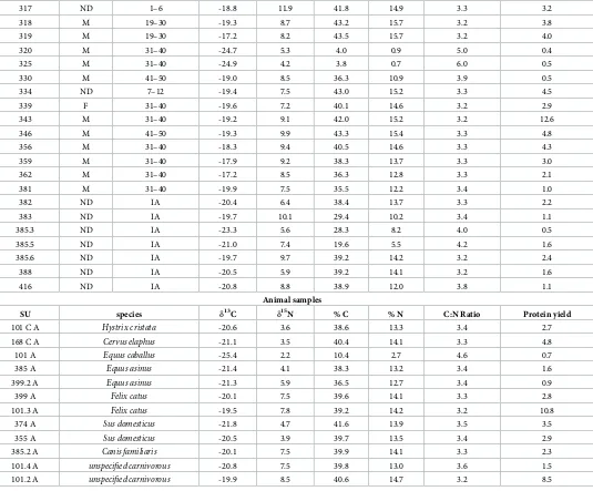
Table 3. Carbon and nitrogen stable isotope values and collagen quality indicators of animals and humans fromLa Bianca.The samples that fell outside the quality range parameters are indicated in red and were excluded from subsequent analysis.
Human samples
SU sex Age at death
(years)
13C 15N % C % N C:N Ratio Collagen yield
110 M 41–50 -17.6 8.1 38.3 13.4 3.3 2.4
135 M 31–40 -19.2 7.9 41.9 15.5 3.2 3.0
139 M 41–50 -20.0 8.1 29.6 10.0 3.5 5.9
144 M 31–40 -19.2 10.0 39.1 14.1 3.2 3.3
147 M 19–30 -18.7 9.6 40.9 15.0 3.2 2.0
158 M 31–40 -18.7 9.4 43.9 16.1 3.2 6.4
159 M 31–40 -20.1 9.0 30.1 10.1 3.5 4.3
169 M 19–30 -18.8 9.7 41.8 15.2 3.2 13.8
173 M 19–30 -19.6 6.7 43.2 15.6 3.2 11.0
176 M 19–30 -17.9 8.2 40.7 14.7 3.2 6.0
179 M 31–40 -21.2 7.8 16.3 4.8 3.9 0.8
182 ND 31–40 -17.1 7.2 40.7 15.1 3.2 2.7
185 M 51–60 -19.5 9.7 39.8 14.1 3.3 2.8
189 M 19–30 -20.1 6.2 36.8 13.3 3.2 3.4
192 M 19–30 -16.4 6.8 40.8 14.8 3.2 8.6
195 M IA -18.1 8.1 39.9 14.5 3.2 2.4
198 M 31–40 -28.1 -0.6 17.8 4.5 4.6 0.4
201 M 31–40 -18.4 7.7 37.7 13.5 3.3 1.0
204 M 31–40 -20.4 7.2 39.4 13.2 3.5 2.9
210 ND IA -20.4 9.1 24.0 8.2 3.4 1.9
213 ND 31–40 -19.4 10.8 39.4 12.7 3.6 2.0
216 F 41–50 -19.0 10.0 37.3 13.6 3.2 1.0
221 M 31–40 -18.8 9.1 40.0 14.1 3.3 7.3
226 ND 13–18 -17.6 8.2 41.8 15.0 3.3 2.2
231 M 41–50 -20.4 8.8 35.3 12.4 3.3 1.7
234 F 31–40 -20.0 8.8 39.3 14.2 3.2 2.1
239 M 19–30 -19.1 10.0 41.3 14.7 3.3 6.8
245 M 51–60 -18.5 12.1 39.1 13.6 3.4 3.0
249 M 19–30 -20.6 8.1 36.8 13.1 3.3 1.8
256 M 19–30 -19.2 7.7 41.6 14.7 3.3 5.8
269 M 19–30 -20.2 6.7 40.3 13.6 3.5 6.0
270 F 19–30 -19.1 9.3 41.0 15.0 3.2 3.8
272 M 31–40 -20.2 7.8 42.6 15.4 3.2 5.7
274 M 19–30 -19.8 7.2 40.4 14.3 3.3 1.4
277 M 41–50 -19.6 6.9 38.5 14.1 3.2 2.5
280 M 41–50 -20.6 8.8 38.8 13.8 3.3 1.2
284 ND 7–12 -19.4 9.3 40.8 15.0 3.2 4.5
290 M 41–50 -19.2 10.7 42.7 15.5 3.2 10.3
293 F 19–30 -20.3 10.4 38.0 13.3 3.3 0.8
296 M 51–60 -18.7 6.5 38.7 13.1 3.5 0.8
303 M 19–30 -19.8 10.9 32.2 11.0 3.4 0.8
304 ND 7–12 -19.3 9.4 41.3 15.0 3.2 2.86
307 M 31–40 -29.2 -1.8 9.0 2.4 4.3 0.6
308 M 19–30 -18.6 9.0 42.6 15.4 3.2 6.3
311 M 19–30 -20.1 7.9 39.0 14.2 3.2 5.0
indicative of the consumption of animal protein. Most of the individuals have diets with low input of animal protein: the wide range in 15N values for the population (6.2‰) indicates that animal protein made a greater or lesser contribution to the diet of particular individuals. On average, the human-animal offset in is 3.5‰, which is at the lower end of the accepted 3–5‰ between trophic levels [90] and spacing of up to 6‰ has been suggested for humans [91]. Therefore, for many individuals, it seems animal protein did not contribute a major part of the diet, which is in keeping with the low status nature of the population. However, for those pos-sessing 15N values of upwards of 9–10‰, animal protein will have had a major input and for the most elevated values, fish may also have made a contribution. High 13C values (>18‰)
for some individuals (e.g. SU 362, 182, 192 and 319) indicate the consumption of C4plants or
[image:14.612.41.576.83.526.2]marine fish consumption. However, the fact that these individuals also tend to have amongst the lowest 15N values would suggest that the former is most likely.
Table 3. (Continued)
317 ND 1–6 -18.8 11.9 41.8 14.9 3.3 3.2
318 M 19–30 -19.3 8.7 43.2 15.7 3.2 3.8
319 M 19–30 -17.2 8.2 43.5 15.7 3.2 4.0
320 M 31–40 -24.7 5.3 4.0 0.9 5.0 0.4
325 M 31–40 -24.9 4.2 3.8 0.7 6.0 0.5
330 M 41–50 -19.0 8.5 36.3 10.9 3.9 0.5
334 ND 7–12 -19.4 7.5 43.0 15.2 3.3 4.5
339 F 31–40 -19.6 7.2 40.1 14.6 3.2 2.9
343 M 31–40 -19.2 9.1 42.0 15.2 3.2 12.6
346 M 41–50 -19.3 9.9 43.3 15.4 3.3 4.8
356 M 31–40 -18.3 9.4 40.5 14.6 3.3 4.3
359 M 31–40 -17.9 9.2 38.3 13.7 3.3 3.0
362 M 31–40 -17.2 8.5 36.3 12.8 3.3 2.1
381 M 31–40 -19.9 7.5 35.5 12.2 3.4 1.0
382 ND IA -20.4 6.4 38.4 13.7 3.3 2.2
383 ND IA -19.7 10.1 29.4 10.2 3.4 1.1
385.3 ND IA -23.3 5.6 28.3 8.2 4.0 0.5
385.5 ND IA -21.0 7.4 19.6 5.5 4.2 1.6
385.6 ND IA -19.7 9.7 39.2 14.2 3.2 2.4
388 ND IA -20.5 5.9 39.2 14.1 3.2 1.6
416 ND IA -20.8 8.8 38.9 12.0 3.8 1.1
Animal samples
SU species 13C 15N % C % N C:N Ratio Protein yield
101 C A Hystrix cristata -20.6 3.6 38.6 13.3 3.4 2.7
168 C A Cervus elaphus -21.1 3.5 40.4 14.1 3.3 4.8
101 A Equus caballus -25.4 2.2 10.4 2.7 4.6 0.7
385 A Equus asinus -21.4 4.1 38.3 13.2 3.4 1.6
399.2 A Equus asinus -21.3 5.9 36.5 12.7 3.4 0.9
399 A Felix catus -20.1 7.5 39.6 14.1 3.3 2.8
101.3 A Felix catus -19.5 7.8 39.2 14.2 3.2 10.8
374 A Sus domesticus -21.8 4.7 41.6 13.9 3.5 3.5
355 A Sus domesticus -20.5 3.9 39.7 13.5 3.4 2.9
385.2 A Canis familiaris -20.1 7.5 39.9 14.1 3.3 2.3
101.4 A unspecified carnivorous -20.8 7.5 39.8 13.0 3.6 1.5 101.2 A unspecified carnivorous -19.9 8.5 40.6 14.7 3.2 8.5
Comparisons of Allumiere with other Medieval published data [92–99] were performed (Fig 4). Significant statistical differences (Kruskal-Wallis test p<0.05), were found between
13C values, mostly with northeastern Italian sites (Cividale, Mainizza, Romans d’Isonzo,
Siena), Cosa from entral Italy and Montella (AV) from Campania in southern Italy. For north-eastern and central Italy sites, the consumption of C4or marine resources were interpreted to
be responsible for enriched 13C values at these sites [95–96]. As regards to Montella [96] the sample was from a Franciscan friary so it is possible to hypothesize that they followed a differ-ent dietary plan. Statistically significant differences (Krustal-Wallis test p<0.05) were found
between 15N with the sites of Siena and Montella. However, only the site of Siena [93] showed higher mean 15N values, although this could also be due to the small sample size (N = 19). These differences describe a heterogeneous dietary landscape of the Medieval Italian commu-nities probably related to geographical position and chronology.
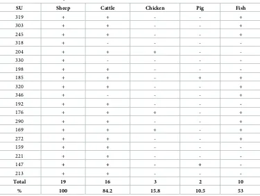
Analysis of aDNA from dental calculus was carried out to attempt to determine which ani-mals were relied upon and to identify the possibility of fish consumption. Successful results were achieved for 19 individuals out of a total of 35 (Table 4).
In accordance with literature data from different Medieval sites [100–102], among theLa
Biancacommunity the majority of individuals seemed to consume sheep and cattle products
with a minimal amount of fish, pig and chicken (Fig 5).
[image:15.612.44.566.84.392.2]The use of the DNA analysis from the dental calculus was useful not only to confirm the stable isotope results but also to understand which of the animal species were predominantly consumed. Fig 3. Plot of carbonvsnitrogen isotopic values for faunal and human remains from Allumiere.Human data are plotted individually and distinguished by age (adults and non-adults) and sex. Animal data are plotted as means and error bars represent±1σ.
Light microscopy analysis
The presence of microfossils in dental calculus was detected in 66% of the individuals. In Table 5, the number of starch granules, pollen grains and other microremains (i.e. phytoliths, calcium oxalate crystals) detected in each sample (lines) was reported, after their taxonomic classification (columns).
The 277 starch granules detected by LM were clustered in 9 different morphotypes, on the basis of morphometric and morphological parameters (Table 6).
Moreover, unidentified remains showing no distinctive features were also recorded. The highest percentage of individuals presented starch of morphotype IX (Fagaceae), followed by miners who consumed morphotype I, II, VI and VII (Poaceae). Particular attention was paid to SU 198 individual, whose dental calculus presented an aggregate containing 220 starch granules, essentially ascribable to morphotype VII (Triticumsp., 192 granules) and to morpho-type III (Myristica fragransHoutt., 24 granules).
[image:16.612.98.572.73.402.2]Beyond starch granules, microscopic analysis also revealed, the presence of pollen grains of Asteraceae andLaurus nobilisL. in the dental calculus of the individuals. In addition, 8 phyto-liths belonging to Poaceae family, 6 calcium oxalates crystals and a Cucurbitaceae fruit epicarp were observed. Examples of images of microremains found in calculus samples are indicated inFig 6.
Fig 4. Plot of carbonvsnitrogen mean isotopic values and error bars represent±1σfor human remains from Allumiere and the other Medieval Italian sites.Triangles represent the north Italian populations and circles the central Italian populations. Trino Vercellese (Piedmont) [92]; Pava Piene Siena (Tuscany) [93]; Piazza Madonna di Loreto (Rome) [94]; Cividale, Romans d’Isonzo and Mainizza (Friuli-Venezia Giulia) [95]; Cosa Grosseto (Tuscany) [96]; Albano Laziale Rome (Lazio) [97]; Montella Avellino (Campania) [98]; Colonna Rome (Lazio) [99].
GC-MS analysis
GC-MS analysis produced results for 23 individuals. The analysis was repeated for each sample at least 3 times and, in all cases, it always showed similar profiles. InS6 Tablethe chemical com-pounds identified in the calculus by GC-MS are listed, clustered by biochemical class. For each chromatographic profile, specific foods (i.e.Artemisia dracunculusL.) or food categories (i.e. milk and derivatives, plant oils), consumed in life at least once by the community ofLa Bianca were extrapolated, associating the detected compounds as reported in literature and scientific food databases [103–104]. Generally, markers of Brassicaceae (i.e. desulfoglucobrassicin; iso-thiocyanatoacetaldehyde dimethyl acetal; isothiocyanic acid, propyl ester; 13-docosenoic acid, methyl ester) and milk and its derivatives (i.e. beta-lactose) were the most recurrent molecules in the dental calculus of the population, followed by plant oils (i.e. oleic acid; 8,11,14-eicosatrie-noic acid, methyl ester, (Z,Z,Z); 9,12-octadecadie8,11,14-eicosatrie-noic acid, methyl ester, (E,E)) and theophyl-line, an alkaloid produced in leaves ofCamellia sinensis(L.) Kuntze. The detection of other plant chemical compounds (i.e. lactones, terpens) suggested the key role of plants (i.e. spices, herbs, fruits of Rosaceae) in the miners’ diet. Moreover, evidence of pharmacologically active alkaloids (i.e. stachydrine; securinine; conhydrin) supported the knowledge and application of medicinal plants (i.e.Ephedrasp.,Stachys officinalis(L.) Trevis.,Artemisia dracunculus,Conium
maculatumL. andSecurinega suffruticosa(Pall.) Rehd).
Discussion
[image:17.612.200.575.84.366.2]Archaeological evidence suggests that the archaeological site ofLa Bianca(Allumiere) is rela-tive in date to one of the first recorded human settlements in the area aimed to alum produc-tion. Musculoskeletal stress markers, combined with degenerative disease patterns provided important results in the reconstruction of miners’ working activity. Morphological changes Table 4. Ancient DNA analysis on dental calculus.
SU Sheep Cattle Chicken Pig Fish
319 + + - - +
303 + + - - +
245 + + - - +
318 + - - -
-204 + + + -
-330 + - - -
-198 + + - -
-185 + + - + +
320 + + - - +
346 + - - - +
192 + + - -
-176 + + + - +
290 + + - - +
169 + + + - +
272 + + - - +
159 + + - -
-221 + + - -
-147 + + - +
-213 + + - -
-Total 19 16 3 2 10
% 100 84.2 15.8 10.5 53
depend on repeated daily exercises that stimulate bone remodeling at the attachment sites, increasing blood flow as a consequence [105–106]. However, although the macroscopic analy-sis of bone morphological modifications can be related to muscles subjected to a high bio-mechanical stress, often the absence of archaeological data or documents confirming the validity of these assumptions does not allow determining the exact activity carried out in the individual’s lifetime [107].
[image:18.612.123.570.78.452.2]The present work represents a unique case study, providing evidence of a relationship between occupational stress markers of bone tissue and working activities [78]. This oddity is partly due to the accurate and detailed description of the four phases of alum production that is available from historical written sources [1–3]. The excavation procedure represented the most physically taxing phase, where picks were used to extract alunite from the rocks. This activity may have also involved climbing with narrow strings on the chest. Calcination was another complex phase, where alunite stones were roasted for 12–14 hours at 600–700˚C. At the end of this process, alunite was cooled through a generous watering, in order to break the “roasted” stones and obtain a doughy solution: this phase, definedmoistening, could last up to 60 days. The last step was the lixiviation during which the moistened solution was purified by mixing for 24 hours. Even if all four phases (i.e. excavation, calcination, moistening and Fig 5. Percentages of consumption of the different animal dietary resources.
lixiviation) markedly involved the shoulder girdle, the different scheme of enthesal changes and osteoarthritis pattern observed on the specimens seemed to differ in relation to the bio-mechanical stress caused by the different tasks; thus allowing the classification of the individu-als in four main clusters, each one corresponding to the working phase they were probably Table 5. Amount of starch granules (per taxonomic group or total) and other microremains detected in dental calculus.The numbers reflect the quantity of each microremain counted by direct microscopy observation in the samples.
SU I II III IV V VI VII VIII IX UN TS PO PG
110 1 1 1 3
139 1 1 1 1 4 2 P+ 1 OCa
147 3 2 1 1 7
158 4 1 5 1 OCa
159 0
169 1 3 1 1 6 1 P
173 0
176 1 1
185 1 1 2 1 A
192 1 2 3
198 1 24 1 194 220
204 1 1 2
213 0
221 1 1
239 1 1 3 P
245 1 1 1 P
256 0
269 1 1
272 0
290 1 1 1 3
303 0
304 1 1
307 1 1 2 1 P
308 0 1 OCa
317 1 1 1 OCa
318 0 1 OCa
319 2 2
320 Tb.47 2 1 3
325 1 1
330 0
333 1 1 2 2 L
334 Tb.51 1 2 2 5 1 OCa + 1C 1 Nd
343 Tb.53 0
346 Tb.54 0
356 0
TOTAL 3 2 30 6 3 6 199 6 11 11 277 4
Stratigraphic unit (SU); Morphotype I,Avenasp. (I); Morphotype II,Hordeumsp.(II); Morphotype III,Myristica fragrans(III); Morphotype IV,Piper nigrum(IV); Morphotype V,Quercus ilex(V); Morphotype VI,Sorghum bicolor(VI); Morphotype VII,Triticumsp. (VII); Morphotype VIII, Fabaceae not determined (VIII); Morphotype IX, Fagaceae not determined (IX); Unidentified starch granules (UN); Total of starches (TS); Phytoliths and other microremain (PO); Pollen grains (PG); Poaceae phytoliths (P); calcium oxalate crystals (OCa); Cucurbitaceae fruit epicarp fragment (C); Asteraceae pollen grain (A);Laurus nobilispollen granules (L); not determined pollen grain (Nd).
most involved in. It is known that osteoarthritis may be a result of traumas or infections, cer-tainly the biomechanical stress suffered from the joint represents one of its causative factors [108] even if the aetiology is far from being fully understood [109]. These data were strongly supported by osteometrics, which reported the existence of different strength and asymmetry patterns among individuals. A tall and robust skeleton could be attributable to a probable selection of individuals to be miners in relation to their adaptability to such type of work. Working tasks and related biomechanical stress seemed to have influenced also the prevalence of skeletal traumas and pathologies. In particular, a comparison with the Latium Medieval populations of Colonna [99] and Santa Severa [110] showed significant differences in the observed pathological pattern. In Colonna and Santa Severa, periostitis was detected in 4.2% and 4.4% of the individuals, respectively,vsthe value of 32% found in Allumiere. Similarly, degenerative diseases affected only 16.7% and 22.3% of the skeletal remains, in the two coeval cases, compared to 86% ofLa Biancacommunity. The morphological examination demon-strated peculiar and unique features on the analyzed sample showing that it is also one of the few Medieval examples that allows a clear identification of the social status of its components. The combined analysis of enthesal changes and osteoarthritis led to interesting results however the exact relationship between morphological variations on bone tissue and the activity carried out should be considered as a plausible hypothesis. It is known that age at death could also play an important role in the morphology of enthesal changes and that the different studies in literature underline the difficulties in finding a real correlation between entheses and activity patterns [111–113].
[image:20.612.202.578.87.385.2]Stable isotope data supported a diet based on terrestrial proteins. The 13C values also sug-gested a contribution of C4plants to the diet, as confirmed by archaeobotanical microscopy
Table 6. Starch granule morphotypes recovered from the dental calculus of the studied individuals.
Morphotype Taxonomic group
Morphologic and morphometric description
I Avenasp. Multifaceted polyhedral shape on one side and dome shaped on the other one; individual granule size: 3–8μm in length and in width.
II Hordeum sp. Granules were rounded or disk shaped; size range: 4–19μm in length and 3–15μm in width; a centrichilumis distinct; close concentriclamellaewere more detectable in the central area. One of them was attributable to theH.vulgare
species as it showed longitudinal fissure located on lateral peripheral margin
III Myristica fragrans
Granules were compound, essentially dimers or trimers. The single subunit is rounded with some peculiar flattened surfaces; size range: 5–9μm in length and 4–5μm in width; multiple fissures radiate from the centrichilum;lamellaeare not detectable.
IV Piper nigrum Polyhedral granules with pentagonal or hexagonal concave faces and acute edges; size range: 1–2μm both in length and in width; typical bright boundary.
V Quercus ilex Grains were drop-shaped; size range: 5–8μm in length and 4–6μm in width;
hilumnot clearly evident; presence of the typical hole at the narrow end.
VI Sorghum bicolor Rounded granules with some peculiar flattened surfaces; size range: 2–15μm in length and 2–10μm in width; deep radial fissures starting from a centrichilum.
Lamellaewere not detectable.
VII Triticumsp. Granules were disc-shaped; size range: 6–21μm in length and 4–18μm in width;
hilumnot visible; weakly concentriclamellaewere present. Three granules were attributable to theT.dicoccumspecies as they showed clear concentric kidney-shapedlamellaedistributed on the whole granule.
VIII Fabaceae Nd Irregular reniform granules; size range: 6–19μm in length and 5–14μm in width;
hilumis not detectable; quite clear concentriclamellae; presence of a longitudinal crack in the amorphous central area.
IX Fagaceae Nd Oblong granules; size range: 5–15μm in length and 3–11μm in width; faintly visiblelamellae; presence of longitudinal fissure.
Fig 6. Examples of plant microremains found in the dental calculus of the samples.Starch granule ofTriticumsp. and relative polarized image (A); starch granule of Fabaceae and relative polarized image (B); Poaceae phytoliths (C); not determined starch granule (D); fragment of Cucurbitaceae fruit epicarp (E); pollen grain of Asteraceae (F); Poaceae phytoliths (G); calcium oxalate crystal (H); starch granules ofMyristica fragrans(I) and relative modern experimental reference (i); starch granule ofAvenasp. (J) and modern experimental reference ofAvena fatua(j); starch granules of
Triticumsp. (K) and modern experimental reference ofTriticum durum(k). The black bar indicates 15μm.
evidence. Indeed, the use of C4plants as food, such as millet, has been documented in Italy
since the Bronze Age [114], and continued in the Medieval period at least in the north east of Italy [95].
DNA analysis revealed the consumption of animal proteins deriving from sheep and cattle meat, whereas pig and chicken use seemed to be negligible. Historical evidence indicates that the consumption of chicken remained low in the Middle Ages, corroborating our results [115]. Similarly, as we observed in the Medieval period, the use of pork meat never reached the elevated consumption of the first century of the Roman Imperial Age [79].
The dietary evidence presented by GC-MS and starch analysis will not be quantifiable in relation to the amounts consumed, however they do give an indication of the types of foods ingested during life, particularly when considered at the population level [20]. The microscopic analysis showed a consumption of Fagaceae, followed by C3Poaceae caryopses (i.e.Hordeum
sp.,Triticumsp.). Acorns, being rich in proteins, unsaturated fat, carbohydrates, minerals and
vitamins, represented a food with high energy value [116–118]. Indeed, they were commonly added to other cereal flours (after elimination of indigestive tannins by thermal treatment in water) or prepared as anti-diarrheal and astringent decoctions [119]. The high number of starch granules that were unable to be taxonomically identified could be due to modifications induced by cooking processes, ptialin enzymimatic activity or grinding procedures. Among the miners, individual SU 198 in particular presented a higher number of starch granules ofTriticumsp.
andMyristica fragrans(nutmeg). This Asian spice was also detected in the dental calculus of
other two individuals, suggesting the use of this plant species in the studied community. Nut-meg trades have been documented in the Mediterranean area since the 6thcentury, although archaeological findings are rare. It was usually grounded and mixed with red wine or used, in medicine, for its anti-inflammatory properties [120–121]. GC-MS analysis revealed that more than half of the individuals consumed Brassicaceae and dairy products, confirming the funda-mental role of these foodstuffs in the Medieval diet. At the population level, the consumption of plant oils, contained for example inOlea europaeafruits or acorns [119,122–123], was obs-erved. In addition, the detection of theophylline, an alkaloid with diuretic power, suggested the use of plant species native of South-East Asia, such asCamellia sinensis(the tea plant). To a lesser extent, the use of foods containing cholesterol (perhaps meat, cheese and eggs),Artemisia
dracunculus(tarragon, spice with antiseptic, anti-inflammatory and digestive properties) [124],
Rosaceae fruits (i.e. lactones) and various herbs were also detected. The identification of second-ary metabolites, essentially alkaloids, with pharmaceutical properties suggested the knowledge of medicinal plant species, such asStachys officinalis(astringent, digestive and sedative) [125]
andEphedrasp. (bronchodilator) [126]. The detection of a metabolite typical of Cucurbitaceae
(cucumber aldehyde) supported the use of species such as cucumber and squash; this evidence was also confirmed by the presence, in the calculus of a specimen, of a fragment of cucumber fruit epicarp [127].
on alum production, a process widely developed in Turkey and more in general in the Eastern Europe [1–3]. It is not possible to exclude the idea that, in order to establish the first Italian alum extraction system, the Pontifical State would have called on foreign experts. This hypoth-esis is corroborated by the presence of four individuals with shovel teeth, a typical feature of Eastern origin individuals [130].
This present combined approach allowed us to obtain a more detailed appreciation of the diet. In particular, two individuals SU 317 and SU 245 showed an enrichment of 15N values
>3.5‰ than the maximum animal measurements which indicated the consumption of protein
enriched in15N, perhaps from aquatic sources [131–132], but note that trophic level enrich-ment for humans has been reported up to 6‰ [133]. Analysis of DNA from dental calculus, performed only on SU 245, suggested marine fish consumption. We hypothesized that this enrichment of 15N value could be also associated to breastfeeding in SU 317 (which was a juvenile individual aged between 2–3 years old) [134] and possibly to nutritional stress in SU 245, who suffered from a compound fracture on the tibia and fibula (Fig 2). It is widely docu-mented that food deficiencies activate gluconeogenesis, a biochemical process, which uses non-carbohydrate sources to produce glucose [96,135–137], and increases 15N values in tis-sues [138]. Furthermore, GC-MS analysis of the dental calculus of SU 245 revealed the pres-ence of markers ofA.dracunculus, a plant documented to be an anesthetic and anti-inflammatory [124]. It is reasonable to believe that a compound fracture, as that detected in SU 245, caused pain and induced the application of drugs and medical treatments to treat this critical condition.
In conclusion, applying an innovative and original multidisciplinary approach, we present a detailed osteobiography of the first Italian community of alum miners. In detail, according to morphological features, we hypothesized the working task of each individual and recon-structed their dietary patterns. Furthermore, archeobotanical analysis demonstrated that min-ers used spices and herbs endemic of Asia, for therapeutic purposes, revealing their knowledge about Eastern traditional medicine.
Supporting information
S1 Table. List of the skeletal remains analyzed in the present research housed at the Department of Biology of the University of Rome “Tor Vergata”.
(DOCX)
S2 Table. Primer sequences used to amplify animal different DNA regions, size of the PCR product obtained and annealing temperature.
(DOCX)
S3 Table. Results of the post-cranial indices. (nr: not recordable). (DOCX)
S4 Table. Presence of pathology/stress markers for each individual.Neoplasia (NP), Infec-tious diseases (periostitis, PO; osteomyelitis, OM; infections, IN), trauma and stress markers (fractures, FR; grasping GR), degenerative pathologies (degenerative diseases, DG; and axial degereative diseases (Schmo¨rl’s nodes, SN)) and congenital disorders (CO), and inflammation (IF).
(DOCX)
S6 Table. The molecules identified in dental calculus by GC-MS analysis were listed and clustered in biochemical classes for each sample.
(DOCX)
Acknowledgments
The authors sincerely thankGruppo Archeologico Romano(GAR) for their valuable support to the excavation of the analyzed skeletal remains, Marianna D’Amico for the cooperation in the osteological study and Domitilla Tibaldi for her technical collaboration in the molecular paleo-diet analyses. The authors would also like to thank Dr. Riccardo Polini for the photographs, Dr. Simone Greco and Dr. Guglielmo Manenti for performing X-rays atAzienda Ospedaliera Universitaria Policlinico Tor Vergata, Matt von Tersch (BioArCh, University of York) for iso-tope mass spectrometry, and Abigail Sequeira and Sophie Gart for their technical assistance. Gabriele Scorrano is supported by the Marie Skłodowska-Curie Individual Fellowship “PALAEO-ENEO”, a project funded by the European Union’s EU Framework Programme for Research and Innovation Horizon 2020 (Grant Agreement number 751349). The authors thank the editor and the anonymous reviewer for their insightful and constructive comments, which contributed improvements to the present paper.
Author Contributions
Conceptualization:Antonella Canini, Olga Rickards, Cristina Martı´nez-Labarga.
Data curation:Marica Baldoni, Gabriele Scorrano, Angelo Gismondi, Fabrizio Vallelonga, Cristina Martı´nez-Labarga.
Formal analysis:Marica Baldoni, Gabriele Scorrano, Angelo Gismondi, Alessia D’Agostino, Michelle Alexander, Luca Gaspari, Cristina Martı´nez-Labarga.
Investigation:Marica Baldoni, Gabriele Scorrano, Angelo Gismondi, Alessia D’Agostino, Cristina Martı´nez-Labarga.
Methodology:Marica Baldoni, Gabriele Scorrano, Angelo Gismondi, Alessia D’Agostino, Michelle Alexander, Luca Gaspari, Fabrizio Vallelonga, Cristina Martı´nez-Labarga.
Resources:Fabrizio Vallelonga, Antonella Canini, Olga Rickards.
Supervision:Cristina Martı´nez-Labarga.
Validation:Cristina Martı´nez-Labarga.
Visualization:Cristina Martı´nez-Labarga.
Writing – original draft:Marica Baldoni, Gabriele Scorrano, Angelo Gismondi, Cristina Mar-tı´nez-Labarga.
Writing – review & editing:Marica Baldoni, Gabriele Scorrano, Angelo Gismondi, Alessia D’Agostino, Michelle Alexander, Luca Gaspari, Fabrizio Vallelonga, Antonella Canini, Olga Rickards, Cristina Martı´nez-Labarga.
References
1. Delumeau J. L’allume di Roma XV-XIX secolo. Civitavecchia: La Litografica; 2003.
2. Ait I. Dal governo signorile al governo del capitale mercantile: i Monti della Tolfa e ’le lumere’del papa. MEFRM 2014, 126:187–200. Italian.
4. Vallelonga F. Ricerche archeologiche nel territorio di Allumiere: gli scavi della Farnesiana e della Bianca. In: Contardi A, editor. Notiziario IX. Allumiere: Petruzzi Stampa. 2012; p. 47–71.
5. Ambrose SH. Isotopic analysis of paleodiets: methodological and interpretative considerations. In: Sandford K. editor. Investigation of ancient human tissue, chemical analyses in anthropology. Lan-ghorne: Gordon and Breach; 1993. p. 59–130.
6. Katzenberg MA. Stable isotope analyses: a tool for studying past diet, demography and life history. In: Katzenberg MA, Saunders SR, editors. The biological anthropology of the human skeleton. New York: Wiley; 2000. p. 305–27.
7. Schoeninger MJ. Diet reconstruction and ecology using stable isotope ratios. In: Larsen CS, editor. A comparison to biological anthropology. Chichester: Wiley- Blackwell; 2011. p. 445–64.
8. Henry AG, Brooks AS, Piperno DR. Microfossils in calculus demonstrate consumption of plants and cooked foods in Neanderthal diets (Shanidar III, Iraq; Spy I and II, Belgium). PNAS. 2011; 108: 486–91.
https://doi.org/10.1073/pnas.1016868108PMID:21187393
9. Buckley S, Usai D, Jakob T, Radini A, Hardy K. Dental calculus reveals unique insights into food items, cooking and plant processing in Prehistoric Central Sudan. PLoS One. 2014; 9(7): e100808.https:// doi.org/10.1371/journal.pone.0100808PMID:25028938
10. Warinner C, Rodrigues JF, Vyas R, Trachsel C, Shved N, Grossmann J, et al. Pathogens and host immunity in the ancient human oral cavity. Nat Genet. 2014; 46: 336–44.https://doi.org/10.1038/ng. 2906PMID:24562188
11. Lindhe J. Parodontologia ed implantologia dentale. Milano: Edi-Ermes; 1984.
12. Radini A, Nikita E, Buckley S, Copeland L, Hardy K. Beyond food: The multiple pathways for inclusion of materials into ancient dental calculus. American journal of physical anthropology 2017; 162: 71–83.
https://doi.org/10.1002/ajpa.23147PMID:28105717
13. Fumière O, Dubois M, Baeten V, von Holst C, Berben G. Effective PCR detection of animal species in highly processed animal byproducts and compound feeds. Anal Bioanal Chem. 2006; 385: 1045–54.
https://doi.org/10.1007/s00216-006-0533-zPMID:16761123
14. Henry AG, Piperno DR. Using plant microfossils from dental calculus to recover human diet: a case study from Tell al-Raq ’i, Syria. J Archaeol Sci. 2008; 35: 1943–50.
15. Piperno DR, Ranere AJ, Holst I, Iriarte J, Dickau R. Starch grain and phytolith evidence for early ninth millennium BP maize from the Central Balsas River Valley, Mexico. PNAS. 2009; 106: 5019–24.
https://doi.org/10.1073/pnas.0812525106PMID:19307570
16. Cawthraw S, Saunders GC, Martin TC, Sawyer J, Windl O, Reaney SD. Real-Time PCR detection and identification of prohibited mammalian and avian material in animal feeds. J Food Protect. 2009; 72: 1055e1062.
17. Yancy HF, Washington JD, Callahan L, Mason JA, Deaver CM, Farrell DE, et al. Development, evalua-tion, and peer verification of a rapid real-time PCR method for the detection of animal material. J Food Prot. 2009; 72: 2368–74. PMID:19903402
18. Wesolowski V, de Souza SMFM, Reinhard KJ, Ceccantini G. Evaluating microfossil content of dental calculus from Brazilian sambaquis. J Archaeol Sci. 2010; 37: 1326–38.
19. Hardy K, Buckley S, Collins MJ, Estalrrich A, Brothwell D, Copeland L, et al. Neanderthal medics? Evi-dence for food, cooking, and medicinal plants entrapped in dental calculus. Naturwissenschaften. 2012; 99: 617–26.https://doi.org/10.1007/s00114-012-0942-0PMID:22806252
20. Leonard C, Vashro L, O’Connell JF, Henry AG. Plant microremains in dental calculus as a record of plant consumption: A test with Twe forager-horticulturalists. J Archaeol Sci Rep. 2015; 2: 449–57.
21. Cristiani E, Radini A, Edinborough M, BorićD. Dental calculus reveals Mesolithic foragers in the Bal-kans consumed domesticated plant foods. PNAS USA 2016; 113: 10298–10303.https://doi.org/10. 1073/pnas.1603477113PMID:27573829
22. Cummings LS, Yost C, Sołtysiak A. Plant microfossils in human dental calculus from Nemrik 9, a Pre-Pottery Neolithic site in Northern Iraq. Archaeol Anthropol Sci. 2016; 1–9.
23. Hardy K, Radini A, Buckley S, Sarig R, Copeland L, Gopher A, et al. Dental calculus reveals potential respiratory irritants and ingestion of essential plant-based nutrients at Lower Palaeolithic Qesem Cave Israel. Quatern Int. 2016; 398: 129–35.
24. Walker PL, Johnson JR, Lambert PM. Age and Sex Biases in the Preservation of Human Skeletal Remains. Am J Phys Anthropol. 1988; 76: 183–88.https://doi.org/10.1002/ajpa.1330760206PMID:
3046371
25. Todd TW. Age changes in the pubic bone: I. The white male pubis. Am J Phys Anthropol. 1920a; 3: 285–334.