Int. J. Electrochem. Sci., 9 (2014) 3431 - 3439
International Journal of
ELECTROCHEMICAL
SCIENCE
www.electrochemsci.orgShort Review
Electrochemical Microarray for Identification Pathogens: A
Review
Miguel Angel Merlos Rodrigo1,2, Ondrej Zitka1,2, Ludmila Krejcova2, David Hynek2, Michal Masarik2, Jindrich Kynicky3, Zbynek Heger2, Vojtech Adam1,2, Rene Kizek1,2*
1
Department of Chemistry and Biochemistry, Faculty of Agronomy, Mendel University in Brno, Zemedelska 1, CZ-613 00 Brno, Czech Republic, European Union
2
Central European Institute of Technology, Brno University of Technology, Technicka 3058/10, CZ-616 00 Brno, Czech Republic, European Union
3
Karel Englis College, Sujanovo nam. 356/1, CZ-602 00, Brno, Czech Republic, European Union
*
E-mail: [email protected]
Received: 18 January 2014 / Accepted: 17 March 2014 / Published: 14 April 2014
Numerous analytical methods are used to detect pathogens agents including methods based on the direct isolation followed by real time-polymerase chain reaction, or immunology tests. There have not been published numerous papers on the using of electrochemical microarrays for detection of viruses and/or bacteria but there is a great potential. Electrochemical detection has been developed and assay performances studied for the CombiMatrix oligonucleotide microarray platform. CombiMatrix core technology is based on a specially modified semiconductor adapted for biological applications, which contains arrays of platinum microelectrodes. The ElectraSenseTM principle is the detection of redox active chemistries proximal to specific electrodes. The target molecules are labelled with a redox enzyme via biotin–streptavidin (or avidin) interaction. Enzymatic oxidation of an electron donor substrate then occurs. The detection current is generated due to electro-reduction of the enzymatic reaction product. Thus, the ElectraSense™ platform has been used to develop nucleic acid assays for highly accurate genotyping of a variety of pathogens including bio-threat agents. In this review we summarize recent studies on the detection and identification of pathogens by electrochemical microarrays technology and discuss the advantages and disadvantages.
1. COMBIMATRIX OLIGONUCLEOTIDE MICROARRAY PLATFORM 1.1. Overview of electrochemical biosensors for identification of pathogenic agents
The methods of enzyme-linked immunoSorbent assay, reverse transcription-polymerase chain reaction (RT-PCR) or PCR are commonly used techniques for diagnosing of viruses or bacteria [1-6]. Despite the relatively high cost of equipment, microarray-based detection methods are highly attractive due to their high sensitivity and rapidity, because these DNA/RNA microarrays are an important tool in gene expression studies, genotyping, pharmacogenomics, pathogen classification, drug discovery, sequencing and molecular diagnostics. Application of oligonucleotide microarrays in different areas of molecular clinical diagnostic and infectious disease monitoring has been rapidly growing during the last years [7-10]. Electrochemical biosensor arrays have been described by different authors [11-13]. The impedance-based horseradish peroxidase (HRP)-labelled immunosensor Ag-PSA-based DNA sensors was developed for identification Hepatitis B (HBsAg), as a model sample, and confirmed the feasibility of applying electrochemical impedance spectroscopy to the electrode array [14]. Magnetically-assisted impedimetric with phage-modified carbon microarrays method was described for more specific detection of bacteria in milk [15]. We can highlight different methods based microarrays sensor as interdigitated array microelectrodes (IDA) amplication-based RNA/DNA microarray sensor [16-18], screen-printed electrode (SPE) amplication-based RNA/DNA microarray sensor [19,20], esterase 2 amplication-based DNA array sensor [21], Hoechst 33258 amplication-based DNA array sensor [22] and HRP amplication-based DNA microarray sensor (CombiMatrix ElectroSenseTM. In this review, we attempted to summarize the utilization of CombiMatrix for detection or/and identification pathogenic agents in biological samples.
1.2. Component of CombiMatrix ElectroSense TM
CombiMatrix core technology is based on a specially modified semiconductor adapted for biological applications, which contains arrays of platinum microelectrodes. The CombiMatrix CustomArray™ and CatalogArray microarrays are currently used with fluorescent detection [23,24].
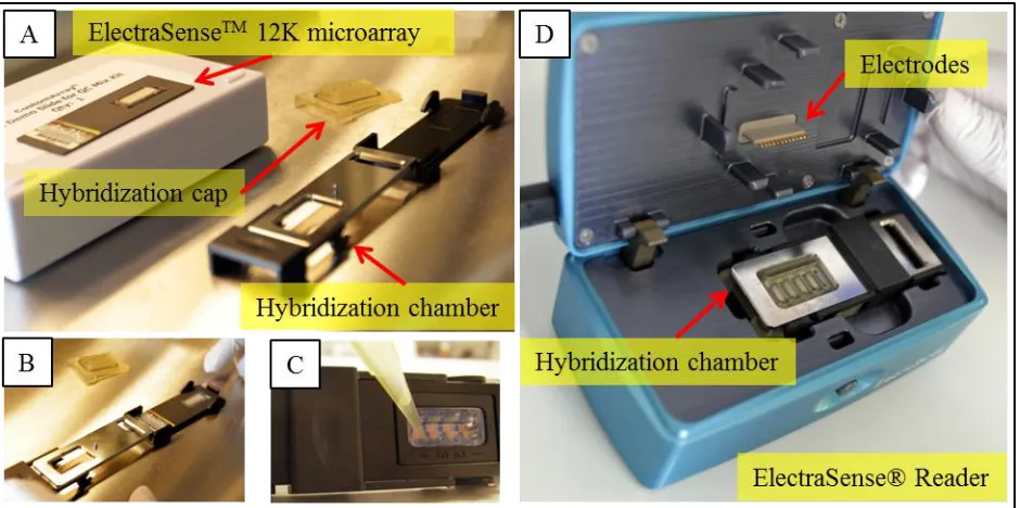
the whole microarray (Fig. 1B). The range of the highest sensitivity is from 0.1 to 200 nA, and it can be configured to detect higher signals [25]. The ElectraSenseTM microarray reader electrochemically measures thousands of probes in less than one minute (Fig. 1C). The ElectraSenseTM reader electronically reads the data by performing amperometric detection of the current flux for each individual spot through the underlying platinum microelectrode. The resulting data provides direct numeric quantification of the hybridization signals for all spots. The ElectraSenseTM Software takes as input a chip design file, which gives detailed information about the placement of probes on the microarray and data from the ElectraSenseTM reader (Fig. 1D). This system provides an optimized and completely automated data extraction, background subtraction and data normalization solution. Results can be further visualized in Excel for species identification. Another major advantage of is the ability to successfully strip and re-use each array (up to 4 times). The microarray re-using procedure is performed using the CombiMatrix CustomArrayTM Stripping Kit.
Figure 1. Component of CombiMatrix ElectroSenseTM. (A) ElectraSenseTM 12K microarray, hybridization cap and hybridization chamber. (B) Assembly of the ElectraSenseTM 12K microarray in hybridization chamber. (C) Load samples into the hybridization cap. (D) Different part of The ElectraSense™ Reader.
1.3. Electrochemical detection method
[image:3.596.65.535.301.535.2]
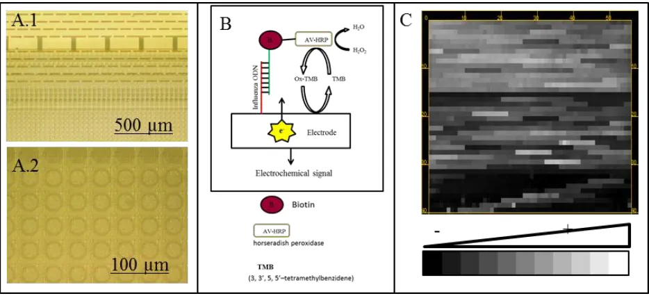
hybridized to the microarray probes. HRP catalyses the oxidation of a substrate, TMB (3,3´,5,5´-tetramethylbenzidene), and the electro-reduction of the oxidized TMB generates a current flux (Fig. 2C). The ElectraSense™ Reader performs amperometric detection of this current flux for each individual spot through the underlying platinum microelectrode [26]. The data provides direct numeric quantification of the hybridization signals for all spots. Thus, in contrast to fluorescent imaging, image data extraction is not required for the ElectraSense™ detection system. Ghindilis et al. compared the ElectraSense TM platform to the standard fluorescent detection, and good agreement was observed between these two different detection techniques. A lower detection limit of 0.75 pM was obtained for ElectraSense TM as compared to the detection limit of 1.5 pM obtained for fluorescent detection [25].
Figure 2. (A) Microphotograph of microarray chip 4x12 K from microscope. (A.1) Microphoto of electrodes and spots from 40x optical magnification. (A.2) Microphoto of spots with oligonucleotides probes from 200x optical magnification. (B) Schematic illustration the electrochemical detection method using TMB and AV-HRP. (C) Image of microarray of influenza viruses, and the position and intensity signal of oligonucleotide probes (image from ElectraSenseTM Reader).
[image:4.596.65.532.250.463.2]
2. IDENTIFICATION OF VIRUSES BY COMBIMATRIX ELECTROSENSETM 2.1. Influenza Viruses
Influenza is an infectious disease caused by RNA viruses of the family Orthomyxoviridae. Among the three types of influenza viruses (A, B, and C), the influenza A and B viruses produce seasonal epidemics in humans [28]. There have not been published numerous papers on the using of electrochemical microarrays for detection of influenza, but there is great potential. This novel electrochemical method has been used by different authors for the identification of influenza virus during the last years. CombiMatrix Corp. fabricated a chip for influenza identification that addresses a central criterion for containment of a potential pandemic: timeliness. The chips can electronically identify the binding events that represent a match between a sample of DNA and the DNA from the flu strain found in the body, which can identify any flu strain within app. four hours. Stevens et al. recently focused on novel glycan microarray technologies that can rapidly assess virus receptor specificity and the potential emergence of human-adapted H5N1 viruses [29].Roth et al. demonstrated well-performed detection of avian influenza subtype H5N1 by application of electrochemical detection of Combimatrix microarrays and support the fact that the ElectraSense reader is a robust and sensitive platform for the detection of DNA hybridization from influenza viruses [23]. Lodes et al. identified upper respiratory tract pathogens by electrochemical detection using oligonucleotide microarray, including A and B influenza viruses [30]. Bolotin et al. showed that the sensitivity of the CombiMatrix influenza detection system was 95.2 % and the specificity was 100 % for influenza A subtype testing on patients diagnosed in the 2007–2008 influenza season in Toronto, Canada [31]. Straight et al. identified 23 of 24 samples of laboratory-confirmed pandemic (H1N1) 2009 Influenza by the ElectraSense influenza A assay. The assay identified all samples of influenza A/H1N1 and A/H3N2, and differentiated these from pandemic (H1N1) 2009 Influenza in all cases. Therefore, the ElectraSense influenza A assay proved to be a useful assay to quickly and accurately differentiate pandemic (H1N1) 2009 influenza from seasonal influenza [32]. Wojciechowski et al. introduced and described super avidin-biotin system (SABS) and secondary enzymatic enhancement (SEE) as viable methods to enhance microarray performance and sensitivity on influenza sequences. In spite of the fact these techniques did not yield functional improvements, in most cases the target-specific detection limit was substantially reduced from control values following SABS or SEE treatment. These post-hybridization amplification techniques are appealing ways to enhance assay performance because they are easy, fast and low cost.
2.2. Human papillomavirus
employed the Combimatrix® B3 CustomArray™ for the synthesis of reusable, bespoke microarrays for the purpose of discerning multiple Human Papilloma Virus strains [34].
2.3. Other viruses
Virus detection and identification is also important for farmers, plant pathologists and others involved in plant protection activities of quarantine and certification programs. Tiberini et al. showed the potentiality of an oligonucleotide diagnostic array based on the Combimatrix platform for detection and identification of 14 artichoke-infecting viruses [35]. In another study, the authors detected and identified viral populations in The Internal Active Thermal Control System (IATCS) samples obtained from the Kennedy Space Center (aboard the International Space Station) as a first step towards characterizing and understanding potential risks associated with viruses’ presence. Benardini et al. used one Combimatrix panvira 12K microarray containing probes for ∼1,000 known human viruses. Positive hybridizations were observed for probes from the Adenoviridae, Mononegavirales, Poxovirade, Orthomyxoviridae, Flaviviridae, Herpesviridae, Papillomaviridae, Parvoviridae, and Reoviridae families. The data generated in this study using both traditional and molecular techniques confirm that viral particles are present within the IATCS [36]. Tiberini et al. prepared an array based on the Combimatrix platform for detection of 37 viruses belonging to 13 families, one of which is unassigned, together with six pospiviroid species, genus Pospiviroid, family Pospiviroidae. Most of the virus probes were highly specific and were able to identify tomato viruses. Most Pospiviroid probes, however, were non-specific in terms of species, but were specific at the genus level as they hybridized to members of the genus Pospiviroid. Only one probe of the Tomato apical stem viroid was species specific [37]. Various enteric viruses including norovirus, rotavirus, adenovirus, and astrovirus are the major etiological agents of food-borne and water-borne disease outbreaks and frequently cause non-bacterial gastroenteritis worldwide. A Combimatrix platform oligonucleotide probe was sensitive and high-throughput detection method for detection of these viral pathogens [9].
3. IDENTIFICATION OF BACTERIA BY COMBIMATRIX ELECTRASENSETM 3.1. Neisseria gonorrhoeae
Gonorrhoea continues to seriously impact human society with an estimated 106 million new infections per annual. The consequences of gonorrhoea on reproductive and neonatal health are serious as its role in the spread of human immunodeficiency virus. Jackson et al. described the use of a custom 12K CombiMatrix ElectraSenseTM oligonucleotide microarray format for assessing global gene expression profiles in Neisseria spp [10].
3.2. Helicobacter pylori
mucosa-associated lymphoid. A whole-genome CombiMatrix Custom oligonucleotide tiling microarray with 90000 probes covering six sequenced H. pylori genomes was designed for one study. This microarray was used to compare the genomic profiles of eight unsequenced strains isolated from patients with different gastroduodenal diseases in Heilongjiang province of China. The results of this study promote further understanding of specific disease-associated genes that can serve as novel biomarkers for identification of gastroduodenal diseases [41].
3.3. Other bacteria
Wojciechowski et al. described application of the CombiMatrix antibody microarray system designed for detection of the bacterial pathogens Bacillus anthracis (B. anthracis), Yersinia pestis (Y. pestis), and bacterial toxin staphylococcal enterotoxin B (SEB) [42]. The ElectraSenseTM platform was used to develop nucleic acid assays for highly accurate genotyping of a variety of pathogens including bio-threat agents (such as B. anthracis, Y. pestis, and other microorganisms including Escherichia coli, Bacillus subtilis, etc.) [25] and for detection of four bacterial pathogens (Bordetella pertussis, Streptococcus pyogenes, Chlamydia pneumoniae and Mycoplasma pneumonia) [30]. CombiMatrix technology makes it as an attractive solution for detection needs of a variety of sensor-based areas including identification pathogens, disease diagnosis and genetic screenings.
4. CONCLUSIONS
CombiMatrix ElectraSenseTM technique is applicable for several microarray-based assays including genotyping and gene expression. On the other hand, the ElectraSenseTM platform has high potential for further development of low cost and easy-to-use identification of pathogens. The electrochemical arrays are new and novel method for fast and convenient monitoring of pathogens. In summary, we show in this review that electrochemistry array method have great advantages compared conventional method for detection and/or identification pathogens. One may also see the potential of these arrays for strain typing or identifying the source of an epidemic.
ACKNOWLEDGEMENTS
The authors are grateful to NanoBioMetalNet CZ.1.07/2.4.00/31.0023 for financial support. Special thanks are dedicated to Hana Buchtelova for perfect technical assistance.
CONFLICT OF INTEREST
The authors declare no conflicts of interest
References
1. A. J. Jaaskelainen and L. Maunula, J. Virol. Methods, 136 (2006) 210.
3. H. Mirghani, F. Amen, F. Moreau, J. Guigay, M. Ferchiou, A. E. Melkane, D. M. Hartl and J. L. St Guily, Oral Oncol., 50 (2014) 1.
4. R. F. O. Franca, C. C. da Silva and S. O. De Paula, Eur. J. Clin. Microbiol. Infect. Dis., 32 (2013) 723.
5. V. Pazienza, G. A. Niro, R. Fontana, M. Vinciguerra and A. Andriulli, Clin. Chem. Lab. Med., 51 (2013) 1707.
6. S. R. Silva, I. S. S. Katz, E. Mori, P. Carnieli, Jr., L. F. P. Vieira, H. B. C. R. Batista, L. B. Chaves and K. C. Scheffer, Biologicals, 41 (2013) 217.
7. J. Hinton, R. Callan, C. Bodine, W. Glasgow, S. Brower, S. W. Jiang and J. P. Li, Expert Rev. Mol. Diagn., 13 (2013) 431.
8. J. Lacroix, F. Schlund, B. Leuchs, K. Adolph, D. Sturm, S. Bender, T. Hielscher, S. M. Pfister, O. Witt, J. Rommelaere, J. R. Schlehofer and H. Witt, Int. J. Cancer, 134 (2014) 703.
9. J. M. Kim, S. Y. Kim, Y. B. Park, H. J. Kim, B. S. Min, J. C. Cho, J. M. Yang, Y. H. Cho and G. Ko, J. Microbiol., 50 (2012) 970.
10. L. A. Jackson and D. W. Dyer, Diagnosis of Sexually Transmitted Diseases: Methods and Protocols, 903 (2012) 343.
11. M. Seidel and R. Niessner, Anal. Bioanal. Chem., 391 (2008) 1521.
12. P. Arora, A. Sindhu, H. Kaur, N. Dilbaghi and A. Chaudhury, Appl. Microbiol. Biotechnol., 97 (2013) 1829.
13. S. Cagnin, M. Caraballo, C. Guiducci, P. Martini, M. Ross, M. SantaAna, D. Danley, T. West and G. Lanfranchi, Sensors, 9 (2009) 3122.
14. X. B. Yu, R. Lv, Z. Q. Ma, Z. H. Liu, Y. H. Hao, Q. Z. Li and D. K. Xu, Analyst, 131 (2006) 745. 15. A. Shabani, C. A. Marquette, R. Mandeville and M. F. Lawrence, Talanta, 116 (2013) 1047. 16. B. Elsholz, A. Nitsche, J. Achenbach, H. Ellerbrok, L. Blohm, J. Albers, G. Pauli, R. Hintsche and
R. Worl, Biosens. Bioelectron., 24 (2009) 1737.
17. Y. L. Liu, B. Elsholz, S. O. Enfors and M. Gabig-Ciminska, Anal. Biochem., 382 (2008) 77. 18. B. Elsholz, R. Worl, L. Blohm, J. Albers, H. Feucht, T. Grunwald, B. Jurgen, T. Schweder and R.
Hintsche, Anal. Chem., 78 (2006) 4794.
19. S. Laschi, I. Palchetti, G. Marrazza and M. Mascini, J. Electroanal. Chem., 593 (2006) 211. 20. F. Farabullini, F. Lucarelli, I. Palchetti, G. Marrazza and M. Mascini, Biosens. Bioelectron., 22
(2007) 1544.
21. C. Pohlmann, Y. R. Wang, M. Humenik, B. Heidenreich, M. Gareis and M. Sprinzl, Biosens. Bioelectron., 24 (2009) 2766.
22. K. Goto, H. Horiuchi, H. Shinohara, K. Motegi, K. Hashimoto, S. Hongo, N. Gemma, N. Hayashimoto, T. Itoh and A. Takakura, J. Microbiol. Methods, 69 (2007) 93.
23. K. M. Roth, K. Peyvan, K. R. Schwarzkopf and A. Ghindilis, Electroanalysis, 18 (2006) 1982. 24. A. Tiberini, L. Tomassoli, M. Barba and A. Hadidi, J. Virol. Methods, 168 (2010) 133.
25. A. L. Ghindilis, M. W. Smith, K. R. Schwarzkopf, K. M. Roth, K. Peyvan, S. B. Munro, M. J. Lodes, A. G. Stover, K. Bernards, K. Dill and A. McShea, Biosensors & Bioelectronics, 22 (2007) 1853.
26. K. Maurer, N. Yazvenko, J. Wilmoth, J. Cooper, W. Lyon and D. Danley, Sensors, 10 (2010) 7371. 27. J. Cooper, N. Yazvenko, K. Peyvan, K. Maurer, C. R. Taitt, W. Lyon and D. L. Danley, PLoS One,
5 (2010) 1.
28. T. Y. Lin and A. L. Brass, Curr. Opin. Virol., 3 (2013) 531.
29. J. Stevens, O. Blixt, J. C. Paulson and I. A. Wilson, Nat. Rev. Microbiol., 4 (2006) 857.
30. M. J. Lodes, D. Suciu, J. L. Wilmoth, M. Ross, S. Munro, K. Dix, K. Bernards, A. G. Stover, M. Quintana, N. Iihoshi, W. J. Lyon, D. L. Danley and A. McShea, PLoS One, 2 (2007) 1.
31. S. Bolotin, E. Lombos, R. Yeung, A. Eshaghi, J. Blair and S. J. Drews, Virol. J., 6 (2009) 1.
33. F. X. Bosch, T. R. Broker, D. Forman, A. B. Moscicki, M. L. Gillison, J. Doorbar, P. L. Stern, M. Stanley, M. Arbyn, M. Poljak, J. Cuzick, P. E. Castle, J. T. Schiller, L. E. Markowitz, W. A.
Fisher, K. Canfell, L. A. Denny, E. L. Franco, M. Steben, M. A. Kane, M. Schiffman, C. Meijer, R. Sankaranarayanan, X. Castellsague, J. J. Kim, M. Brotons, L. Alemany, G. Albero, M. Diaz and S. de Sanjose, Vaccine, 31 (2013) 1.
34. D. Ayers, M. Platt, F. Javad and P. J. R. Day, in B.D.M. Theophilus, R. Rapley (Editors), Pcr Mutation Detection Protocols, Second Edition, Humana Press Inc, Totowa, 2011, p. 75. 35. A. Tiberini and M. Barba, J. Plant Pathol., 95 (2013) 145.
36. J. Benardini Iii, E. Hagerman, T. Green, R. L. Crawford, R. Sumner and K. Venkateswaran, SAE Technical Papers, 1 (2007) 1.
37. A. Tiberini and M. Barba, J. Virol. Methods, 185 (2012) 43.
38. J. Y. Park, D. Forman, E. R. Greenberg and R. Herrero, Curr. Oncol. Rep., 15 (2013) 517. 39. P. Malfertheiner, M. Venerito and M. Selgrad, Curr. Opin. Gastroenterol., 29 (2013) 669. 40. J. S. Muhammad, T. Sugiyama and S. F. Zaidi, J. Pak. Med. Assoc., 63 (2013) 1528.
41. Y. H. You, L. H. He, M. J. Zhang, J. Y. Fu, Y. X. Gu, B. H. Zhang, X. X. Tao and J. Z. Zhang, PLoS One, 7 (2012) 1.
42. J. Wojciechowski, D. Danley, J. Cooper, N. Yazvenko and C. R. Taitt, Sensors, 10 (2010) 3351. © 2014 The Authors. Published by ESG (www.electrochemsci.org). This article is an open access