Int. J. Electrochem. Sci., 7 (2012) 5100- 5114
International Journal of
ELECTROCHEMICAL
SCIENCE
www.electrochemsci.orgSurface Effect of Assembling Enzyme and Modulation of
Surface Enzyme Activity with Electric Potential Stress
Cheng-Han Chao1,2, Kun-Lin Li1, Chung-Shu Wu1, Cheng-Che Lee1, Han-Ping Chiang3, Yuh-Shyong Yang3, Tung-Ming Pan4, Fu-Hsiang Ko1,*
1
Department of Materials Science and Engineering, National Chiao-Tung University, Hsinchu 300, Taiwan
2
Division of Nephrology, Department of Internal Medicine, National Taiwan University Hospital Hsin-Chu Branch, Hsinchu 300, Taiwan
3
Department of Biological Science and Technology, National Chiao Tung University, Hsinchu 300, Taiwan
4
Department of Electronics Engineering, Chang Gung University, Taoyuan 333, Taiwan
*
E-mail: [email protected]
Received: 20 April 2012 / Accepted: 10 May 2012 / Published: 1 June 2012
The fluorescent marker of rhodamine B amine is successfully used to evaluate the immobilization capability onto silicon-based patterns fabricated by semiconductor manufacturing. Only the silicon dioxide surface, by means of fluorescent observation, can immobilize the rhodamine molecule by the sequential linkage of (3-aminopropyl)triethoxysilane (APTES) and glutaraldehyde. The phenol sulfotransferase enzyme is also successfully immobilized onto the silicon dioxide surface by the linking molecules of APTES and sulfosuccinimidyl 4-(N-maleimidomethyl)-cyclohezane-1-carboxylate in the home-made apparatus. The enzyme activity of the sulfotransferase is determined from the absorbance of 4-nitrophenol at 400 nm wavelength. The surface immobilized enzyme remains its activity for catalytic reaction at least 120-min duration. The surface saturation effect on the activity of immobilized enzyme is explained and ascribed to the surface diffusion effect of electric double layers. We can success control the surface immobilized enzyme by electric potential stress. The activity of enzyme is reduced under negative potential, while is enhanced under positive potential. The electric potential can induce the enzyme structure variation and modulate the enzyme activity due to the electrostatic effect.
1. INTRODUCTION
The interdisciplinary study of biology, chemistry, and electronics becomes more and more important than ever before. Combining the biotechnology and semiconductor technology, various types of biochips or biosensors have now been developed to detect and monitor the specific binding of biomolecules on the solid-state substrates [1]. The choice of suitable chip surface for the purpose of biomolecular immobilization has gradually received attraction in recent years. For example, (3-mercaptopropyl)trimethoxysilane linker first assembles onto the silicon dioxide film. Through the assistant of gold nanoparticles, the deoxyribonucleic acid (DNA) molecule can be tethered [2]. DNA molecule can also be assembled onto the platinum-silicide surface through the linkers of (3-aminopropyl)trimethoxysilane, N-cyclohexyl-N’-[2-(N-methylmorpholino)-ethyl]-carbodiimide-4-toluene sulfonate and imidazole [3]. Genomics and proteomics research has elucidated many new biomarkers that exhibit the potential to greatly improve the correctness of disease diagnosis [4-5].
Various enzymes immobilized onto different electrodes can be used to detect herbicide, triglyceride or glucose [6-8]. Sulfotransferase is an important enzyme in a variety of organisms and can specifically catalyze the transfer of the sulfuryl group (SO3). The reaction involves the transfer of
the donor of universal sulfuryl group (3’-phosphoadenosine 5’-phosphsulfate, PAPS) to the variety of nucleophiles ranging from endobiotics (i.e., monoamines, phenyl compounds, hormones, proteins and carbohydrate) to xenobiotics [9,10]. Hence, sulfonation catalyzed by sulfotransferase is an important pathway in detoxification of a broad range of endobiotics and xenobotics. They have been implicated in the activation and deactivation of hormones and carcinogens through the formation of sulfate conjugates [11]. In the previous review [12], membrane-associated proteins have been used in several influential biological processes, including viral entry of cells, leukocyte adhesion, and anticoagulation. More generally, enzymatic transformations of cell-surface proteoglycans by sulfotransferases appear to trigger vital molecular-recognition and signal-transduction events. Redox modification of Cys residues provides a mechanism for protein regulation [13,14]. Proteins can be glutathionylated or S-nitrosylated, especially for the environment of oxidative stress. Oxidative stress is involved in the pathogenesis of various degenerative diseases including cancer [15].
form in the continuous phase, the morphology of binding enzyme will be changed due to pH, temperature, ionic strength or electric field. As to the electrochemical effect from electric field, very few literature mentions the morphology change to control the enzyme activity. Zhao and Yang mention lysozyme and pepsin enzymes will loss activity once the dosage of pulsed electric field is higher than the specific critical value [21]. Wu and coworker suggest various metal cations have different effects on the activity for sulfotransferase [22].
In this study, we firstly evaluate the pattern surface often encountered in the semiconductor technology to assemble the fluorescent rhodamine molecule. The suitable pattern material is adopted for the selective immobilization of an important active enzyme, i.e. the phenol sulfotransferase. The activity of the enzyme after surface immobilization is evaluated by the spectrophotometer at 400 nm wavelength. A surface diffusion model is proposed to explain the activity for the surface immobilized enzyme. Finally, we design an apparatus to control the electrochemical behavior and activity of sulfotransferase enzyme.
2. EXPERIMENTAL
2.1 Pattern formation processes
P-type Si(100) wafers (14-21 -cm, MEMC, MO, USA) with 15 cm diameter were deposited and etched to form the structure with silicon dioxide pattern on poly-Si film, and silicon nitride pattern on poly-Si film, respectively. They were cut into 2 cm x 2 cm pieces to serve as test samples. To prepare the silicon oxide pattern on poly-Si film, the poly-Si film was first deposited with silane gas (SiH4) at 60 cm3/min and 620oC. Prior to photolithography, the silicon oxide film was grown by wet
oxidation with a gas mixture of hydrogen (8000 cm3/min) and oxygen (5000 cm3/min) at 978oC. The mask with the pattern of interest was used to define the photoresist (TMER-iP3650, Tokyo Ohka Kogyo, Tokyo, Japan) pattern. A 365-nm light emitted from high pressure mercury lamp (SUV-2001CIL, USHIO, Tokyo, Japan) induced the photo-active reaction for the photoresist film. After the dissolution of exposure area with 2.38% tetramethylammonium hydroxide, the plasma was used to etch the silicon oxide film without passivation by photoresist pattern. The reactive-ion etch system (TE5000, Tokyo Electron Limited, Tokyo, Japan) was operated at 500 W RF power under 0.2 Torr high vacuum, and the gas mixture of 20 cm3/min of CF4, 20 cm3/min of CHF3, and 400 cm3/min Ar.
Finally, the residual photoresist was removed and cleaned by the mixing chemical of H2SO4 and H2O2
(volume ratio = 3:1) at 120oC for 10min. The chemicals used were of higher grade from Merck (Darmstadt, Germany).
Similar with the processes for the above sample of silicon oxide pattern on poly-Si film, the silicon nitride film was deposited with a mixture of ammonium (130 cm3/min) and dichlorosilane (30 cm3/min) at 780oC. The silicon nitride film with photoresist pattern passivation was etched by the same reactive-ion etch system. The instrument was operated at 250W RF power under 0.8 Torr high vacuum, and the gas mixture of 50 cm3/min of CF4, 20 cm3/min of O2, and 1000 cm3/min Ar. After the surface
2.2 Chemicals, apparatus and procedures for immobilization
Prior to immobilization, the pattern sample should be carefully cleaned for 30 min. The temperature of the solution of H2SO4 and H2O2 (volume ratio is 3:1) must be maintained above 85oC to
possess the oxidative power. If the temperature was dropped, hydrogen peroxide was required to replenish. It should be noted that the cleaning solution was very corrosive and dangerous; we must handle it carefully. After pure water rinsing and drying, the pattern of interest was immersed in the (3-aminopropyl)triethoxysilane (APTES) solution for 30 min in room temperature. The APTES (Sigma-Aldrich, MO, USA) solution was prepared by the following procedures: (1) mixing pure water with acetone (volume ratio is 5:1), (2) adjusting the above solvent pH to 3.5 by 1 M HCl, and (3) preparing the 5% APTES solution by diluting with above solvent. Then, the pattern sample was rinsed with pure water thoroughly. The succeeding step was to bake the pattern sample at 120oC for 30 min. Then, the sample was immersed in the linker solution (2.5 % glutaraldehyde, i.e. pentane-1,5-dial) for 30 minutes in room temperature. The 2.5% glutaraldehyde solution was diluted with phosphate buffered saline solution (pH 7.4 PBS buffer, containing 120 mM NaCl, 2.7 mM KCl, and 10 mM phosphate buffer, Sigma-Aldrich) from 25% glutaraldehyde (in water, Sigma-Aldrich). Then, the pattern sample was rinsed with PBS. Finally, the pattern sample was immersed in the rhodamine B amine (C28H31N3O3, Sigma-Aldrich) solution for 16 h at room temperature. The powder of rhodamine B
amine was first weighted and then dissolved in the dimethylformamide (DMF, Sigma-Aldrich). Then, it was diluted by 0.1 M carbonate buffer (pH 9.6) to reach the concentration of 0.1 mM rhodamine B amine solution. The rhodamine B amine is a light-sensitive molecule. Hence, the solution should be prepared freshly and the vessel should be covered with aluminum foil. Prior to observing these pattern samples by fluorescent microscope (BX51, OLYMPUS, PA, USA), all the samples were cleaned sequentially by 0.1 M carbonate buffer, pure water, and drying.
The home-made apparatus in Fig. 1 was designed and used to evaluate the enzyme activity on the sample surface of interest. The Teflon ring tightly contacted with the substrate and sealed with the silicon resin glue. Prior to conducting the enzyme immobilization, we needed to test the reliability of the home-made apparatus to avoid leakage problem. The clean and APTES immobilization methods for enzyme immobilization were conducted in the home-made apparatus with the same procedures as rhodamine mentioned above. We prepared the linker solution by dissolving sulfosuccinimidyl 4-(N-maleimidomethyl)-cyclohezane-1-carboxylate (sulfo-SMCC, Sigma-Aldrich) into the 50 mM sodium borate buffer (Sigma-Aldrich).
[image:5.596.148.426.212.446.2]
phosphoadenosine 5’-phosphate (PAP) into the solvent of 100 mM 1,3-bis[tris(hydroxymethyl)methylamino]propane buffer at pH 7. Then, we added the above solution into the made apparatus. At the reaction time of interest, the liquid was siphoned out from the home-made apparatus to an UV-Vis spectrophotometer (Hitachi UV-Vis-3300, Tokyo, Japan) for characterization. We analyzed the absorbance of 4-nitrophenol (the catalytic product of 4-nitrophenyl sulfate) at 400 nm wavelength to determine the activity of sulfotransferase.
Figure 1. Schematic diagram of the home-made apparatus for immobilization of sulfotransferase onto the silicon oxide surface.
3. RESULTS AND DISCUSSION
3.1 Immobilization of fluorescent material onto various pattern substrates
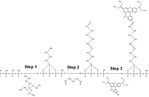
Rhodamine and its derivatives are popular fluorescent materials for labeling all types of biomolecules. The integration of the rhodamine derivatives onto the semiconductor-based pattern structures is very important for developing the biosensors. In Fig. 2, the successful immobilization of rhodamine B amine onto silicon oxide surface requires three mail steps. In first step, the ethoxysilane group (CH3CH2O-Si) in APTES reacts with the surface silanol group (Si-OH) on silicon dioxide film.
Figure 2. The immobilization steps for rhodamine B amine onto silicon dioxide surface.
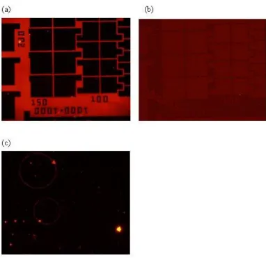
Two types of pattern structures (silicon dioxide on poly-Si, and silicon nitride on poly-Si) are used to evaluate the immobilization capability for the rhodamine B amine. The successful immobilization can be evaluated from the red fluorescent rhodamine molecule. The pattern sample in Fig. 3a has finished the whole procedures mentioned in Fig. 2. We find only the red color emitted from the silicon dioxide pattern, while no color in poly-Si region. This observation clearly suggests the rhodamine molecule can only be immobilized in silicon dioxide pattern, but not in poly-Si. This result is beneficial for future biosensor construction by means of semiconductor process. As to the same pattern but skip the APTES and glutaraldehyde procedures in Fig. 2, no image contrast can be seen from Fig. 3b. This observation suggests the rhodamine molecule has not yet assembled onto the sample surface. The amine group of rhodamine molecule cannot react either with the silicon dioxide or the poly-Si film.
Figure 3. (a) The sample has previously assembled linkers of APTES and glutaraldehyde. The red region of fluorescent image represents the rhodamine molecule has immobilized onto the silicon dioxide surface, and the black region represents no rhodamine molecule has immobilized onto the poly-Si surface. (b) The sample omits the linker processes of APTES and glutaraldehyde. The rhodamine molecule can be immobilized onto the silicon dioxide and poly-Si surface without the aid of linkers. (c) The sample has previously tried to assemble linkers of APTES and glutaraldehyde. The black color means the rhodamin molecule cannot immobilize onto silicon nitride and poly-Si surface, irrespective of linking molecules.
3.2 Enzyme immobilization and its activity
[image:7.596.102.484.70.441.2]
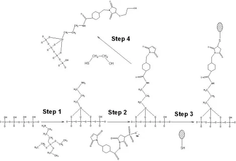
Hence, the PST is successfully immobilized onto the surface of silicon dioxide film. Prior to testing the activity of surface enzyme, the region without enzyme immobilization, but still has terminal maleimide group, is deactivated by the 2-mercaptoethanol (step 4 in Fig. 4).
Figure 4. The immobilization steps for sulfotransferase onto silicon oxide surface.
[image:8.596.64.534.142.462.2]
Figure 5. The couple reactions in the home-made apparatus are catalyzed by phenol sulfotransferase.
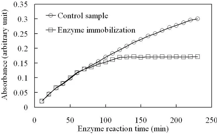
observed during the time period of 230 min. The trend of enzyme reaction meets the enzyme kinetics in the solution. On the contrary, the surface immobilized enzyme exhibits a quite different tendency. The behavior of surface immobilized enzyme exhibits the same behavior with the control sample for the duration within 80 min, but gradually loss the activity hereafter.
Figure 6. The absorbance of 4-nitrophenol at various enzyme reaction times for the control sample (sulfotransferase in solution) and the enzyme target sample (sulfotransferase immobilizes onto the silicon oxide surface), n=3.
[image:10.596.127.475.167.384.2][image:11.596.119.468.125.483.2]
effect during the time period of 230 min cannot be seen for the solution enzyme (control sample) in Fig. 6.
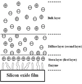
Figure 7. The surface diffusion model for the catalytic reaction of sulfotransferase.
Figure 8. The absorbance of 4-nitrophenol at various measurement times for the sample with immobilization of sulfotransferase onto the silicon oxide surface, n=3. The marked area means the solution is removed from the home-made apparatus. The other samples are catalyzed by the sulfotransferase immobilized in the home-made apparatus.
3.3 Modulation of surface enzyme activity with electric potential stress
An electric enzyme regulator has been designed from Fig. 1, where the Pt is top electrode covering the solution and gold is the backside electrode around substrate. The electric potential stress is applied from both the electrodes to generate a controllable electric field. As anticipated, we use electric potential/field to modulate the enzyme activity. PST is used as target enzyme and is covalently immobilized onto patterned Si/SiO2 substrates as mentioned in Fig. 4. In a specific redox environment,
[image:12.596.139.453.68.326.2]
activity is promoted under +5 V, +10 V and +20 V stress. This observation in Figs. 9 and 10 demonstrates the capability to regulate enzyme activity in an electric field environment.
Figure 9. The effect of negative electric potential stress on the absorbance of 4-nitrophenol under various enzyme reaction times for sulfotransferase immobilizes onto the silicon oxide surface.
[image:13.596.93.507.144.392.2] [image:13.596.93.506.472.722.2]
The enzyme under both negative stress and positive stress demonstrates the similar trend. All the experiment runs suggest the catalytic activity of enzyme is increased from initial experiment to 120 min, and the reaction is approaching the saturation level. Different electrochemical stress exhibits different saturation level, and this effect is mainly attributed to the binding structure of surface enzyme. The reason to explain the saturation level is ascribed to the surface diffusion model mentioned earlier.
4. CONCLUSIONS
In this study, we have successfully immobilized rhodamine B amine and phenol sulfotransferase onto the silicon dioxide pattern, while the silicon nitride and poly-Si surface cannot immobilize the enzyme due to the lack of suitable functional group. The surface immobilized sulfotransferase has the similar activity within the reaction period of 80 min. As to the reaction time of catalytic reaction higher than 80 min, the product concentration in the home-made apparatus restricts the enzyme activity. The surface enzyme can restore its activity once the reaction solution is homogeneous mixing. The surface diffusion model is proposed to explain the catalytic reaction of sulfotransferase immobilized onto the silicon dioxide surface. The electric potential stress can successfully modulate the enzyme activity through the home-made apparatus. The modulation of surface bound enzyme from either positive potential or negative potential stressing demonstrates different trends.
ACKNOWLEDGMENTS
The authors would like to thank the National Taiwan University Hospital Hsin-Chu Branch and Chang Gung Memorial Hospital for financially supporting this research under contract of HCH101-07 and CMRPD290031, respectively.
References
1. T. Sakata, Y. Miyahara, Biosens. Bioelectron., 21 (2005) 827.
2. F.-H. Ko, Z.-H. Yeh, C.-C. Chen, T.-F. Liu, J. Vac. Sci. Technol. B, 23 (2005) 3000.
3. C.-C. Chen, C.-Y. Tsai, F.-H. Ko, C.-C. Pun, H.-L. Chen, P.-H. Chen, Jpn. J. Appl. Phys., 43 (2004) 3843.
4. S. Zhang, N. Wang, Y. Niu, C. Sun, Sens. Actuators B, 109 (2005) 367.
5. D. v. d. Voort, C.A. McNeil, R. Renneberg, J. Korf, W.T. Hermens, J.F.C. Glatz, Sens. Actuators B, 105 (2005) 50.
6. X. Wang, L. Chen, S. Xia, Z. Zhu, J. Zhao, J.-M. Chovelon, N. J. Renaul, Int. J. Electrochem. Sci., 1 (2006) 55.
7. M. R. Ganjali, F. Faridbod, E. Nasli-Esfahani, B. Larijani, H. Rashedi, P.Norouzi, Int. J. Electrochem. Sci., 5 (2010) 1422.
8. M. Stoces, K. Kalcher, I. Svancara, K. Vytras, Int. J. Electrochem. Sci., 6 (2011) 1917.
9. M. W. Duffel, A. D. Marshall, P. McPhie, V. Sharma, W. B. Jakoby, Drug Metab. Rev., 33 (2001) 369.
11. C. Clarke, P. Thorburn, D. McDonald, J. B. Adams, Biochim. Biophys. Acta, 707 (1982) 28. 12. E. Chapman, M. D. Best, S. R. Hanson, C. H. Wong, Angew. Chem. Int. Ed., 43 (2004) 3526. 13. C. Lind, R. Gerdes, Y. Hamnell, I. Schuppe-Koistinen, H. B. von Lowenhielm, A. Holmgren, I. A.
Cotgreave, Arch. Biochem. Biophys., 406 (2002) 229.
14. Y. Ji, V. Toader, B. M. Bennett, Biochem. Pharmacol., 63 (2002) 1397. 15. R. Gopalakrishna, S. Jaken, Free Radical Biol. Med., 28 (2000) 1349.
16. A. Deep, U. Tiwari, P. Kumar, V. Mishra, S. C. Jain, N. Singh, P. Kapur, L. M. Bharadwaj, Biosens. Bioelectron., 33 (2012) 190.
17. P. Norouzi, F. Faridbod, B. Larijani, M. R. Ganjali, Int. J. Electrochem. Sci., 5 (2010) 1213. 18. C.-S. Wu, C.-C. Lee, C.-T. Wu, Y.-S. Yang, F.-H. Ko, Chem. Commun., 47 (2011) 7446.
19. C.-C. Wu, T.-M. Pan, C.-S. Wu, L.-C. Yen, C.-K. Chuang, S.-T. Pang, Y.-S. Yang, F.-H. Ko, Int. J. Electrochem. Sci., 7 (2012) 4432.
20. C.-C. Lin, F.-H. Ko, C.-C. Chen, Y.-S. Yang, F.-C. Chang, C.-S. Wu, Electrophoresis, 30 (2009) 3189.
21. W. Zhao, R. Yang, J. Phys. Chem. B, 112 (2008) 14018. 22. S.-C. G. Wu, K. D. Straub, J. Biol. Chem., 251 (1976) 6529.
23. P. C. Hiemenz, Principles of Colloid and Surface Chemistry, 2nd ed., Marcel Dekker, New York, 1986, pp. 677-684 (Chapter 12).
24. P. H. Rieger, Electrochemistry, Prentice-Hall, New Jersey, 1987, pp. 70-72 (Chapter 2). 25. T. M. Glendening, J. E. Poulton, Plant Physiol., 94 (1990) 811.