fraction.
White Rose Research Online URL for this paper: http://eprints.whiterose.ac.uk/135912/
Version: Accepted Version
Article:
Bowen, TS, Herz, C, Rolim, NPL et al. (7 more authors) (2018) Effects of endurance training on detrimental structural, cellular, and functional alterations in skeletal muscles of heart failure with preserved ejection fraction. Journal of Cardiac Failure, 24 (9). pp.
603-613. ISSN 1071-9164
https://doi.org/10.1016/j.cardfail.2018.08.009
© 2018 Elsevier Inc. This manuscript version is made available under the CC-BY-NC-ND 4.0 license http://creativecommons.org/licenses/by-nc-nd/4.0/.
[email protected] https://eprints.whiterose.ac.uk/ Reuse
This article is distributed under the terms of the Creative Commons Attribution-NonCommercial-NoDerivs (CC BY-NC-ND) licence. This licence only allows you to download this work and share it with others as long as you credit the authors, but you can’t change the article in any way or use it commercially. More
information and the full terms of the licence here: https://creativecommons.org/licenses/
Takedown
If you consider content in White Rose Research Online to be in breach of UK law, please notify us by
Accepted Manuscript
Effects of endurance training on detrimental structural, cellular, and functional alterations in skeletal muscles of heart failure with
preserved ejection fraction
T. Scott Bowen , Christian Herz , Natale P.L. Rolim ,
Anne-Marie Ormbostad Berre , Martin Halle , Angela Kricke , Axel Linke , Gustavo Justo da Silva , Ulrik Wisloff , Volker Adams
PII: S1071-9164(18)30966-7
DOI: https://doi.org/10.1016/j.cardfail.2018.08.009 Reference: YJCAF 4192
To appear in: Journal of Cardiac Failure
Received date: 25 January 2018 Revised date: 4 May 2018 Accepted date: 14 August 2018
Please cite this article as: T. Scott Bowen , Christian Herz , Natale P.L. Rolim , Anne-Marie Ormbostad Berre , Martin Halle , Angela Kricke , Axel Linke , Gustavo Justo da Silva , Ulrik Wisloff , Volker Adams , Effects of endurance training on detrimental structural, cellular, and functional alterations in skeletal muscles of heart failure with preserved ejection fraction, Journal of Cardiac Failure(2018), doi:https://doi.org/10.1016/j.cardfail.2018.08.009
A
CCE
P
T
E
D
M
A
N
U
S
CRIP
T
Effects of endurance training on detrimental structural, cellular, and functionalalterations in skeletal muscles of heart failure with preserved ejection fraction
T. Scott Bowen1*, Christian Herz2*, Natale P.L. Rolim3, Anne-Marie Ormbostad Berre3,
Martin Halle4,Angela Kricke2, Axel Linke5, Gustavo Justo da Silva3, Ulrik Wisloff3,6, Volker
Adams5
School of Biomedical Sciences, University of Leeds, Leeds, United Kingdom1; Department of Internal Medicine and Cardiology, Leipzig University-Heart Center, Leipzig, Germany2; K.G. Jebsen Center of Exercise in Medicine, Department of Circulation and Medical Imaging, Faculty of Medicine and Health Sciences, Norwegian University of Science and Technology, Trondheim, Norway3; Technische Universität München, Munich Germany4; Department of Internal Medicine and Cardiology at the Technische Universität Dresden5;
School of Human Movement & Nutrition Sciences, University of Queensland, Australia6 :
*Equal Contribution
Running head: Skeletal muscle alterations in HFpEF
Word Count: 2732
Corresponding Author: T. Scott Bowen, PhD
Address: School of Biomedical Sciences, Univ. of Leeds, Leeds, LS2 9JT, United
Kingdom,
Email: [email protected]
A
CCE
P
T
E
D
M
A
N
U
S
CRIP
T
AbstractBackground: HFpEF is underpinned by detrimental skeletal muscle alterations that
contribute to disease severity, yet underlying mechanisms and therapeutic treatments
remain poorly established. This study used an animal model of HFpEF to better
understand whether skeletal muscle abnormalities were: 1) fiber-type specific; and 2)
reversible by various exercise training regimes.
Methods and Results: Lean controls were compared to obese ZSF1 rats at 20 weeks,
and 8 weeks later following sedentary, high-intensity interval training, or
moderate-continuous treadmill exercise. Oxidative-soleus and glycolytic-extensor digitorum longus
(EDL) muscles were assessed for fiber size, capillarity, glycolytic metabolism, autophagy,
and contractile function. HFpEF reduced fiber size and capillarity by 20-50% (P<0.05) in
both soleus and EDL, but these effects were not reversed by endurance training. In
contrast, both endurance training regimes in HFpEF attenuated the elevated lactate
dehydrogenase activity observed in the soleus. Autophagy was downregulated in EDL and
upregulated in soleus (P<0.05), with no influence following endurance training. HFpEF
impaired contractile forces of both muscles by ~20 % (P<0.05) and these were not
reversed by training.
Conclusion: Obesity-related HFpEF was associated with detrimental structural, cellular,
and functional alterations to both slow-oxidative and fast-glycolytic skeletal muscles that
could not be reversed by endurance training.
Word count: 198 of 200
A
CCE
P
T
E
D
M
A
N
U
S
CRIP
T
Introduction
The increasing prevalence of heart failure patients with preserve ejection fraction (HFpEF)
despite no breakthrough in effective treatments is now recognized as a major unmet
problem in cardiovascular medicine [1]. Cardiac-orientated drug interventions in
large-scale clinical trials in HFpEF have proven largely ineffective in terms of patient outcomes
[2]. This has shifted focus towards possible treatment of “non-cardiac organs”, such as the
skeletal muscle, endothelial, and pulmonary systems. Recent studies have now
established functional and structural impairments are present in skeletal muscle from both
patients and animal models of HFpEF [3-7]. Muscle-specific changes include fiber atrophy,
adipose infiltration, oxidative to glycolytic fiber type shift, reduced capillary density, and
mitochondrial dysfunction, which have also been closely correlated to the main symptom
of exercise intolerance [5-8]. Interestingly, new evidence has confirmed the presence of a
specific obesity-related HFpEF patient cohort [9]. Whether skeletal muscle alterations
occur in this distinct phenotype is yet to be fully explored. Nevertheless, skeletal muscle
maladaptations induced by HFpEF predispose patients towards a rapid depletion of
high-energy phosphates and earlier accumulation of fatigue inducing metabolites [10, 11].
Therefore identification of interventions to reverse such processes are a key therapeutic
target in cardiovascular medicine.
While evidence remains scarce, numerous skeletal muscle alterations induced by HFpEF
can be prevented following exercise training. One study using a hypertensive rat model of
HFpEF documented that high-intensity interval training (HIIT) attenuated mitochondrial
impairments, which was associated with normalized contractile function and
fatigue-resistance [4]. However in this study exercise training was initiated before the onset of
HFpEF (i.e. primary intervention) [4], which precluded answering the more
A
CCE
P
T
E
D
M
A
N
U
S
CRIP
T
Further, HFpEF was induced by hypertension alone rather than the multiple co-morbidities
(e.g., obesity, diabetes, renal dysfunction, and hypertension) that typically drive the
development of this disease in most patients - particular in those with obesity-related
HFpEF [9].
This study, therefore, aimed to further characterize the underlying structural, metabolic,
and molecular skeletal muscle alterations that occur in obesity-related HFpEF and
determine whether endurance training could reverse impairments (i.e. secondary
prevention). In this study, we used a recently established cardiometabolic obese-driven rat
model of HFpEF [3, 12, 13]. The model closely resembles a typical patient phenotype,
where cardiac impairments and exercise intolerance are underpinned by numerous
co-morbidities including obesity, type II diabetes, hypertension, and kidney dysfunction [14,
15]. We specifically compared two skeletal muscles with differing metabolic profiles: the
oxidative slow-twitch soleus in addition to the glycolytic fast-twitch extensor digitorum
longus (EDL), in order to determine whether HFpEF induces fiber-type specific
maladaptations.
Methods
Study design and animal model of HFpEF
Procedures and experiments in this study were approved by the Norwegian Animal
Research Authority, in accordance the European Directive 2010/63/EU. A full description
of the present study design and animal cohort as well as the methods employed were
recently described elsewhere [3]. Briefly, obese diabetic Zucker fatty/Spontaneously
hypertensive heart failure F1 hybrid (ZSF1) rats (Charles River, Kingston, US) were used
as a model to induce HFpEF, which occurs by 20 weeks of age [3, 12, 13]. Non-invasive
echocardiography as well as invasive hemodynamics were used to confirm the presence
of HFpEF, where left ventricular diastolic impairments and a preserved ejection fraction
A
CCE
P
T
E
D
M
A
N
U
S
CRIP
T
exercise intolerance (as described in detail elsewhere [3]). The first part of the study
assessed male obese ZSF1 rats (i.e., HFpEF, n=12) rats at 20 weeks of age compared to
their lean strain-matched counterparts (i.e., controls; n=8). In the second part of the study,
a further 4 groups were assessed at 28 weeks of age, which included lean rats compared
to 3 groups of obese ZSF1 rats that performed either no exercise training (i.e., sedentary;
n=13), high-intensity interval training (i.e., HFpEF+HIIT; n=11) or moderate continuous
training (i.e., HFpEF+MCT; n=11). Exercise training was performed for 8 weeks (from
20-28 weeks of age), which included HIIT (3 x per week; 4 intervals at 90% peak oxygen
uptake ( peak) for 4 min, with 3 min recovery at 60% peak) or MCT (5 x per week at
60% peak for 1 h) for a total of 8 weeks on a treadmill at a gradient of 25°, as previously
described [3].
Muscle function
The right EDL and soleus were dissected and mounted vertically mounted in a buffer-filled
organ bath between a hook and force transducer (FORT250, World Precision Instruments
Inc., Berlin, Germany), with the output continuously recorded and digitized (PowerLab
8/30, ADInstruments, Oxford, UK). In vitro muscle function was assessed by platinum
electrodes stimulating the muscle with a supramaximal current (700 mA; 500 ms train
duration; 0.25 ms pulse width) from a base stimulator (Grass S88, Grass Technologies,
Warwick, USA) amplified via a high power bi-polar stimulator (701C, Aurora Scientific Inc.,
Aurora, Canada). The muscle bundle was set at an optimal length (Lo) equivalent to the
maximal twitch force produced, after which bath temperature was increased to 25°C and a
15 min thermo-equilibration period followed. A force-frequency protocol was then
performed at 1, 15, 30, 50, 80, and 120 Hz respectively, separated with 1 min rest
intervals. Following a 5 min rest period muscles then underwent a fatigue protocol over 5
min (40 Hz every 2 s). Forces generated during the fatigue protocol were normalized to the
A
CCE
P
T
E
D
M
A
N
U
S
CRIP
T
Tissue analysesOne portion of the left EDL and soleus were frozen immediately in liquid N2 for biochemical
analyses. Frozen muscle samples were subsequently homogenized in lysis buffer (50 mM
Tris, 150 mM sodium chloride, 1 mM EDTA, 1% NP-40, 0.25% sodium-deoxycholate,
0.1% SDS, 1% Triton X-100; pH 7.4) containing a protease inhibitor mix (Inhibitor mix M,
Serva, Heidelberg, Germany), sonicated, and centrifuged at 16,000 g for 5 min. The
supernatant was isolated and protein content determined (BCA assay, Pierce, Bonn,
Germany). Muscle homogenates (5 - 20 g) mixed with loading buffer (126 mM Tris-HCl,
20% glycerol, 4% SDS, 1.0% 2-mercaptoethanol, 0.005% bromophenol blue; pH 6.8) were
separated by SDS-polyacrylamide gel electrophoresis for 1.5 h at 90 V. Proteins were
transferred to a polyvinylidene fluoride membrane (PVDF) and incubated overnight at 4°C
with primary antibodies for the LC3 (1:1000, Novus Biologicals, Cambridge, UK), where
the relative ratio of LC3 in its content form of II/I was used as a marker of autophagy.
Membranes were subsequently incubated with a horseradish peroxidase-conjugated
secondary antibody and specific bands visualized by enzymatic chemiluminescence
(Super Signal West Pico, Thermo Fisher Scientific Inc., Bonn, Germany) and densitometry
quantified using a 1D scan software package (Scanalytics Inc., Rockville, USA). In
addition, enzyme activities for lactate dehydrogenase (LDH) were measured
spectrophotometrically as a measure of glycolytic metabolism, as previously described
[16]. Protein and enzyme activity data are presented as fold change relative to the
respective control group. In addition, a portion of the EDL and soleus fixed in 4%
PBS-buffered formalin and paraffin-embedded were used for histological analyses of
cross-sectional area (CSA). Briefly, sections of 3 µm were cut, mounted on glass cover slips,
and stained for H&E for fiber CSA to be calculated. In addition, sections were stained with
an antibody against von Willebrand factor (vWF; 1/500, Santa Cruz) for capillaries to be
A
CCE
P
T
E
D
M
A
N
U
S
CRIP
T
Statistical analysesAnalyses were performed by SPSS version 22 (SPSS Inc., Chicago, IL, USA). Data are
presented as mean±SEM. Between-group differences were assessed by unpaired
Student’s t-test at 20 weeks or by one-way ANOVA at 28 weeks, while data presented as
ratios were analyzed by appropriate non-parametric tests (Mann-Whitney or
Kruskal-Wallis). Muscle function (force-frequency and force-time relationships) was assessed by
two-way repeated measures ANOVA (group X frequency or group X time). Post hoc
comparisons were calculated using Bonferroni-corrected tests.
Results
Animal model of HFpEF
Obese ZSF1 rats developed typical signs of HFpEF at 20 and 28 weeks, independent of
exercise training. These data have already been published elsewhere and the reader is
referred to this manuscript for more expansive details characterizing the current cohort of
rats [3]. Briefly, at 28 weeks compared to lean controls all HFpEF groups (sedentary, MCT,
HIIT) demonstrated evidence (P<0.05) of diastolic dysfunction (E/E’: 15±1 vs. 25±1, 25±2,
23±1, respectively; P<0.0001), LV hypertrophy (23±1 vs. 28±1, 28±1, 29±1 mg/mm of tibia
length, respectively; P<0.0001), obesity (body weight: 465±1 vs. 590±15, 591±17, 599±12
g, respectively; P<0.0001), diabetes (fasting blood glucose; 4.1±0.2 vs. 6.4±0.5, 5.5±0.3,
7.8±0.7 mmol/L, respectively; P<0.0001), and hypertension (systolic blood pressure:
158±14 vs. 181±15, 176±13,183±18 mmHg, respectively; P=0.008), in the presence of
normal LVEF (66±2 vs. 75±2, 74±2, 72±3~70 %, respectively). Importantly, exercise
capacity as assessed by peak was reduced (P<0.0001) in sedentary HFpEF rats
compared to controls by ~30% (38±1 vs. 49±1 mL kg-1min-1, respectively). However, 8
weeks of exercise training resulted in a ~15 % peak increase (P=0.004) in both the MCT
-A
CCE
P
T
E
D
M
A
N
U
S
CRIP
T
1, respectively). All of the changes mentioned above between control and sedentary
HFpEF rats were also present at 20 weeks.
In addition, compared to control rats, muscle wet-weights at 20 weeks were reduced in
HFpEF for both the EDL (258±29 vs. 174±21 mg, respectively; P<0.0001) and soleus
(219±11 vs. 180±19 mg, respectively; P<0.0001). Similarly, at 28 weeks control rats
maintained higher (P<0.05) muscle wet-weights compared to all HFpEF groups
(sedentary, MCT, HIIT) such that training had no impact on muscle mass for the EDL
(241±8 vs. 156±2, 164±3, 161±3 mg, respectively; P<0.0001) or soleus (221±4 vs. 170±4,
184±6, 188±4 mg, respectively; P<0.0001).
Glycolytic and oxidative skeletal muscle alterations in HFpEF
At 20 weeks, we observed numerous structural, metabolic, and molecular alterations in the
skeletal muscles of HFpEF rats, both in the glycolytic fast-twitch EDL as well as the
oxidative slow-twitch soleus muscle. Representative muscle sections of the EDL are
presented in Fig. 1A-B for control and HFpEF rats, respectively. Compared to controls,
HFpEF rats demonstrated fiber atrophy by ~50 % (P<0.0001; Fig. 1C) and a reduction in
capillarity by ~20 % (P=0.001; Fig. 1D) in the EDL muscle. While not significant, the
activity of the glycolytic enzyme LDH tended to be higher in the EDL of HFpEF rats
(P=0.07; Fig. 1E), with protein levels of LC3 II/I ratio (an index of autophagy) showing a
reduction of ~15 % in HFpEF compared to the control rats (P=0.049; Fig. 1F). These
structural and molecular changes likely contributed to the lower force generated in the EDL
muscle, with maximal forces reduced by ~15 % (P=0.001; Fig. 1G) while relative fatigue
was unaffected (P=0.401; Fig. 1H). Similarly, the predominately oxidative soleus muscle
also showed numerous alterations in HFpEF, but to a lesser degree. Representative
muscle sections of the soleus from control and HFpEF rats is presented in Fig. 2A-B.
A
CCE
P
T
E
D
M
A
N
U
S
CRIP
T
fiber cross sectional area and capillarity, respectively (P<0.0001 and P=0.002,
respectively; Fig. 2B-C). In contrast, while LDH activity and LC3 II/I protein levels were not
different between groups (P=0.791 and P=0.621; Fig. 2E-F), maximal soleus force was
reduced by ~20 % in HFpEF compared to control rats (P=0.006; Fig. 2G) yet fatigability
remained unchanged (P=0.767; Fig. 2H).
Can exercise training reverse skeletal muscle impairments in HFpEF?
At 28 weeks, following 8 weeks of either sedentary, HIIT, or MCT regimes, the EDL
muscle (see Fig. 3A for representative sections) in HFpEF rats showed no apparent
benefit from exercise training: the observed fiber atrophy (P<0.0001) and reduced
capillarity (P=0.016) were not reversed compared to control values (Fig. 3B-C), while
limited effects were observed on markers of glycolytic metabolism (P=0.316) and
autophagy (P=0.066; Fig. D-E). As one would expect, therefore, EDL muscle weakness
and fatigability were not influenced with exercise training in HFpEF, with evidence of
impaired force production (P<0.004) and greater fatigability (P<0.009) in the HFpEF
groups compared to controls (Fig. 3F-G). In line with these findings, the oxidative soleus
muscle (see Fig. 4A for representative sections) also showed limited benefits following
exercise training in the HFpEF rats: HIIT or MCT were unable to increase fiber
cross-sectional area (P=0.003) or capillarity (P=0.010) back towards control values (Fig.4B-C). In
contrast, the exercise training regimes attenuated activity of the glycolytic enzyme LDH,
which remained elevated in the soleus of sedentary HFpEF rats relative to controls
(P=0.004; Fig. 4D). Yet exercise training did not reduce autophagy activation, with protein
levels of the LC3 II/I ratio elevated across all three HFpEF groups compared to controls in
the soleus (P=0.016; Fig. 4E). As such, the lower contractile forces observed in the soleus
from HFpEF animals compared to healthy rats at 20 weeks were not overcome by 8 weeks
of exercise training (P=0.003; Fig. 4F), which was also similar for fatigability (P=0.001; Fig
A
CCE
P
T
E
D
M
A
N
U
S
CRIP
T
DiscussionThis is the first study to characterize fiber type-specific skeletal muscle alterations in a rat
model of HFpEF and to further assess the efficacy of various exercise training regimes to
reverse these deficits. The main finding of this study is that HFpEF caused detrimental
alterations to both slow-oxidative and fast-glycolytic skeletal muscles including fiber
atrophy, impaired capillarity, a shift towards glycolytic metabolism, and autophagy
dysregulation, which expands upon our current knowledge collected from both patient and
animal studies [3-8]. These structural, metabolic, and proteostasis changes likely
contributed, at least in part, to the greater muscle weakness and fatigability that we also
observed in HFpEF rats. Somewhat surprisingly 8 weeks of exercise training (either high-
or moderate-intensity treadmill running) was unable to attenuate many of these
muscle-specific deficits, which likely provides a rationale for why muscle dysfunction could not be
overcome. Overall, therefore, these data: 1) provide novel underlying mechanisms that
may be responsible for skeletal muscle dysfunction in HFpEF: 2) confirm that glycolytic
and oxidative muscles are both affected in HFpEF; 3) demonstrate exercise training to be
an ineffective tool in reversing skeletal muscle impairments in this experimental model.
Skeletal muscle dysfunction in HFpEF: oxidative vs. glycolytic
Data from vastus lateralis biopsies have shown that when compared to age-matched
controls, patients with HFpEF demonstrate reduced mitochondrial content, lower
mitochondrial fusion proteins, a shift from oxidative to glycolytic fibers, and impaired
capillarity [5-7]. Recent data have also suggested that a distinct obesity-related HFpEF
patient cohort exists [9], but no data are available to support whether skeletal muscle
alterations specific to this phenotype develop. In the current study, therefore, we used an
obese-specific rat model of HFpEF to provide novel evidence that skeletal muscle
A
CCE
P
T
E
D
M
A
N
U
S
CRIP
T
show that both oxidative as well as glycolytic muscles are afflicted. While both the soleus
and EDL were severely affected by fiber atrophy and lower capillarity, it is important to
note that the glycolytic EDL demonstrated greater susceptibility. Indeed, previous data
have shown the oxidative soleus to be better protected against impairments induced by
heart failure with reduced ejection fraction (HFrEF) compared to glycolytic muscles [17],
with our data tending to support this view in HFpEF. Our data also showed a strong trend
for the glycolytic enzyme lactate dehydrogenase activity to be increased in HFpEF (both in
soleus and EDL), which supports recent magnetic resonance imaging studies where an
earlier depletion of high-energy phosphates occurred during the onset of exercise in
HFpEF patients [10, 11].
An additional novel finding in the present study was revealing that other molecular
signaling pathways may be involved in the muscle remodeling process induced by HFpEF.
For example autophagy (as assessed by the ratio of LC3 II/I protein expression) was
differentially regulated between fiber types during HFpEF. Autophagy is a catabolic
process that degrades cytoplasmic proteins, however basal flux is key to maintain cell
homeostasis. Previous studies in mice have shown that either excessive or deficient levels
of autophagy can induce skeletal muscle atrophy and functional deficits [18, 19].
Collectively, therefore, our data suggest that HFpEF could modulate autophagy levels in a
fiber-type specific manner via a signaling pathway not yet characterized in this disease.
We speculate the significant increase of autophagy in the soleus but decrease in the EDL
may have contributed, at least in part, to the muscle atrophy and dysfunction observed in
HFpEF. Interestingly, previous data have shown that autophagic flux differs between
oxidative and glycolytic muscles, which supports the suggestion that HFpEF could
modulate this process in a fiber-dependent manner [20]. Moreover our findings also
suggest that HFpEF could induce muscle alterations in both a fiber- and time-dependent
A
CCE
P
T
E
D
M
A
N
U
S
CRIP
T
susceptible to impairments earlier in the disease (i.e., 20 weeks), whereas the oxidative
soleus only exhibited alterations at a later time point (i.e. 28 weeks). Interestingly, only at
28 weeks did the soleus demonstrate a significant increase in LDH activity and autophagic
flux in HFpEF rats, which were not apparent at 20 weeks. The mechanism underlying
these time-dependent changes remain unclear, however it may be related to the chronic
pro-inflammatory state that accompanies the HFpEF phenotype [21], which given enough
time would be expected to impact even the most resistant of all muscle fibers.
Exercise training in HFpEF to reverse skeletal muscle impairments
In the present study, exercise training conferred no significant benefits to either glycolytic
or oxidative muscles, which was generally consistent in terms of structural, metabolic,
autophagic, and functional measures. We did observe that glycolytic enzyme activity of
lactate dehydrogenase was significantly attenuated in the soleus muscle of HFpEF rats
that performed HIIT or MCT, which suggests a shift towards oxidative metabolism
following exercise training. While the effects of exercise training to reverse skeletal muscle
impairments in HFpEF remain largely unestablished, previous data in a hypertensive rat
model of HFpEF showed endurance training was protective against reductions in soleus
mitochondrial content and fatigability [4]. As the current study’s main focus was not on
muscle mitochondrial adaptations, we cannot rule out the possibility that exercise training
induced a shift towards an oxidative phenotype in the trained HFpEF rats, such that
improvements in mitochondrial function/content or a shift towards increased oxidative
slow-twitch fibers occurred. However, as part of another study using the same HFpEF rats
[3], we have previously documented that citrate synthase activity (a marker of
mitochondrial content) was not significantly increased in the soleus post-training. These
findings, together with those we present here, seem to support therefore that a metabolic
inflexibility may exist in this specific animal model with obesity-related HFpEF. Indeed,
A
CCE
P
T
E
D
M
A
N
U
S
CRIP
T
current HFpEF experimental model remains unknown, but the potential co-morbidities that
occur in these animals such as obesity, diabetes, hypertension and renal dysfunction may
prove too overwhelming for exercise training to be beneficial in only 8 weeks. In support,
we recently reported the diaphragm was unable to benefit from exercise training in the
same model [3], suggesting skeletal muscle adaptations in this model are not achieved
with 8 weeks of endurance exercise training. Whether these findings, therefore, translate
to patients with a specific obese-related HFpEF phenotype remains to be confirmed, but
warrants further attention.
Clinical translation and new insights
In terms of clinical translation, our findings strongly support a number of muscle-specific
changes reported to occur in HFpEF patients, as assessed from leg biopsies (e.g., fiber
atrophy, reduced capillarity) [5-7], but also extend current knowledge in HFpEF by
providing new findings on fiber-type specific adaptations (i.e., oxidative vs. glycolytic) as
well as new mechanistic insight into the underlying signaling pathways (e.g., dysregulation
in autophagy, upregulation of glycolytic enzymes). Up until now, patient and animal studies
in HFpEF had drawn conclusions based upon a single muscle group (i.e., the vastus
lateralis in patients and the soleus in animal models) [3-8]. As such, it was unknown
whether other limb muscles composed of different fiber types showed similar impairments
in HFpEF. The current study, therefore, provides new evidence that shows skeletal muscle
remodeling in HFpEF is fiber-type dependent, with fast-twitch glycolytic fibers impacted
earlier and to a greater degree. This adds additional insight by suggesting muscle groups
composed of fast-twitch fibers should be primarily targeted during cardiac rehabilitation in
HFpEF patients, as these may be more susceptible to impairments. Future patient studies
should therefore focus on sampling various muscle groups in order to characterize the role
A
CCE
P
T
E
D
M
A
N
U
S
CRIP
T
Perhaps the most novel yet controversial finding of the present study is that it challenges
the prevalent view that endurance training can be used as a treatment for muscle
dysfunction in HFpEF [22]. In fact, our data support the recent suggestion that more work
in the area is required to establish the benefit of exercise training in treating muscle
dysfunction in HFpEF [23]. Specifically, here we found endurance training was unable to
rescue muscle dysfunction and attenuate detrimental structural remodeling induced by
HFpEF, irrespective of the exercise regime performed. While it is clearly established that
endurance training in patients with HFpEF improves exercise capacity and quality of life
[23-29], the role for this in the disease to rescue muscle dysfunction still remains unclear.
Our current experimental data also found 8 weeks of endurance training (either HIIT or
MCT) in HFpEF was able to increase peak by ~15% (which mirrors the increase seen in
patients after training [23], thus supporting clinical translation of our findings). Clearly,
however, the lack of improvements we found in both oxidative and glycolytic skeletal
muscles post-training in HFpEF would suggest a limited role of improved muscle function
as a central mechanism underlying the increased exercise capacity. One key question is
whether our animal findings closely reflects the patient scenario? Unfortunately, as of yet,
no patient studies have evaluated skeletal muscle biopsies following endurance training in
HFpEF. While data from an experimental rat model have shown HIIT attenuates
mitochondrial and contractile impairments in the soleus [4], this study induced HFpEF by
hypertension alone (rather than multiple comorbidities) and the endurance training was
extensive lasting a total of 28 weeks. As such, limitations to both patient and animal
studies preclude the degree to which we can conclude how much endurance training
benefits skeletal muscle in HFpEF, but clearly this remains a ‘hot-topic’ of investigation.
By inference, therefore, our data do not support the concept that an increases in peak in
A
CCE
P
T
E
D
M
A
N
U
S
CRIP
T
terms, peak is determined by products of the Fick principle (i.e. cardiac output and
arterial-venous O2 difference). As our in vivo and in vitro measures of cardiac function
were not improved post-training in HFpEF (which in fact agrees with many patient studies;
as reviewed in ref. [23]), we speculate the main mechanism responsible for the increase in
peak likely resides at the peripheral vascular level [30, 31]. Indeed patients with HFpEF
are known to have endothelial dysfunction [32] and we have previously shown endurance
training can reverse in vitro endothelial dysfunction in aortic rings isolated from
hypertensive rats with HFpEF [33]. However, endothelial dysfunction as a main
mechanism of limitation in HFpEF remains controversial based on a key patient study
showing endurance training over 16 weeks failed to improve endothelial function [27].
Nevertheless, the same authors haven also provided data to support that an increase in
the arterial-mixed venous O2 difference is likely the primary mechanism underlying the
higher peak achieved post-training in HFpEF [26, 27], which indicates a peripheral
limitation is present in HFpEF. Based on these data and together with our findings, we
speculate improved microvascular perfusion may be the primary mechanism leading to an
improvements in peak following endurance training in HFpEF (which is known to be
impaired [30, 31]), as this would be predicted to elevate O2 diffusion delivery from capillary
to mitochondria (as dictated by Fick’s Law) [34].
Conclusion
Obesity-related HFpEF caused detrimental alterations to both slow-oxidative and
fast-glycolytic skeletal muscles, which included substantial fiber atrophy, a reduction in
capillarity, a shift towards glycolytic metabolism, autophagy dysregulation, as well as
impaired contractile functional measures. The intervention of exercise training (either high-
A
CCE
P
T
E
D
M
A
N
U
S
CRIP
T
which poses the intriguing question of whether endurance training can be used to combat
skeletal muscle impairments induced by obesity-related HFpEF at the patient level.
Acknowledgments
This study was funded by the European Commission, The Seventh Framework
Programme for Research (FP7-Health/602405).
Conflicts of interest
A
CCE
P
T
E
D
M
A
N
U
S
CRIP
T
References[1] Butler J, Fonarow GC, Zile MR, Lam CS, Roessig L, Schelbert EB, et al. Developing
therapies for heart failure with preserved ejection fraction: current state and future
directions. JACC Heart failure. 2014;2:97-112.
[2] Sharma K, Kass DA. Heart failure with preserved ejection fraction: mechanisms, clinical
features, and therapies. Circ Res. 2014;115:79-96.
[3] Bowen TS, Brauer D, Rolim NPL, Baekkerud FH, Kricke A, Ormbostad Berre AM, et al.
Exercise Training Reveals Inflexibility of the Diaphragm in an Animal Model of Patients
With Obesity-Driven Heart Failure With a Preserved Ejection Fraction. Journal of the
American Heart Association. 2017;6: e006416.
[4] Bowen TS, Rolim NP, Fischer T, Baekkerud FH, Medeiros A, Werner S, et al. Heart
failure with preserved ejection fraction induces molecular, mitochondrial, histological, and
functional alterations in rat respiratory and limb skeletal muscle. European journal of heart
failure. 2015;17:263-72.
[5] Haykowsky MJ, Kouba EJ, Brubaker PH, Nicklas BJ, Eggebeen J, Kitzman DW.
Skeletal muscle composition and its relation to exercise intolerance in older patients with
heart failure and preserved ejection fraction. The American journal of cardiology.
2014;113:1211-6.
[6] Kitzman DW, Nicklas B, Kraus WE, Lyles MF, Eggebeen J, Morgan TM, et al. Skeletal
muscle abnormalities and exercise intolerance in older patients with heart failure and
preserved ejection fraction. American journal of physiology Heart and circulatory
physiology. 2014;306:H1364-70.
[7] Molina AJ, Bharadwaj MS, Van Horn C, Nicklas BJ, Lyles MF, Eggebeen J, et al.
Skeletal Muscle Mitochondrial Content, Oxidative Capacity, and Mfn2 Expression Are
Reduced in Older Patients With Heart Failure and Preserved Ejection Fraction and Are
A
CCE
P
T
E
D
M
A
N
U
S
CRIP
T
[8] Haykowsky MJ, Brubaker PH, Morgan TM, Kritchevsky S, Eggebeen J, Kitzman DW.
Impaired aerobic capacity and physical functional performance in older heart failure
patients with preserved ejection fraction: role of lean body mass. J Gerontol A Biol Sci Med
Sci. 2013;68:968-75.
[9] Obokata M, Reddy YNV, Pislaru SV, Melenovsky V, Borlaug BA. Evidence Supporting
the Existence of a Distinct Obese Phenotype of Heart Failure With Preserved Ejection
Fraction. Circulation. 2017;136:6-19.
[10] Bhella PS, Prasad A, Heinicke K, Hastings JL, Arbab-Zadeh A, Adams-Huet B, et al.
Abnormal haemodynamic response to exercise in heart failure with preserved ejection
fraction. European journal of heart failure. 2011;13:1296-304.
[11] Weiss K, Schar M, Panjrath GS, Zhang Y, Sharma K, Bottomley PA, et al. Fatigability,
Exercise Intolerance, and Abnormal Skeletal Muscle Energetics in Heart Failure.
Circulation Heart failure. 2017;10:e004129.
[12] Hamdani N, Franssen C, Lourenco A, Falcao-Pires I, Fontoura D, Leite S, et al.
Myocardial titin hypophosphorylation importantly contributes to heart failure with preserved
ejection fraction in a rat metabolic risk model. Circulation Heart failure. 2013;6:1239-49.
[13] van Dijk CG, Oosterhuis NR, Xu YJ, Brandt M, Paulus WJ, van Heerebeek L, et al.
Distinct Endothelial Cell Responses in the Heart and Kidney Microvasculature
Characterize the Progression of Heart Failure With Preserved Ejection Fraction in the
Obese ZSF1 Rat With Cardiorenal Metabolic Syndrome. Circulation Heart failure.
2016;9:e002760.
[14] Lewis GA, Schelbert EB, Williams SG, Cunnington C, Ahmed F, McDonagh TA, et al.
Biological Phenotypes of Heart Failure With Preserved Ejection Fraction. Journal of the
A
CCE
P
T
E
D
M
A
N
U
S
CRIP
T
[15] Shah SJ, Kitzman DW, Borlaug BA, van Heerebeek L, Zile MR, Kass DA, et al.
Phenotype-Specific Treatment of Heart Failure With Preserved Ejection Fraction: A
Multiorgan Roadmap. Circulation. 2016;134:73-90.
[16] Seiler M, Bowen TS, Rolim N, Dieterlen MT, Werner S, Hoshi T, et al. Skeletal Muscle
Alterations Are Exacerbated in Heart Failure With Reduced Compared With Preserved
Ejection Fraction: Mediated by Circulating Cytokines? Circulation Heart failure. 2016; 9:
e003027.
[17] Li P, Waters RE, Redfern SI, Zhang M, Mao L, Annex BH, et al. Oxidative phenotype
protects myofibers from pathological insults induced by chronic heart failure in mice. The
American journal of pathology. 2007;170:599-608.
[18] Masiero E, Agatea L, Mammucari C, Blaauw B, Loro E, Komatsu M, et al. Autophagy
is required to maintain muscle mass. Cell metabolism. 2009;10:507-15.
[19] Mammucari C, Milan G, Romanello V, Masiero E, Rudolf R, Del Piccolo P, et al.
FoxO3 controls autophagy in skeletal muscle in vivo. Cell metabolism. 2007;6:458-71.
[20] Pare MF, Baechler BL, Fajardo VA, Earl E, Wong E, Campbell TL, et al. Effect of
acute and chronic autophagy deficiency on skeletal muscle apoptotic signaling,
morphology, and function. Biochimica et biophysica acta. 2017;1864:708-18.
[21] Paulus WJ, Tschope C. A novel paradigm for heart failure with preserved ejection
fraction: comorbidities drive myocardial dysfunction and remodeling through coronary
microvascular endothelial inflammation. Journal of the American College of Cardiology.
2013;62:263-71.
[22] Fleg JL, Cooper LS, Borlaug BA, Haykowsky MJ, Kraus WE, Levine BD, et al.
Exercise training as therapy for heart failure: current status and future directions.
A
CCE
P
T
E
D
M
A
N
U
S
CRIP
T
[23] Tucker WJ, Nelson MD, Beaudry RI, Halle M, Sarma S, Kitzman DW, et al. Impact of
Exercise Training on Peak Oxygen Uptake and its Determinants in Heart Failure with
Preserved Ejection Fraction. Card Fail Rev. 2016;2:95-101.
[24] Edelmann F, Gelbrich G, Dungen HD, Frohling S, Wachter R, Stahrenberg R, et al.
Exercise training improves exercise capacity and diastolic function in patients with heart
failure with preserved ejection fraction: results of the Ex-DHF (Exercise training in Diastolic
Heart Failure) pilot study. Journal of the American College of Cardiology.
2011;58:1780-91.
[25] Fu TC, Yang NI, Wang CH, Cherng WJ, Chou SL, Pan TL, et al. Aerobic Interval
Training Elicits Different Hemodynamic Adaptations Between Heart Failure Patients with
Preserved and Reduced Ejection Fraction. Am J Phys Med Rehabil. 2016;95:15-27.
[26] Haykowsky MJ, Brubaker PH, Stewart KP, Morgan TM, Eggebeen J, Kitzman DW.
Effect of endurance training on the determinants of peak exercise oxygen consumption in
elderly patients with stable compensated heart failure and preserved ejection fraction.
Journal of the American College of Cardiology. 2012;60:120-8.
[27] Kitzman DW, Brubaker PH, Herrington DM, Morgan TM, Stewart KP, Hundley WG, et
al. Effect of endurance exercise training on endothelial function and arterial stiffness in
older patients with heart failure and preserved ejection fraction: a randomized, controlled,
single-blind trial. Journal of the American College of Cardiology. 2013;62:584-92.
[28] Kitzman DW, Brubaker PH, Morgan TM, Stewart KP, Little WC. Exercise training in
older patients with heart failure and preserved ejection fraction: a randomized, controlled,
single-blind trial. Circulation Heart failure. 2010;3:659-67.
[29] Smart NA, Haluska B, Jeffriess L, Leung D. Exercise training in heart failure with
preserved systolic function: a randomized controlled trial of the effects on cardiac function
A
CCE
P
T
E
D
M
A
N
U
S
CRIP
T
[30] Lee JF, Barrett-O'Keefe Z, Garten RS, Nelson AD, Ryan JJ, Nativi JN, et al. Evidence
of microvascular dysfunction in heart failure with preserved ejection fraction. Heart.
2016;102:278-84.
[31] Lee JF, Barrett-O'Keefe Z, Nelson AD, Garten RS, Ryan JJ, Nativi-Nicolau JN, et al.
Impaired skeletal muscle vasodilation during exercise in heart failure with preserved
ejection fraction. Int J Cardiol. 2016;211:14-21.
[32] Borlaug BA, Olson TP, Lam CS, Flood KS, Lerman A, Johnson BD, et al. Global
cardiovascular reserve dysfunction in heart failure with preserved ejection fraction. Journal
of the American College of Cardiology. 2010;56:845-54.
[33] Adams V, Alves M, Fischer T, Rolim N, Werner S, Schutt N, et al. High-intensity
interval training attenuates endothelial dysfunction in a Dahl salt-sensitive rat model of
heart failure with preserved ejection fraction. J Appl Physiol (1985). 2015;119:745-52.
[34] Poole DC, Richardson RS, Haykowsky MJ, Hirai DM, Musch TI. Exercise limitations in
heart failure with reduced and preserved ejection fraction. J Appl Physiol (1985).
A
CCE
P
T
E
D
M
A
N
U
S
CRIP
T
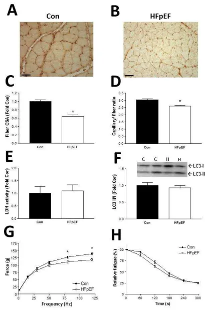
Figure legendsFig 1. Characterization of the glycolytic EDL muscle at 20 weeks of age in control (Con) and to
HFpEF rats, with representative muscle sections stained with an antibody against vWF to visualize
capillaries presented for control (A) and HFpEF (B) rats (scale bar represents 100 m). Mean fiber
cross-sectional area (CSA) was severely reduced in HFpEF compared to control rats (C), as was
the capillary-to-fiber ratio (D). As a marker of glycolytic metabolism, lactate dehydrogenase (LDH)
activity was assessed, which tended to be higher in HFpEF rats (E), while LC3 II/I protein levels
were assessed to provide an index of autophagy and these were depressed in HFpEF compared
to controls (F). In vitro muscle function of the EDL was also assessed and this was impaired in
HFpEF rats, with lower absolute forces generated compared to controls at higher stimulation
frequencies (G) while relative fatigue was not influenced (H). n = 8 for Con and n = 12 for
HFpEF. *P<0.05 vs. Con.
Fig 2. Characterization of the oxidative soleus muscle at 20 weeks of age in control (Con) and to
HFpEF rats, with representative muscle sections stained with an antibody against vWF to visualize
capillaries presented for control (A) and HFpEF (B) rats (scale bar represents 100 m). Mean fiber
cross-sectional area (CSA) was lower in HFpEF compared to control rats (C), as was
capillary-to-fiber ratio (D). While lactate dehydrogenase (LDH) activity (E) and LC3 II/I protein levels (F) were
not different between groups, in vitro absolute muscle forces were lower in HFpEF compared to
control rats (G) but relative fatigue remained unchanged (H). n = 8 for Con and n = 12 for
HFpEF. *P<0.05 vs. Con.
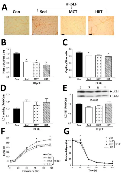
Fig 3. Characterization of the glycolytic EDL muscle at 28 weeks of age following exercise training
on a treadmill for the last 8 weeks, in control (Con) and HpEF rats that were assigned to either
sedentary, high-intensity interval training (HIIT), and moderate-continuous training (MCT) groups
for 8 weeks. Representative muscle sections (A) for each group are presented and have been
stained with an antibody against vWF to visualize capillaries (scale bar represents 100 m). Mean
A
CCE
P
T
E
D
M
A
N
U
S
CRIP
T
(B), as was the capillary-to-fiber ratio (C). Lactate dehydrogenase (LDH) was not different between
groups, (D), however the protein levels of the autophagy marker LC3 tended to be lower in the
HFpEF groups compared to controls (E). Absolute contractile forces were impaired in all HFpEF
groups compared to controls (F), while fatigability was also greater in the HFpEF groups compared
to controls early during the onset of repeated stimulations (G). *P<0.05 vs. Con.
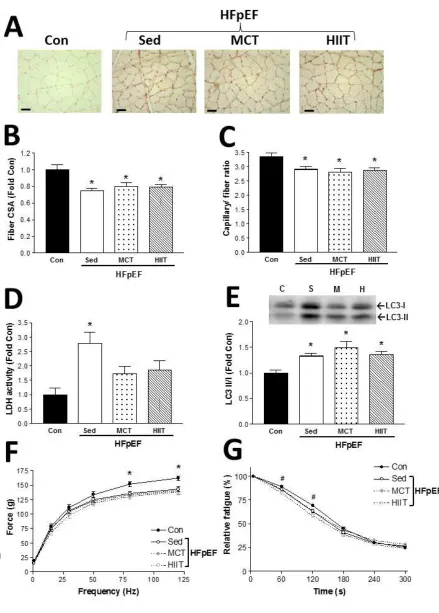
Fig 4. Characterization of the oxidative soleus muscle at 28 weeks of age following exercise
training on a treadmill for 8 weeks, in control (Con) and HpEF rats that were assigned to either
sedentary, high-intensity interval training (HIIT), and moderate-continuous training (MCT) groups.
Representative muscle sections (A) for each group are presented and have been stained with an
antibody against vWF to visualize capillaries (scale bar represents 100 m). Mean fiber cross
-sectional area (CSA) was lower in all HFpEF groups compared to control rats (B), as was the
capillary-to-fiber ratio (C). Lactate dehydrogenase (LDH) activity, as an index of reliance on
glycolytic metabolism, was elevated only in sedentary HFpEF rats compared to controls (D),
however autophagic flux, as measured by LC3 protein content, was increased in all the HFpEF
groups (E). While absolute muscle forces were impaired in all HFpEF groups at the higher
stimulation frequencies compared to control rats (F), fatigability was shown only to be greater in
A
CCE
P
T
E
D
M
A
N
U
S
CRIP
T
Figures