HIV
Thesis by Kenneth K. Yu
In Partial Fulfillment of the Requirements for the Degree of
Doctor of Philosophy
CALIFORNIA INSTITUTE OF TECHNOLOGY Pasadena, California
2012
2012
Kenneth Yu
ACKNOWLEDGEMENTS
Some people say that science is an intensely personal enterprise, bringing to mind the image of a scientist working alone long into the night, in a solo quest to make Nature yield its secrets. There is some truth to that image, but it’s far from the whole story. For me, I would have to say that science is an inherently social experience. It’s hard to engage in it without becoming connected to people, and having one’s self enriched by their presence. And thus it’s a tremendous honor for me to write this acknowledgement, to express, perhaps inadequately, my deepest gratitude to the people without whom this work would not have been possible.
I want to thank my parents for their unconditional love, and for always supporting me to pursue my dreams, even when sometimes they appear seemingly incoherent. Dad, thank you for being my inspiration—you are the first physician I got to know in my life, and you are the one who taught me that caring for and helping those around us is what gives meaning to our lives. And thank you for showing me the meaning of Hebrews 11:1. Mom, thank you for putting up with being separated from Dad across an ocean, so that we kids could pursue the American dream. And thank you for standing by me and encouraging me even when you did not understand the reasons for my choices. To you both I dedicate this thesis.
I want to thank my sister and brother, Karen and Kevin, for always believing in me, even at moments when I doubted myself. And thank you Kevin for introducing me to “Still Alive” and writing “Prepping Cells”.
I want to thank my first-grade science teacher, Dr. Yu-wen Zhuang. You first spotted in me the curiosity to ask questions about Nature, and nutured it by teaching me to do experiments to answer them, in the process leaving me more full of wonder than when I started. You inspired me to be a scientist.
I want to thank my high-school friend Morgan Gough, who took time off from work and came to take care of me when I nearly went blind in one eye in the second year of grad school.
And to the many wonderful and talented people that are the Baltimore Lab, thank you for sharing with me your excitement about science and showing me how to do it. Lili Yang, thank you for your critical input and discussions. Your creativity and discipline will always be an inspiration to me. Dinesh Rao, thank you for being a great bay-mate, friend, and role model. And I will always treasure the times we spent pondering questions about biology, or for that matter, the mysteries of life. Jonathan Tsai, thank you for being my first student. I enjoyed doing science with you as much as learning from you about the unique species known as the Caltech Undergrads. Kiefer Aguilar, thank you for being a student, teacher, and friend; science is best when shared with good company. Alex Balazs, thank you for your tips on how to do experiments, thoughtful discussions, and the pHAGE vectors. Your rationality is an inspiration. Ryan O’Connell, thank you for your help with the HIS mice. Dev Majumdar, thank you for keeping me on my toes! Your curiosity is impeccable. And thanks for sharing with me your optimism and
should do more trips to the Shakespeare Theatre! Alex Sigal, thanks for enlightening me with your sense of humor. Evgenij Raskatov, great business plan; let’s have fun with some more mice!
And to all the other members of the Baltimore family, Arnav Mehta, Jimmy Zhao, Geoffrey Lovely, Aadel Chaudhuri, Param Ramakrishnan, Chee-Kwee Ea, Shengli Hao, Mark Boldin, Konstantin Taganov, Alex So, Jocelyn Kim, Yvette Garcia, Vanessa Jonsson, Yang Yu, Claret Liu, Stella Ouyang, Christin Hong, Joyce Chen, Eric Santiestevan, Joanne Laurence, and Julie Kelly, thank you for making my experience here a memorable and enjoyable one. The lab wouldn’t be the same without you.
I want to thank the members of my committee for your support and insights. Dr. Ellen Rothenberg, thank you for sharing with me your love and passion for science. Your immunology class was a wonderful window into the process of science and discovery. Your excitement about science is contagious! Thank you for all the thoughtful
discussions we’ve had. Dr. Sarkis Mazmanian, thank you for your encouragement and discernment about what paths one should explore when doing science. Dr. Pamela
Bjorkman, thank you for your insight and support and making me feel as much a member of the Bjorkman lab as the Baltimore lab.
Last but not least, I want to thank my advisor Dr. David Baltimore, who is the ultimate source of my inspiration. You have assembled an environment for learning, doing, and experiencing science that is unparalleled. It could only be the embodiment and
for the lab, it was a little past 5 pm, the normal time they’d lock the doors to the
buildings. As we neared Braun’s east entrance, I started fumbling for my keys. Like a true experimentalist, you reached for the door handle and pulled the unlocked door open. With a smile on your face, you said to me, “Do the experiment!” To a theoretical
ABSTRACT
TABLE OF CONTENTS
Acknowledgements ... iii
Abstract ... iv
Table of Contents ...v
Chapter 1: Introduction ...1
AIDS at 30 ...1
Toward an AIDS Vaccine: The Broadly Neutralizing Antibodies ...5
Engineering Immunity Against HIV ...7
Overview of Thesis ...10
References ...12
Chapter 2: Use of Mutated “Self-Cleaving” 2A Peptides as “Molecular Rheostats” to Direct Simultaneous Formation of Membrane and Secreted Immunoglobulins ...14
Abstract ...14
Introduction ...15
Materials and Methods ...17
Results ...22
Discussion ...30
References ...44
Chapter 3: In Vivo Characterization of the Molecular Rheostat Immunoglobulins ...46
Materials and Methods ...47
Results and Discussion ...49
References ...63
Chapter 4: The Use of Non-Lymphoid Hematopoietic Cells for Antibody Production ...64
Introduction ...64
Materials and Methods ...65
Results and Discussion ...66
References ...77
Chapter 5: Lentiviral-Vector-Mediated-Broadly-Neutralizing Antibody Production from Muscle ...78
Introduction ...78
Materials and Methods ...79
Results and Discussion ...80
References ...92
Chapter 5: Looking Ahead ...98
Summary and Future Directions ...98
Concluding Remarks ...101
C
HAPTER1:
I
NTRODUCTIONAIDS at 30
The year 2011 marks the 30th anniversary of the first formal report of the disease that came to be known as AIDS (Acquired Immune-Deficiency Syndrome) caused by the human immunodeficiency virus (HIV). According to the most recent statistics available through UNAIDS, it is estimated that 34 million people globally were living with HIV/AIDS at the end of 2010, with 2.6 million new infections, and 1.8 million deaths directly attributed to AIDS (UNAIDS 2011). While the number of new infections has stabilized, the burden of the disease continues to grow globally.
would lose his mother, and whose own life was threatened by the disease. I never wished more fervently for a vaccine.
Here in the United States, the CDC estimates that 1.2 million people are living with HIV infection and over half a million people have died since the epidemic began (National Center for HIV/AIDS 2011). The annualized medical costs per HIV infection in the U.S. was estimated to be approximately $24,000 per person per year (Farnham, Holtgrave et al. 2010). These numbers do not begin to capture the magnitude of the human suffering caused by HIV/AIDS; they say nothing of the fear and stigma associated with the disease.
Toward an AIDS Vaccine: The Broadly Neutralizing Antibodies
Vaccines are antigen preparations that elicit immune responses against pathogens. The utility of vaccines is limited by the kinds of antibodies (Abs) that are made by the host immune system after vaccination. Most currently used anti-viral vaccines work by stimulating production of neutralizing antibodies (NAbs), which block viral infection (Zinkernagel, LaMarre et al. 2001; Burton 2002). HIV is an enveloped retrovirus that presents problems for antibody-based vaccine strategies. The virus rapidly mutates to change residues on its surface, sheds immunodominant decoy epitopes, masks
immunogenic sites on its surface with host-derived carbohydrates, and/or hides conserved regions in the interfaces of oligomeric proteins (Burton, Stanfield et al. 2005; Berkley and Koff 2007). While neutralizing antibodies against HIV do emerge in the natural course of infection, they occur too late, after an infection has already been established, and are thwarted by rapid genetic mutation of the virus (Richman, Wrin et al. 2003). For all of these reasons, it has been exceedingly difficult to design an immunogen that would elicit an anti-HIV antibody response of sufficient quality and breadth to be protective (Burton, Desrosiers et al. 2004; Flynn, Forthal et al. 2005; Pitisuttithum, Gilbert et al. 2006; Johnston and Fauci 2007; Fauci, Johnston et al. 2008).
Broadly neutralizing antibodies (bNAbs) against HIV do exist; they are an unusual class of antibodies that neutralize a broad range of HIV variants (Burton,
Engineering Immunity Against HIV
It occurred to Dr. Baltimore that if we could directly instruct the immune system to produce broadly neutralizing anti-HIV antibodies, we might be able to use them as “vaccines” to prevent HIV infections. Gene therapy technology provides the means for us to genetically program immune cells. Using gene therapy to deliver broadly neutralizing antibodies, we would be able to provide them to people as a prophylaxis against HIV infections. We call this approach “Engineering Immunity”.
rearrangement of endogenous heavy and light chain genes (allelic exclusion), and guides the ordered development of functional B cells with specificity defined by the transgene (Spanopoulou, Roman et al. 1994; Young, Ardman et al. 1994).
The mature B cells patrol the body in the general and lymphatic circulations, using their BCRs as antigen sensors. When a cognate antigen engages the BCR, the B cell becomes activated and enters into germinal center reactions in the lymph node or spleen in a dance of mutual activation with T cells; this process leads to further
development into memory B cells or differentiation into antibody-producing plasma cells. The memory B cells will provide a more rapid and higher quality antibody response in the future when the same antigens are encountered again. The plasma cells produce antibodies against the inciting antigens, which leads to their eventual clearance from the body (McHeyzer-Williams and McHeyzer-Williams 2005).
As B cells differentiate into plasma cells, they switch from producing the membrane-bound BCR to making a soluble, secreted antibody. The switch is
Some of the cells in the germinal center reactions also go through a process called isotype switching, in which the heavy chain constant regions of the initial IgM BCR are replaced with that of another isotype, which encodes different effector functions. This involves a DNA rearrangement mediated by the enzyme activation-induced cytidine deaminase (AID) (Muramatsu, Kinoshita et al. 2000). While the IgM BCR is required for the normal development of B cells in the bone marrow, and the IgM antibody is generally the first antibody isotype produced against an antigen, alternate isotypes provide additional effector functions that enhance the ability of the antibody to clear certain types of pathogens or to function in different body compartments. For example, in addition to fixing complements on target cells, as IgM antibodies can, IgG antibodies also have the ability to direct the killing of antibody-bound infected cells by engaging Fcγ
receptors on NK cells (termed ADCC, or antibody-dependent cell-mediated cytotoxicity). IgA antibodies are produced by plasma cells in mucosal areas and are transported across the epithelial barriers of the lung, gut, and genital tracts by binding the polymeric
immunoglobulin receptors (pIgR) with their Fc portions. These antibodies are critical in the defense of mucosal surfaces from pathogens.
Our understanding of the humoral immune response as summarized above forms the framework for my efforts to engineer the immune system. This framework suggests to us that by delivering a cleverly designed, synthetic immunoglobulin gene to the
the development of B cells that would produce broadly neutralizing antibodies against HIV with pre-programmed specificity and effector functions. Specifically, the synthetic immunoglobulin gene should 1) encode a mechanism that directs the production of both an IgM-like membrane-bound BCR and a secreted immunoglobulin isotype that has the desired effector properties, such as those of an IgG antibody, and 2) it should bind and neutralize HIV with the specificity and affinity of an anti-HIV broadly neutralizing antibody. Those were the two objectives that I set out to accomplish in my work when I joined Dr. Baltimore’s team in 2006, and this work will be described in detail below. As the project proceeds, I also explored a few other alternatives to this original approach by looking at cell types other than B cells as targets for engineering to produce anti-HIV antibodies, and the results obtained are summarized in separate chapters of this thesis.
Overview of Thesis
Chapter 1 gives a short, personal introduction to the HIV/AIDS epidemic and briefly reviews some significant scientific advances that have been made in the fight against HIV/AIDS. It then gives a succinct review of the broadly neutralizing antibodies against HIV and the biology of B cells that form the background of the Engineering Immunity project.
and characterization of this technology. I will show that the Molecular Rheostats provides a useful tool for manipulating B cell specificity and gives us the ability to program them to produce a bNAb against HIV.
Chapter 3 describes my attempt to use the Molecular Rheostats to program B cells
in vivo. It summarizes what we have learned from testing the system in both the
human-immune-system (HIS) mouse model and the murine bone marrow adoptive transfer model. We describe certain limitations of the lentiviral vector system we developed and suggest what we might do to overcome the limitations
In Chapters 4 and 5 I pivot to look at alternative approaches to make broadly neutralizing antibodies in vivo. Chapter 4 describes the use of retroviral vectors to program non-lymphoid hematopoietic cells to produce antibody long-term. Chapter 5 describes my effort to study the feasibility of using lentiviral vectors to program muscle.
References
Armbruster, C., G. M. Stiegler, et al. (2002). "A phase I trial with two human monoclonal antibodies (hMAb 2F5, 2G12) against HIV-1." AIDS 16(2): 227-233.
Baltimore, D. (2002). "Steering a course to an AIDS vaccine." Science 296(5577): 2297. Barre-Sinoussi, F., J. C. Chermann, et al. (1983). "Isolation of a T-lymphotropic
retrovirus from a patient at risk for acquired immune deficiency syndrome (AIDS)." Science 220(4599): 868-871.
Berkley, S. F. and W. C. Koff (2007). "Scientific and policy challenges to development of an AIDS vaccine." Lancet 370(9581): 94-101.
Burrows, P. D. and M. D. Cooper (1993). "B-cell development in man." Curr Opin Immunol 5(2): 201-206.
Burton, D. R. (2002). "Antibodies, viruses and vaccines." Nat Rev Immunol 2(9): 706-713.
Burton, D. R., R. C. Desrosiers, et al. (2004). "HIV vaccine design and the neutralizing antibody problem." Nat Immunol 5(3): 233-236.
Burton, D. R., R. L. Stanfield, et al. (2005). "Antibody vs. HIV in a clash of evolutionary titans." Proc Natl Acad Sci U S A 102(42): 14943-14948.
Cavacini, L. A., M. H. Samore, et al. (1998). "Phase I study of a human monoclonal antibody directed against the CD4-binding site of HIV type 1 glycoprotein 120." AIDS Res Hum Retroviruses 14(7): 545-550.
Chen, J. and F. W. Alt (1993). "Gene rearrangement and B-cell development." Curr Opin Immunol 5(2): 194-200.
Farnham, P. G., D. R. Holtgrave, et al. (2010). "Medical costs averted by HIV prevention efforts in the United States, 1991-2006." J Acquir Immune Defic Syndr 54(5): 565-567.
Fauci, A. S., M. I. Johnston, et al. (2008). "HIV vaccine research: the way forward." Science 321(5888): 530-532.
Ferrantelli, F., R. A. Rasmussen, et al. (2002). "Do not underestimate the power of antibodies—lessons from adoptive transfer of antibodies against HIV." Vaccine 20 Suppl 4: A61-65.
Flynn, N. M., D. N. Forthal, et al. (2005). "Placebo-controlled phase 3 trial of a
recombinant glycoprotein 120 vaccine to prevent HIV-1 infection." J Infect Dis 191(5): 654-665.
Gallo, R. C., P. S. Sarin, et al. (1983). "Isolation of human T-cell leukemia virus in acquired immune deficiency syndrome (AIDS)." Science 220(4599): 865-867. Johnston, M. I. and A. S. Fauci (2007). "An HIV vaccine--evolving concepts." N Engl J
Med 356(20): 2073-2081.
Mascola, J. R. (2002). "Passive transfer studies to elucidate the role of antibody-mediated protection against HIV-1." Vaccine 20(15): 1922-1925.
McHeyzer-Williams, L. J. and M. G. McHeyzer-Williams (2005). "Antigen-specific memory B cell development." Annu Rev Immunol 23: 487-513.
Muramatsu, M., K. Kinoshita, et al. (2000). "Class switch recombination and
hypermutation require activation-induced cytidine deaminase (AID), a potential RNA editing enzyme." Cell 102(5): 553-563.
National Center for HIV/AIDS, V. H., STD, and TB Prevention (CDC) (2011). "HIV in the United States."
Peterson, M. L. (2007). "Mechanisms controlling production of membrane and secreted immunoglobulin during B cell development." Immunol Res 37(1): 33-46. Peterson, M. L., E. R. Gimmi, et al. (1991). "The developmentally regulated shift from
membrane to secreted mu mRNA production is accompanied by an increase in cleavage-polyadenylation efficiency but no measurable change in splicing efficiency." Mol Cell Biol 11(4): 2324-2327.
Pilgrim, A. K., G. Pantaleo, et al. (1997). "Neutralizing antibody responses to human immunodeficiency virus type 1 in primary infection and
long-term-nonprogressive infection." J Infect Dis 176(4): 924-932.
Pitisuttithum, P., P. Gilbert, et al. (2006). "Randomized, double-blind, placebo-controlled efficacy trial of a bivalent recombinant glycoprotein 120 HIV-1 vaccine among injection drug users in Bangkok, Thailand." J Infect Dis 194(12): 1661-1671. Richman, D. D., T. Wrin, et al. (2003). "Rapid evolution of the neutralizing antibody
response to HIV type 1 infection." Proc Natl Acad Sci U S A 100(7): 4144-4149. Spanopoulou, E., C. A. Roman, et al. (1994). "Functional immunoglobulin transgenes
guide ordered B-cell differentiation in Rag-1-deficient mice." Genes Dev 8(9): 1030-1042.
Stiegler, G., C. Armbruster, et al. (2002). "Antiviral activity of the neutralizing antibodies 2F5 and 2G12 in asymptomatic HIV-1-infected humans: a phase I evaluation." Aids 16(15): 2019-2025.
UNAIDS (2011). AIDS at 30: Nations at Crossroads.
Wolfe, E. J., L. A. Cavacini, et al. (1996). "Pharmacokinetics of F105, a human monoclonal antibody, in persons infected with human immunodeficiency virus type 1." Clin Pharmacol Ther 59(6): 662-667.
Xiao, Y., X. Dong, et al. (2002). "Neutralizing antibodies mechanism of neutralization and protective activity against HIV-1." Immunol Res 25(3): 193-200.
Young, F., B. Ardman, et al. (1994). "Influence of immunoglobulin heavy- and light-chain expression on B-cell differentiation." Genes Dev 8(9): 1043-1057. Zinkernagel, R. M., A. LaMarre, et al. (2001). "Neutralizing antiviral antibody
CHAPTER 2:USE OF MUTATED “SELF-CLEAVING”2APEPTIDES AS “MOLECULAR
RHEOSTATS” TO DIRECT SIMULTANEOUS FORMAION OF MEMBRANE AND SECRETED
IMMUNOGLOBULINS
Abstract
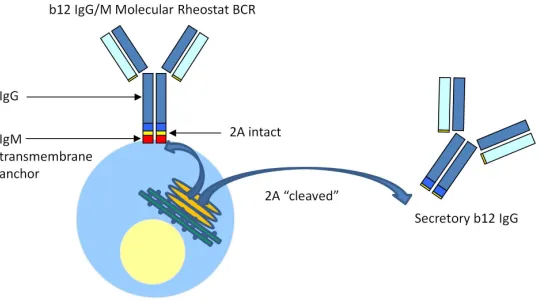
In nature, B cells produce surface immunoglobulin and secreted antibody from the same immunoglobulin gene via alternative splicing of the pre-messenger RNA. Here we present a novel system for genetically programming B cells to direct the simultaneous formation of membrane-bound and secreted immunoglobulins that we term a “Molecular Rheostat” Immunoglobulin gene, based on the use of mutated “self-cleaving” 2A
peptides. The Molecular Rheostats are designed so that the ratio of secreted to membrane-bound immunoglobulins can be controlled. Lentiviral transgenesis of the Molecular Rheostat constructs into B cell lines enables the expression of functional b12-based BCRs that signal to the cells and mediate the secretion of b12 IgG broadly
Introduction
B cells are responsible for the production of antibodies in response to foreign antigens. The ability to manipulate the antigen specificity of B cells and that of the antibody produced by these cells could be useful for achieving immunization against deadly pathogens such as HIV. In this chapter, I describe a novel way of programming B cells by using mutated 2A peptides to direct the simultaneous formation of an IgM-like BCR and IgG antibody. The system is designed so that the ratio of surface-to-secreted immunoglobulins can be controlled by appropriate choice of mutations. We call this system a “Molecular Rheostat” for immunoglobulin gene expression.
B cells begin their life in the bone marrow as descendants of the more primitive common hematopoietic stem and progenitor cells. As these cells develop into B cells, they undergo sequential RAG1/2-mediated DNA rearrangement of the heavy and light chain immunoglobulin gene loci in a process called V(D)J rearrangment. Cells that successfully complete this process and assemble a functional B cell receptor (BCR) of the IgM isotype on their surface are able to leave the bone marrow to continue further
IgM heavy chain and light chain transgene shuts down the rearrangement of endogenous heavy and light chain genes (allelic exclusion), and guides the ordered development of functional B cells with specificity defined by the transgene (Spanopoulou, Roman et al. 1994; Young, Ardman et al. 1994). See Chapter 1, pp. 7-9, for more details on the process of B cell development and developmentally regulated switch from membrane to secreted Ig production.
2A peptides are “self-cleaving” peptides that are derived from animal viruses and multicellular parasites of mammals (de Felipe 2004; Szymczak and Vignali 2005). They are involved in the processing and expression of polyproteins. Mechanistically, these peptides do not really undergo a “self-cleaving” event in the sense of breaking a
pre-existing peptide bond; rather the presence of the 2A element in the mRNA causes the
stop-and-restart event. Several 2A peptides appear to have near 100% cleavage efficiency in their native contexts, but they can be made to cleave at lower efficiencies when they are mutated at key amino acid residues or introduced into non-native sequences (Ryan and Drew 1994; Donnelly, Hughes et al. 2001; Donnelly, Luke et al. 2001). By engineering the peptides with reduced efficiency of cleavage, we show that we can co-express the BCR and antibody molecule simultaneously. We will call the system a “Molecular Rheostat” for immunoglobulin genes.
Materials and Methods
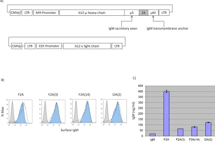
Constructs
The Molecular Rheostat constructs were created by cloning a transgene containing the EEK promoter, the b12 light and heavy chains, the 2A sequences, and and the 3’ region of the human IgM BCR gene corresponding to the last 41 amino acids into either a pHAGE2 or pHAGE6 vector system. The Igα and Igβ genes were cloned into a FUW vector.
Transfections
293T cells were grown to 50–75% confluence on 30 cm dishes and were transfected in 15 ml D10 media (DMEM plus 10% heat-inactivated fetal bovine serum, supplemented with 20 mM L-glutamine, 1000 IU/ml penicillin, and 1000 µg/ml streptomycin, filtered
TransIT-293 reagent (Mirus Bio, Madison WI) or BioT (Bioland Scientific, Paramount CA) according to manufacturer's instructions using a total of 40 µ g DNA.
Lentiviral Vector Production
293T cells were transfected with lentiviral vectors. After 24 h of incubation, the supernatant was pipetted off the cells and filtered through a 0.22 µm PES membrane bottle-top filter into a collection bottle. 15 ml of fresh D10 media was then filtered through the bottle-top filter into the collection bottle to reduce virus waste from
supernatant that the filter absorbed. The collected supernatant was stored at 4⁰C, and 30
ml of fresh D10 media was added to the dish. This collection process into the same collection bottle was repeated 4 to 5 additional times at 12 h intervals. All of the
collected supernatant was centrifuged at 10000 rpm for 12–24 h at 4⁰C to pellet the virus,
and the supernatant was poured off the pellet. The pellet was re-suspended in 500– 1000µL DMEM media (for 293T transductions) or RPMI media 1640 (for OCI-Ly7 or
EU12 transductions) and incubated on ice at 4⁰C for 12 h.
Lentiviral Transductions
OCI-Ly7 or EU12 transductions in 12 well plates, and 400–600µL of virus re-suspensions or dilutions thereof was added to each well. 10 mg/mL polybrene (Millipore, Billerica, MA) was added so that the final polybrene concentration was 10 µg/mL in each well. The transductions were incubated for 24 h before the cells were passaged.
Cell Line
The 293T-Igα/β cell line was created from a vector carrying the Igα and Igβ genes using the transfection, lentiviral production, and lentiviral transduction procedures above.
Tissue Culture
293T and 293T Ig-αβ cells were grown in D10 media. The cells were passaged 1:5 every other days. OCI-Ly7 and EU12 cells were grown in C10 media. The cells were passaged 1:5–1:10 every other day to maintain a density between 105–106 cells/ml.
Flow Cytometry
Cell Sorting
Cells were prepared as in flow cytometric analysis and were sorted with the assistance of Sylvia Chavira at the University of Southern California’s Clinical Pathology Laboratory using a MoFlo FACS cell sorter.
Calcium Flux Assay
Calcium flux measurements were made essentially using the protocol described by Bondada, et al. [29], with the following modifications: cells were washed, pelleted, and resuspended in dye loading buffer (HBSS with Ca2+ and Mg2+ plus 4% 100mM
probenecid, 2% 1 M HEPES buffer, and 1% heat-inactivated fetal bovine serum) and were incubated with 4 µg/mL Fluo-3 AM and 1 µg/mL FuraRed AM dyes in the presence of 0.02% (w/v) pluronic F-127 for 30 m. The cells were again washed, pelleted, and resuspended in dye loading buffer and were kept at room temperature until they were analyzed on a BD FACSCalibur flow cytometer equipped with a circulating 37⁰C water
bath on the sample port. During analysis, cells were stimulated with goat F(ab’)2 anti-human IgG γ Fc-specific antibodies (Invitrogen, Carlsbad, CA) or with goat F(ab’)2 anti-human IgM µ Fc-specific antibodies (Southern Biotech, Birmingham, AL) and a
ELISA
Supernatants from cultured cells were analyzed using Human IgG ELISA Quantitation Set (Bethyl Laboratories, Montgomery, TX) according to manufacturer’s instructions.
Biacore Binding Assay
Biacore binding assays were performed as previously described by Klein et al. (2009), with the following modifications: All experiments were done in-house. b12 antibody supernatants were produced from transfection of 293T cells.
In Vitro Neutralization Assay
In vitro neutralization assays were performed as previously described by West et al. [30],
Results
IgM Molecular Rheostat Immunoglobulin Genes Mediate Co-Expression of IgM-Like
BCR and Secreted IgM Antibody
As a pilot experiment to test whether the mutated 2A peptides can mediate co-expression of surface and secreted immunoglobulins, we constructed the first-generation Molecular Rheostat Immunoglobulin genes by joining the secreted version of the b12 IgM heavy chain to the transmembrane domain of the IgM BCR via a mutated 2A peptide. The transmembrane domain is defined as the M1 and M2 exons from the human IgM locus and comprises the last 41 amino acids of the membrane bound IgM BCR (Figure 2.1A). We call these “IgM Molecular Rheostats”. We chose the wild type F2A and two mutant peptides as well as another F2A-like element derived from a silk-worm virus, based on previous work by Donnelly et al. (Donnelly, Hughes et al. 2001), in which they observed reduced cleavage efficiencies when certain mutations are introduced. The four mutants we chose are designated F2A, F2A(3), F2A(14), and I2A(2). See Table 2.1 for the nomenclature and the amino acid sequence for each of the 2A elements.
light chain (FEEK-b12L) and a mammalian expression vector carrying the human Igα
and Igβ genes (phIgαβ) into 293T cells (Figure 2.1A). We analyzed the cells and their supernatants by FACS and human IgM ELISA 48 hours later. All transfected cells showed surface expression of the IgM Molecular Rheostat BCR and secreted IgM into their supernatants (Figure 2.1B and 2.1C).
IgG Molecular Rheostat Mediates Expression of an IgG/M Chimeric BCR and Secreted
IgG Antibody
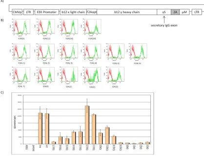
We next attempted to adapt the Molecular Rheostat format to the production of an IgG antibody in an effort to mimic an isotype-switched secretory IgG while preserving the signaling properties of an IgM, which is required for normal B cell development. Furthermore, we wished to explore whether we could manipulate the ratio of surface-bound to secreted immunoglobulins by making appropriate mutations in the 2A elements. To test these ideas, we constructed a library of chimeric IgG Molecular Rheostat
immunoglobulins, in which a complete secretory b12 IgG is joined to the transmembrane anchor of the IgM BCR via different 2A peptides (Figure 2.2). The library includes all 2A peptides listed in Table 2.1.
To reduce the number of vectors that need to be transfected and anticipating the need to use the vectors in the context of lentiviral transduction, where it would be advantageous to work with a single vector, we fused the b12 light chain with the
different F2A element, F2Aopt. F2Aopt is codon-optimized for human expression and contains a furin cleavage site before the 2A element.
Additionally, to ensure consistency of Igα and Igβ expression across the cells used to test the Molecular Rheostat constructs and reduce the number of vectors that need to be transfected, we engineered 293T cells that express human Igα and Igβ by repeatedly co-infecting 293T cells with two lentiviral vectors, FUW-Igα and FUW- Igβ, which carry the Igα and Igβ transgenes, repectively, under the control of a ubiquitin C promoter. The resulting cells are denoted 293T-Igαβ cells.
IgG Molecular Rheostat Mediates Expression of a Range of Ratios of Surface BCR to
Secretory IgG in the Human B-Cell Line OCI-Ly7
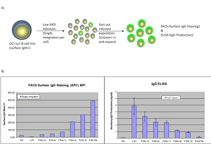
To validate the results that the IgG/M Molecular Rheostat constructs can mediatea range of expression ratios of surface BCR to secreted antibodies in human B cells, we used lentiviral vectors to deliver the constructs into the OCI-Ly7 B cell line, which expresses an endogenous IgM BCR on its surface and therefore should possess the necessary machinery (such as Igα and Igβ co-receptors) for BCR surface expression. To provide an independent marker of lentiviral transduction than the expression of the Molecular Rheostat immunoglobulins, we constructed a lentiviral vector, pHAGE2-EEK-IRES-ZsGreen, which contains an Internal Ribosomal Entry Site (IRES) driving a
ZsGreen fluorescent protein gene. Based on the results in Figure 2.2B, we selected six of the IgG/M Molecular Rheostat genes and cloned them into the first position (before the IRES-ZsGreen) of the pHAGE2-EEK-IRES-ZsGreen vector. We then infected OCI-Ly7 cells with the IgG Molecular Rheostat vectors at low MOI (~ 0.1) to ensure that every cell that was infected had at most one copy of the transgene (Figure 2.3A). 48 hours after infection, we FACS-sorted out the ZsGreen positive cells and allowed these cells to expand for another 48 hours.
between the amount of IgG Molecular Rheostat BCR expressed on the surface of the cells vs. the amount of IgG antibody that was detected in the supernatants, indicating that the mutant 2A elements were behaving like a “rheostat”, tuning the ratios of secreted immunoglobulins. Also notably, the rank order of the ratios of surface-to-secreted immunoglobulin expression recapitulates what was observed from the
transfection into 293T-Igαβ cells (see Figure 2.2B and C). For example, from Figure 2.2B and C, F2A(-2) would be expected to make more secreted IgG than F2A(-4), and this was indeed the case when the constructs were expressed in the OCI-Ly7 B cell line.
Furthermore, F2A(-2) made less surface Molecular Rheostat BCR than F2A(-4), as would be expected if the F2A(-2) peptide mediated more efficient cleavage than the F2A(-4) peptide. The library of mutants thus gives us a Molecular Rheostat system that we can use to direct tunable ratios of expression of surface vs. secreted immunoglobulins.
IgG Molecular Rheostat Constructs Produce Functional b12 IgG/M Chimeric BCRs are
Signaling Competent and Bind to HIV GP120
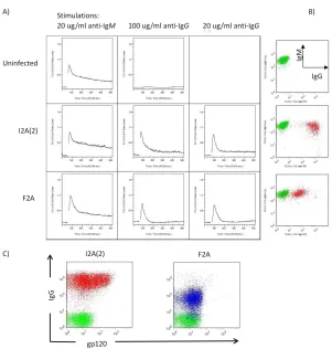
As the ZsGreen protein interferes with the Fluo-3 calcium-sensitive dye used in the assay, we cloned those two IgG Molecular Rheostat immunoglobulins into lentiviral vectors that do not have the IRES-ZsGreen marker gene. Lentiviral infections of OCI-Ly7 B cells with these vectors resulted in a variegated pattern of expression of the BCRs. The vector containing the I2A(2) element showed generally higher levels of surface BCR expression than F2A, as expected. While both populations responded to BCR stimulation using a control anti-IgM antibody (Southern Biotech, Birmingham, AB) and an anti-IgG antibody (Sigma, St Louis, MO), the responses were detectable but modest (data not shown). We believe the modest response was due to the effect of averaging the calcium signals over the large range of surface expressions. To ensure we have more homogenous populations for use in BCR stimulations, we FACS sorted out the top 10% of IgG positive cells from each of the populations (Figure 2.4B), and performed the calcium flux assays on the sorted cells. The cells responded robustly to anti-BCR stimulation (Figure 2.4A), with a dose-response correlating with the levels of surface IgG Molecular Rheostat BCR expression and the concentrations of anti-Ig used. The higher anti-IgG dose (100 ug/ml) gives a stronger calcium signal than the lower dose (20 ug/ml); the higher amount of surface Molecular Rheostat BCR also generates a stronger and more lasting response.
chain constant region of b12 IgG, respectively (Figure 2.4C). We found that the Molecular Rheostat BCRs on the cells bound to HIV GP120.
IgG Molecular Rheostat Constructs Produce b12 IgG Antibody that Neutralizes HIV
Pseudovirus with Same Potency as Unmodified b12 IgG
To determine whether secreted b12 IgG from the Molecular Rheostat system can neutralize infectious virus, we performed an in vitro pseudovirus neutralization assay using an Env SF162 pseudotyped HIV-1 pseudovirus on the TMZ-b1 reporter cell line with supernatants from 293T cells transfected with several different IgG Molecular Rheostat constructs according to a protocol previously described by Klein et al. (Klein, Gnanapragasam et al. 2009). The neutralization curves demonstrated that secreted Molecular Rheostat b12 IgG antibodies neutralized the Env SF162 pseudovirus as potently as the control b12 IgG antibody (L+H), with IC50 values nearly identical to that of the control b12 IgG (Figure 2.5A). We also performed a surface-plasmon resonance GP120-binding assay. The antibodies tested bound GP120 as well as the control b12 IgG antibody, consistent with the neutralization assay results (Figure 2.5B).
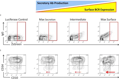
Expression of IgG Molecular Rheostat Immunoglobulins Promote Maturation of EU12
Cells in an In Vitro Model of B Cell Development
Molecular Rheostat Immunoglobulin BCR can direct B cell development, we adopted a model of human B cell development using the EU12 system (Zhang, Wang et al. 2003; Zhang 2007). The EU12 cells are derived from a B cell leukemia patient, and the cells are CD19+ and exist in a spectrum of primitive (CD34+ and CD10-, or CD34+ and CD10+) to more mature (CD34- and CD10+, or CD34- and CD10-) states. These cells lack a
functional BCR, but rarely an IgM BCR is generated spontaneously and the cells proceed to acquire a more mature phenotype.
We isolated early-stage, CD 34+ EU12 cells by FACS sorting. These cells were then transduced with lentiviral vectors carrying IgG Molecular Rheostats that give rise to respectively low, intermediate, and high surface BCR expression. A luciferase-carrying vector was used as a control. The cells were allowed to expand, and 4 weeks after transduction the surface expression of IgG Molecular Rheostat BCR and maturation markers were analyzed by FACS (Figure 2.6). The EU12 cells transduced with Molecular Rheostat constructs tuned for different levels of surface BCR vs. secreted antibody
Discussion
To provide a compact system for genetically manipulating the BCR and antibody specificity of B cells with a lentiviral vector, we created the Molecular Rheostat
Immunoglobulins to direct tunable simultaneous formation of the membrane-bound and secreted immunoglobulins by using mutant 2A “self-cleaving” peptides (Figure 2.7).
This system provides a synthetic approximation to the natural process of the mRNA alternative splicing-mediated switch to make membrane and secreted
immunoglobulins. By fusing an IgG to the membrane anchor of IgM through a mutant 2A peptide that functions as a Molecular Rheostat, we constructed both IgM and IgG/M chimeric versions of Molecular Rheostat immunoglobulins. We showed that such a design could produce both membrane bound and secreted immunoglobulins and
forms of gp120 and possibly HIV spike complexes. Finally, we provided evidence suggesting that the chimeric BCR produced by the Molecular Rheostat system can direct maturation of B cells using a cell line model of B cell maturation. In EU12 cells
transduced with vectors carrying the Molecular Rheostat Immunoglobulins, we observed increasing CD10-/CD 34- populations in the cells that received increasingly more surface-biased Molecular Rheostat constructs, suggesting that the chimeric IgG Molecular Rheostat BCRs are capable of directing the normal B cell maturation progression from CD10-/CD34+ to CD10+/CD34+ to CD10+/CD34- to CD10-/CD34-. We note, however, that the CD10-/CD34+ populations were also greater in cells that were treated with Molecular Rheostat immunoglobulins biased toward higher surface BCR expression. At first glance, this might be explained by the downregulation of CD10 alone as a result of the expression of chimeric BCR. However, that the ratio of the most mature CD10-, CD34- double negative population to the most primitive CD10-,CD34+ population also increases with the use of surface-baised Molecular Rheostat immunoglobulins suggests that the chimeric Molecular Rheostat BCR gives the more mature cells a proliferative advantage over the more primitive cells. This is consistent with the hypothesis that the Molecular Rheostat immunoglobulin genes promoted maturation of the cells.
earlier to remove the introns of the heavy chain locus, except for the one required for the alternative splicing of the secreted and transmembrane exons. Our efforts to get those constructs to splice were not successful. We thus created the Molecular Rheostat system to mimic the natural system, incorporating the additional feature of expressing isotype-switched IgG antibodies while maintaining the signaling properties of an IgM
Figure 2.4 Molecular Rheostat BCRs generate calcium signals in response to anti-BCR
stimulations and bind to HIV gp120. A) Calcium response of cells to anti-BCR stimulation. First column: response of endogenous IgM BCR to anti-IgM stimulation. Second column: high dose (100 ug/ml) anti-IgG stimulation. Third column: low dose (20 ug/ml) anti-IgG stimulation. B) BCR expression post-sorting. Endogenous IgM
and Blue: I2A(2) and F2A Molecular Rheostat Immunoglobulin vector transduced cells, repectively. Green: untransduced control cells.
2A
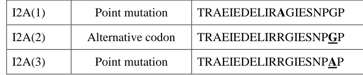
Mutant Mutation Type Amino Acid Sequence
F2A Wild-type QLLNFDLLKLAGDVESNPGP
F2A(-7) 7aa N-terminal deletion
LKLAGDVESNPGP
F2A(-6) 6aa N-terminal deletion
LLKLAGDVESNPGP
F2A(-5) 5aa N-terminal deletion
DLLKLAGDVESNPGP
F2A(-4) 4aa N-terminal deletion
FDLLKLAGDVESNPGP
F2A(-3) 3aa N-terminal deletion
NFDLLKLAGDVESNPGP
F2A(-2) 2aa N-terminal deletion
LNFDLLKLAGDVESNPGP
F2A(-1) 1aa N-terminal deletion
LLNFDLLKLAGDVESNPGP
F2A(3) Point mutation QLLNFDLLKLAGDVQSNPGP F2A(11) Point mutation QLLNFDLLKLAGDVEINPGP F2A(14) Point mutation QLLNFDLLKLAGDVESEPGP F2A(19) Point mutation QLLNFDLLKLAGDVESNPAP
Figure 2.1 A) Schematic representation of the IgM Molecular Rheostat
Figure 2.2 A) Schematic representation of the IgG/M Molecular Rheostat constructs.
2A: location of mutant self-cleaving 2A elements. 2Aopt: optimized 2A element with a furin cleavage site at 5’ end. CMVp: CMV promoter. LTR: long terminal repeat. EEK: internal B cell specific promoter. b12 γ heavy chain: IgG heavy chain with the variable region corresponding to that of the b12 broadly neutralizing antibody. B) Surface staining for human IgG. Green: Molecular Rheostat Contructs. Red: Secretory IgG (L+H)
Figure 2.3 A) Experimental design for measuring the ratioed expression of
Figure 2.5 Secreted b12 IgG from the Molecular Rheostat constructs neutralized Env
SF162 pseudovirus and bound to GP120 as well as control b12 IgG. A) In vitro
Figure 2.6 Molecular Rheostat BCRs promote maturation of EU12 cells. A) CD 34+
Figure 2.7. A model of how the b12 IgG Molecular Rheostat immunoglobulin system
References
Burrows, P. D. and M. D. Cooper (1993). "B-cell development in man." Curr Opin Immunol 5(2): 201-206.
Chen, J. and F. W. Alt (1993). "Gene rearrangement and B-cell development." Curr Opin Immunol 5(2): 194-200.
de Felipe, P. (2004). "Skipping the co-expression problem: the new 2A "CHYSEL" technology." Genet Vaccines Ther 2(1): 13.
de Felipe, P., L. E. Hughes, et al. (2003). "Co-translational, intraribosomal cleavage of polypeptides by the foot-and-mouth disease virus 2A peptide." J Biol Chem 278(13): 11441-11448.
Donnelly, M. L., L. E. Hughes, et al. (2001). "The 'cleavage' activities of foot-and-mouth disease virus 2A site-directed mutants and naturally occurring '2A-like'
sequences." J Gen Virol 82(Pt 5): 1027-1041.
Donnelly, M. L., G. Luke, et al. (2001). "Analysis of the aphthovirus 2A/2B polyprotein 'cleavage' mechanism indicates not a proteolytic reaction, but a novel translational effect: a putative ribosomal 'skip'." J Gen Virol 82(Pt 5): 1013-1025.
Doronina, V. A., P. de Felipe, et al. (2008). "Dissection of a co-translational nascent chain separation event." Biochem Soc Trans 36(Pt 4): 712-716.
Doronina, V. A., C. Wu, et al. (2008). "Site-specific release of nascent chains from ribosomes at a sense codon." Mol Cell Biol 28(13): 4227-4239.
Kitamura, D. and K. Rajewsky (1992). "Targeted disruption of mu chain membrane exon causes loss of heavy-chain allelic exclusion." Nature 356(6365): 154-156.
Kitamura, D., J. Roes, et al. (1991). "A B cell-deficient mouse by targeted disruption of the membrane exon of the immunoglobulin mu chain gene." Nature 350(6317): 423-426.
Klein, J. S., P. N. Gnanapragasam, et al. (2009). "Examination of the contributions of size and avidity to the neutralization mechanisms of the anti-HIV antibodies b12 and 4E10." Proc Natl Acad Sci U S A 106(18): 7385-7390.
Ryan, M. D. and J. Drew (1994). "Foot-and-mouth disease virus 2A oligopeptide mediated cleavage of an artificial polyprotein." EMBO J 13(4): 928-933. Spanopoulou, E., C. A. Roman, et al. (1994). "Functional immunoglobulin transgenes
guide ordered B-cell differentiation in Rag-1-deficient mice." Genes Dev 8(9): 1030-1042.
Szymczak, A. L. and D. A. Vignali (2005). "Development of 2A peptide-based strategies in the design of multicistronic vectors." Expert Opin Biol Ther 5(5): 627-638. Wagner, S. D., G. T. Williams, et al. (1994). "Antibodies generated from human
immunoglobulin miniloci in transgenic mice." Nucleic Acids Res 22(8): 1389-1393.
Zhang, Z. (2007). "VH replacement in mice and humans." Trends Immunol 28(3): 132-137.
CHAPTER 3:IN VIVO CHARACTERIZATION OF THE MOLECULAR RHEOSTAT
IMMUNOGLOBULINS
Introduction
Materials and Methods
Lentiviral Vector Constructs and Viral Vector Production
The constructs we used were described in detail in Chapter 2. 293T cells were
transfected with lentiviral vectors. After 24 h of incubation, the supernatant was pipetted off the cells and filtered through a 0.22 µm PES membrane bottle-top filter into a
collection bottle. 15 ml of fresh D10 media was then filtered through the bottle-top filter into the collection bottle to reduce virus waste from supernatant that the filter absorbed. The collected supernatant was stored at 4⁰C, and 30 ml of fresh D10 media was added to
the dish. This collection process into the same collection bottle was repeated 4 to 5 additional times at 12 h intervals. All of the collected supernatant was centrifuged at 25,000g for 90 mins at 4⁰C to pellet the virus, and the supernatant was aspirated. The
pellet was re-suspended in 500–1000µL DMEM media (for 293T transductions) or RPMI media 1640 (for OCI-Ly7 or EU12 transductions) and incubated on ice at 4⁰C for 12 h.
The resuspended vectors were aliquoted and frozen at -80°C until use.
Tranduction of CD34+ Cells
Transduction of CD34+ cells was performed using a modified protocol described by Luo et al. (Luo, Maarschalk et al. 2009). Briefly, The CD34+ cells were cultured for 24 hours before transduction in StemSpan media (Stemcell Technologies, Vancouver, BC,
SCF and 20 ng/ml TPO (from R&D Systems, Mineapolis, MN), and 25 ng/ml Flt3 ligand (from eBioscience, San Diego, CA). Spin-infection was performed in Retronectin-coated 24-well plates (~4000 cells per well) for each construct with 1 ml/well of StemSpan media with the human cytokines at an MOI of between 50–100 for the three constructs, at 2500 rpm at 30°C for 90 mins. The cells were washed once with StemSpan media and resuspended in 250 ul of PBS and kept on ice until injection.
CD34+ Cell Transplantation
Newborn pups were given 300 rads of radiation from a Cs137 irradiator. The human cells were resuspended in sterile PBS and injected intrahepatically by using a 27 gauge needle in 50 ul of PBS per mouse.
Transduction of Murine Bone Marrow Cells and Transplantation of Transduced Cells
Transduction of murine bone marrow cells and transplantation of the transduced cells were carried as described by Yang et al. (Yang and Baltimore 2005). Briefly, we
recepient mice one day after the last spin-infection. The Rag1 mice were pre-conditioned with 1100 rads of radiation from a Cs137 source.
Mouse Serum Collection and Human IgG ELISA
75 ul of blood was collected from each mouse using a heparin-coated microcapillary tube by retro-orbital bleeding and transferred into a microcentrifuge tube and kept on ice. The blood was then incubated at 37°C for 30 mins, and then spun down at 1150g at 4°C. The serum was collected from the top of the tube. Sera were analyzed using Human IgG ELISA Quantitation Kit (Bethyl Laboratories Inc., Montgomery, TX) according to manufacturer’s instructions.
Results and Discussion
Using Human-Immune System Mice to Study Human B Cell Development under the
Influence of the Molecular Rheostat Immunoglobulin Genes
centrifugation process I developed in which the viral pellet obtained from a previous stage of centrifugation was combined with the supernatant from the next stage and re-spun.
The supernatants were spun at 10,000 rpm for 8 hours at 4°C. The total volume of supernatant processed for each lentiviral vector was approximately 1.5 L. The viral pellet from the final spin was resuspended in approximately 500 uL of the residual media in the centrifuge bottle after pouring off the supernatant. The vectors were aliquoted in 50 ul volumes and flash-frozen in an ethanol/dry ice mixture and stored at -80°C. The resulting vectors were titered on EU12 cells. The titers were on the order of 108 TU/ml (1.32, 4.59, and 3.22 × 10⁸ TU/ml for F2A(-4), F2A(11), and luciferase control,
respectively).
Frozen CD34+ cord blood cells were purchased from Lonza (Lonza Walkersville Inc., Walkersville, MD). We transduced 100,000 CD34+ cells for each experimental group (no vector control, F2A(-4), F2A(11), and luciferase), which will be injected into four Rag2/γC knockout pups per group. The newborn pups were given 300 rads of
radiation from a Cs137 irradiator and the human cells were injected intrahepatically by Dr. Ryan O’Connell using a 27 gauge needle. We lost one group of pups (F2A(11)) as the box containing them fell off a cart used to transport them and the pups died. The remaining groups were placed with their mothers.
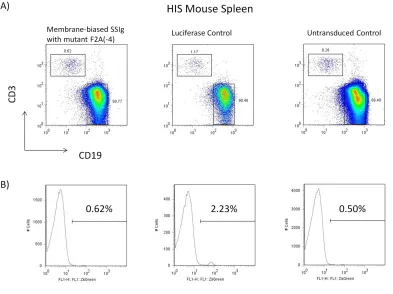
reconstitution based on the human CD45 marker. The mice that had the best
reconstitution were sacrificed (F2A(-4) mice, luciferase, and untransduced control) and their spleen and bone marrow were analyzed. Despite the low proportion of human cells, the mice showed reconstitution of the human B and T cell compartment. Gating on CD19, we found that the number of ZsGreen+ cells in the mice that carried the Molecular Rheostat immunoglobulin genes was much lower than those of the luciferase control, and close to the level of the untransduced cells (Figure 3.1A). The bone marrow of these mice was analyzed (Figure 3.2B). We found that there were many fewer ZsGreen+ than ZsGreen- cells, which might partially explain why there were also few ZsGreen+ cells in the periphery. However, it does not explain why there were many fewer ZsGreen+ cells in the mice that received the Molecular Rheostat vectors. We therefore gated on the
ZsGreen+ and ZsGreen- cells. We found that in the ZsGreen- compartment, both
Figure 3.1 FACS analysis of the bone marrow of HIS mice reconstituted from human
Figure 3.2 FACS analysis of the bone marrow of HIS mice reconstituted from human
Using the Murine Bone Marrow Transplant Model to Study B Lymphopoiesis under the
Control of Molecular Rheostat Immunoglobulin Genes
To study the Molecular Rheostat immunoglobulins in an in vivo system that provided more consistent engraftment and is more amenable to detailed analysis than the HIS mouse model, we elected to use the murine bone marrow radiation chimera model. We harvested HSC-enriched bone marrow from wild-type C57/BL6 mice that had been injected with 5-fluorouracil five days prior, and transduced the bone marrow cells in culture by spin-infection with VSVg-pseudotyped concentrated pHAGE2-EEK-IRES-ZsGreen lentiviral vectors that carry four different Molecular Rheostats genes and a luciferase control gene, respectively (Figure 3.3A). Gating on the c-Kit+ CD19- cells, it could be seen that we achieved high levels of transduction in the enriched marrow (Figure 3.3B), as measured by the shift of the ZsGreen+ populations as compared to the control, untransduced bone marrow. The amount of vectors we gave was normalized such that the bone marrow cells all received equivalent, ~ 50 MOI of the vectors. We observed that the expression of ZsGreen is approximately eightfold higher in the
luciferase vector infected cells than the vectors that carried the Molecular Rheostats. This was consistent with what we observed when the same vectors were used to transduce cell lines. We also note that over the course of the culture prior to injection into mice, we observed by fluorescence microscopy increasing numbers of doublet cells (cells
integrated vectors, as opposed to a speckled pattern characteristic of pseudo-infected cells, where pre-formed fluorescent protein was carried into the cells by the vectors. Taken together, these suggested to us that the high levels of transduction we achieved were not pseudo-infection.
Taken together, the immunophenotypic analysis coupled with the molecular data
suggested to us that the lentiviral vector driven by the EEK promoter is either silenced or that the EEK promoter is inactive. However, our earlier results with the bone marrow culture suggested to us that the promoter was active in vitro. Thus, the lentiviral vector backbone and/or the EEK promoter might be silenced in vivo through a yet unknown mechanism.
Figure 3.3 Pre-transplant FACS analysis of HSC-enriched bone marrow. A) FACS
Figure 3.4 FACS analysis peripheral blood of Rag1/WT radiation chimera. Each column
Figure 3.5 ZsGreen expression in peripheral blood of Rag1/WT radiation chimera. Each
//
Figure 3.6 Detection of lentiviral vector elements in the peripheral blood of the bone
marrow transplant recipient mice. The bolded numbers in the lengend indicate the animals that were sacrificed for analysis of the spleen and bone marrow. For each of the Molecular Rheostat constructs, the animals with the highest detected amount of
Figure 3.7 FACS analysis of the bone marrow of mice with the highest level of
References
Brainard, D. M., E. Seung, et al. (2009). "Induction of robust cellular and humoral virus-specific adaptive immune responses in human immunodeficiency virus-infected humanized BLT mice." J Virol 83(14): 7305-7321.
Luo, X. M., E. Maarschalk, et al. (2009). "Engineering human hematopoietic
stem/progenitor cells to produce a broadly neutralizing anti-HIV antibody after in vitro maturation to human B lymphocytes." Blood 113(7): 1422-1431.
Traggiai, E., L. Chicha, et al. (2004). "Development of a human adaptive immune system in cord blood cell-transplanted mice." Science 304(5667): 104-107.
Yang, L. and D. Baltimore (2005). "Long-term in vivo provision of antigen-specific T cell immunity by programming hematopoietic stem cells." Proc Natl Acad Sci U S A 102(12): 4518-4523.
C
HAPTER4:
T
HEU
SE OFN
ON-L
YMPHOIDH
EMATOPOIETICC
ELLS FORA
NTIBODYP
RODUCTIONIntroduction
Given the challenges we faced in programming B cells via lentiviral modification of the HSCs to produce HIV-responsive B cells that can produce anti-HIV broadly
neutralizing antibodies, we asked whether it might be possible to make use of non-B cells to accomplish this task. The advantage of a direct gene therapy approach to the
production of antibodies is its relative simplicity, compared to designing a system that could “slot in” to the normal B cell developmental process. Furthermore, use of non-B lineage cells avoids altogether the “mispairing problem”, which arises when a transgenic antibody is co-expressed with an endogenous antibody in the absence of allelic exclusion, resulting in a combinatorial pairing of the light chains and heavy chains from two
marrow transplant models and the established use of bone marrow transplantation in human therapies against cancer and other hematopoeitic disease; and we decided to examine muscle cells as a target for antibody production due to their easy accessibility and high protein synthetic capacity. This chapter will focus results on the work done on the hematopoietic cells that are not of the B or T lineage. The next chapter will describe our work on muscle cells.
Materials and Methods
pMIG-aHEL Vector
The aHEL IgG1 antibody is contructed by PCR cloning the entire κ light chain and the
heavy-chain-variable region of the anti-HEL antibody from the MD4 mouse genomic DNA. The light chain is fused to the heavy-chain-variable-region DNA via a F2Aopt element (described in Chapter 2). The light chain-F2Aopt-heavy-chain-variable-region DNA is then grafted onto a murine anti-human CD34 IgG1 antibody by SOE (splicing-by-overlapping-extension) PCR. The cassette is then cloned into the pMIG vector between the Not1 and BamH1 sites.
Tranduction of Murine Bone Marrow Cells and Transplantation of the Transduced Cells
heat-inactivated fetal bovine serum supplemented with recombinant murin IL-3 (20 ng/ml), IL-6 (50 ng/ml), and SCF (50 ng/ml). The bone marrow cells were transduced with the pMIG-aHEL retroviral vector by spin-infection once per day for three days beginning 24 hours after the harvest. The cells were injected into irradiated Rag1
recepient mice one day after the last spin-infection. The Rag1 mice were pre-conditioned with 300 rads of radiation from a Cs137 source.
Serum Collection and Mouse IgG ELISA
75 ul of blood was collected from each mouse each time using a heparin-coated
microcapillary tube by retro-orbital bleeding and transferred into a microcentrifuge tube and kept on ice. The blood was then incubated at 37°C for 30 mins, and then spun down at 1150g at 4°C. The serum was collected from the top of the tube. Murine IgG ELISA was performed using a Mouse IgG ELISA Kit (Bethyl Laboratories Inc., Montgomery, TX) per manufacturer’s instructions.
Results and Discussion
The Use of Non-Lymphoid Hematopoietic Cells to Produce Antibodies
We asked whether it was possible to produce antibodies in non-lymphoid hematopoietic cells. We employed an in vitro and in vivo models in parallel.
antibodies. For the ex vivo studies, we chose to use bone-marrow-derived-macrophages (BMMs) as a representative myeloid lineage cell because the culture conditions for them are well established (Zhang, Goncalves et al. 2008). They were chosen also because they up-regulate the Xbp-1 gene on activation by LPS (Martinon, Chen et al. 2010; Zeng, Liu et al. 2010; Dickhout, Lhotak et al. 2011), a gene that is required for and similarly induced in the B-cell-to-plasma-cell transition, and required for the secretion of immunoglobulins (Reimold, Iwakoshi et al. 2001; Calfon, Zeng et al. 2002; Iwakoshi, Lee et al. 2003).
cells taking on an activated, more spread out morphology and many fewer cells staying in the spherical morphology. We quantitated the amounts of antibody in a 24-hour period for two days by aspirating the media, washing the cells, and adding fresh media to the cultures. We found that the macrophages were able to produce a substantial amount of antibody (Figure 4.2). To give a sense of scale, OCI-Ly7 cells, a DLBCL (diffuse large B cell lymphoma) B cell line, when transduced with a human antibody gene by lentiviral vectors, are capable of producing on the order of 50 ng/ml of IgG in a culture volume of 10 ml containing a total of roughly 5 million cells over a period of 48 hours, giving an output of 50 ng/ml × 10 ml / 5 million cells / 2 days = 5 × 10-14 ng/cell/day. In the case of the BMMs, the number of cells at the end of the 10-day culture approximately quardruples the initial number of input bone marrow cells at the start of the culture, giving us approximately 2 million cells in each 1 ml culture in the 24-well plate. The number of cells reaches a plateau at this time due to near confluent growth. Thus, the per-cell antibody output from the macrophages can be estimated to be 50 ng/ml × 1ml / 2 million cells / 1 day = 2.5 ×10⁻¹⁴ ng/cell/day. This estimate of the antibody production capacity of an activated macrophage is on the same order of magnitude as that of a B-lineage cell that constitutively secretes antibodies. This is rather impressive.
secretion of other crucial proteins, such as secreted cytokines. Extensive in vivo characterization would be required to fully address this concern, but to get an initial handle on the issue, we quantitated the amount of the inflammatory cytokines IL-6 and TNFα secreted by the transduced BMMs on LPS-challenge as a measure of the overall function of these cells. We found that the amounts of cytokines secreted by the antibody-vector transduced cells were not significantly different from those produced by cells transduced by control-vectors carrying either IRES-GFP alone or a Luciferase-IRES-GFP cassette (Figure 4.3). Taken together, these data suggest that we might be able to use non-lymphoid cells to produce a substantial amount of antibody in vivo. To test this
hypothesis, we turned now to a bone marrow adoptive transfer model.
Figure 4.1 A) Schematic representation of the pMIG-aHEL-mIgG1 retroviral vector. B)
Figure 4.2 Antibody production by bone marrow-derived-macrophages transduced with
Figure 4.3 Cytokine production from vector-transduced
To study whether non-lymphoid hematopoietic cells could be made to produce antibody in vivo, we chose to use the Rag-1 knockout (Rag1) mouse radiation chimera model. The Rag1 mouse does not produce any functional T or B cells due to
homozygous deletion of the RAG1 gene. Therefore, antibody measured in blood would have to have been derived from the antibody vector. The strain of Rag1 mice that we propagated in the lab was more sensitive to ionizing radiation than a Rag1 strain based on C57/BL6 mice, commercially available from Jackson Laboratory (Bar Harbor, ME). The lethal dose for the strain we used was achieved at 450 rads, due to modes of radiation death from organ damage not related to bone-marrow failure; this is in contrast to wild-type C57/BL6 mice and C57/BL6-based Rag1 mice, which can tolerate a lethal dose of up to 900 rads, and die due to bone-marrow failure. We therefore chose to use a dose of 400 rads to maximize clearance of endogneous marrow while preserving the viability of the transplant recepients. This “sublethal” dose of radiation is not myeloablative in the Rag1 mice.
Figure 4.4 A) An outline of experimental design for studying antibody production from
non-lymphoid hematoipoietic cells in pMIG-aHEL transduced Rag1 mice. B) Serum antibody levels post-transplant. The time shown is the number of weeks after bone marrow transplant. UTS: Untransduced control mice. 1130 A–D: pMIG-aHEL
The normal concentrations of IgG antibody present in mouse serum is
approximately between 1–10 mg/ml, depending on a variety of factors, including strain, gender, and age. Thus the levels of antibody we achieved represented less than 0.035% of the total serum antibody concentration in the mouse. To give additional perspective, a previous attempt by Dr. Lili Yang in our laboratory to express antibodies in murine bone marrow transplant models that also employed the pMIG vector resulted in a serum concentration of approximately 1 ug/ml in wild-type mice, which had normal B and T cells. We were thus able to achieve approximately 40% of that level using non-lymphoid cells alone. We note that this value was achieved with only 10% of chimerism. We believe that the antibody concentrations could be higher if the levels of engraftment could be improved in wild-type animals that can tolerate myeloablative preconditioning prior to transplantation.
In summary, we showed that non-lymphoid hematopoietic cells are viable alternative cell types to lymphocytes for engineering antibody production. The
References
Calfon, M., H. Zeng, et al. (2002). "IRE1 couples endoplasmic reticulum load to secretory capacity by processing the XBP-1 mRNA." Nature 415(6867): 92-96. Dickhout, J. G., S. Lhotak, et al. (2011). "Induction of the unfolded protein response after
monocyte to macrophage differentiation augments cell survival in early atherosclerotic lesions." FASEB J 25(2): 576-589.
Iwakoshi, N. N., A. H. Lee, et al. (2003). "Plasma cell differentiation and the unfolded protein response intersect at the transcription factor XBP-1." Nat Immunol 4(4): 321-329.
Martinon, F., X. Chen, et al. (2010). "TLR activation of the transcription factor XBP1 regulates innate immune responses in macrophages." Nat Immunol 11(5): 411-418.
Reimold, A. M., N. N. Iwakoshi, et al. (2001). "Plasma cell differentiation requires the transcription factor XBP-1." Nature 412(6844): 300-307.
Yang, L. and D. Baltimore (2005). "Long-term in vivo provision of antigen-specific T cell immunity by programming hematopoietic stem cells." Proc Natl Acad Sci U S A 102(12): 4518-4523.
Zeng, L., Y. P. Liu, et al. (2010). "XBP-1 couples endoplasmic reticulum stress to augmented IFN-beta induction via a cis-acting enhancer in macrophages." J Immunol 185(4): 2324-2330.