STRUCTURE-FUCTION STUDIES OF
NICOTINIC ACETYLCHOLINE RECEPTORS
USING UNNATURAL AMINO ACIDS
Thesis by
Joanne (Xinan) Xiu
In Partial Fulfillment of the Requirements
for the degree of
Doctor of Philosophy
California Institute of Technology
Pasadena, California
2007
To my parents, Ruiling Xiu and Changfan Guo,
my husband, Gang Duan,
ACKNOWLEDGEMENTS
The five and half years at Caltech have been the most memorable time in my 28
years of life so far. I owe many thanks for my experience here. First of all, I have to thank
Prof. Dennis Dougherty, who has been a most wonderful advisor to me. The enthusiasm
and persistence of Dr. Dougherty regarding science have motivated me and encouraged
me from the very beginning, when I joined the lab in the summer of 2003, and I am sure
will benefit me for my whole life. His inspiration and encouragements over the past years
were always there whenever I needed them. Talking with Dennis has always been
enlightening, and I count myself among the luckiest to have benefited from his broad
knowledge in various areas of science. Dennis has also been very accessible and patient,
which is extremely important for me to learn along the way of research.
It has been great to work with Dr. Henry Lester as well. Collaborations with the
Lester group have been very fruitful and enjoyable. Here I would also like to thank my
committee, Prof. Dough Rees, Prof. Erin Schuman, and Prof. Carl Parker, for their time
Neurion, Inc., on experiments that I couldn’t have performed without her. Dr. James
Pettersson was a good resource to go to whenever I had questions about anything in the
lab. Dr. Amanda Cashin helped me with numerous experiments in molecular biology and
electrophysiology recordings and her delightful personality made it always a pleasure to
be around her. I cannot thank Dr. Tingwei Mu enough for his warm words and valuable
suggestions throughout the years, even after he left the lab. Dr. David Dahan was a
knowledgeable and warm-hearted scientist and talking with him was always a pleasure. I
also want to thank Dr. Steve Spronk and Dr. Lori Lee, who are both postdocs at
University of Michigan right now. Steve worked on computational simulation which was
a new area to me. Lori helped me with the few proline analog incorporation experiments
and I appreciate her input very much.
My daily life at Caltech could not have been such a pleasant experience if it hadn’t
been for the wonderful people in the Dougherty group. I have truly enjoyed the happy,
friendly, active, and warm atmosphere in the lab, which has undoubtedly helped with my
academic work as well. I thank Amy Eastwood for helping me with dCA coupling
O-Me-Ser and for being a great listener and helper with my problems. I thank Michael Torrice for
being a great Opus captain and a great FPLC captain. I thank Erik Rodriguez for being a
strict safety officer and for the countless suggestions on various experiments. These are
people from my year and I feel lucky to have them go through candidacies, committee
meetings, and graduations together with me. People from the next year from us are a fun
group as well. Kiowa Bower was a great neighbor in the office and always a fun person to
Hanek is also a great Opus captain and she also impresses me for never hesitating to go out
her way to do her job excellently. Working with her on my first publication on nAChR
gating mechanism was a great pleasure. Katie McMenimen has interests in numerous areas
in chemistry, biology, and computation, and she never ceases to learn new things. I would
also like to thank the newer people in the lab. Jai Shanata has taught me a lot about
single-channel recordings and also his interest in philosophy has stimulated my curiosity as well.
Kristin Rule is not only a dedicated researcher but also a great dancer, and I will always
remember her infectious laughter. Kay Limapichat and I sat together for the last few
months and I enjoyed being her neighbor in the office. Angela Blum is a hard worker and
she holds interest in various aspects in chemistry and biology. Sean Kedrowski is a very
devoted researcher and his love for science is apparent to all.
The last but certainly not the least, I owe my deepest thanks to my family. I thank my
parents for always being there for me whenever I needed them. I thank my husband for
being supportive of me all the time. I also want to thank my new son, Ryan Duan, for all
ABSTRACT
Nicotinic acetylcholine receptors (nAChR) are an important family of ligand gated ion
channels found throughout the CNS and the PNS. They have been indicated in a series of
physiological functions and pathological states. nAChRs have received extensive study in
the past as a prototype of the Cys loop LGIC member. Growing interest in developing
subtype specific agents targeting nAChRs to treat neurological diseases require more
detailed structural and functional information in the numerous members of the nAChR
family.
We performed structure-function studies on the chemical scale of several of the most
important members of this family using a powerful combination of conventional
mutagenesis and unnatural amino acid incorporations. Chapter 2 describes our research in
studying the channel gating mechanism of the prototypic nAChR, the muscle type
(α1)2βγδ. We studied thoroughly the gating interface of the receptor and concluded that the overall charging pattern of the gating interface, and not any specific pairwise electrostatic
interactions, controls the gating process in the Cys loop superfamily. Chapter 3 reports our
studies in the ligand binding mechanism of the most prevalent neuronal type α4β2 and α7 nAChR. We identified a cation-π interaction and a hydrogen bond employed by nicotine with the α4β2 receptor. These two key interactions are absent or significantly diminished in both the muscle type receptors and in the α7 form of neuronal receptor. In Chapter 4 we studied the ligand binding mechanism of a relatively newly characterized neuronal
From these studies, we found that in the Cys loop superfamily, homology in amino
acid sequences and structures do not translate into a shared functional mechanism. In fact,
different sets of chemical interactions are adopted between ligands and the receptor, and
between amino acids within the ion channel proteins, both in ligand binding and channel
gating.
Ion channels are membrane bound multi-subunit macromolecules. We are able to
carry out such exhaustive detailed structure-function studies by means of the
fast-developing methodology of unnatural amino acid incorporation by nonsense suppression.
This thesis also describes our effort to improve the efficiency of nonsense suppression. In
particular, we designed multiple 21nt small interfering RNA (siRNA) targeting release
factor 1 (eRF1) in both HEK cells and Xenopus oocytes, and monitored the nonsense
suppression efficiency change in vivo and in vitro by RNA PCR, Western blotting,
TABLE OF CONTENTS
Acknowledgements ...iv
Abstract ...vii
Table of Contents...ix
List of Figures...xiii
List of Tables ... xv
Chapter 1: Introduction... 1
1.1 Molecular neurobiology and chemistry ... 1
1.2 Muscle type and neuronal nAChR receptors... 3
1.3 Gating of the Cys loop ligand gated ion channels... 8
1.4 Nonsense suppression to incorporate unnatural amino acids... 12
1.5 Thesis summary... 16
1.6 References ... 18
Chapter 2: A unified view of the role of electrostatic interactions in modulating the gating of Cys loop receptors ... 22
2.1 Abstract... 22
2.2 Introduction ... 23
2.3 Results... 28
2.3.1 Electrostatics at the gating interface ... 28
2.3.2 Studies of the muscle-type nAChR α subunit... 33
2.3.3 Studies of the muscle-type nAChR non-α subunits... 47
2.3.4 Studies of a partial agonist ... 48
2.3.5 Previous work on the GABAA and glycine receptors ... 51
2.4 Discussion... 55
2.5 Materials and methods ... 58
2.5.1 Mutagenesis and mRNA synthesis ... 58
2.5.2 Electrophysiology and data analysis... 58
2.5.4 Bungarotoxin binding... 59
2.6 References ... 60
Chapter 3: Nicotine in the CNS vs. the neuromuscular junction: a cation-π interaction and a hydrogen bond make the difference... 67
3.1 Abstract... 67
3.2 Introduction ... 67
3.3 Results... 70
3.3.1 Background and approach... 70
3.3.2 Challenges in studying neuronal nAChRs... 75
3.3.3 A cation-π interaction in the α4β2 receptor ... 79
3.3.4 A strong hydrogen bond in the α4β2 receptor ... 82
3.3.5 Single channel measurements... 83
3.3.6 Differential binding behavior in the α7 receptor... 84
3.4 Discussion... 88
3.5 Experimental procedures... 94
3.5.1 mRNA synthesis and mutagenesis... 94
3.5.2 Ion channel expression... 94
3.5.3 Unnatural amino acid / α-hydroxy acid incorporation... 95
3.5.4Electrophysiological characterizations of the channels ... 95
4.2.5 TrpD...108 4.3 Discussion...109 4.4 References ...114 Chapter 5: Improving nonsense suppression by suppressing eRF-1 with siRNA...115
5.1 Abstract...115 5.2 Introduction ...115 5.2.1 Unnatural amino acid incorporation by nonsense suppression.115 5.2.2 Nonsense suppression efficiency ...117 5.2.3 Improving nonsense suppression efficiency in Xenopus
oocytes ...118 5.2.4 Improving nonsense suppression efficiency in mammalian cells ...119 5.2.5Improving nonsense suppression efficiency by suppressing eRF1...120 5.2.6 Using RNA interference (RNAi) to suppress eRF1 in
mammalian cells...122 5.2.7 Using RNAi to suppress endogenous proteins in Xenopus
laevis oocytes...122 5.2.8 RNA interference ...123 5.3 Experimental design...124 5.3.1 siRNA molecules designed to target human and Xenopus
laevis eRF1 ...124 5.3.2 Coinjection of siRNA molecules with nonsense suppression experiments into Xenopus oocytes...126 5.3.3 Recording currents in Xenopus oocytes of nAChR introduced by 50 μM ACh and 250 μM ACh. ...126 5.3.4RNA PCR on eRF1 both in Xenopus oocytes and in HEK
5.3.6Co-electroporation of siRNA with TAG29 GFP-TAG plasmid DNA and HSAS tRNA and microscopy in
HEK cells...131
5.4 Results and discussion...132
5.4.1 Electrophysiology of siRNA treated oocytes ...132
5.4.2 RNA PCR results of uninjected oocytes and siRNA treated oocytes ...135
5.4.3 Western blots for siRNA treated oocytes. ...138
5.4.4HSAS/TAG EGFP experiments. ...138
5.5 Conclusion and future direction...139
5.6 References ...140
LIST OF FIGURES
Figure 1.1 A simplified view of a chemical synapse... 3 Figure 1.2 Topology and diversity of nAChR subtypes... 5 Figure 1.3 Unnatural amino acid incorporation by nonsense suppression.14 Figure 1.4 Expression system mostly used in our lab for structure-
function of ion channels ... 15 Figure 2.1 Views of the gating interface... 26 Figure 2.2 Plot of log(EC50) vs. side chain physicochemical properties
at site α V46.. ... 38 Figure 2.3 A variety of mutations at α E45 are well tolerated... 40 Figure 2.4 Plot of log(EC50) vs. side chain physicochemical properties
at site α E45... 41 Figure 2.5 Charge reversal at α D138 of the nAChR is rescued by
charge swap at either of two sites: R429 (postM4) or K276 (M2-M3).. ... 44
Figure 2.6 Relative efficacy (ε) of succinylcholine for several variants of the nAChR... 50 Figure 2.7 Analysis of previously published results for the GABAA
receptor α1 subunit... 52 Figure 3.1Sequence alignments in the immediate vicinity of the
agonist binding sites from AChBP, mouse muscle type,
with the agonists in the muscle nAChR... 74
Figure 3.5 Rectification behaviors of A2B3 and A3B2 α4L9’Aβ2
nAChR in Xenopus oocytes.. ... 78 Figure 3.6Fluorination plots demonstrating cation-π interactions of TrpB in α4β2 nAChR with nicotine (left) and ACh (right)... 81 Figure 3.7 Fluorination plots demonstrating cation-π interactions of TyrA in α7 nAChR with ACh (left) and Epibatidine (right)... . 85 Figure 3.8 Fluorination plots demonstrating cation-πinteractions of
TyrC2 in α7 nAChR with epibatidine.. ... 86 Figure 4.1 Compounds tested in the current study...103 Figure 4.2Tryptophan and phenylalanine analogs incorporated in the current study ...105 Figure 5.1 Schematics of the unnatural amino acid incorporation in vivo by nonsense suppression.... ...116
LIST OF TABLES
Table 2.1 Selected sequences in the gating interface, highlighting
cationic (blue) and anionic (red) residues... 30 Table 2.2 Charge characteristics of the gating interface. ... 30 Table 2.3 Mutations in the nAChR α subunits... ... 35 Supplementary Table S 2.1: Sequences in the gating interface of the whole LGIC family, highlighting cationic (blue) and anionic (red) residues. ... 63 Table 3.1 Injection of various mRNA ratios α4(L9’A):β2 resulting in receptors of different stoichiometry.. ... 77
Table 3.2 EC50 values (μM), Hill coefficients, and normalized current
size at +70mV (normalized to current size at -100mV as -1)
for selected mutants in α4L9’Aβ2 nAChR A2B3 receptors....80 Table 3.3 EC50 values (μM) and Hill coefficients for selected mutants in
α7T6’S nAChR.. ... 87 Table 4.1 Amino acids in the aromatic box of the agonist binding site in five different Cys-loop receptors. ...103
Table 4.2 Comparison of muscle type nAChR (α1)2β1γδ and the
neuronal nAChR α4β4 EC50s in Xenopus oocytes...105
Table 4.3 Ratios of mutant EC50 over the wild type EC50 for selected
Chapter 1
INTRODUCTION
1.1.
Molecular Neurobiology and Chemistry
The brain is the most complex organ of the human body and it is comprised of about 1011
neurons [1]. A synapse is the basic unit of signal transmission between the neurons and between
one neuron to non-neural cells such as those in muscles or glands. Each neuron usually has
multiple dendrites; serving as the postsynaptic component, but only one axon, which forms the
presynaptic component. It is estimated that one neuron forms 103~104 synapses with other neurons.
The vast volume of information transmission between these numerous neural cells is largely
dependent on small molecules called neurotransmitters, the most studied of which is acetylcholine
[2].
At rest, neurotransmitter-containing vesicles are stored in the presynaptic cell either close to
the presynaptic membranes in places called active zones, or further away from the active zones by
The released neurotransmitters diffuse across the thin synaptic cleft where they bind their
corresponding receptors embedded in the postsynaptic membrane of the neighboring neuron. This
binding causes a conformational change in the receptors, opening an ion pore in the receptor,
allowing a selection of ions to flow through. This ion flow changes the membrane potential of the
postsynaptic cell, either exciting it or inhibiting it. Thus the electric-chemical-electric transmission
of the information is completed from one cell to the next.
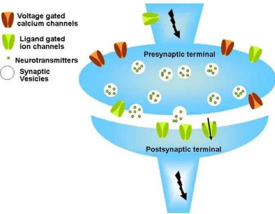
In this process, a ligand gated ion channel could play important roles at two locations: the
presynaptic part and the postsynaptic part. The presynaptic channels are usually regulatory, the
opening of which contributes to raising the membrane potential of the presynaptic cell, thus easing
the opening of the voltage gated calcium channels, finally resulting in the transmitter release [3-5].
The postsynaptic channels contribute to the fast electric signal transmission between two cells,
converting the chemical signal carried by the small molecule neurotransmitter into an electric
signal. Despite the variable locations and functions of different families of ligand ion channels, the
ability to bind a small molecule neurotransmitter and to allow ion flow upon opening is essential
Figure 1.1 A simplified view of a chemical synapse. Both presynaptic and postsynaptic ligand
gated ion channels are shown in green and the voltage gated calcium channels are shown in brown. The bolts show the direction of electric signal transmission. The arrow in the ion channel shows the direction of ion flow in the case of an open postsynaptic nAChR.
The structure-function study of these highly specialized ion channels at the molecular level
requires chemistry to combine with structural biology, molecular biology, and electrophysiology.
In our lab, we take advantage of recent advances in high-resolution structural data, fast-developing
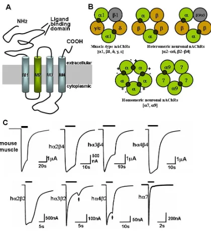
resolved into four different subunits designated α, β, γ(ε), and δ. These subunits were later
sequenced and cloned, paving the way for the molecular analysis of nicotinic receptors, especially
for those at the neuromuscular junction, which show a stoichiometry of α2βγ(ε)δ, γ in the fetal
form and ε in the adult form [6].
The muscle type nicotinic receptor turns out to be the best studied and thus the prototypic
nAChR so far. This pentameric ion channel carries two inequivalent ligand binding sites at the
large extracellular N-terminal domain subunit interfaces. Following the N-terminal domain are
four α-helices within the membrane, the third and the fourth of which are separated by a large
cytoplasmic loop carrying the phosphorylation sites and the trafficking signals for the receptor
transport [7] (Figure 1.2 A). The α subunits differ from the others by the two adjacent cysteines
In the 1980s, almost a decade after the muscle type nAChRs were discovered, nAChRs were
characterized in the central nervous system with a much more complex subunit composition.
Although subunits from only the α and β families were found, the pentameric combinations of α
2-α10 and β2-β4 make possible the existence of a great number of neuronal nicotinic receptors [10,
11] (Figure 1.2 B and C). Adding to this complexity is the variable stoichiometry of many neuronal
nAChRs, the most well known example being the existence of at least two subgroups of α4β2
receptors: (α4)2(β2)3and (α4)3(β2)2 [9, 12].
While distributed throughout the nervous system, neuronal receptors are preferentially located
presynaptically, regulating the release of neurotransmitters, in contrast to the mainly postsynaptic
location of the muscle type nAChR. A particular kind of neurotransmitter can be regulated by
different neuronal nAChR subtypes at different CNS regions. For instance, α4β2-containing
receptors are important both at the striatum and the thalamus in regulating the release of the
pleasure-inducing dopamine; α6β2β3-containing receptors were found to also play an important
role at the striatum [5, 12].
More than 90 percent of the nAChR in the CNS contain the α4 and β2 subunits, and the other
major group contains α7. In the PNS, the most abundant subunits are α3 and β4, which colocalize
with α5 and/or β4; α7 are also found in the ganglia in the PNS. The α4β2-containing, α
7-containing, and α3β4-containing receptors are the best characterized subtypes in terms of ligand
selectivity, as they can be purified from animal tissues and studied by radio-labeled ligands. [3
H]-cytisine, [3H]-nicotine, or [3H]-epibatidine can label α4β2-containing receptors, and [125I]-Bgtx or
complication could be caused by the lack of high selectivity of these ligands. For example, α-Bgtx
binds to α7-containing, α8-containing, and α9-containing homomeric and heteromeric receptors,
and MLA also recognizes receptors that contain α3 or α6 subunits. Also, epibatidine binds α2−α4
and β2-β4 subunits with high affinity [9, 12].
The addictive properties of nicotine lead to over 5,000,000 smoking-related deaths annually,
producing the largest source of preventable mortality in the western world. One of the earliest uses
of nicotinic drugs dates back to 1932 when the antinociceptive activity of nicotine was discovered.
Later epibatidine was discovered, displaying outstanding analgesic properties and high affinity for
the nicotinic receptors [13]. However, lack of selectivity for specific subtypes compromised the
wide use of epibatidine, due to the serious side effects. Other physiological functions and
pathological effects are also mediated by one or a small selection of neuronal nAChR subtypes.
The heterogeneity of the native nAChR populations in the CNS presents major challenges in
developing therapeutics targeting these receptors. Targeting one or a few nAChR subtypes without
affecting other subtypes avoids the cardiovascular, gastrointestinal, and other side effects, making
in vivo studies [15, 16]. There has also been a consistent observation of widespread decline in
nicotinic receptors in aging normal human brains or the brains of neurodegenerative disease
patients [17]. The nicotinic cholinergic systems have been shown to be involved in several
cognitive functions including attention, learning, and memory [18]. Among the many subtypes
present in the brain, the α4β2-containing and α7-containing subtypes seem particularly important.
In the post-mortem brains of schizophrenic patients, it was found that nicotinic receptors, in
particular the α7-containing receptors, are reduced in number [19]. Association between smoking
and major depression and anxiety has also been shown by several studies. Despite the complex
effects of nicotine on these pathological psychiatric states, cholinergic agonists and antagonists
aiming to treat several psychiatric disorders have been proposed recently [20].
In addition to the therapeutic potentials of targeting nicotinic receptors mentioned above,
using nicotinic drugs including nicotinic antagonists such as mecamylamine or partial agonists
such as varenicline are the most popular strategies to aid smoking cessation [21]. The possibility of
treating epilepsy and Tourette’s syndrome has been explored as well [18, 22].
1.3
Gating of the Cys loop ligand gated ion channels
Ligand gated ion channels are membrane-bound macromolecules capable of 1) binding
ligands of various sizes and properties, 2) structural changes that produce dramatically different
functional states identified as open, closed, or desensitized, and 3) selectively allowing ions to flow
through a pore region usually far away from the ligand binding region. Ion channels perform these
functions on the time scale of milliseconds, enabling them to fine-tune the fast electric transmission
When we talk about the gating of an ion channel, we generally refer to the channel opening
and closing processes (desensitization is usually considered part of the gating, but is not stressed
here), which involves multiple structural elements in the protein, including the ligand binding site,
the channel gate, and many other structural elements of the protein [24]. There are several critical
and important questions to ask when studying the gating mechanisms. Which structural elements
are involved in this process? What is the nature of the chemical interactions employed to transmit
these interactions from one amino acid to another? In what order or fashion do these changes
occur? Are these changes conserved in structurally/functionally related proteins, and so on and so
forth.
The Cys loop superfamily of neurotransmitter-gated ion channels includes receptors for
acetylcholine (nicotinic ACh receptor, nAChR), serotonin (5-HT3 receptor), γ-aminobutyric acid
(GABA, typesA and C receptors), and glycine. These receptorsare classified as excitatory
(cation-conducting; nAChR and 5-HT3) or inhibitory (anion-conducting; GABA and glycine) [25].
Tremendous efforts have been put into revealing structures at different states of the receptor or
receptor analogs, aiming to find changes that may help in defining different functional stages of the
in particular, AChBP structures from multiple organisms with various ligands bound have provided
great insight into the ligand binding mechanism of the nicotinic receptors for the past few years
[26-33]. Agonist and antagonist bound forms of AChBP show dramatic differences in certain
structural elements. This protein lacks completely the transmembrane part where the ion pore is
located, however it was shown to be a “gate-able” protein when conjugated with the
transmembrane part of the serotonin receptor [34]. Unwin and co-workers refined the cryo-EM
model of the whole receptor from the electric organ of Torpedo to a resolution of 4Ǻ [35].
However, the model still lacks the resolution to draw any conclusion at the mobile loops which are
of critical importance when it comes to the channel gating study [35-37]. Furthermore, it has not
been determined whether the agonist bound form of AChBP structures is in the active or
desensitized state; therefore, conclusions from the structural data alone seem imprudent [38].
By looking at the several AChBP crystal structures with or without different ligands bound,
loop C seems to be a structural unit that moves significantly. An “uncapped” conformation of C
loop corresponds to a closed or resting state of the receptor, while a “capped” loop C corresponds
to an open or desensitized receptor. Other elements indicated to have moved include β1−β2 loop
(loop 2), Cys loop (loop 7) and β8−β9 loop (loop 9) [38].
Recently, a 1.94 Ǻ crystal structure of the mutated single monomer of the extracellular
domain of mouse muscle α1 nAChR subunit bound with α-bungarotoxin provided interesting new
results suggesting important structural features. A water molecule buried in the core of the subunit
and a well ordered carbohydrate chain are seen. Functional studies indicate that both features are
In addition to structural biology, chemistry, biochemistry, chemical labeling, and
electrophysiology come into play to study the gating of this family of receptors. In earlier days,
using SCAM (substituted cystein accessibility mutagenesis), Karlin suggested that during gating
the α M2 domain undergoes significant changes in secondary structure, from a nonhelical to an α
-helical conformation [40]. Backbone amide-to-ester mutations disrupting backbone hydrogen
bonds performed in our lab supported this mechanism, with a few modifications to Karlin’s model
of the exact locations where the largest conformational changes are seen [41].
From mutational studies, it was proposed by Sine et al. that after ligand binding, a conserved
tyrosine residue (Y185, Lymnaea AChBP) in the C-loop is drawn closer to a conserved lysine
residue (K139) in the β7 strand, breaking an interaction between this lysine and an aspartate
residue (D194) in the β10 strand (K145 and D200 in the mouse muscle α subunit) [34, 42].
For the 5-HT3 receptor, Lummis and co-workers from our lab have proposed that structural
changes induced by ligand binding lead to the cis-trans isomerization of a conserved proline
residue (Pro 8*) on the M2-M3 linker and subsequent channel opening [43]. Thus it has been
“A stepwise mechanism for channel gating” or what was previously called a “conformational
wave” has become more complete as more regions are explored by a combination of point
mutation and single channel recording in the Auerbach lab [49-52]. By looking at a “Linear Free
Energy Relationship”, Auerbach and co-workers applied model-based kinetic analyses to quantify
the effects of mutations made on various domains of the mouse α1 subunit of nAChR. It is
proposed that nAChR gating occurs as a series of stepwise movements of such domains that link
the low-to-high affinity conformational change in the agonist binding site with the low-to-high
conductance conformational change in the pore. Specifically, they suggested blocks of coordinated
motions starting with the β4-β5 linker, the β7-β8 linker, and loop C, through the Cys loop and the
β1-β2 linker to the pore region M2, and finally the gating of the channel. This stepwise motion
propagates throughout the nAChR via Brownian motion [49, 53]. This model provides a more
complete view of nAChR gating than previous studies.
1.4
Nonsense suppression to incorporate unnatural amino acids
Site-specific incorporation of unnatural amino acids in vitro or in vivo is a powerful extension
of conventional site-directed mutagenesis to study structure-function relations of macromolecules,
and also it enables addition of biophysical probes and photo-reactive cross-linking reagents when
the mutant proteins are produced in large enough quantities [54, 55]. The chemically synthesized
unnatural amino acids are either chemically ligated to a “suppressor tRNA”, an engineered
orthogonal tRNA that cannot be acylated by an endogenous aminoacyl-tRNA synthetase (aaRS ) in
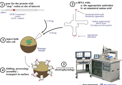
Our lab uses the first methodology, so that a wide variety of amino acids with distinctly
different properties can be incorporated in a similar fashion (Figure 1.3). The limitation of this
method is that the chemically ligated tRNA is a stoichiometric reagent, so that the quantities of
protein produced are limited to the amount of tRNA introduced. Using the high-sensitivity assay of
electrophysiology, large electric currents can be seen from Xenopus oocytes that are expressing as
little as 10 attomol of receptor on their surface. A patch clamp experiment has also made the
detection of a single channel possible [57, 58].
Our lab described the first incorporation of an unnatural amino acid into a protein expressed
in a living cell [59]. In this method, the unnatural amino acid is introduced after it is chemically
acylated to an in vitro transcribed tRNA, transferred to the site of interest, which is engineered as a
stop codon. Specifically, both mRNA and tRNA can be injected into Xenopus oocyte (Figure 1.4).
More recently, unnatural amino acids are site-specifically expressed into neuroreceptors expressed
in mammalian cells [60]. Other labs have previously reported the application of this method in
Figure 1.3 Unnatural amino acid incorporation by nonsense suppression (adapted from [55]). A:
Fig 1.4 Expression system mostly used in our lab for structure-function studies of ion channels. 1:
yeast system or from E.coli [61, 62]. Chamberlin and co-workers engineered an E. coli tRNA
suppressor in a rabbit reticulocyte system [63]. In our lab, great nonsense efficiency increase was
seen after a mutation U73G was engineered into a Tetrahymena thermophila amber suppressor
tRNA, producing THG73 and reducing recognition by the oocyte’s endogenous glutamine
acyltransferease [64].
Suppressor tRNAs that recognize the amber stop codon (UAG) have been the most widely
used both in vitro and in vivo [59, 65]. In the Chamberlin group [62, 63], the Schultz group [62],
RajBhandary group [66] and in our group [55, 67], opal (UGA) and ochre (UAA) suppressor have
been explored. Four-base codons and corresponding frame shift suppressor tRNAs have also
been developed [54, 68, 69] This frameshift suppression method can avoid the competition
between the suppressor tRNA and endogenous release factors. It has also enabled the ability to
incorporate multiple unnatural amino acids simultaneously. In our lab, we established the
simultaneous incorporation of three amino acids in vivo by frameshift suppression [54].
Our most recent developments in nonsense suppression include the development of a
Tetrahymena thermophila Gln amber suppressor (TQAS) tRNA library with increased
orthogonality and nonsense suppression efficiency, which allows for screening in eukaryotic cells.
The creation of a T. thermophila opal suppressor TQOpS’ shows about 50 percent suppression
efficiency relative to THG73, allowing for multiple incorporation of unnatural amino acids [55, 67].
1.5
Thesis summary
Nicotinic acetylcholine receptors (nAChR) are an important family of ligand gated ion
physiological functions and pathological states. nAChRs have received extensive study in the
past as a prototype of the Cys loop LGIC member. Growing interest in developing subtype-
specific agents targeting nAChRs to treat neurological diseases require more detailed structural
and functional information in the numerous members of the nAChR family.
This thesis contains structure-function studies on the chemical scale of several of the most
important members of this family: the muscle type (α1)2βγδ, which has been the representative
receptor of the family (Chapter 2); the most prevalent neuronal types α4β2 and α7 (Chapter 3);
and a relatively newly characterized neuronal receptor α4β4 (Chapter 4). A large amount of
research has been performed on other Cys loop receptors which share significant structural
resemblance to the nicotinic receptors. Using a powerful combination of conventional
mutagenesis and unnatural amino acid incorporations, we aim to look at the potential
conservation of important channel functions, particularly ligand binding mechanism (Chapter 3
and 4) and channel opening mechanism (Chapter 2). We found that homology in amino acid
sequences and structures does not translate into a shared functional mechanisms. In fact,
different sets of chemical interactions are adopted between ligands and the receptor, and between
oocytes, and monitored the nonsense suppression efficiency change in vivo and in vitro by RNA
PCR, western blotting, fluorescence, and electrophysiology (Chapter 5).
1.6 References
1. Kandel, E.R.S., J. H.; Jessell, T. M., The McGraw-Hill Companies, Inc, Principles of Neural Science. . 2000.
2. Nestler, E.J.H., S. E.; Malenka, R. C., The McGraw-Hill Companies, Inc.: . Molecular Neuropharmacology: A Foundation for Clinical Neuroscience. . 2001.
3. Boehm, S. and H. Kubista, Fine tuning of sympathetic transmitter release via ionotropic and metabotropic presynaptic receptors. Pharmacol Rev. 2002 Mar;54(1):43-99.
4. Ghijsen, W.E. and A.G. Leenders, Differential signaling in presynaptic neurotransmitter release. Cell Mol Life Sci. 2005 May;62(9):937-54., 2005.
5. Wonnacott, S., et al., Nicotinic receptors modulate transmitter cross talk in the CNS: nicotinic modulation of transmitters. J Mol Neurosci. 2006;30(1-2):137-40.
6. Romanelli, M.N., et al., Central Nicotinic Receptors: Structure, Function, Ligands, and Therapeutic Potential. ChemMedChem. 2007 Jun 11;2(6):746-767.
7. Keller, S.H. and P. Taylor, Determinants responsible for assembly of the nicotinic acetylcholine receptor. J Gen Physiol. 1999 Feb;113(2):171-6.
8. Corringer, P.J., N. Le Novere, and J.P. Changeux, Nicotinic receptors at the amino acid level. Annu Rev Pharmacol Toxicol, 2000. 40: p. 431-58.
9. Jensen, A.A., et al., Neuronal nicotinic acetylcholine receptors: structural revelations, target identifications, and therapeutic inspirations. J Med Chem. 2005 Jul 28;48(15):4705-45., 2005.
10. Gotti, C. and F. Clementi, Neuronal nicotinic receptors: from structure to pathology. Prog Neurobiol. 2004 Dec;74(6):363-96.
11. Le Novere, N., P.J. Corringer, and J.P. Changeux, The diversity of subunit composition in nAChRs: evolutionary origins, physiologic and pharmacologic consequences. J Neurobiol. 2002 Dec;53(4):447-56. 12. Gotti, C., M. Zoli, and F. Clementi, Brain nicotinic acetylcholine receptors: native subtypes and their
relevance. Trends Pharmacol Sci. 2006 Sep;27(9):482-91. Epub 2006 Jul 31.
13. Decker, M.W., L.E. Rueter, and R.S. Bitner, Nicotinic acetylcholine receptor agonists: a potential new class of analgesics. Curr Top Med Chem. 2004;4(3):369-84.
14. Dani, J.A., Overview of nicotinic receptors and their roles in the central nervous system. Biol Psychiatry. 2001 Feb 1;49(3):166-74.
15. Nashmi, R., et al., Chronic nicotine cell specifically upregulates functional alpha 4* nicotinic receptors: basis for both tolerance in midbrain and enhanced long-term potentiation in perforant path. J Neurosci. 2007 Aug 1;27(31):8202-18.
16. Scott, W.K., et al., Family-based case-control study of cigarette smoking and Parkinson disease. Neurology, 2005. 64(3): p. 442-447.
17. Levin, E.D., et al., Hippocampal alpha 7 and alpha 4 beta 2 nicotinic receptors and working memory.
Neuroscience. 2002;109(4):757-65.
18. Newhouse, P.A., A. Potter, and A. Singh, Effects of nicotinic stimulation on cognitive performance. Curr Opin Pharmacol. 2004 Feb;4(1):36-46.
20. Araki, H., K. Suemaru, and Y. Gomita, Neuronal nicotinic receptor and psychiatric disorders: functional and behavioral effects of nicotine. Jpn J Pharmacol. 2002 Feb;88(2):133-8.
21. Coe, J.W., et al., Varenicline: an alpha4beta2 nicotinic receptor partial agonist for smoking cessation. J Med Chem. 2005 May 19;48(10):3474-7.
22. Hogg, R.C. and D. Bertrand, Neuronal nicotinic receptors and epilepsy, from genes to possible therapeutic compounds. Bioorg Med Chem Lett. 2004 Apr 19;14(8):1859-61.
23. Hille, B., Ion Channels of Excitable Membranes (3rd Edition) 2001.
24. Colquhoun, D., Binding, gating, affinity and efficacy: the interpretation of structure- activity relationships for agonists and of the effects of mutating receptors. Br J Pharmacol, 1998. 125(5): p. 924-47.
25. Xiu, X., et al., A unified view of the role of electrostatic interactions in modulating the gating of Cys loop receptors. J Biol Chem. 2005 Dec 16;280(50):41655-66. Epub 2005 Oct 10.
26. Smit, A.B., et al., Acetylcholine-binding proteins: functional and structural homologs of nicotinic acetylcholine receptors. J Mol Neurosci. 2006;30(1-2):9-10.
27. Hansen, S.B., et al., Structures of Aplysia AChBP complexes with nicotinic agonists and antagonists reveal distinctive binding interfaces and conformations. EMBO J. 2005 Oct 19;24(20):3635-46. Epub 2005 Sep 29.
28. Celie, P.H., et al., Crystal structure of nicotinic acetylcholine receptor homolog AChBP in complex with an alpha-conotoxin PnIA variant. Nat Struct Mol Biol. 2005 Jul;12(7):582-8. Epub 2005 Jun 12. 29. Celie, P.H., et al., Crystal structure of acetylcholine-binding protein from Bulinus truncatus reveals the
conserved structural scaffold and sites of variation in nicotinic acetylcholine receptors. J Biol Chem. 2005 Jul 15;280(28):26457-66. Epub 2005 May 16.
30. Bourne, Y., et al., Crystal structure of a Cbtx-AChBP complex reveals essential interactions between snake alpha-neurotoxins and nicotinic receptors. EMBO J. 2005 Apr 20;24(8):1512-22. Epub 2005 Mar 24. 31. Celie, P.H., et al., Nicotine and carbamylcholine binding to nicotinic acetylcholine receptors as studied in
AChBP crystal structures. Neuron. 2004 Mar 25;41(6):907-14.
32. Smit, A.B., et al., Structure and function of AChBP, homologue of the ligand-binding domain of the nicotinic acetylcholine receptor. Ann N Y Acad Sci. 2003 Sep;998:81-92.
33. Brejc, K., et al., Crystal structure of an ACh-binding protein reveals the ligand-binding domain of nicotinic receptors. Nature. 2001 May 17;411(6835):269-76.
43. Lummis, S.C., et al., Cis-trans isomerization at a proline opens the pore of a neurotransmitter-gated ion channel. Nature. 2005 Nov 10;438(7065):248-52.
44. Lee, W.Y. and S.M. Sine, Principal pathway coupling agonist binding to channel gating in nicotinic receptors.
Nature. 2005 Nov 10;438(7065):243-7.
45. Schofield, C.M., A. Jenkins, and N.L. Harrison, A Highly Conserved Aspartic Acid Residue in the Signature Disulfide Loop of the α1 Subunit Is a Determinant of Gating in the Glycine Receptor. J. Biol. Chem., 2003. 278(36): p. 34079-34083.
46. Kash, T.L., et al., Evaluation of a proposed mechanism of ligand-gated ion channel activation in the GABAA and glycine receptors. Neurosci Lett, 2004. 371: p. 230-234.
47. Kash, T.L., et al., Coupling of agonist binding to channel gating in the GABA(A) receptor. Nature, 2003. 421(6920): p. 272-5.
48. Kash, T.L., et al., Charged Residues in the β2 Subunit Involved in GABAA Receptor Activation. J. Biol.
Chem., 2004. 279(6): p. 4887-4893.
49. Grosman, C., M. Zhou, and A. Auerbach, Mapping the conformational wave of acetylcholine receptor channel gating. Nature. 2000 Feb 17;403(6771):773-6.
50. Purohit, P., A. Mitra, and A. Auerbach, A stepwise mechanism for acetylcholine receptor channel gating.
Nature. 2007 Apr 19;446(7138):930-3.
51. Zhou, Y., J.E. Pearson, and A. Auerbach, Phi-value analysis of a linear, sequential reaction mechanism: theory and application to ion channel gating. Biophys J. 2005 Dec;89(6):3680-5. Epub 2005 Sep 23. 52. Chakrapani, S., T.D. Bailey, and A. Auerbach, Gating dynamics of the acetylcholine receptor extracellular
domain. J Gen Physiol. 2004 Apr;123(4):341-56.
53. Chakrapani, S. and A. Auerbach, A speed limit for conformational change of an allosteric membrane protein. Proc Natl Acad Sci U S A. 2005 Jan 4;102(1):87-92. Epub 2004 Dec 23.
54. Rodriguez, E.A., H.A. Lester, and D.A. Dougherty, In vivo incorporation of multiple unnatural amino acids through nonsense and frameshift suppression. Proc Natl Acad Sci U S A. 2006 Jun 6;103(23):8650-5. Epub 2006 May 25.
55. Rodriguez, E.A., H.A. Lester, and D.A. Dougherty, Improved amber and opal suppressor tRNAs for incorporation of unnatural amino acids in vivo. Part 1: Minimizing misacylation. RNA. 2007 Oct;13(10):1703-14. Epub 2007 Aug 13.
56. England, P.M., Unnatural amino acid mutagenesis: a precise tool for probing protein structure and function.
Biochemistry. 2004 Sep 21;43(37):11623-9.
57. Dougherty, D.A., Cys-Loop Neuroreceptors: Structure to the Rescue? Chemical Reviews, 2008. In press.
58. Dougherty, D.A., Unnatural amino acids as probes of protein structure and function. Curr Opin Chem Biol. 2000 Dec;4(6):645-52.
59. Nowak, M.W., et al., Nicotinic receptor binding site probed with unnatural amino acid incorporation in intact cells. Science, 1995. 268(5209): p. 439-442.
60. Monahan, S.L., H.A. Lester, and D.A. Dougherty, Site-specific incorporation of unnatural amino acids into receptors expressed in Mammalian cells. Chem Biol. 2003 Jun;10(6):573-80.
61. Noren, C.J., et al., A general method for site-specific incorporation of unnatural amino acids into proteins.
Science, 1989. 244(4901): p. 182-188.
62. Cload, S.T., et al., Development of improved tRNAs for in vitro biosynthesis of proteins containing unnatural amino acids. Chem Biol. 1996 Dec;3(12):1033-8.
64. Saks, M.E., et al., An engineered Tetrahymena tRNAGln for in vivo incorporation of unnatural amino acids into proteins by nonsense suppression. J Biol Chem. 1996 Sep 20;271(38):23169-75.
65. Noren, C.J., et al., A general method for site-specific incorporation of unnatural amino acids into proteins.
Science. 1989 Apr 14;244(4901):182-8.
66. Kohrer, C., et al., Import of amber and ochre suppressor tRNAs into mammalian cells: a general approach to site-specific insertion of amino acid analogues into proteins. Proc Natl Acad Sci U S A. 2001 Dec 4;98(25):14310-5. Epub 2001 Nov 20.
67. Rodriguez, E.A., H.A. Lester, and D.A. Dougherty, Improved amber and opal suppressor tRNAs for incorporation of unnatural amino acids in vivo. Part 2: Evaluating suppression efficiency. RNA. 2007 Oct;13(10):1715-22. Epub 2007 Aug 13.
68. Hohsaka, T., et al., Incorporation of Nonnatural Amino Acids into Streptavidin through In Vitro Frame-Shift Suppression. J. Am. Chem. Soc., 1996. 118(40): p. 9778-9779.
C h a p t e r 2
A UNIFIED VIEW OF THE ROLE OF ELECTROSTATIC
INTERACTIONS IN MODULATING THE GATING OF CYS LOOP
RECEPTORS
2.1 Abstract
In the Cys loop superfamily of ligand-gated ion channels, a global conformational change, initiated
by agonist binding, results in channel opening and the passage of ions across the cell membrane. The
detailed mechanism of channel gating is a subject that has lent itself to both structural and
electrophysiological studies. Here we defined a gating interface that incorporates elements from the
ligand binding domain and transmembrane domain previously reported as integral to proper channel
gating. An overall analysis of charged residues within the gating interface across the entire superfamily
showed a conserved charging pattern, although no specific interacting ion pairs were conserved. We
utilized a combination of conventional mutagenesis and the high-precision methodology of unnatural
amino acid incorporation to study extensively the gating interface of the mouse muscle nicotinic
acetylcholine receptor. We found that charge reversal, charge neutralization, and charge introduction at
the gating interface are often well tolerated. Furthermore, based on our data and a re-examination of
previously reported data on γ-aminobutyric acid, type A, and glycine receptors, we concluded that the
overall charging pattern of the gating interface, and not any specific pairwise electrostatic interactions,
controls the gating process in the Cys loop superfamily.
2.2 Introduction
The Cys loop superfamily of neurotransmitter-gated ion channels plays a prominent role in
mediating fast synaptic transmission. Receptors for acetylcholine (nicotinic ACh receptor, nAChR),
serotonin (5-HT3 receptor), γ-aminobutyric acid (GABAA and GABAC receptors), and glycine are
known, and the receptors are classified as excitatory (cation-conducting; nAChR and 5-HT3) or
inhibitory (anion-conducting; GABA and glycine). Malfunctions in these receptors are responsible for
a number of “channelopathies”, and the receptors are targets of pharmaceutical efforts toward
treatments for a wide range of neurological disorders, including Alzheimer’s disease, Parkinson’s
disease, addiction, schizophrenia, and depression [1, 2]. The receptors share a common architecture, are
significantly homologous, and are known to have evolved from a single ancestral gene that coded for
an ACh receptor.
The gating mechanism for the Cys loop superfamily is one of the most challenging questions in
the extracellular domain of the nAChR and, by extension, other members of the superfamily, provide a
good sense of the layout of the agonist binding site and its relationship to the rest of the receptor.
Second, continued refinement of cryoEM images of the Torpedo nAChR by Unwin [8-10],
incorporating insights gained from the AChBP structure, has produced a full atomic-scale model, 2BG9,
of the nAChR. It is important to appreciate from the outset that 2BG9, while heuristically quite
valuable, is not a crystal structure of the nAChR. It is a model built from low-resolution data and
homology modeling. Nevertheless, it represents a substantial advance for the field, and all modern
attempts to obtain molecular-scale information on the structure and function of Cys loop receptors must
consider this as a starting point.
The full 2BG9 model of the nAChR [10] immediately suggested ways in which the agonist binding
site could couple to the transmembrane region and thus initiate gating. As summarized in Figure 2.1,
loops 2, 7, and 9 from the AChBP structure are oriented toward the transmembrane region, and, indeed,
in 2BG9 these loops make contacts with parts of the transmembrane domain. Note that loop 7 is the
eponymous Cys loop. The transmembrane region consists of four α helices per subunit, labeled
M1-M4. It is accepted that M2 lines all or most of the channel. Helix M1 extends out of the
transmembrane region toward the extracellular domain, creating a segment termed preM1. While M4
is somewhat separated from the rest of the protein in 2BG9, recent modeling studies produce a more
compact structure in which M4 is more intimately involved [11]. In particular, the C-terminus of M4, a
region we will term postM4, can contact the extracellular domain. A key structure is the M2-M3 loop,
a short connector between the two transmembrane helices. Topological considerations have long
placed this loop at the interface between the transmembrane and extracellular domains. That
expectation was resoundingly confirmed by 2BG9, and many workers have anticipated that this loop
the apex of the M2-M3 loop provides the conformational switch that gates the channel in the 5-HT3
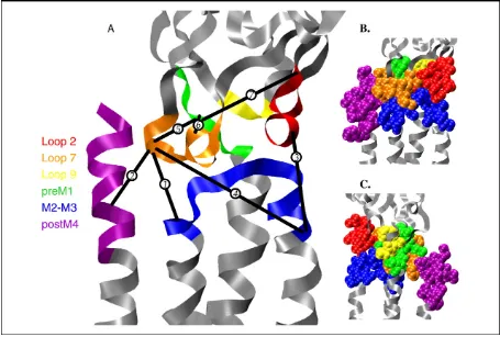
Figure 2.1 Views of the gating interface. Structure is the full model of an α subunit of the Torpedo
nAChR developed by Unwin [10] (Protein Data Bank code 2BG9). Regions of the gating interface, as defined in text, are color-coded. A: Ribbon diagram, also including pairwise interactions from various studies that have been proposed to contribute to the gating mechanism. Even though they are from different receptors and could be important in different states of the receptor, they are mapped onto the
Torpedo structure to provide some sense of relative spatial relationships. Distances range from 6 to 20 Å. Interactions are as follows: 1, Asp-138 to Lys-276 of muscle α nAChR; 2, Asp-138 to Arg-429 of muscle α nAChR; 3, Asp-57 to Lys-279 of GABAAα1 subunit; 4, Asp-149 to Lys-279 of GABAA α1 subunit; 5, Lys-215 to Asp-146 of GABAA β2 subunit; 6, Lys-215 to Asp-139 of GABAA β2
subunit; and 7, Lys-215 to Asp-56 of GABAA β2 subunit. Interactions 1, 2, 4, and 5 all involve the
Several groups have attempted to identify key interactions in the interface between the extracellular
domain and the transmembrane domain, and we discuss some of these results below. This interface
contains a large number of charged residues, and most efforts have focused on these, attempting to find
crucial electrostatic interactions that regulate gating. Specific hydrophobic interactions have also been
proposed [9, 13]. Several interacting pairs have been identified in various receptors [14, 15], and
specific gating models based on critical electrostatic interactions have been proposed [16-19]. We note
from the start, however, the curious fact that none of the proposed interactions is conserved across the
superfamily. We have been puzzled by the notion that in this closely related family of receptors, the
mechanism of action of the essential function of the receptors seems to vary from system to system.
In the present work we argue that specific, pairwise electrostatic interactions at the interface between
the transmembrane and extracellular domains are not critical to gating. Rather, we argue it is the global
charging of this region and the network of interacting ionic residues that are critical to receptor function.
We present an overall analysis of charged interfacial residues in the Cys loop superfamily; extensive
mutagenesis studies of potential electrostatic interactions in the nAChR; and a reconsideration of
previously published data on other receptors to support the model. From such an analysis, a more
2.3. Results
2.3.1 Electrostatics at the gating interface
For the purposes of discussion and analysis, we have defined a “gating interface” between the
extracellular domain and the transmembrane domain. It is comprised of six segments: three from the
extracellular domain (all or parts of loops 2, 7, and 9) and three from the transmembrane domain
(preM1, M2-M3, and postM4). The precise residues considered are given in Table 2.1. Unless
otherwise noted, we will use the residue numbering system accepted for the nAChR α1 subunit. The
selection criterion for the gating interface was geometric; only residues that could reasonably be
considered to experience a meaningful electrostatic interaction with another component of the gating
interface were included. Because of the low resolution of the nAChR structure and the further
uncertainty introduced by extrapolating to other Cys loop receptors, precise distance constraints were
not applied. Rather, as illustrated in Figure 2.1, we chose a contiguous belt of residues in the region
where the extracellular and transmembrane domains meet. Some leeway must be given in selecting
possible interactions, as residues that are not in direct contact in 2BG9 could become so on transit from
the closed state to the open, or on going from one receptor to another. We recognize there is some
arbitrariness to this assignment, but our studies suggest that extending the definition further out from
the interface does not significantly impact the analysis. We will refer to the extracellular component
(from loops 2, 7, and 9) and the transmembrane component (from preM1, M2-M3, and postM4) when
discussing the gating interface.
To search for patterns of charged residues, we considered the sequences of 124 subunits from the
Cys loop superfamily - 74 cationic and 50 anionic channel subunits (see supplementary table S2.1 at
and 11 anionic (inhibitory) channels, and also serves to define the various segments. Table 2.2
summarizes the analysis of the full collection of the 124 subunits. Shown for each segment of the
interface are: the number of cationic residues (K, R); the number of anionic residues (D, E); the net
Loop 2 Loop 7 L9 preM1 M2-M3 linker postM4 Tor α DEVNQI IIVTHFPFDQ EW MQIRP STSSAVPLIGKY FAGRLIELSQEG Tor β NEKIEE IKVMYFPFDW QW IQRKP ETSLSVPIIIRY FLDASHNVPPDN Tor γ NEKEEA IAVTYFPFDW EW IQRKP ETSLNVPLIGKY FLTGHFNQVPEF Tor δ KETDET INVLYFPFDW EW IRRKP ETALAVPLIGKY FVMGNFNHPPAK
nACh α1 DEVNQI IIVTHFPFDE EW MQRLP STSSAVPLIGKY FAGRLIELHQQG nACh β1 NEKDEE IQVTYFPFDW QW IRRKP ETSLAVPIIIKY FLDATYHLPPPE
nACh γ NEREEA ISVTYFPFDW EW IQRKP ETSQAVPLISKY FLMAHYNQVPDL nACh δ KEVEET ISVTYFPFDW EW IRRKP ATSMAIPLVGKF FLQGVYNQPPLQ nACh α4 DEKNQM IDVTFFPFDQ EW IRRLP STSLVIPLIGEY FLPP--WLAGMI nACh α7 DEKNQV IDVRWFPFDV EW MRRRT ATSDSVPLIAQY LMSAPNFVEAVS 5HT3A DEKNQV LDIYNFPFDV EW IRRRP ATAIGTPLIGVY VMLWSIWQYA-- GABA α1 SDHDME MHLEDFPMDA QY LKRKI KVAYATAM-DWF LNREPQLKAPTP GABA α2 SDTDME MHLEDFPMDA QY LKRKI KVAYATAM-DWF LNREPVLGVSP- GABA α3 SDTDME MHLEDFPMDV QY LKRKI KVAYATAM-DWF VNRESAIKGMIR
GABA α4 SDVEME MRLVDFPMDG QY LRRKM KVSYLTAM-DWF LSKDTMEKSESL GABA α5 SDTEME MQLEDFPMDA QY LKRKI KVAYATAM-DWF LNREPVIKGAAS GABA α6 SDVEME MRLVNFPMDG QY LQRKM KVSYATAM-DWF LSKDTMEVSSSV GABA β1 SEVNMD MDLRRYPLDE QF LKRNI KIPY-VKAIDIY VN--- GABA β2 SEVNMD MDLRRYPLDE QF LKRNI KIPY-VKAIDMY VN--- GABA β3 SEVNMD MDLRRYPLDE QF LKRNI KIPY-VKAIDMY VN--- Gly α1 AETTMD MDLKNFPMDV QF LERQM KVSY-VKAIDIW KIVRREDVHNQ- Gly α2 TETTMD MDLKNFPMDV QF LERQM KVSY-VKAIDIW KIIRHEDVHKK-
[image:45.595.70.495.136.421.2]44 49 130 139 175 207 211 266 277 426
Table 2.1 Selected sequences in the gating interface, highlighting cationic (blue) and anionic (red) residues. The abbreviations used are as follows Tor: nAChR from Torpedo californica; nACh: nicotinic ACh receptor; 5-HT3A: 5-HT3 receptor, type A. All sequences are from human receptors
except: Tor and nACh α1, β1, γ, δ, which are mouse muscle.
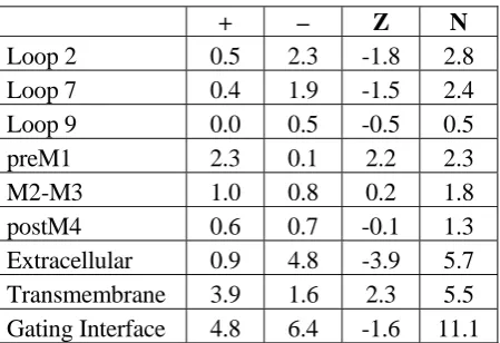
+ – Z N
Loop 2 0.5 2.3 -1.8 2.8
Loop 7 0.4 1.9 -1.5 2.4
Loop 9 0.0 0.5 -0.5 0.5
preM1 2.3 0.1 2.2 2.3
M2-M3 1.0 0.8 0.2 1.8
postM4 0.6 0.7 -0.1 1.3
Extracellular 0.9 4.8 -3.9 5.7
Transmembrane 3.9 1.6 2.3 5.5 Gating Interface 4.8 6.4 -1.6 11.1
[image:45.595.67.292.500.654.2]Although there is some variation, the typical gating interface contains 47 residues: 18 in the
extracellular component and 29 in the transmembrane component. On average, 11.1 or 24% of these
residues are charged. This is not significantly different from expectation based on the overall
frequencies of occurrence of D, E, R, and K in proteins (July, 2004, Swiss Protein Database). Most of
the residues of the gating interface are, or can be easily imagined to be, water exposed to some extent,
therefore this global result is not surprising. Of the ~11 charged residues found in the gating interface
only two are universally conserved: D138 and R209. So, while all Cys loop receptors have a large
number of ionic residues in the gating interface, their locations and absolute charges are variable.
Although the two regions of the gating interface do not have the same number of amino acids, the
total number of charges is essentially the same (5.7 vs. 5.5) for the two regions. There is, however, a
dramatic difference in the net charge of the two components. The extracellular component has an
overall negative charge, averaging -3.9 over the 124 subunits considered. The transmembrane
component has an overall positive charge, averaging +2.3. Thus, there is a global electrostatic
attraction in the interface, holding together the extracellular component and the transmembrane
component. This interfacial electrostatic interaction is not created by simply putting anions in the
There is variability in the charging pattern. Considering only GABAA subunits, α1 shows Z = -6 in
the extracellular component, and Z = +4 in the transmembrane component. In contrast, the α4 subunit
shows Z = -4 in the extracellular component and Z = +2 in the transmembrane component. Despite the
smaller Z values, the α4 subunit actually has more ionic residues overall than α1 (N = 16 vs. 14).
Looking in more detail, it is clear that loop 2 carries the most negative charge per residue, followed
by loop 7. The largest net positive charge is associated with preM1. The total number of charges (N) is
slightly larger for the inhibitory channels (average of 11.8 vs. 10.7). The “additional charge” is usually
cationic, as the net charge is slightly more positive for the inhibitory channels (-1.1 vs. -1.9).
We propose that Cys loop receptors can function as long as the essential features of the electrostatic
network are intact. As we will see below, mutations that alter the charge balance are often well
tolerated, apparently because they can be absorbed by the larger collection of charges. In fact full
charge reversals (replacing a plus with a minus or vice versa) are often quite acceptable. It appears that
2.3.2 Studies of the muscle-type nAChR α subunit
We have evaluated a number of residues in the gating interface by both conventional mutagenesis
and unnatural amino acid mutagenesis [20]. Many mutations are meant to parallel studies in other
receptors, but, as noted above, conservation is not strong across the family. We study the embryonic
mouse muscle nAChR, with a subunit composition of (α1)2β1γδ. This receptor shows extremely high
homology with and is thus directly comparable to the Torpedo receptor modeled by 2BG9. A goal of
this work is to conduct an extensive survey of the gating interface, complementing the statistical
analysis presented above. As such, we report the results of two-electrode voltage clamp determinations
of EC50, rather than the more time-consuming patch clamp studies of single-channel behaviors. Since
EC50 is a measure of channel function, it reflects contributions from agonist binding and gating.
However, the residues studied are not part of the agonist binding site, and so seem unlikely to directly
contribute to binding. Furthermore, we show that representative mutations in the gating interface alter
the relative efficacy of succinylcholine, a partial agonist of the mouse muscle nAChR [21], supporting
a change in the gating of the mutants [14, 22]. In addition, we recently showed that a range of
mutations of a key proline at the heart of the gating interface in the M2-M3 loop of the 5HT3A receptor
mutations occur in both copies of the mouse muscle α subunit; the results are given in Table 2.3. In a
subsequent section we will briefly discuss the non-α subunits.
The 2BG9 model features a particularly intimate interfacial interaction: that between V46 on loop 2
and a portion of the M2-M3 loop [9]. A “pin-into-socket” arrangement was proposed, ascribing a
critical gating role to V46. While it was immediately appreciated by many workers that V46 is not
conserved in the Cys loop superfamily, the proposal merited investigation. Studies by Harrison,
Trudell, and coworkers on analogous residues in the GABAA α1 and β2 subunits and the glycine
receptor α1 subunit (the residue is H, V, and T, respectively) provided no support for the
“pin-into-socket” proposal [13].
We have explicitly evaluated V46 in the nAChR (Table 2.3). We wished to determine whether a
precise geometrical arrangement of the sort implied by a pin-into-socket interaction was essential for
proper receptor function. Not surprisingly, the V46A mutation is substantially deleterious, while the
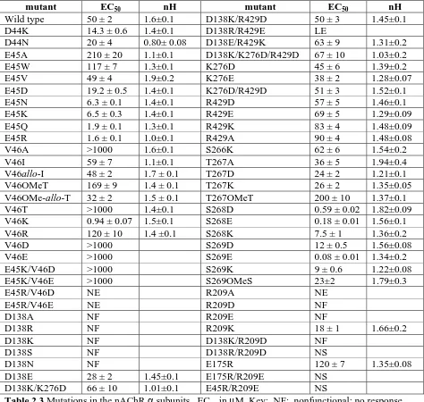
mutant EC50 nH mutant EC50 nH
Wild type 50 ± 2 1.6±0.1 D138K/R429D 50 ± 3 1.45±0.1
D44K 14.3 ± 0.6 1.4±0.1 D138R/R429E LE
D44N 20 ± 4 0.80± 0.08 D138E/R429K 63 ± 9 1.31±0.2
E45A 210 ± 20 1.1±0.1 D138K/K276D/R429D 67 ± 10 1.03±0.2
E45W 117 ± 7 1.3±0.1 K276D 45 ± 6 1.39±0.2
E45V 49 ± 4 1.9±0.2 K276E 38 ± 2 1.28±0.07
E45D 19.2 ± 0.5 1.4±0.1 K276D/R429D 51 ± 3 1.52±0.1
E45N 6.3 ± 0.1 1.4±0.1 R429D 57 ± 5 1.46±0.1
E45K 6.5 ± 0.3 1.4±0.1 R429E 69 ± 5 1.29±0.09
E45Q 1.9 ± 0.1 1.3±0.1 R429K 83 ± 4 1.48±0.09
E45R 1.6 ± 0.1 1.0±0.1 R429A 90 ± 4 1.48±0.08
V46A >1000 1.6±0.1 S266K 62 ± 6 1.54±0.2
V46I 59 ± 7 1.1±0.1 T267A 36 ± 5 1.94±0.4
V46allo-I 48 ± 2 1.7 ± 0.1 T267D 24 ± 2 1.21±0.1
V46OMeT 169 ± 9 1.4 ± 0.1 T267K 26 ± 2 1.35±0.05
V46OMe-allo-T 32 ± 2 1.5 ± 0.1 T267OMeT 200 ± 10 1.37±0.1
V46T >1000 1.4±0.1 S268D 0.59 ± 0.02 1.82±0.09
V46K 0.94 ± 0.07 1.5±0.1 S268E 0.18 ± 0.01 1.56±0.1
V46R 120 ± 10 1.4 ±0.1 S268K 7.5 ± 1 1.36±0.2
V46D >1000 S269D 12 ± 0.5 1.56±0.08
V46E >1000 S269E 0.08 ± 0.01 1.34±0.2
E45K/V46D >1000 S269K 9 ± 0.6 1.22±0.08
E45K/V46E >1000 S269OMeS 23±2 1.79±0.3
E45R/V46D NE R209A NE
E45R/V46E NE R209D NF
D138A NF R209E NF
D138R NF R209K 18 ± 1 1.66±0.2
D138K NF D138K/R209D NF
D138S NF D138R/R209D NS
[image:50.595.65.539.131.581.2]binding pocket, one might anticipate that the isomeric allo-I, in which the side chain methyl and ethyl
groups swap position relative to I, would show a significantly different interaction. In the event, the
difference between I and allo-I is insignificant (Table 2.3). The unnatural amino acid
O-methyl-threonine (OMeT) is isosteric with I, but inserts a more polar O in place of a CH2 group [25]. This
subtle change is deleterious, raising EC50 more than 3-fold. The isomeric OMe-allo-T introduces a
stereochemical swap that parallels the I/allo-I pair, but now the effect is substantial. The difference
between OMeT and OMe-allo-T is roughly 5-fold, corresponding to 1 kcal/mol at room temperature.
This suggests that perhaps the side chain of position 46 is in a sterically well-defined pocket, but one
that can only be probed by polar oxygen atoms, not by hydrophobic groups such as CH2.
Replacing V46 with much more polar groups like T (essentially isosteric to V) and the anionic D
and E seriously compromises receptor function. Radiolabeled α-bungarotoxin binding studies show
that V46D and V46E channels are expressed in large enough quantities to detect macroscopic currents,
but electrophysiology studies show only small (< 300 nA) currents at 1 mM ACh, suggesting a shift to
a much higher EC50. Surprisingly, though, the cationic residues R and K produce, in the first case, only
mutation of the nAChR α1 subunit produces a loop 2 pattern equivalent to those of the α7 and α4
nicotinic and 5-HT3 serotonin receptors, and so perhaps it is not surprising that it can be tolerated.
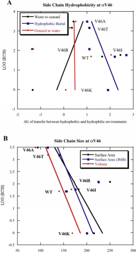
We have sought a correlation between various physicochemical properties of the mutant side chains
and the mutant EC50s at V46 (Figure 2.2) and find that there is no apparent correlation to the side chain
hydrophobicity [26, 27] or size [28, 29]. Auerbach et al. previously reported single channel recordings
on several mutations at V46, and a very rough tendency was observed that more polar side chains have
smaller gating equilibrium constants [23]. However, the new mutations that we made, V46K and V46R,
do not follow this pattern. Our results suggest that while the side chain of V46 may be docked into a
sterically well-defined pocket as implied by the “pin-into-socket” mechanism, a full description of the
Figure 2.2 Plot of log(EC50) vs. side chain physicochemical properties at site αV46. A:log(EC50) vs.
side chain hydrophobicity. B:log(EC50) vs. side chain size. Three hydrophobicity scales were used.
As noted above, Loop 2 is highly charged across the Cys loop superfamily, with an overall negative
charge. As others have done in different receptors within the superfamily, we have evaluated some of
these charged residues. Position 45 is very highly conserved as anionic (D or E), and our studies of E45
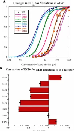
in the muscle α subunit are summarized in Figure 2.3. Quite surprisingly, we find that full charge
reversal (E45K or E45R) substantially lowers EC50. Substitution by a neutral, but polar residue (E45Q
or E45N) also lowers EC50, while conversion to a hydrophobic residue gives a small effect, raising
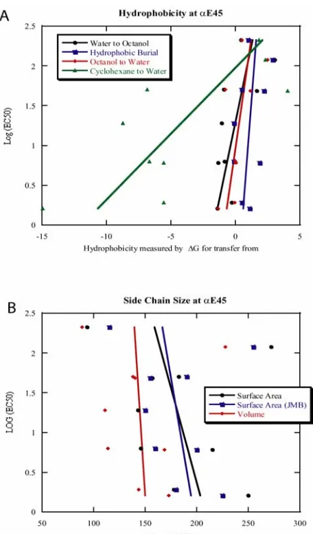
EC50, if anything. The logarithms of the mutant EC50 at E45 were plotted against the physicochemical
properties of the mutant side chains as done for V46, and no apparent correlation was found (Figure
Figure 2.3 A variety of mutations at αE45 are well tolerated. Charge neutralization (N,Q) and charge reversal (K,R) both lower EC50 substantially, while mutations to hydrophobic residues (A,W,V) leave
EC50 little changed. A: Dose-response curves. B: Ratios of mutant to wild type EC50s plotted for
[image:55.595.167.427.143.600.2]Figure 2.4 Plot of log(EC50) vs. side chain physicochemical properties at site αE45. A: log(EC50) vs.
[image:56.595.184.412.115.504.2]N47 in loop 2 of the nAChR α subunit aligns with D57 of the GABAA α1 subunit, which, as
discussed below, has been proposed to experience important electrostatic interactions [15]. Auerbach
and co-workers have studied mutations at this site in the nAChR, including extensive single-channel
measurements [23]. Auerbach found that N47K shows a decrease in EC50, but N47D shows an
increase. As noted above, Auerbach’s single-channel studies of this and other loop 2 residues establish
a role in setting the gating equilibrium for residues in this region.
Thus, at four consecutive residues in loop 2 – D44, E45, V46, and N47 – introducing a positive
charge lowers EC50. At N47 and V46 it has also been shown that introducing a negative charge has the
opposite effect. These various side chains point in quite different directions in 2BG9. While is it
possible that all make specific electrostatic contacts that are being modulated in similar ways by the
mutations introduced, we conclude instead that it is the global negative charge of loop 2
![Figure 1.3 Unnatural amino acid incorporation by nonsense suppression (adapted from [55])](https://thumb-us.123doks.com/thumbv2/123dok_us/13372.904/29.595.103.515.124.433/figure-unnatural-amino-acid-incorporation-nonsense-suppression-adapted.webp)