Parainfluenza Virus Infection Sensitizes Cancer Cells to
DNA-Damaging Agents: Implications for Oncolytic Virus Therapy
Candace R. Fox,aGriffith D. Parksa
aUniversity of Central Florida College of Medicine, Orlando, Florida, USA
ABSTRACT A parainfluenza virus 5 (PIV5) with mutations in the P/V gene (P/V-CPI⫺) is restricted for spread in normal cells but not in cancer cellsin vitroand is effective at reducing tumor burdens in mouse model systems. Here we show that P/V-CPI⫺ infection of HEp-2 human laryngeal cancer cells results in the majority of the cells dying, but unexpectedly, over time, there is an emergence of a population of cells that survive as P/V-CPI⫺persistently infected (PI) cells. P/V-CPI⫺PI cells had elevated levels of basal caspase activation, and viability was highly dependent on the activity of cellular inhibitor-of-apoptosis proteins (IAPs) such as Survivin and XIAP. In chal-lenge experiments with external inducers of apoptosis, PI cells were more sensitive to cisplatin-induced DNA damage and cell death. This increased cisplatin sensitivity correlated with defects in DNA damage signaling pathways such as phosphorylation of Chk1 and translocation of damage-specific DNA binding protein 1 (DDB1) to the nucleus. Cisplatin-induced killing of PI cells was sensitive to the inhibition of wild-type (WT) p53-inducible protein 1 (WIP1), a phosphatase which acts to terminate DNA damage signaling pathways. A similar sensitivity to cisplatin was seen with cells during acute infection with P/V-CPI⫺as well as during acute infections with WT PIV5 and the related virus human parainfluenza virus type 2 (hPIV2). Our results have general implications for the design of safer paramyxovirus-based vectors that cannot establish PI as well as the potential for combining chemotherapy with oncolytic RNA virus vectors.
IMPORTANCE There is intense interest in developing oncolytic viral vectors with in-creased potency against cancer cells, particularly those cancer cells that have gained resistance to chemotherapies. We have found that infection with cytoplasmically replicating parainfluenza virus can result in increases in the killing of cancer cells by agents that induce DNA damage, and this is linked to alterations to DNA damage signaling pathways that balance cell survival versus death. Our results have general implications for the design of safer paramyxovirus-based vectors that cannot estab-lish persistent infection, the repurposing of drugs that target cellular IAPs as antivi-rals, and the combined use of DNA-damaging chemotherapy agents in conjunction with oncolytic RNA virus vectors.
KEYWORDS DNA damage, oncolytic viruses, parainfluenza virus
T
here is intense interest in developing oncolytic viral vectors with increased potency against cancer cells. As examples, a modified herpes simplex virus 1 strain desig-nated talimogene laherparepvec (T-VEC) (Imlygic; Amgen) is the first oncolytic virus approved for human use by the Food and Drug Administration (1), and there has been a large increase in the number of clinical trials for new viruses for tumor therapy (2, 3). A number of paramyxoviruses have shown promise as oncolytic vectors based in part on their inherent cytopathic properties, including measles virus (MV), Newcastle disease virus (NDV), Sendai virus, and mumps virus (MuV) (4–10). The goal of the work described here was to understand how cancer cells infected with a cytopathicparain-Received8 November 2017Accepted5 January 2018
Accepted manuscript posted online17 January 2018
CitationFox CR, Parks GD. 2018. Parainfluenza virus infection sensitizes cancer cells to DNA-damaging agents: implications for oncolytic virus therapy. J Virol 92:e01948-17.https://doi .org/10.1128/JVI.01948-17.
EditorRebecca Ellis Dutch, University of Kentucky College of Medicine
Copyright© 2018 American Society for Microbiology.All Rights Reserved.
Address correspondence to Griffith D. Parks, Griffith.parks@ucf.edu.
VIRUS-CELL INTERACTIONS
crossm
on November 6, 2019 by guest
http://jvi.asm.org/
fluenza virus oncolytic vector respond to external inducers of apoptosis such as cisplatin.
Wild-type (WT) parainfluenza virus 5 (PIV5) has an unusual property among paramyxoviruses of being a poor inducer of host cell responses in most human epithelial and fibroblast cell types (11, 12). This property is related in part to the active evasion of host cell responses. One such evasion mechanism is through the viral V protein hijacking the host protein damage-specific DNA binding protein 1 (DDB1). In virus-infected cells, DDB1 becomes part of a cytoplasmic “V degradation complex,” which induces the degradation of signal transducer and activator of transcription 1 (STAT1) or STAT2, resulting in the inhibition of type I interferon (IFN) signaling (13–15). The PIV5 V protein targets the degradation of STAT1, whereas the related virus human parainfluenza virus type 2 (hPIV2) targets STAT2 degradation (14–17).
For reasons that are not fully understood, WT PIV5 is also largely noncytopathic to most cell typesin vitro. Interestingly, alterations to the PIV5 P/V gene can convert the noncytopathic WT virus into a highly cytopathic mutant (P/V-CPI⫺) (12, 18–22). The PIV5 P/V gene encodes phosphoprotein P and accessory protein V, which share an identical 164-residue amino-terminal domain (the shared P/V region) but have unique C-terminal domains. The P protein is an essential subunit of the viral RNA-dependent RNA polymerase (23). The V protein is thought to function in the regulation of viral RNA synthesis (24) but also has additional roles in blocking IFN signaling (14, 15), disrupting apoptosis pathways (12), and inhibiting IFN- gene expression through binding to mda-5 (25). In the case of the P/V-CPI⫺mutant, the introduction of six amino acid changes in the shared P/V region (listed in Materials and Methods) results in a mutant with properties very different from those of WT PIV5, including the activation of IFN and proinflammatory cytokine responses (18, 26), the overexpression of viral RNA and proteins (18, 27), and the induction of massively cytopathic effects in most cancer cell lines tested so far (18, 21, 22).
The PIV5 P/V-CPI⫺mutant is currently being developed as a novel oncolytic vector. This mutant is restricted for growth in normal cells but is fully capable of growth and spread through a population of tumor cellsin vitro(19, 28). These differences in cell type restriction have been proposed to be due in part to effective IFN responses following P/V-CPI⫺ infection of normal cells but not cancer cells (28). During the transformation process, tumor cells can accumulate specific defects in IFN pathways that contribute to resistance to the antiproliferative effects of IFN (e.g., see references 29 and 30). It has been proposed that these alterations can also confer increased susceptibility to viral infection (3, 31), particularly in the case of mutants such as P/V-CPI⫺, which is defective in blocking IFN.
The mechanism of cell killing by the P/V-CPI⫺mutant is not completely under-stood at this time; however, it could be tied mechanistically to the high-level induction of double-stranded RNA (dsRNA) during replication and the shutoff of host and viral protein synthesis through protein kinase R (PKR) pathways (22). Similarly, cell killing by the P/V-CPI⫺ mutant involves the activation of
caspase-dependent death pathways (32, 33). Importantly, we have also shown that the P/V-CPI⫺mutant is very effective at reducing prostate cancer tumor burdensin vivo in a mouse model system (28).
Here we show that P/V-CPI⫺infection of HEp-2 human laryngeal cancer cells results
in the majority of the cells dying, but unexpectedly, over time, there is an emergence of a population of cells that survive as persistently infected (PI) cells. While testing the hypothesis that PI cells have altered apoptotic pathways, we found that PI cells and cells acutely infected with the P/V-CPI⫺virus show enhanced DNA damage and cell death induced by chemotherapy agents such as cisplatin. Our results have general implications for the design of safer paramyxovirus-based vectors that cannot establish persistent infection as well as the potential for combining chemotherapy with oncolytic RNA virus vectors.
Fox and Parks Journal of Virology
on November 6, 2019 by guest
http://jvi.asm.org/
RESULTS
The cytopathic PIV5 P/V-CPIⴚ mutant is capable of establishing persistent infection.The PIV5 P/V-CPI⫺mutant is highly cytopathic to a number of cancer cell
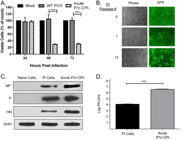
lines (18, 21, 28). This is illustrated in Fig. 1A, where HEp-2 human laryngeal cancer cells were mock infected or infected with WT PIV5 or the P/V-CPI⫺mutant. Cell viability was
monitored over 72 h postinfection (hpi) by using 3-(4,5-dimethyl-2-thiazolyl)-2,5-diphenyl-2H-tetrazolium bromide (MTT) assays. In contrast to WT PIV5, the P/V-CPI⫺
mutant induced⬃70% cell death at 48 and 72 hpi. Unexpectedly, at these later time points, a substantial percentage of viability remained. Cells that survived this persistent infection could be passaged and retained green fluorescent protein (GFP) expression (Fig. 1B). Western blotting confirmed that PI cells expressed all viral proteins examined so far (Fig. 1C). PI cells produced infectious virions (Fig. 1D) albeit with a⬃3-log-lower yield in PFU per milliliter than with acute P/V-CPI⫺infection. These data indicate that
in some cell types, the cytopathic P/V-CPI⫺mutant can establish a PI cell line that still
expresses viral proteins and produces virus.
PI cells have elevated basal levels of apoptotic markers and are highly depen-dent on inhibitors of apoptosis for survival.The above-described findings raised the question of whether PI cells had downregulated apoptotic pathways in order to survive P/V-CPI⫺infection. To determine the levels of steady-state stress, naive and PI HEp-2
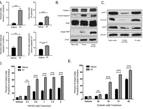
cells were examined for cell viability and apoptotic effector proteins. As shown in Fig. 2A (top),⬃10 and⬃20% of PI cells stained positive for annexin V and propidium iodide under steady-state conditions, respectively, compared to 2 to 3% of naive HEp-2 cells. The levels of steady-state propidium iodide staining of PI cells varied between exper-iments but were always in the range of⬃10 to 20%, a variability which may reflect differences in cell passage number, confluence, or time after plating or other differ-ences in cell culture conditions. In addition, a higher fraction of PI cells showed cleaved caspase 3 and active caspase 3/7 than in naive cells (Fig. 2A, bottom, and B). PI cells and
FIG 1Persistent infection with a cytopathic oncolytic virus. (A) Naive HEp-2 cells were mock infected or infected with rPIV5-WT or the P/V-CPI⫺mutant at an MOI of 10 PFU/cell. Cell viability was determined by an MTT assay at the indicated hours postinfection. Values are the means for three samples, with***
indicating aPvalue of⬍0.001 comparing WT and P/V-CPI⫺infections. (B) Cells that survived P/V mutant infection were examined by fluorescence microscopy for GFP expression. (C) Lysates from naive, PI, and acutely P/V-CPI⫺-infected cells (24 hpi) were analyzed by Western blotting for the indicated viral proteins. (D) Titers of virus released from PI HEp-2 cells or after 24 h of acute infection with P/V-CPI⫺were determined.***indicates aPvalue of⬍0.001.
Parainfluenza Virus Infection and DNA Damage Response Journal of Virology
on November 6, 2019 by guest
http://jvi.asm.org/
[image:3.585.48.361.71.313.2]acutely P/V-CPI⫺-infected cells showed similar levels of cleaved caspase 3 (Fig. 2B). In
contrast, PI cells did not contain substantially higher levels of downstream apoptotic markers such as cleaved poly(ADP-ribose) polymerase (PARP) (Fig. 2B). Together, these data suggest that PI cells have high basal levels of cell stress, indicated by caspase activation; however, this stress does not lead to substantial levels of cell death.
Cellular inhibitor-of-apoptosis proteins (IAPs) have been found to have distinct mechanisms of action to block apoptosis. For example, XIAP can directly bind and inhibit cleaved caspases 3, 7, and 9 from activating their substrates, whereas Survivin binds to and inhibits cleaved caspases 3 and 7 (34, 35). Our finding of high levels of caspase activation and low levels of cell death in PI cells suggested that IAPs may be important for survival of P/V-CPI⫺infection. To test this hypothesis, levels of IAPs were
determined by Western blotting. As shown in Fig. 2C, levels of c-IAP1, Survivin, and XIAP expression in PI cells were very similar to those in naive HEp-2 cells. In contrast, acute infection with P/V-CPI⫺led to reduced levels of all three of these IAPs, a result
which may be due to PKR activation and a global reduction of protein synthesis (22). To determine the functional importance of IAPs in PI cell survival, naive and PI cells were treated with increasing concentrations of a Survivin inhibitor (Fig. 2D) or an XIAP inhibitor (Fig. 2E) for 24 h before determining viability by propidium iodide staining.
FIG 2PI cells have elevated basal levels of apoptotic markers and are highly dependent on inhibitors of apoptosis for survival. (A) Naive HEp-2 and PI HEp-2 cells were stained with annexin V and propidium iodide or with antibodies to cleaved caspase 3 or active caspase 3/7 before analysis by flow cytometry. Values are the means for three samples, with***indicating aPvalue of⬍0.001. (B and C) Lysates from naive HEp-2 cells, PI cells, or acutely P/V-CPI⫺-infected cells were analyzed by Western blotting for the indicated apoptotic proteins (B) or for cellular inhibitors of apoptosis (C). (D and E) Naive and PI HEp-2 cells were treated with the Survivin inhibitor YM155 (D) or with the XIAP inhibitor Embelin (E) for 24 h. Cells were stained with propidium iodide before analysis by flow cytometry. Values are the means for three samples, with**and***indicatingPvalues of⬍0.01 and⬍0.001, respectively.
Fox and Parks Journal of Virology
on November 6, 2019 by guest
http://jvi.asm.org/
[image:4.585.44.538.68.444.2]The viability of PI cells was sensitive to IAP inhibition compared to naive HEp-2 cells, suggesting that PI cells are highly dependent on IAPs in order to survive the high levels of cell stress under steady-state conditions.
PI cells have increased sensitivity to DNA-damaging agents.Given the survival of PI cells harboring the cytopathic P/V-CPI⫺mutant and the increased sensitivity of PI
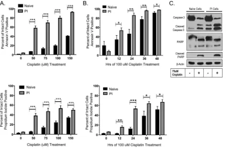
cells to IAP inhibitors, we hypothesized that PI cells have increased resistance to inducers of cell death. To test this, naive and PI cells were challenged with the DNA-damaging agent cisplatin, and cell viability was measured by annexin V and propidium iodide staining. As shown in Fig. 3, annexin V staining of PI cells showed enhanced sensitivity to cisplatin compared to that of naive cells, and this sensitivity was both dose dependent (Fig. 3A) and time dependent (Fig. 3B). This was also observed with propidium iodide staining, whereby 24 h of cisplatin treatment resulted in about 60% propidium iodide-positive PI cells, compared to ⬃10% of naive cells (Fig. 3B). Although P/V-CPI⫺is defective at blocking IFN signaling, IFN treatment of naive Hep2
cells did not alter sensitivity to cisplatin (data not shown).
Western blotting was carried out to confirm that cisplatin activated apoptotic pathways in PI cells. As shown in Fig. 3C, cisplatin treatment of PI cells resulted in higher levels of cleaved caspase 3 and cleaved PARP than those detected in untreated PI cells. Higher levels of cleaved PARP were also detected in cisplatin-treated PI cells than in naive cisplatin-treated cells (Fig. 3C). Levels of cleaved caspase and PARP were similar in PI and naive cells. Taken together, these data indicate that PI cells are more sensitive to cisplatin-induced cell death than are naive cells.
To test whether cisplatin treatment resulted in more DNA damage in PI cells, naive and PI cells were treated with increasing concentrations of cisplatin, andin situterminal
FIG 3PI cells have increased sensitivity to DNA-damaging agents. (A and B) Naive and PI HEp-2 cells were treated with the indicated concentrations of cisplatin for 24 h (A) or with 100M cisplatin for the indicated times (B). The remaining cell viability was determined by annexin V and propidium iodide staining followed by flow cytometry. Values are the means for three samples, with*,**, and***indicatingPvalues of
⬍0.05,⬍0.01, and⬍0.001, respectively. (C) Naive and PI HEp-2 cells were treated with 75M cisplatin for 24 h, and lysates were analyzed by Western blotting for levels of caspase 3 and PARP.
Parainfluenza Virus Infection and DNA Damage Response Journal of Virology
on November 6, 2019 by guest
http://jvi.asm.org/
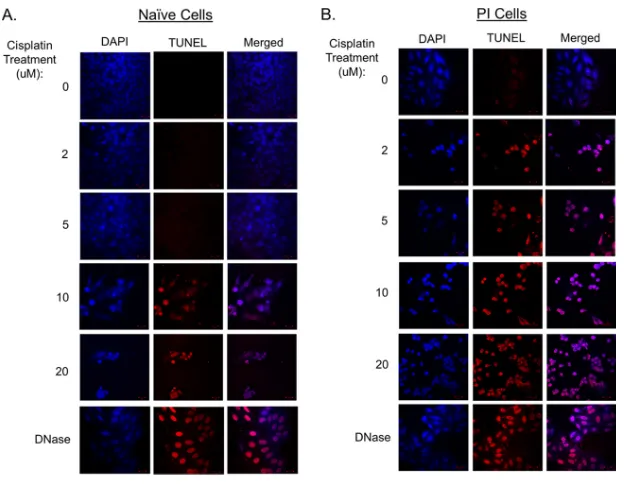
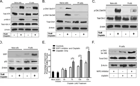
[image:5.585.41.497.70.373.2]deoxynucleotidyltransferase-mediated dUTP-biotin nick end labeling (TUNEL) assays were used to visualize DNA fragmentation by microscopy. As shown in Fig. 4A (left), naive cells did not show substantial DNA damage until treatment with 10M cisplatin. In contrast, treatment of PI cells with the lowest tested cisplatin concentration (2M) resulted in the majority of cells staining TUNEL positive (Fig. 4B). These data indicate that PI cells have enhanced sensitivity to cisplatin-induced cell death and DNA damage. PI cells have altered DNA damage repair pathways.DNA adducts induced by cisplatin are mainly recognized by the nucleotide excision repair (NER) pathway (36), which can lead to cell cycle arrest to efficiently repair the DNA or can lead to cell death based on the extent of DNA damage. To examine DNA damage repair pathways, naive and PI cells were treated with and without cisplatin and examined by Western blotting for the phosphorylation of upstream sensors of damaged DNA, such as the ataxia telangiectasia- and Rad3-related (ATR) protein and repair mediator protein gamma-histone H2A.X. As shown in Fig. 5A, both naive and PI cells responded to cisplatin treatment in similar fashions, as evidenced by similar levels of phosphorylation of ATR. The level of phosphorylation of H2A.X was slightly lower in PI cells treated with cisplatin than in naive cells treated with cisplatin.
Downstream of H2A.X, checkpoint kinase 1 (Chk1) and Chk2 are two DNA repair effector proteins, which, when activated by phosphorylation, can lead to cell cycle arrest and the repair of damaged DNA (37). As shown in Fig. 5B, phosphorylation of Chk1 at Ser317 and Ser345 was detected in naive cells treated with cisplatin, whereas phosphorylation at these sites was not detected in PI cells treated with cisplatin. Similarly, phosphorylation of Chk2 at Ser516 was detected in naive cisplatin-treated cells but was greatly diminished in PI cells (Fig. 5C). Taken together, these data indicate that DNA damage is sensed in PI cells, as evidenced by the phosphorylation of H2A.X; however, PI cells are altered in downstream Chk1 and Chk2 signaling, resulting in a defect in DNA repair.
Wild-type p53-inducible protein 1 (WIP1) is a stress-responsive phosphatase that acts as a negative-feedback loop to terminate the DNA repair pathways (38) through dephosphorylation of signaling proteins, including Chk1 and Chk2 (39–42). As shown in
FIG 4PI cells have enhanced DNA damage following cisplatin treatment. Naive (A) and PI (B) HEp-2 cells were treated with the indicated concentrations of cisplatin for 18 h. Alternatively, cells were treated with DNase as a positive control (bottom). All samples were also treated with DAPI to visualize DNA. Cells were analyzed by anin situTUNEL assay to determine levels of DNA fragmentation.
Fox and Parks Journal of Virology
on November 6, 2019 by guest
http://jvi.asm.org/
[image:6.585.48.363.68.312.2]the Western blot in Fig. 5D, levels of p53 and WIP1 were elevated in cisplatin-treated PI cells compared to those in naive cells. This finding raised the hypothesis that WIP1 activity against DNA repair proteins contributed to the enhanced sensitivity of PI cells to cisplatin. A prediction of this hypothesis is that WIP1 inhibition should result in decreased cell killing by cisplatin treatment. To test this, PI cells were pretreated with a WIP1 inhibitor, followed by treatment with increasing concentrations of cisplatin and cell viability assays. As shown in Fig. 5E, treatment of PI cells with 125 M cisplatin resulted in⬃45% of the population being positive for propidium iodide staining. In the presence of the WIP1 inhibitor, cisplatin treatment resulted in⬃25% of cells staining positive for propidium iodide. Consistent with a decrease in phosphatase activity, WIP1 inhibition of PI cells followed by cisplatin treatment restored the phosphorylation of Chk1 (Fig. 5F). These data are consistent with the hypothesis that an elevated expres-sion level of WIP1 in PI cells results in alterations in DNA repair pathway effector proteins, which are unable to effectively repair damaged DNA.
Acute infection with the PIV5 P/V-CPIⴚmutant sensitizes cancer cells to DNA damage-induced death. In the above-described studies, we sought to understand how PI cells were sensitized to stress pathways leading to cell death. Our results raised the question of whether the phenotype of enhanced sensitivity to cisplatin-induced death was limited to P/V-CPI⫺-derived PI cells or was also seen during acute infections
with the P/V-CPI⫺virus. To test this, naive cells were mock infected or infected with the
P/V-CPI⫺mutant, followed by treatment for 24 h with increasing doses of cisplatin and
cell viability assays. As shown in Fig. 6A, treatment of mock-infected cells with 75M cisplatin resulted in ⬃12% annexin V-positive cells and ⬃18% propidium iodide-positive cells. In contrast, acute infection with the P/V-CPI⫺ mutant followed by
treatment with 75M cisplatin resulted in⬎50% annexin V-positive cells and⬃40%
FIG 5PI cells have altered DNA repair pathways. (A to D) Naive and PI HEp-2 cells were treated with 75M cisplatin for 24 h, and lysates were analyzed by Western blotting for the DNA damage repair sensors ATR and H2A.X (A), the effector proteins Chk1 (B) and Chk2 (C), or p53 and WIP1 (D). (E) PI HEp-2 cells were treated with or without a WIP1 inhibitor for 24 h, followed by an additional 24 h of cisplatin treatment with or without the WIP1 inhibitor before analysis by flow cytometry. Values are the means for three samples, with**and***indicatingPvalues of⬍0.01 and 0.001, respectively. (F) PI HEp-2 cells were treated with or without a WIP1 inhibitor for 24 h, followed by an additional 24 h of cisplatin treatment with or without the WIP1 inhibitor, and lysates were analyzed by Western blotting for the DNA damage repair effector protein Chk1.
Parainfluenza Virus Infection and DNA Damage Response Journal of Virology
on November 6, 2019 by guest
http://jvi.asm.org/
[image:7.585.42.497.72.354.2]propidium iodide-positive cells. Under similar conditions, the DNA damage repair pathway proteins Chk1 and Chk2 were phosphorylated in mock-infected cisplatin-treated cells, as expected, but not in the case of cells acutely infected with P/V-CPI⫺or
in PI cells (Fig. 6B). These data indicate that acute infection with the P/V-CPI⫺mutant
can sensitize cancer cells to cisplatin-induced death and alter DNA damage response pathways similarly to what is seen with PI cells.
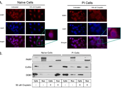
PI cells show a difference in the distribution of the DDB1 protein following treatment with a DNA-damaging agent.Upon DNA damage, DDB1 binds to DDB2, forming the DNA damage binding (DDB) complex that can then translocate to the nucleus. DDB1 is also known to function in a cytoplasmic complex with the rubulavirus V protein to induce STAT degradation (14–17). As such, we hypothesized that the DDB1 protein in PI cells would be localized to the cytoplasm and altered in its translocation to the nucleus in response to DNA damage. To test this hypothesis, naive and PI cells were mock treated or treated with 100M cisplatin before immunostaining for DDB1. As shown in Fig. 7A for naive cells, DDB1 appeared in both the nucleus and cytoplasm but had exclusively nuclear staining after cisplatin treatment. While untreated PI cells had staining similar to that of untreated naive cells, cisplatin treatment of PI cells resulted in a perinuclear localization with very little nuclear accumulation. Thus, these data suggest that DDB1 is unable to enter the nucleus when PI cells are treated with cisplatin.
FIG 6Acute infection by the P/V mutant sensitizes cells to a DNA-damaging agent and results in deficient DNA repair signaling. (A) Naive HEp-2 cells were mock infected or infected with the P/V-CPI⫺mutant for 18 h. Cells were treated with the indicated concentrations of cisplatin for 24 h before analysis for annexin V and propidium iodide staining by flow cytometry. Values are means for three samples, with
***indicatingPvalues of⬎0.001. (B) Naive cells, PI HEp-2 cells, or acutely P/V-CPI⫺-infected cells were treated with 75M cisplatin for 24 h. Cell lysates were analyzed by Western blotting for the indicated DNA repair proteins.
Fox and Parks Journal of Virology
on November 6, 2019 by guest
http://jvi.asm.org/
[image:8.585.44.476.72.416.2]As an alternative approach to localize DDB1 after DNA damage, naive and PI cells were treated with 50M cisplatin, and cell extracts were fractionated into nuclear and cytoplasmic fractions, followed by Western blotting for a nuclear control protein, PARP; a cytoplasmic control protein, cofilin; and DDB1. As shown in Fig. 7B, PARP localized to the nucleus as a high-molecular-weight protein in untreated cells and as a cleaved smaller form in cisplatin-treated cells. Cofilin largely localized to the cytoplasm, al-though PI cells had some of this protein detected in the nucleus. Most importantly, DDB1 was detected in the nuclear extract of cisplatin-treated naive cells but was not detected in the nuclear extract of cisplatin-treated PI cells. Taken together, these data support the proposal that DDB1 is defective in translocating to the nucleus upon DNA damage in PI cells.
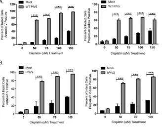
Acute infections with WT PIV5 and hPIV2 sensitize cancer cells to DNA damage-induced death.We tested the hypothesis that WT viruses in the Rubulavirusfamily could also sensitize cancer cells to DNA-damaging agents. Naive HEp-2 cells were mock infected or infected with WT PIV5 or hPIV2, followed by 24 h of treatment with doses of cisplatin and cell viability assays. Treatment of WT PIV5-infected cells with 100M cisplatin resulted in about 90% annexin V-positive cells and 65% propidium iodide-positive cells, whereas about 20% of the mock-infected cells were annexin V iodide-positive (Fig. 8A). As shown in Fig. 8B, treatment of cells with 50M cisplatin after acute hPIV2 infection resulted in⬃80% annexin V-positive and 75% propidium iodide-positive cells, values much higher than those seen for cisplatin-treated mock-infected cells or infected cells that did not receive cisplatin treatment. Taken together, these data indicate that acute infections with at least two WT rubulaviruses can sensitize cells to DNA damage-induced cell killing.
FIG 7PI cells show a difference in the distribution of the DDB1 protein following treatment with a DNA-damaging agent. (A) Naive and PI HEp-2 cells were treated with 150M cisplatin for 18 h and stained with antibody to DDB1 and with DAPI. Representative pictures are shown. (B) Naive and persistently P/V mutant-infected HEp2 cells were treated with 50M cisplatin for 18 h prior to cell lysis and separation into nuclear (Nuc) and cytoplasmic (Cyto) fractions. Samples were analyzed for the indicated proteins by Western blotting.
Parainfluenza Virus Infection and DNA Damage Response Journal of Virology
on November 6, 2019 by guest
http://jvi.asm.org/
[image:9.585.44.463.68.373.2]DISCUSSION
During our analysis of the capacity of the P/V-CPI⫺mutant to kill a range of cancer
cell lines, we discovered that infection results in the majority of the cells dying, but in some cases, there is an emergence of a population of cells that survive as P/V-CPI⫺PI
cells. The goal of the work described here was to understand how cells are altered to survive harboring a cytopathic virus. Our most striking finding, which emerged from challenge experiments aimed at testing resistance to apoptosis, is that cells either acutely or persistently infected with P/V-CPI⫺have an enhanced sensitivity to
DNA-damaging agents. This was evidenced in PI cells by increased cisplatin-induced DNA damage and death relative to those of naive cells. This was also reflected in alterations in PI cell DNA damage response signaling pathways in the nucleus (e.g., Chk1) as well as in alterations of the translocation of key DNA damage response factors from the cytoplasm to the nucleus (e.g., DDB1). Based on these findings, we present a model below for how cytoplasmically replicating RNA viruses such as PIV5 and hPIV2 alter cellular responses to DNA damage. These results with PI cells and acute infections have implications for the design of oncolytic RNA virus-based vectors and the possible use of combinatorial virus and chemical approaches to cancer therapy.
The mechanisms that lead to the transition from a cytopathic acute infection to a noncytopathic persistent infection are not completely understood (reviewed in refer-ence 43). This can involve apparently unrelated changes in cell morphology, such as with a persistent infection with foot-and-mouth disease virus (FMDV), where there are alterations in cell shape and increased growth, as well as acquired resistance to acute FMDV infection (44). In the case of MuV, previous studies have shown that PI cellsin vitro continually express MuV antigen and are resistant to the cytopathic effects of parental MuV (45). Host cell type can also be key in developing a persistent paramyxo-virus infection (46). Consistent with this, a persistent NDV infection was successfully established in one ovarian cancer cell line, OVCAR3, while other cell lines, such as OAW28, CAL27, FaDu, and PE/CA PJ15, were unable to establish a persistent infection
FIG 8Acute WT PIV5 and hPIV2 infections sensitize cells to a DNA-damaging agent. Naive HEp-2 cells were mock infected or infected with WT PIV5 (A) or hPIV2 (B) at an MOI of 10 for 18 h. Cells were treated with the indicated concentrations of cisplatin for 24 h, followed by analysis for annexin V (left) and propidium iodide (right) staining by flow cytometry. Values are the means for three samples, with***indicating aP
value of⬍0.001.
Fox and Parks Journal of Virology
on November 6, 2019 by guest
http://jvi.asm.org/
[image:10.585.43.371.73.330.2](47). PI cells can have an altered antiviral state induced by low levels of IFN production and the presence of IFN-stimulated genes, as seen in the case of NDV infection (47, 48). The P/V-CPI⫺virus is a potent inducer of IFN (18), and work is in progress to understand
the role that this cytokine plays in the differential ability to establish P/V-CPI⫺PI cells
in different cancer cell lines.
IFN induction could also play a role in the lower level of virus production from PI cells than that during acute infection (Fig. 1D), but other possibilities include lower cell growth rates, elevated cell stress responses (e.g., translation arrest), or the accumulation of mutations in the virus population. Consistent with this, virus derived from persis-tently MV-infected cells leading to subacute sclerosing panencephalitis (SSPE) had mutations in the M, H, and F genes due to polymerase errors and hypermutation events (49). Since the level of virus released from P/V-CPI⫺PI cells is lower than that with acute
P/V-CPI⫺infection (Fig. 1D), we hypothesize that the persistently infecting virus
ac-quired mutations that impair virus production and enhance persistence. Future studies will examine mutations in the virus genome derived from P/V-CPI⫺PI cells.
Under standard cell culture conditions, P/V-CPI⫺PI cells survive with elevated levels
of stress markers (e.g., annexin V) and cleaved caspases compared to those in naive cells. This survival was at least partially dependent on the activities of IAPs, since the inhibition of Survivin and XIAP dramatically increased cell death of PI cells relative to naive cells. These findings support a model where the continuous production of inducers of cell death during low-level P/V-CPI⫺replication (e.g., dsRNA) is countered
by IAP activity, and once IAP activity is inhibited, PI cells are induced to die. This raises the attractive hypothesis that screening tumors for IAP expression could provide a tool to determine their susceptibility to the establishment of a persistent infection by an oncolytic virus. In addition, the findings that PI cells are sensitized to IAP inhibitors, which are currently in various phases of clinical trials (50), raise the possibility of their repurposing as antivirals and as combined chemotherapy agents in conjunction with oncolytic viruses.
Since P/V-CPI⫺PI cells survive while harboring a cytopathic virus, we anticipated
that they would be more resistant to cell killing after challenge with external inducers of apoptosis. Unexpectedly, cisplatin-treated PI cells had more DNA damage than did naive cells and showed dose- and time-dependent enhancement of cisplatin-induced cell death. While the phosphorylation of “upstream” sensor molecules such as H2A.X appeared normal, PI cells were defective in the phosphorylation of “downstream” molecules such as Chk1 and -2. The phosphatase WIP1 was upregulated in PI cells by cisplatin treatment, and WIP1 inhibitors decrease cisplatin-induced cell death. WIP1 has also been reported to be upregulated and to have enhanced activity by the human T cell leukemia virus type 1 Tax protein. Here WIP1 upregulation results in suppressed DNA repair capabilities and cell cycle progression in the presence of DNA damage, although the exact mechanism has yet to be elucidated (51). We have found that the combination of P/V-CPI⫺and cisplatin is effective in HEp-2 (Fig. 3) and A549 human
non-small-cell lung carcinoma cells (data not shown), both of which have wild-type p53 (52, 53). Future studies will determine the roles of wild-type versus mutated p53 and WIP1 in P/V-CPI⫺-enhanced sensitivity to cisplatin.
How does an RNA virus that replicates in the cytoplasm sensitize cells to DNA-damaging agents and enhanced cell killing? The cellular protein DDB1 normally shuttles from the cytoplasm to the nucleus in response to DNA damage and acts to assemble sensors and effectors to efficiently repair DNA damage (54). This includes DNA repair signaling proteins such as Chk1 and -2, which are activated by phosphor-ylation (reviewed in reference 55). Once DNA repair is completed, WIP1 can be induced to deactivate the DNA damage signaling pathways by dephosphorylation for continued cell cycle progression and return the cell to homeostasis (38). PIV5 (and other rubula-viruses) hijacks DDB1 as part of the “V degradation complex,” which targets the cytoplasmic degradation of STAT proteins to inhibit IFN signaling pathways (14–17). Our microscopy and cell fractionation data on P/V-CPI⫺PI cells show that DDB1 is
localized largely in the cytoplasm in structures distinct from those in naive cells, and it
Parainfluenza Virus Infection and DNA Damage Response Journal of Virology
on November 6, 2019 by guest
http://jvi.asm.org/
is altered in accumulating in the nucleus in response to cisplatin treatment. We propose a model whereby the treatment of PI cells with cisplatin induces a typical DNA damage response, including the phosphorylation of ATR and H2A.X (Fig. 5A). The retention of DDB1 in the cytoplasm of PI cells results in the reduced sensing of the extent of DNA damage, in effect telling the cell that DNA damage is not extensive. The finding that WIP1 is induced in cisplatin-treated PI cells is consistent with our finding of unphos-phorylated Chk1 and Chk2 and with a model where PI cells shut down signaling pathways due to the lack of the DDB1-mediated ability to sense the extent of DNA damage. In our model, ultimately, the cell is unable to recognize the extent of DNA damage, which is then not repaired, and cells undergo apoptosis.
Our studies raise the possibility of using combination therapies with oncolytic paramyxoviruses and chemotherapies, as were studied previously with other viruses. For example, a phase II clinical trial has shown that the combination of the oncolytic adenovirus ONYX-015, cisplatin, and 5-fluorouracil was more effective than these therapies alone in patients with recurrent head and neck cancers (56). A phase I/II study investigated the combination of the oncolytic virus T-VEC, radiotherapy, and cisplatin for the treatment of patients with head and neck cancers. These clinical trials have found that combination therapy with an oncolytic virus and chemotherapy decreases disease progression and the rate of relapse and improves overall survival (57).
The finding that acute infection with the oncolytic virus P/V-CPI⫺sensitizes cells to DNA-damaging agents raises a practical application for our work in terms of the resistance of some cancer cells to DNA-damaging agents. Although cisplatin is the gold standard for numerous cancers, chemoresistance is a major concern (58). For example, cisplatin treatment of ovarian cancer patients is initially very effective, but tumor recurrence occurs in up to 75% of cases, resulting in chemotherapy-resistant tumors (59). Previous studies have shown that cisplatin resistance can result when cells increase DNA repair capabilities (36, 60–63). Conversely, cisplatin sensitivity has been associated with a lower capacity of DNA repair pathways in cells (64–67). Thus, an attractive property of an oncolytic virus would be its ability to impair DNA damage repair pathways and increase cisplatin sensitivity, especially in the case of drug-resistant tumors that have upregulated DNA repair pathways. This is in line with what we report here for the ability of P/V-CPI⫺to reduce DNA repair capacities.
A number of paramyxoviruses and rhabdoviruses are being developed as oncolytic vectors for tumor therapy, including MV, mumps virus, Sendai virus, and NDV (4–10). Our finding that the highly cytopathic PIV5 P/V mutant can establish persistent infection in some cells raises concern about the potential use of RNA viruses for oncolytic therapy. Future work will focus on the extent to which the establishment of PI cells occurs during treatment of tumors in animal model systems as well as the potential for combinations of cisplatin and P/V-CPI⫺to reduce tumor burdens.
MATERIALS AND METHODS
Cells, viruses, and plaque assays.Cultures of HEp-2, MDBK, Vero, and CV-1 cells were grown in Dulbecco modified Eagle medium (DMEM) supplemented with 10% heat-inactivated fetal bovine serum (HI FBS; HyClone, Logan, UT). WT recombinant PIV5 (rPIV5)-GFP was recovered as described previously from a cDNA plasmid (33) kindly provided by Robert Lamb (Northwestern University) and Biao He (University of Georgia) and was grown in MDBK cells. The P/V mutant rPIV5-P/V-CPI⫺ (P/V-CPI⫺) expressing GFP was generated and grown in Vero cells as described previously (18). PIV5 P/V-CPI⫺ encodes 6 naturally occurring mutations in the amino-terminal region of the P/V gene, resulting in amino acid changes of Y26H, V32I, T33I, L50P, L102P, and S157F (21). Human parainfluenza virus type 2 was grown in CV-1 cells. Viral titers were determined on CV-1 cells as described previously (18).
To obtain PI cell lines, HEp-2 cells were infected with the P/V-CPI⫺mutant at a multiplicity of infection (MOI) of 10, and the medium was replaced every 3 days postinfection (p.i.) for 2 weeks. P/V mutant-infected cells were sorted for high GFP expression levels by using a FACSCalibur flow cytometer (BD Bioscience, San Diego, CA) and were designated PI HEp-2 cells.
Fluorescence microscopy.Media were removed from cell monolayers and replaced with phosphate-buffered saline (PBS) before analysis by microscopy using a Zeiss Axiovert fluorescence microscope with a 10⫻objective lens. Exposure times were 46 ms for bright field and 500 ms for fluorescence. Slides were imaged on a Zeiss 710 confocal microscope with a 40⫻objective lens.
Fox and Parks Journal of Virology
on November 6, 2019 by guest
http://jvi.asm.org/
Cell viability assays.MTT cell viability assays were performed in 96-well dishes using Cell Titer 96 Aqueous One solution reagent (Promega) according to the manufacturer’s instructions. Data are ex-pressed as a percentage of the value for mock-infected cells analyzed in parallel.
Alternatively, PI or naive cells were treated as indicated in each figure legend (concentration of drug and time). Media and trypsinized adherent cells were centrifuged and analyzed for annexin V binding (BD Bioscience) and propidium iodide staining (BD Bioscience) as described by the manufacturer. Cells were analyzed by flow cytometry using the CytoFLEX system (Beckman Coulter), and 10,000 independent events were recorded and analyzed by using CytExpert software.
Western blotting.Dishes (60-mm diameter) of cells were treated as described in the figure legends, followed by lysis in 1⫻protein lysis buffer (Cell Signaling Technology). The cell lysate was resolved on 10 or 15% sodium dodecyl sulfate-polyacrylamide gel electrophoresis (SDS-PAGE) gels and transferred onto nitrocellulose membranes. Samples were probed with the antibodies indicated in the figure legends (Cell Signaling) or with anti--actin antibody (catalog number A5316; Sigma) and rabbit polyclonal antisera to the PIV5 NP, hemagglutinin-neuraminidase (HN), and P proteins (18). Blots were visualized with horseradish peroxidase-conjugated antibodies and chemiluminescence (Pierce ThermoScientific).
Chemical challenge experiments.YM155 was purchased from EMD Millipore and reconstituted in sterile dimethyl sulfoxide (DMSO) at a stock concentration of 3 mM. Embelin was purchased from Tocris and reconstituted in sterile DMSO at a stock concentration of 5 mM. Cisplatin was purchased from Sigma-Aldrich and reconstituted in sterile water at a stock concentration of 5 mM. The WIP1 inhibitor (GSK 2830371) was purchased from Tocris and reconstituted in sterile DMSO at a stock concentration of 5 mM. PI and naive cells cultured in 24-well plates (diameter of 2 cm) were challenged as indicated in the figure legends and treated with drugs that were diluted in DMEM containing 10% HI FBS.
Immunostaining and TUNEL staining. Cells grown on 8-chamber slides (ThermoFisher) were treated with cisplatin as indicated in the figure legends and analyzed by staining with anti-DDB1 (1:250 dilution; Zymed, Invitrogen) followed by goat anti-rabbit Alexa Fluor 568 (1:2,000 dilution; Invitrogen). 4=,6-Diamidino-2-phenylindole (DAPI) was included to stain for nuclei. To visualize DNA damage, cells were treated with cisplatin as indicated in the figure legends and analyzed by using the Click-iT TUNEL Alexa Fluor 647 assay kit according to the manufacturer’s instructions (Invitrogen).
Nuclear extraction.Cells were grown in 6-well dishes and treated with cisplatin as indicated in the figure legends. Nuclear and cytoplasmic extracts were obtained by using a kit according to manufac-turer’s guidelines (Active Motif). Ten micrograms of extracts was lysed in 1⫻protein lysis buffer (Cell Signaling Technology) and analyzed by Western blotting.
Statistical analyses.Statistical analysis was performed by using GraphPad Student’sttest.
ACKNOWLEDGMENTS
We thank members of the Parks laboratory for input, Namita Varudkar and Kritika Kedarinath for excellent technical assistance, and Annie Mayer for the original obser-vations leading to this study.
This work was supported in part by a grant from Circle of Hope Cancer Research.
REFERENCES
1. Kaufman HL, Kohlhapp FJ, Zloza A. 2015. Oncolytic viruses: a new class of immunotherapy drugs. Nat Rev Drug Discov 14:642– 662.https://doi .org/10.1038/nrd4663.
2. Fukuhara H, Ino Y, Todo T. 2016. Oncolytic virus therapy: a new era of cancer treatment at dawn. Cancer Sci 107:1373–1379.https://doi.org/10 .1111/cas.13027.
3. Russell SJ, Peng KW, Bell JC. 2012. Oncolytic virotherapy. Nat Biotechnol 30:658 – 670.https://doi.org/10.1038/nbt.2287.
4. Elankumaran S, Rockemann D, Samal SK. 2006. Newcastle disease virus exerts oncolysis by both intrinsic and extrinsic caspase-dependent path-ways of cell death. J Virol 80:7522–7534.https://doi.org/10.1128/JVI .00241-06.
5. Kinoh H, Inoue M, Washizawa K, Yamamoto T, Fujikawa S, Tokusumi Y, Iida A, Nagai Y, Hasegawa M. 2004. Generation of a recombinant Sendai virus that is selectively activated and lyses human tumor cells expressing matrix metalloproteinases. Gene Ther 11:1137–1145.https://doi.org/10 .1038/sj.gt.3302272.
6. Lorence RM, Reichard KW, Katubig BB, Reyes HM, Phuangsab A, Mitchell BR, Cascino CJ, Walter RJ, Peeples ME. 1994. Complete regression of human neuroblastoma zenografts in athymic mice after local Newcastle disease virus therapy. J Natl Cancer Inst 86:1228 –1233.https://doi.org/ 10.1093/jnci/86.16.1228.
7. Myers R, Greiner S, Harvey M, Soeffker D, Frenzke M, Abraham K, Shaw A, Rozenblatt S, Federspiel MJ, Russell SJ, Peng KW. 2005. Oncolytic activities of approved mumps and measles vaccines for therapy of ovarian cancer. Cancer Gene Ther 12:593–599.https://doi.org/10.1038/ sj.cgt.7700823.
8. Peng KW, Donovan KA, Schneider U, Cattaneo R, Just JA, Russell SJ. 2003. Oncolytic measles viruses displaying a single-chain antibody against CD38, a myeloma cell marker. Blood 101:2557–2562.https://doi.org/10 .1182/blood-2002-07-2195.
9. Yu N, Puckett S, Antinozzi PA, Cramer SD, Lyles DS. 2015. Changes in susceptibility to oncolytic vesicular stomatitis virus during progression of prostate cancer. J Virol 89:5250 –5263. https://doi.org/10.1128/JVI .00257-15.
10. Matveeva OV, Guo ZS, Senin VM, Senina AV, Shabalina SA, Chumakov PM. 2015. Oncolysis by paramyxoviruses: preclinical and clinical studies. Mol Ther Oncolytics 2:150017.https://doi.org/10.1038/mto.2015.17. 11. Choppin PW. 1964. Multiplication of a myxovirus (Sv5) with minimal
cytopathic effects and without interference. Virology 23:224 –233. https://doi.org/10.1016/0042-6822(64)90286-7.
12. Sun M, Rothermel TA, Shuman L, Aligo JA, Xu S, Lin Y, Lamb RA, He B. 2004. Conserved cysteine-rich domain of paramyxovirus simian virus 5 V protein plays an important role in blocking apoptosis. J Virol 78: 5068 –5078.https://doi.org/10.1128/JVI.78.10.5068-5078.2004. 13. Andrejeva J, Poole E, Young DF, Goodbourn S, Randall RE. 2002. The
p127 subunit (DDB1) of the UV-DNA damage repair binding protein is essential for the targeted degradation if STAT1 by the V protein of the paramyxovirus simian virus 5. J Virol 76:11379 –11386.https://doi.org/ 10.1128/JVI.76.22.11379-11386.2002.
14. Didcock L, Young DF, Goodbourn S, Randall RE. 1999. The V protein of simian virus 5 inhibits interferon signaling by targeting STAT1 for proteasome-mediated degradation. J Virol 73:9928 –9933.
15. Ulane CM, Kentsis A, Cruz CD, Parisien JP, Schneider KL, Horvath CM.
Parainfluenza Virus Infection and DNA Damage Response Journal of Virology
on November 6, 2019 by guest
http://jvi.asm.org/
2005. Composition and assembly of STAT-targeting ubiquitin ligase complexes: paramyxovirus V protein carboxyl terminus is an oligomer-ization domain. J Virol 79:10180 –10189.https://doi.org/10.1128/JVI.79 .16.10180-10189.2005.
16. Parisien JP, Leu JF, Rodriguez JJ, Sullivan BM, Moscona A, Parks GD, Lamb RA, Horvath CM. 2001. The V protein of human parainfluenza virus 2 antagonizes type I interferon responses by destabilizing signal trans-ducer and activator of transcription 2. Virology 283:230 –239.https://doi .org/10.1006/viro.2001.0856.
17. Young DF, Didcock L, Goodbourn S, Randall RE. 2000. Paramyxoviridae use distant virus-specific mechanisms to circumvent the interferon re-sponse. Virology 269:383–390.https://doi.org/10.1006/viro.2000.0240. 18. Wansley EK, Parks GD. 2002. Naturally occurring substitutions in the P/V
gene convert the noncytopathic paramyxovirus simian virus 5 into a virus that induces alpha/beta interferon synthesis and cell death. J Virol 76:10109 –10121.https://doi.org/10.1128/JVI.76.20.10109-10121.2002. 19. Wansley EK, Dillon PJ, Gainey MD, Tam J, Cramer SD, Parks GD. 2005.
Growth sensitivity of a recombinant simian virus 5 P/V mutant to type 1 interferon differs between tumor cell lines and normal primary cells. Virology 335:131–144.https://doi.org/10.1016/j.virol.2005.02.004. 20. Young VA, Dillon PJ, Parks GD. 2006. Variants of the paramyxovirus
simian virus 5 with accelerated or delayed viral gene expression activate proinflammatory cytokine synthesis. Virology 350:90 –102. https://doi .org/10.1016/j.virol.2006.01.006.
21. Dillon PJ, Wansley EK, Young VA, Alexander-Miller MA, Parks GD. 2006. Exchange of P/V genes between two non-cytopathic simian virus 5 variants results in a recombinant virus that kills cells through death pathways that are sensitive to caspase inhibitors. J Gen Virol 87: 3643–3648.https://doi.org/10.1099/vir.0.82242-0.
22. Gainey MD, Dillon PJ, Clark KM, Manuse MJ, Parks GD. 2008. Paramyxovirus-induced shutoff of host and viral protein synthesis: role of the P and V proteins in limiting PKR activation. J Virol 82:828 – 839. https://doi.org/10.1128/JVI.02023-07.
23. Lamb RA, Parks GD. 2007.Paramyxoviridae: the viruses and their repli-cation, p 1449 –1496.InKnipe DM, Howley PM, Griffin DE, Lamb RA, Martin MA, Roizman B, Straus SE (ed), Fields virology, 5th ed. Lippincott Williams & Wilkins, Philadelphia, PA.
24. Lin Y, Horvath F, Aligo JA, Wilson R, He B. 2005. The role of simian virus 5 V protein on viral RNA synthesis. Virology 338:270 –280.https://doi .org/10.1016/j.virol.2005.05.014.
25. Childs K, Stock N, Ross C, Andrejeva J, Hilton L, Skinner M, Randall R, Goodbourn S. 2007. mda-5, but not RIG-I, is a common target for paramyxovirus V proteins. Virology 359:190 –200. https://doi.org/10 .1016/j.virol.2006.09.023.
26. Young VA, Parks GD. 2003. Simian virus 5 is a poor inducer of chemokine secretion from human lung epithelial cells: identification of viral mutants that activate interleukin-8 secretion by distinct mechanisms. J Virol 77:7124 –7130.https://doi.org/10.1128/JVI.77.12.7124-7130.2003. 27. Dillon PJ, Parks GD. 2007. Role for the phosphoprotein P subunit of the
paramyxovirus polymerase in limiting induction of host cell antiviral responses. J Virol 81:11116 –11127.https://doi.org/10.1128/JVI.01360-07. 28. Gainey MD, Manuse MJ, Parks GD. 2008. A hyperfusogenic F protein enhances the oncolytic potency of a paramyxovirus simian virus 5 P/V mutant without compromising sensitivity to type I interferon. J Virol 82:9369 –9380.https://doi.org/10.1128/JVI.01054-08.
29. Wong LH, Krauer KG, Hatzinisiriou I, Estcourt MJ, Hersey P, Tam ND, Edmondson S, Devenish RJ, Ralph SJ. 1997. Interferon-resistant human melanoma cells are deficient in ISGF3 components, STAT1, STAT2, and p48-ISGF3. J Biol Chem 272:28779 –28785.https://doi.org/10.1074/jbc .272.45.28779.
30. Xu B, Grander D, Sangfelt O, Einhorn S. 1994. Primary leukemia cells resistant to alpha-interferon in vitro are defective in the activation of the DNA-binding factor interferon-stimulated gene factor 3. Blood 84: 1942–1949.
31. Bell JC, Lichty B, Stojdl D. 2003. Getting oncolytic virus therapies off the ground. Cancer Cell 4:7–11.https://doi.org/10.1016/S1535-6108(03) 00170-3.
32. Wansley EK, Grayson JM, Parks GD. 2003. Apoptosis induction and interferon signaling but not IFN-beta promoter induction by an SV5 P/V mutant are rescued by coinfection with wild-type SV5. Virology 316: 41–54.https://doi.org/10.1016/S0042-6822(03)00584-1.
33. He B, Paterson RG, Ward CD, Lamb RA. 1997. Recovery of infectious SV5 from cloned DNA and expression of a foreign gene. Virology 237: 249 –260.https://doi.org/10.1006/viro.1997.8801.
34. Deveraux QL, Takahashi R, Salvesen GS, Reed JC. 1997. X-linked IAP is a direct inhibitor of cell-death proteases. Nature 388:300 –304.https://doi .org/10.1038/40901.
35. Tamm I, Wang Y, Sausville E, Scudiero DA, Vigna N, Oltersdorf T, Reed JC. 1998. IAP-family protein survivin inhibits caspase activity and apoptosis induced by Fas (CD95), Bax, caspases, and anticancer drugs. Cancer Res 58:5315–5320.
36. Siddik ZH. 2003. Cisplatin: mode of cytotoxic action and molecular basis of resistance. Oncogene 22:7265–7279. https://doi.org/10.1038/sj.onc .1206933.
37. Hurley PJ, Bunz F. 2007. ATM and ATR, components of an integrated circuit. Cell Cycle 6:414 – 417.https://doi.org/10.4161/cc.6.4.3886. 38. Lu X, Nguyen TA, Moon SH, Darlington Y, Sommer M, Donehower LA.
2008. The type 2C phosphatase Wip1: an oncogenic regulator of tumor suppressor and DNA damage response pathways. Cancer Metastasis Rev 27:123–135.https://doi.org/10.1007/s10555-008-9127-x.
39. Shreeram S, Demidov ON, Hee WK, Yamaguchi H, Onishi N, Kek C, Timofeev ON, Dudgeon C, Fornace AJ, Anderson CW, Minami Y, Appella E, Bulavin DV. 2006. Wip1 phosphatase modulates ATM-dependent signaling pathways. Mol Cell 23:757–764.https://doi.org/ 10.1016/j.molcel.2006.07.010.
40. Cha H, Lowe JM, Li H, Lee JS, Belova GI, Bulavin DV, Fornace AJ. 2010. Wip1 directly dephosphorylates gamma-H2A.X and attenuates the DNA damage response. Cancer Res 70:4112– 4122.https://doi.org/10.1158/ 0008-5472.CAN-09-4244.
41. Lu X, Nannenga B, Donehower LA. 2005. PPM1D dephosphorylates Chk1 and p53 and abrogates cell cycle checkpoints. Genes Dev 19:1162–1174. https://doi.org/10.1101/gad.1291305.
42. Olivia-Trastoy M, Berthonaud V, Chevalier A, Ducrot C, Marsolier-Kergoat MC, Mann C, Leteurtre F. 2007. The Wip1 phosphatase (PPM1D) antag-onizes activation of the Chk2 tumour suppressor kinase. Oncogene 26:1449 –1458.https://doi.org/10.1038/sj.onc.1209927.
43. Rima BK, Martin SJ. 1976. Persistent infection of tissue culture cells by RNA viruses. Med Microbiol Immunol 162:89 –118.https://doi.org/10 .1007/BF02121320.
44. de la Torre JC, Martínez-Salas E, Diez J, Villaverde A, Gebauer F, Rocha E, Dávila M, Domingo E. 1988. Coevolution of cells and viruses in a persis-tent infection of foot-and-mouth disease virus in cell culture. J Virol 62:2050 –2058.
45. Walker DL, Hinze HC. 1962. A carrier state of mumps virus in human conjunctiva cells. I. General characteristics. J Exp Med 116:739 –750. https://doi.org/10.1084/jem.116.5.739.
46. Holmes KV, Choppin PW. 1966. On the role of the response of the cell membrane in determining virulence. Contrasting effect of the parain-fluenza virus SV5 in two cell types. J Exp Med 124:501–520.https://doi .org/10.1084/jem.124.3.501.
47. Rangaswamy US, Wang W, Cheng X, McTamney P, Carroll D, Jin H. 2017. Newcastle disease virus establishes persistent infection in tumor cells in vitro: contribution of the cleavage site of fusion protein and second sialic acid binding site of hemagglutinin-neuraminidase. J Virol 91: e00770-17.https://doi.org/10.1128/JVI.00770-17.
48. Schneider WM, Chevillotte MD, Rice CM. 2014. Interferon-stimulated genes: a complex web of host defenses. Annu Rev Immunol 32:513–545. https://doi.org/10.1146/annurev-immunol-032713-120231.
49. Billeter MA, Cattaneo R, Spielhofer P, Kaelin K, Huber M, Schmid A, Baczko K, ter Meulen V. 1994. Generation and properties of measles virus mutations typically associated with subacute sclerosing panencephalitis. Ann N Y Acad Sci 724:367–377.https://doi.org/10.1111/j.1749-6632.1994 .tb38934.x.
50. Fulda S, Vucic D. 2012. Targeting IAP proteins for therapeutic interven-tion in cancer. Nat Rev Drug Discov 11:109 –124.https://doi.org/10.1038/ nrd3627.
51. Dayaram T, Lemoine FJ, Donehower LA, Marriott SJ. 2013. Activation of WIP1 phosphatase by HTLV-1 Tax mitigates the cellular response to DNA damage. PLoS One 8:e55989.https://doi.org/10.1371/journal .pone.0055989.
52. Abdulkarim B, Sabri S, Deutsch E, Chagraoui H, Maggiorella L, Thierry J, Eschwege F, Vainchenker W, Chouaïb S, Bourhis J. 2002. Antiviral agent cidofovir restores p53 function and enhances the radiosensitivity in HPV-associated cancers. Oncogene 21:2334 –2346. https://doi.org/10 .1038/sj.onc.1205006.
53. Yao CJ, Chow JM, Lin PC, Hu TS, Kuo HC, Huang JS, Bai KJ, Lai GM. 2016. Activation of p53/miR-34a tumor suppressor axis by Chinese herbal formula
Fox and Parks Journal of Virology
on November 6, 2019 by guest
http://jvi.asm.org/
JP-1 in A549 lung adenocarcinoma cells. Evid Based Complement Alternat Med 2016:5989681.https://doi.org/10.1155/2016/5989681.
54. Iovinea B, Iannellaa ML, Bevilacqua MA. 2011. Damage-specific DNA binding protein 1 (DDB1): a protein with a wide range of functions. Int J Biochem Cell Biol 43:1664 –1667.https://doi.org/10.1016/j.biocel.2011 .09.001.
55. Gabriele S, Raffaella DM, Fagagna FD. 2012. Crosstalk between chroma-tin state and DNA damage response in cellular senescence and cancer. Nat Rev Cancer 12:709 –720.https://doi.org/10.1038/nrc3344. 56. Khuri FR, Nemunnaitis J, Ganly I, Arseneau J, Tannock IF, Romel L, Gore
M, Ironside J, MacDougall MR, Heise C, Randlev B, Gillenwater AM, Bruso P, Kaye SB, Hong WK, Kirn DH. 2000. A controlled trial of intratumoral ONYX-015, a selectively replicating adenovirus, in combination with cisplatin and 5-fluorouracil in patients with recurrent head and neck cancer. Nat Med 6:879 – 885.https://doi.org/10.1038/78638.
57. Harrington KJ, Hingorani M, Tanay MA, Hickey J, Bhide SA, Carke PM, Renouf LC, Thway K, Sibtain A, McNeish IA, Newbold KL, Goldsweig H, Coffin R, Nutting CM. 2010. Phase I/II study of oncolytic HSV GM-CSF in combination with radiotherapy and cisplatin in untreated stage III/IV squamous cell cancer of the head and neck. Clin Cancer Res 16: 4005– 4015.https://doi.org/10.1158/1078-0432.CCR-10-0196.
58. Dasari S, Tchounwou PB. 2014. Cisplatin in cancer therapy: molecular mechanisms of action. Eur J Pharmacol 5:364 –378.https://doi.org/10 .1016/j.ejphar.2014.07.025.
59. Agarwal R, Kaye SB. 2003. Ovarian cancer: strategies for overcoming resistance to chemotherapy. Nat Rev Cancer 3:502–516.https://doi.org/ 10.1038/nrc1123.
60. Parker RJ, Eastman A, Bostick-Bruton F, Reed E. 1991. Acquired cisplatin resistance in human ovarian cancer cells is associated with enhanced repair of cisplatin-DNA lesions and reduced drug accumulation. J Clin Invest 87:772–777.https://doi.org/10.1172/JCI115080.
61. Masuda H, Ozols RF, Lai GM, Fojo A, Rothenberg M, Hamilton TC. 1988. Increased DNA repair as a mechanism of acquired resistance to cis-diamminedichloroplatinum(II) in human ovarian cancer cell lines. Cancer Res 48:5713–5716.
62. Pooter CMD, Oosterom ATV, Scalliet PG, Maes RA, Brujin EAD. 1996. Correlation of the response to cisplatin of human ovarian cancer cell lines, originating from one tumor but with different sensitivity, with the recovery of DNA adducts. Biochem Pharmacol 51:629 – 634.https://doi .org/10.1016/S0006-2952(95)02229-5.
63. Ferry KV, Hamilton TC, Johnson SW. 2000. Increased nucleotide excision repair in cisplatin-resistant ovarian cancer cells: role of ercc1-xpf. Biochem Pharmacol 60:1305–1313.https://doi.org/10.1016/S0006-2952 (00)00441-X.
64. Rosell R, Taron M, Barnadas A, Scagliotti G, Sarries C, Roig B. 2003. Nucleotide excision repair pathways involved in cisplatin resistance in non-small-cell lung cancer. Cancer Control 10:297–305.https://doi.org/ 10.1177/107327480301000404.
65. Welsh C, Day R, McGurk C, Masters JR, Wood RD, Köberle B. 2004. Reduced levels of XPA, ERCC1 and XPF DNA repair proteins in testis tumor cell lines. Int J Cancer 110:352–361.https://doi.org/10.1002/ijc .20134.
66. Calsou P, Barret JM, Cros S, Salles B. 1993. DNA excision-repair synthesis is enhanced in a murine leukemia L1210 cell line resistant to cisplatin. Eur J Biochem 211:403– 409.https://doi.org/10.1111/j.1432-1033.1993 .tb17563.x.
67. Hill BT, Scanlon KJ, Hansson J, Harstrick A, Pera M, Fichtinger-Schepman AMJ, Shellard SA. 1994. Deficient repair of cisplatin-DNA adducts iden-tified in human testicular teratoma cell lines established from tumours from untreated patients. Eur J Cancer 30:832– 837.https://doi.org/10 .1016/0959-8049(94)90301-8.
Parainfluenza Virus Infection and DNA Damage Response Journal of Virology