A STUDY ON THE CLINICAL PROFILE OF TOXIC
AND NUTRITIONAL OPTIC NEUROPATHY
DISSERTATION SUBMITTED FOR
MS ( BRANCH III ) OPHTHALMOLOGY
THE TAMIL NADU DR. M.G.R. MEDICAL UNIVERSITY
CHENNAI
MAY 2018
CERTIFICATE
This is to certify that the dissertation entitled “A STUDY ON THE CLINICAL PROFILE OF TOXIC AND NUTRITIONAL OPTIC NEUROPATHY” is a bonafide work done by Dr. KeerthanaSoundararajan under our guidance and supervision in the Neuro –Ophthalmology Department of Aravind Eye Hospital and Post Graduate Institute of Ophthalmology, Madurai during the period of her post graduate training in Ophthalmology for
June 2015 – May 2018.
GUIDE:
Dr.A.Kowsalya Thiruvadi Dr.Venkatesh Prajna
Consultant Prof.&Head of the Department
Department of Neuro Ophthalmology Aravind Eye Hospital & Aravind Eye Hospital & PG Institute of Ophthalmology PG Institute of Ophthalmology Madurai
Madurai
Dr.R.Rathinam, Principal
Aravind Eye Hospital&
PG Institute of Ophthalmology
DECLARATION
I, Dr. Keerthana Soundararajan, hereby declare this dissertation,
“A STUDY ON THE CLINICAL PROFILE OF TOXIC AND NUTRITIONAL OPTIC NEUROPATHY” is being submitted in partial fulfilment for the award of M.S.in Ophthalmology Degree by the Tamil Nadu DR.MGR. Medical University in the examination to be held in May 2018.
I declare that this dissertation is my original work and has not formed the basis for the award of any other degree or diploma awarded to me previously.
Dr. Keerthana Soundararajan, Aravind Eye Hospital,
Madurai
ACKNOWLEDGEMENT
I am grateful to The Almighty for His blessings and for giving me
the opportunity to be a part of the medical profession and Aravind Eye
Care System.
I take this opportunity to pay my respect and homage to
Dr.G.Venkatasamy, the founder of this great institution and the visionary, whose spiritual realisation and dynamism in the material world
has led this institute against all odds to its epitome.
I extend my heartfelt words of gratitude to my esteemed guide
Dr. A. Kowsalya Thiruvadi, Professor , Neuro Ophthalmology, Aravind Eye Hospital, Madurai for her constant support and guidance throughout
the duration of my thesis.
I am deeply indebted to Dr.N.V.Prajna, Director of Medical Education for his dynamic and overwhelming support during my
residency programme.
I would like to thank my co guide Dr. S. Mahesh Kumar, Head of the Department, Neuro Ophthalmology, Aravind Eye Hospital for his
I would like to thank our Chairman Dr.R.D.Ravindran whose words of encouragement and guidance have always been a source of
inspiration for all of us in this institution.
A word of gratitude to Dr.P. Namperumalsamy, Chairman Emeritus and Dr.G.Natchiar, Director Emeritus (Human Resource Department) , Dr.M.Srinivasan , Director-Emeritus, and other scholars of Ophthalmology at Aravind Eye Care System.
My very sincere thanks to Mr. Govindarajan, Librarian, and all other staff of the library for the immediate responses in providing all the
articles and academic support required in the completion of thesis.
I would also like to thank the timely assistance of the biostatistician
Mrs.Iswarya, who has helped with the task of compiling the statistics and enabled in the preparation of results.
I thank the patients who have patiently listened to and compiled
with this study, for without them, this study would not have been
possible.
I would like to thank my parents, my husband Dr.M.Raghul and my
friends for their unwavering support and love towards me.
CONTENTS
NO
TITLE
PAGE
1
Introduction
1
2
Review Of Literature
2
3
Aim and Objectives
47
4
Materials and Methods
48
5
Results
51
6
Discussion
69
7
Conclusion and Limitations
75
Bibliography
ANNEXURE
Proforma
Informed Consent Form
Institutional Review Board- Approval
Plagiarism Report
INTRODUCTION
Nutritional deficiency may also be the cause of a genuine optic
neuropathy, which is sometimes associated with involvement of the
peripheral nervous system.1With the exception of vitamin B12, no
specific nutrient deficiency has been conclusively proved to cause optic
neuropathy in humans.2
The pathophysiological mechanisms which are involved in toxic
and nutritional optic neuropathies affect biochemical pathways involved
in cell energetic production, correction of oxidative stress and quenching
of free radicals causing papillomacular bundle damage.1,3In the current
scenario , the treatment is limited to the intensive use of vitamins with
variable results in individual cases, and also to the implementation of
preventive measures, when feasible.1,4,5
As this entity is mostly a disease of exclusion, with a blanket cover
diagnosis; and with prompt probing into the etiology with directional
history, clinical examination and specific investigations , the underlying
cause can be identified and addressed for maximal patient benefits and
REVIEW OF LITERATURE
The optic nerve is susceptible to damage from various toxins,
including drugs, methanol, metals, organic solvents and probably some
sort of tobacco.6This group of disorders is named as toxic optic
neuropathy (TON) and is characterized by bilateral visual loss, reduction
of colour vision and central or cecocentralscotoma. Toxic optic
neuropathy may be triggered or just aggravated by nutritional deficits,
including the vitamins cobalamin (B12), folic acid, thiamine (B1),
riboflavin (B2), niacin (B3), pyridoxine (B6) and proteins with amino
acids.
For example, among malnourished and intoxicated patients, both
factors usually play an equal role contributing to the TON. Because of
people’s greater exposure to toxic substances in environment and food, and coexisting malnutrition.they are more prevalent in developing
countries,. No age, gender and racial predilections have been
demonstrated. The existence of a nutritional deficiency often constitutes
the predisposing factor that produces symptoms of optic nerve
involvement in the presence of toxins, including iatrogenic ones, or in a
The disruption of biochemical mechanisms involved in the
correction of oxidative stress appear to be a common mechanism in these
different forms of optic neuropathies11. It should be noted that the earliest
possible diagnosis allows better treatment of the nutritional deficit or the
toxic disorder and helps in the prevention of late sequelae. In fact, with
the earliest diagnosis, the optic nerve injury tends to be more reversible.
Tobacco alcohol amblyopia is an outdated term that was used to
describe visual impairment as a result of tobacco and alcohol abuse
accompanied usually by nutritional deficiencies12,13. The visual
impairment usually presents as a centrocecalscotoma with
dyschromatopsia. Its incidence is declining likely with time, secondary to
nutritional supplementation which seems to indicate a major role of
vitamin deficiency. Hence the disease process was renamed as Nutritional
MECHANISM OF OPTIC NEUROPATHIES
The mechanism of nutritional or toxic optic neuropathies is
certainly not unique. Several substances have been suggested including
riboflavin, glutathione, cyanides or zinc . The mechanisms involvedare
complex and includes a number of effective hydrosolubleor liposoluble
quenchers of oxygen and free radical orthe cofactors of these reactions.
Theconcentration or availability of some of the anti-oxidantsplay a
majorprotective role against the development of optic neuropathies
despite exposure to toxins.This corrective system of oxidative stress play
a role, in the worsening of opticneuropathies secondary to glaucoma,
directly or indirectly. On theone hand, oxidative stress interferes
remodelling the collagen and elastin of the extracellular matrix and
affects the structure of thetrabeculum . On the other hand, the existence of
an oxidative stress expresses the production of large quantities offree
radicals. It can be postulated that free radicals interferein the modulation
of apoptosis of ganglion neurons. The combination of anti-oxidant and
nutritional factors appearsto have future therapeutic implications.
However, the pathophysiology responsible for this condition is still
poorly understood and multiple mechanisms have been suggested.
Tobacco, alcohol and vitamin deficiencies all contribute either in
Neuropathy (LHON) gene mutation are more prone to acquire tobacco
alcohol amblyopia. The common underlying mechanism postulated is the
impairment of mitochondrial oxidative phosphorylation which explains
the overlap between the two conditions14-16. Nicotine and cyanide from
tobacco play a role in causing the optic neuropathy17. In addition both the
alcohol and tobacco lead to production of free radicals exerting a direct
toxic effect. This may also explains the therapeutic effects of vitamins
such as riboflavin which acts as an antioxidant., Malnutrition, thiamine
deficiency, gradual accumulation of formic acid, poor B12 absorption
secondary to alcohol and toxic byproducts like cyanide act together to
cause neuropathy. It is considered to be different from other toxic
amblyopia due to its insidious onset and reversibility of symptoms.
The optic nerve is susceptible to various pathologic processes such
astraumatic vascular, inflammatory, toxic, nutritional, compressive,
infiltrative, hereditary, and. The clinical findings such as visual field,
visual acuity, color vision help to distinguish among the various causes
and guide in the workup. A metabolic optic neuropathy is defined as
dysfunction of optic nerve caused by an exogenous substance that causes
relatively selective damage to the optic nerve (toxic optic neuropathy) or
a nutritional deficiency that impairs optic nerve function (nutritional
nerve damage that is secondary to an underlying genetic disease that
includes optic neuropathy as a major component of its phenotype.These
conditions are classifiedunder a broader category of mitochondrial optic
neuropathies.
Impaired papillomacular bundle causes specific clinical findings,
including (1) decreased visual acuity, (2) impaired color perception, (3)
central (involving fixation only) or cecocentral (involving fixation and
the blind spot) visual field defects and (4) temporal disc pallor.
Toxic optic neuropathy is a complex multi-factorial disease affecting
individuals of all ages and races worldwide. Etiologically it includes
nutritional factors like B-complex vitamins deficiency and toxic factors,
especially alcohol abuse and tobacco use. The condition also leads to
morbid metabolic, neurologic effects and even death.
Methanol which is an organic solvent,is cheap and easily available.
It is used frequently in adulterated alcoholic beverages18. The major
factor that is responsible for the adverse effects is not methanol itself but
its metabolite formic acid, which due to slow clearance produces toxic
effect. Patients with alcohol related toxic optic neuropathy present with a
bilateral progressive painless loss of visual acuity, dyschromatopsia,
is no specific treatment; early detection and prompt management may
decrease visual impairment.
CLINICAL APPROACH TO A CASE OF OPTIC NEUROPATHY
TIME COURSE OF VISUALLOSS
Time course is an useful feature for determining pathogenesis.
Abrupt onset upon awakening is characteristic of ischemic optic
neuropathy. Acute onset over a few days suggests optic neuritis.
Compressive lesions causesslowly progressive visual loss over months to
years. Subacute visual loss ranging from weeks to months is
characteristic of toxic/nutritional optic neuropathies. Subacute loss that is
bilateral and sequential (i.e.,one eye then the other) is the typical mode of
onset of Leber’s Hereditary Optic Neuropathy. Congenital anomalies of the optic disc, remote trauma, and amblyopia present static visual loss.
Optic nerve dysfunction due to papilledema is often preceded by episodes
of transient visual obscuration, typically precipitated by postural change.
PAIN
The presence of eye pain, especially pain on eye movement, is
highly suggestive of optic neuritis and helps to distinguish this condition
from Non Arteritic Anterior Ischemic Optic
Patients with raised intracranial pressure due to
pseudotumorcerebri usually have headache but some may complain of
mild eye ache and pressure sensation.
Patients with AION dueto Giant Cell Arteritis usuallycomplain of
scalp tenderness, rather than headache.
VISUAL FIELD DEFECTS
Altitudinal defects are characteristic of AION and of glaucoma. A central
scotoma is the most common field defect in optic neuritis. Bilateral
central or centrocecalscotomas is characteristic of toxic-nutritional optic
neuropathies and of LHON. Intracranial optic nerve and/or chiasmal
compression should be suspected in patients with temporal hemianopic
defects, either unilateral or bilateral,. Chronic papilledema causes
physiological blindspotenlargement and inferior nasal constriction early,
and generalized constriction in the late stages.
DISC APPEARANCE
Optic disc changes are nonspecific, but associated features can be
helpful. Segmental edema is characteristic of AION. The combination of
edema and pallor, when acute, should be considered for arteritic AION.
reduced. Pallor may be diffuse or focal. Segmental pallor suggests prior
disc ischemia. Band atrophy (temporal and nasal pallor) is usually
associated with temporal hemianopic loss (usually compressive). Pallor
with optociliary shunt vessels is mostly seen in long-standing optic nerve
sheath meningiomas and with chronic disc edema of any cause.
IMAGING
Although in most optic neuropathies the diagnosis is made from
the clinical features, a few ancillary tests can be useful. Computerized
tomographic (CT) scanning is usually effective in demonstrating
intraorbital optic nerve compression, but is less sensitive for intracranial
mass lesions visualization. CT is particularly well-suited for detecting the
calcification sometimes seen with optic nerve sheath meningiomas and
optic disc drusen. Magnetic resonance imaging (MRI) provides better
spatial resolution in visualizing intracranial optic nerve compression, but
is inferior to CT scanning for orbital imaging. Hence an adequate MRI
should include axial and coronal views with fat suppression and
gadolinium. Visual Evoked Potential (VEP) testing may yield useful
information to a certain extent. The VEP is particularly sensitive for
demyelination but relatively insensitive for most other types of optic
neuropathy.
The conditions included under this category are
Heriditary optic neuropathy
Toxic – Nutritional optic neuropathy
HEREDITARY OPTIC NEUROPATHIES
Inherited optic neuropathies commonly affect the papillomacular
nerve fiber bundle, causing central vision loss, decreased visual acuity,
dyschromatopsia, and central or cecocentralscotomas. The time course is
the most useful feature in distinguishing specific forms. The most
common hereditary optic neuropathies are Dominant Optic Atrophy and
Leber’s Hereditary Optic Neuropathy.
Dominant Optic Atrophy
Dominant optic atrophy (DOA), or Kjers optic neuropathy,
typically begins during the school-age years and produces slowly
progressive loss of central vision over a lifetime. There is enormous
variability in presentation, both inter- and intrafamilial, including
occasional individuals with 20/20 visual acuity and only mild
dyschromatopsia20.
Consequently, patients with this disorder may be not aware of other
this condition can be mistaken for glaucoma causing further diagnostic
confusion. Bygenetic linkage studies the responsible gene ws mapped to
an area on the long arm of chromosome 3, termed the OPA 1 locus.The
biochemical consequences of this mutation are unknown yet and there is
stil no known treatment.21-24
Leber’s Hereditary Optic Neuropathy
Leber’s hereditary optic neuropathy (LHON) is one among a multitude of conditions that bear his name. Mostly it presents as subacute
painless loss of central vision followed by similar loss in the fellow eye
weeks to months later. It usually affects young men. Recent advances in
molecular genetics have played a major role in our understanding of
LHON and have led to the development of a reliable blood test for this
disorder. Previously, this entity was defined purely on clinical grounds,
hence cases that differed from our idea of what “classic” LHON should present like were not included.
LHON is due to a point mutation within the mitochondrial genome that
eventually leads to breakdown of cellular energy production. Certain
mutations are associated with a better visual prognosis. The trait is
transmitted exclusively by women which is consistent with mitochondrial
inheritance,. All offspring of female carriers would receive the mutation
Men express the trait more likely than women, for which reasons
remain unclear. Examination typically shows bilateral central scotomas
and varied optic disc appearance. Absence of leakage on fluorescein
angiography distinguishes true disc edema from characteristic thickening
of the nerve fiber layer (termed pseudoedema) and capillary hyperemia
(termed circumpapillarymicroangiopathy).
This ophthalmoscopic appearance is sometimes even present for
years prior to the onset of visual loss and is also seen in some unaffected
carriers of the LHON mutation. In some cases, the optic nerve head
appears normal even during the acute phase of visual loss, with eventual
onset of pallor. Late phase LHON may present as optic atrophy which
may be difficult to distinguish from other causes of optic neuropathy.
In most affected individuals, visual loss is considered to be the
only clinical manifestation of the LHON mutation. Some of the patients
demonstrate cardiac conduction defects and some may show minor
neurological abnormalities. In addition, there are well-described group of
patients, mostly women, with LHON who develop clinical and laboratory
signs of multiple sclerosis. The basis for this association is unclear.
Multiple sclerosis patients in whom the severity of visual loss is out of
proportion to other clinical manifestations should be tested for the LHON
Magnetic Resonance Imaging is especially suggested to rule out
demyelinating disease and compressive lesions
The basis behind male predominance (80–90% of cases) is stillunclear. The long and varying latency period before the onset of
visual loss, the rapid decline of vision once it begins to fail, and the
subsequent long-term stability of vision after the acute phase is still not
understood. Environmental factors such as smoking, diet, and toxic
exposure may play an aggravating role in the expression of the LHON
mutation. Affected individuals and carriers should be strongly advised
regarding these lifestyle issues.
TOXIC-NUTRITIONAL OPTIC NEUROPATHIES
Toxic and nutritional optic neuropathies are frequently grouped
together since they share common clinical features and because of the
difficulty involved in distinguishing between their relative contributions
in many cases. For example, the visual loss of “tobacco-alcohol amblyopia” can be reversed by supplying appropriate vitamin supplements in the absence of smoking and alcohol consumption.
Optic neuropathies due to toxins and to nutritional deficiencies
usually present with subacute onset of bilateral central visual loss which
dyschromatopsia. The optic discs may appear hyperemic in the initial
stage; temporal pallor is a late finding. Similar clinical features also occur
in Leber’s hereditary optic neuropathy and it has been suggested that each of these optic neuropathies may share a common pathophysiological
basis. The basis behind the selective vulnerability of the papillomacular
bundle in these syndromes is not entirely understood. It may be
associated with the high concentration of parvocellular ganglion cells in
this region. These cells exhibit a higher maintained firing rate and a
greater surface to volume ratio when compared tomagnocellular ganglion
cells and hence may be more susceptible to deficits in energy metabolism.
More specifically, a deficiency of available adenosine triphosphate may
contribute to the visual loss in these forms of optic neuropathy.
The concept of a failure of energy production within retinal
ganglion cells, being the cause of visual loss would be consistent with the
capacity for delayed recovery in some cases rather than a primary
demyelination of the optic nerve,.Mitochondrial injury and imbalance of
intracellular and extracellular-free radical homoeostasis is generally
accepted to be the common pathway for toxic and nutritional optic
neuropathies, for at least some of the toxins,.
Drug or toxin exposure: in a workplace (e.g. heavy metals, fumes,
solvents, etc), ingestion of materials or food and use of a systemic
medication;
Social history and habits (e.g. amount and sort of tobacco and
alcohol used), diet (e.g. any specific diets used), anorexia nervosa,
gastrointestinal disease or surgery or anaemia.25
Some metabolic diseases, including diabetes mellitus, kidney
failure and thyroid disease, might play a role in influencing TON
due to the accumulation of toxins.
A family history should also be taken into consideration to identify
hints on hereditary optic nerve disorders. On the other hand, if
alcohol or drug addiction is suspected, information from family
members or friends might contribute.
History of presenting illness should be probed towards symptoms that
include sensory disturbances in the extremities and gait problems,
indicating toxic peripheral neuropathy and/or toxic effect upon the
cerebellum. Patients’ complaints include the following: loss of visual acuity (acute or chronic), decreased contrast perception, a blur in the
centre when reading (continuously and slowly progressing), faded colours
(particularly red) or a general loss of colour perception. All of these
Clinical presentation
This diagnosis should be suspected in cases with decrease of the
visual acuity of variable intensity ranging from simple blurriness to
severe visual loss. Cases with blindness are rare or very late and those are
irreversible. Course of involvement is slowly progressive over several
months, definitely painless and classically bilateral. Nevertheless, it is not
rare to observe a unilateral, or at least asymmetric deficit, particularly
when the nutritional deficit and the intoxication are recent. Anisocoria is
usually absent; Mostlythere is no relative afferent defect in these cases
because the optic neuropathy is always bilateral and symmetric.
However, in most patients, pupils are bilaterally sluggish in reacting to
light. The pupillary light reflex, both direct and consensual, is discreetly
impaired with presence of an afferent pupillary defect (Marcus Gunn
phenomenon) is more evident when the optic nerve involvement is
asymmetric.
Nevertheless, there are many atypical forms. Hence, the ingestion
of small quantities of methanol (methyl alcohol), most often in the form
of illicit alcoholic drinks, is in fact responsible for an optic neuropathy
Visual loss is most severe and is also sometimes irreversible
resulting in optic nerve atrophy. In addition to the signs and symptoms
suggestive of this intoxication (nausea, vomiting, and disturbances of
consciousness leading to coma, severe acidosis with a large anion gap
requiring intensive care treatment), the early loss of the light reflex is an
additional hint in favor of this etiology.
The concept of the optic nerve head change is not contributory in
the context of toxic and nutritional optic neuropathies. It is mostusually
normal indicating the existence of a retrobulbar optic neuritis.
Nevertheless, optic nerve head changes consistent with an anterior
ischemic optic neuropathy with peripapillary hemorrhages or even
papilledema could be suggestive of nutritional optic neuropathies
associated with toxins. The presence of obvious optic atrophy remains
rare in the context of these “nutritional” neuropathies, isolated or not. This presentation as optic atrophy is suggestive of a late stage which is
irreversible. The functional prognosis becomes reserved despite of
returning to a normal food balance and the stop of an eventual
intoxication.
It depends not only on the intensity of the alimentary insufficiency
but also on personal and social factors. Thus, a nutritional optic
up of patients receiving high-risk treatments or subjects exposed to
dangerous environments.
Early decrease in mesopic visual acuity may be found and scotopic
vision (rod mediated monochromatic vision) is usually low, while the
photopic visual acuity (i.e., under well-lit conditions) is preserved.
But, these tests are not performed routinely. Color vision or
chromatic sense is abnormal even in the earliest stages of the disease. The
color vision defect usually involves first the “blue– yellow” axis preceding a characteristic “red–green” axis. In a similar way , visual fields are always abnormal. The type of abnormalities found varies
according to the pathophysiological mechanism leading to optic nerve
neuropathy and this is clearly correlated to the causal etiology. Examples
include the enlargement of the blind spots of Mariotte which may result
from papilledema; a cuneiform scotoma leaving the papilla is due to
peripheral fasciculus involvement; bilateral and symmetrical
cecocentralscotomas, sometimes associated with a concentric narrowing
of the visual field, may result from papillomacular bundle involvement
and this constitutes the most characteristic aspect of the nutritional optic
neuropathies, either pure or in association with toxins26. The study of
visual evoked potentials (VEP)confirms the presence of subclinical
confirm the involvement of the optic pathways. The ophthalmologic
examination does not rule out the other etiology definitely to explain the
functional deficit. But still the VEP abnormalities such as lengthening of
peak times and reduction of amplitudes are non-specific and do not orient
towards a specific etiology. Similar changes can occur in a wide range of
clinical settings such as toxic, hereditary or inflammatory conditions27.
Varioustests must be performed in order to confirm nutritional
etiology of the neuropathy. It is also done to eliminate certain specific
causes that would need to be addressed. Neuroimaging must be obtained
when optic nerve involvement is suspected and eventually repeated
looking for a compressive lesion of the optic pathways with such an
atypical clinical presentation that it could resemble a nutritional
deficiency or a toxic component as etiology with progressive and
insidious evolution. Cerebrospinal fluid (CSF) studies are not routinely
performed but must be done to rule out certain inflammatory, tumoral or
toxic etiologies. In fact these lesions are very frequent in the context of
the nutritional deficits. Finally, other ocular (i.e., keratitis) or systemic
manifestations (i.e., skin lesions) must be searched for because they are
frequently observed with dietary or vitamin deficiencies.
The course of loss of visual acuity is usually progressive, painless
and bilateral. It begins with the blur at the point of fixation (a relative
scotoma) and is followed by a progressive decline. It ranges from
minimal reduction to no light perception (NLP) in rare cases (e.g.
methanol ingestion). Most patients reach 20 of 200 or better. Relative
afferent pupillary defect (RAPD) is usually not present because the optic
neuropathy is always bilateral and symmetric. The pupils often react only
to strong light bilaterally with an adequate constriction, but they usually
respond normally to near stimulation.
Clinically, in most cases pupillary reaction does not differ visibly
from a normal population. Dyschromatopsia is a typical feature which
can be tested with colour vision tests such as Ishihara plates or, still better
because also blue deficits are included and Panel D 15 test. However, it
has to be kept in mind that at least 7% of men and 0.3% of women have a
colour vision defect which is congenital. In these cases, the anomaloscope
frequently shows typical results, different from those seen in acquired
colour vision deficits that cannot be designed to a classical congenital
colour vision disorder.
In the early stages, most of the patients have normal-appearing
optic discs, but disc oedema and hyperaemia may occur in some acute
poisonings. Disc haemorrhages may also be present. Thereafter,
papillomacular bundle loss and optic atrophy develop,which initially
present as temporal pallor of the optic disc.
Visual field (VF) test is of major importance in any patient
suspected of having TON. It reveals symmetrically central or
cecocentralscotoma, initially as relative scotoma, with preservation of the
peripheral field. Defects are characterized by soft margins, which are
easier to plot for coloured targets, such as red, compared to a white
stimuli.
INVESTIGATIONS
Laboratory studies include complete blood count, blood biochemistries,
urinalysis and a serum lead level; identification of a specific toxin (such
as methanol) or its metabolites in the patient’s blood or fluids; screening of the blood and urine for other toxins if exposure to a particular one is
not identified based on history (e.g. heavy metal screening). Serum B-12
(pernicious anaemia) and red cell folate levels (marker of general
nutritional status) need to be obtained to exclude NON. Other tests that
reveal nutritional imbalance include direct or indirect vitamin assays,
serum protein concentrations and antioxidant levels. Serologic testing for
syphilis is also recommended. Altered liver enzymes may indicate
alcoholism. It is better advisable to contact a legal medical and/or
industrial medical centrewho is experienced with toxicological tests.
NEUROIMAGING
Imaging studies include MRI of the optic nerves, chiasm with and
without gadolinium enhancementand is usually performed to exclude a
compressive lesion, and it is normal intypical case of TON. In methanol
intoxication, it may reveal degeneration of the basal ganglia.
ELECTROPHYSIOLOGICAL TESTS
Visual-evoked potentials (VEP) and the pattern electroretinography
subclinical optic neuropathy. Itis used to differentiate from
demyelinating disease. VEP usually reveals normal or near normal
latency with significantly reduced amplitude of P100. In most cases,
demyelinating disease would become visible in MRI as well. The P50 and
N95 components of PERG reflect macular and retinal ganglion cell
function, respectively. PERG is useful in a patient with abnormal VEP to
identify a macular lesion. ERG or still better multifocal ERG may be used
to exclude a retinal disease. ERG, contrast sensitivity (CS) measurements
and retinal nerve fibre layer thickness by OCT are also recommended to
detect subclinical toxicity of drugs, such as antibiotic, antimetabolite or
antituberculosis medicines in an early stage.
DIAGNOSIS
Diagnosis is based on the identification of a toxic factor and
exclusion of any other pathologies giving a very similar clinical picture.
Differential diagnosis include LHON, dominantly inherited optic
neuropathy, compressive or infiltrative lesion of optic chiasm, bilateral
inflammatory ordemyelinative optic neuropathy, maculopathies, macular
dystrophies, retinal degenerations such as cone dystrophy, syphilitic optic
neuritis, Graves disease, radiation optic neuropathy, diabetic papillopathy
Toxic optic neuropathy is usually a diagnosis of exclusion. Hence,
the procedures necessary for the diagnostic approach could not be easily
specified.
Treatment includes removing the toxic substance (e.g.
discontinuation of the drug), cessation of smoking and alcohol
consumption. Follow up examinations should be continued initially every
4–6 weeks and include visual acuity, colour vision, visual field, pupil reaction and optic disc examination.
Prognosis usually depends on the dosage and duration of exposure
to toxic substance. In most cases, after discontinuation, vision improves
to normal over several days or weeks. It is known that TON is dose and
duration dependent and occurs more often with antituberculosis drugs
such as ethambutol and isoniazid, some antimicrobial agents such as
linezolid, ciprofloxacin, cimetidine and chloramphenicol, antiepileptic
drugs (vigabatrin), disulfiram (for chronic alcoholism), halogenated
hydroquinolones (amebicidal medications), antimetabolites (e.g.
methotrexate, cisplatin, carboplatin, vincristine and cyclosporin),
tamoxifen and sildenafil. In these cases, especially when drugs are used
for longer periods or with higher dosage, patients should be informed
about possible toxicity in prior and educated to report for any visual
Ethambutol
Ethambutol is used in treatment against tuberculosis and
Mycobacterium avium infections. Optic neuropathy belongs to most
serious adverse effects related to ethambutol use. It occurs in up to 6% of
patients who taking the drug. In general the clinical picture is similar to
other toxic optic neuropathies, including early occurrence of
dyschromatopsia.
Common field defect encountered is central or centrocecalscotoma.
Less common field defects include peripheral constriction, altitudinal
field defects, and bitemporal field defects .29,30
.Visual problems do usually not occur during the first 2 months of
ethambutol treatment, and they generally appear between 4 and 12
months. Renal dysfunction might shorten this period by decreasing the
excretion of the drug and increasing its serum levels. It was proposed that
the risk of optic neuropathy was higher at that dosages of 25 mg/kg/day
or more. In such a case, the dosage should be reduced to 15
mg/kg/day.This is considered both relatively safe and effective. However,
even with much lower doses, cases of vision loss have been reported. The
exact pathophysiology mechanism of ethambutol induced optic
The ocular toxicity is dose and duration dependent; hence, early
recognition of TON and prompt cessation of the therapy is important in
preventing further progression of vision loss. Standard methods used for
monitoring ethambutol use include visual acuity assessment, visual field
testing, funduscopy, colour vision testing, contrast sensitivity
measurement, OCT and VEP. It was shown that the use of 3D
computer-automated threshold Amsler grid testing (3DCTAG) with 0.5° grid
spacing revealed relative scotomas that had been missed by both standard
visual field testing methods and 3DCTAG with 1° grid spacing . Visual
acuity and colour vision testing are some of the easiest tests that can also
be performed by non-ophthalmologists, especially important in countries
where only few specialists are available.
International guidelines on prevention and early detection of
ethambutol –induced ocular toxicity have been published. But still, opinion of the clinical effectivenessof regular vision tests to enable early
detection of toxicity is divided.Other than cessation of the the drug, no
specific treatment is available for optic neuropathy caused by ethambutol.
Once this is accomplished, many patients will recover, and this may take
weeks to months.However, there arecase reports that vision may still
decline or fail to recover even the drug is stopped if damage is severe
Isoniazid
This is another antitubercular drug able to induce optic neuropathy.
This TON may be associated with bilateral optic disc swelling with
concurrent bitemporalhemianopicscotomas. Vision improves when
administration of the drug is ceased.Pyridoxine 25-100mg/day may help
stabilize or even reverse isoniazid induced toxic optic neuropathy.
Though it has been used to reverse the toxicity of isoniazid, the
improvement may simply be due to stopping the drug and not the
pyridoxine.
Because both ethambutol and isoniazid may be given concurrently
in the treatment of tuberculosis, and both may produce a TON, clinicians
should remember that if stopping one does not result in improvement of a
patient’s vision, then the other drug should be stopped.
Methanol
Methanol is metabolized to formic acid and this is responsible for
its toxicity . This originates from a combined effect of metabolic acidosis
and an intrinsic toxicity of the formiate anion itself. Severe poisoning
causes nausea, vomiting and abdominal pain and affects the central
It is believed that visual symptoms are due to mitochondrial
damage and suppression of oxidative metabolism by inhibiting
cytochrome oxidase activity resulting in hyperaemia,oedema and optic
nerve atrophy. It was also shown that methanol causes retina damage,
including change in the molecular structure and orientation of rhodopsin
in cell membranes of the retina and an impairment of ERG.
Ocular symptoms occur in half of the patients. They may develop
after 6 hours or more after ingestion and include: blurred vision;
photophobia; visual hallucinations, such as ‘a snowstorm, partial to total visual loss and, rarely, eye pain. Ocular examination usually reveals
normal to constricted visual field, sluggish or non-reactive pupils,
nystagmus, hyperaemic optic discs, papilloedema, retinal oedema and
haemorrhages and reduced to absent vision.
The most common acute field defect is a central scotoma.
Methanol poisoning is a life-threatening disease; therefore, its ocular
complications are usually of secondary importance. However, the visual
symptoms help to make the diagnosis, thus leading to an early start of the
appropriate therapy. Prognosis is correlated with the severity of the
acidosis rather than with serum methanol concentrations.
Linezolid
Linezolid belongs to the group of oxazolidinone antibiotics. It
shows activity against methicillin-resistant Staphylococcus species,
penicillin resistant Streptococcus species and vancomycin-resistant
enterococci. Recommended therapy duration extends upto 28 days.
Usually, it is well tolerated with only a few adverse effects. In most cases
after discontinuation of the therapy, optic neuropathy improved, but a
residual deficit in central visual acuity remained. The outcome is
comparatively favourable. Mitochondrial dysfunction is suspected to be
the cause of neurotoxicity. Specifically, low folate levels cause the
plasma homocysteine level to increase, which may inhibit neuronal
mitochondrial function.
Antitumor Necrosis Factor Alpha (TNF-a) Agents
Antitumor necrosis factor alpha (TNFa) agents have been used in
treatment of a wide range of inflammatory diseases, including rheumatoid
arthritis, inflammatory bowel disease, psoriatic arthritis and refractory
uveitis. The drugs licensed for clinical use are as follows: etanercept, a
dimeric fusion protein blocking the effect of TNF-a; infliximab, a
chimeric monoclonal receptor antibody; and adalimumab, a human
Other Drugs
Vigabatrin
This is an antiepileptic medicine which is used for infantile spasms
and refractorycomplex partial seizures. Visual field defects occur in 15– 31% infants, 15% children and 25–30% adults. They usually begin as bilateral nasal defects and progress to concentric bilateral field
constriction with preservation of central vision. Vigabatrin-associated
visual field defects may be irreversible. The time of onset of visual field
defects is age and duration dependent: 3 months in infants, 11 months in
children and 9 months in adults. These visual field changes are probably
related to both retinal and optic nerve toxicities. It is recommended to
monitor visual fields before starting the treatment with vigabatrin and at
regular 6-month intervals thereafter (infants in 3-month intervals).
Taurine deficiency is a cause of vigabatrin- induced retinal phototoxicity.
Ciprofloxacin
Ciprofloxacin is an antibiotic used to treat a wide range of
infections. Ocular adverse effects are rare. Blurred vision, changes in
colour perception, decreased visual acuity and ocular pain are noted to
The mechanism of ciprofloxacin induced optic neuropathy remains
unknown. It is important to be aware of adverse effects including the
possibility of toxic optic neuropathy, particularly in those patients who
are on high dose or prolonged treatment courses. After discontinuation of
ciprofloxacin therapy, a 6-month testing of visual acuity, visual field and
colour vision is recommended.
Dapsone
There are also two cases reported in the literature
ondapsone-induced optic neuropathy.
Amiodarone
Amiodarone is an iodinated benzofurane derivative used initially for the
treatment of refractory ventricular arrhythmias and more recently for
supraventricular rhythm disturbances as well. Its amphiphilic nature
causes the drug to accumulate in lysozymes, thus altering their function.
Neurological side effects include peripheral neuropathy, ataxia and
tremor, and generally indicate toxicity. The most common ocular
manifestation is a form of feathery crystalline deposit in the cornea
termed cornea verticellata and is termed vortex keratopathy.Such corneal
changes occur at therapeutic levels and are usually asymptomatic. Less
disc edema that may be segmental or generalized with normal or only
mildly reduced optic nerve function.Most cases are bilateral and most
resolve when the drug is reduced or discontinued. In some cases,
significant visual loss occurs, perhaps due to underlying anatomic
predisposition (crowded discs) and/or atherosclerotic changes in disc
vasculature. The presumed mechanism for amiodarone optic neuropathy
involves diffusion of the drug into choroidal interstitial fluid via
fenestrated capillaries in the peripapillary region (i.e., through a gap in
the blood–retinal barrier).
A similar clinical presentation (bilateral disc edema with normal
optic nerve function and normal intracranial pressure) has been attributed
to cyclosporine neurotoxicity.
Digoxin
Cardiac glycosides affect many different tissues including eye and
brain. Visual symptoms occur in 95% of patients with digitalis toxicity
and are thought to be due to inhibition of sodium-potassium-activated
ATP-ase in retinal cones and/or ganglion cells. Xanthopsia (yellow
vision) is the most common manifestation of digitalis toxicity.
Patients with symptomatic digitalis toxicity often complain “frosty” or
sparkles, flashes, flickering, and shimmering.Some patients complain of
glare or dazzle. These visual phenomena may be mistakenly attributed to
a variety of other causes, such as cataracts, vitreous traction, migraine, or
vertebrobasilar insufficiency. Visual symptoms may occur in the absence
of any other manifestations of digitalis toxicity and may occasionally
occur even with therapeutic levels. Digitalis toxicity can occur without a
recent change in dosage.
Tobacco-Alcohol Amblyopia– Misnomer
The term ‘tobacco-alcohol amblyopia’ is often misleading. It is not an amblyopia but an optic neuropathy and it has never been proven that it
is really caused by an interaction or synergism of alcohol and tobacco.
However, there are patients who are heavily smoking and alcohol
addicted that develop a characteristic optic neuropathy with bilateral
visual loss.
In developed countries, this type of toxic-nutritional optic
neuropathy most commonly occurs in the setting of alcohol abuse. Most,
though not all, thiscondition occur in patients who also smoke; rare cases
of optic neuropathy occur in patients with excessively high tobacco
consumption (usually cigar or pipe smokers) unassociated with
alcoholism or nutritional deficiency. Patients with tobacco-alcohol optic
the basis of decreased intake (obtaining most of their caloric requirements
from alcohol). Tobacco use may contribute by increasing levels of
cyanide and also of free radicals although there is yet no firm
experimental data to support this theory.
Patients suspected of tobacco-alcohol amblyopia should also be
evaluated for B-12 deficiency, which is an important, treatable cause of
optic neuropathy. Of note, serum B-12 levels in the low-normal range can
occasionally cause neurological dysfunction, including visual loss. In
these circumstances, bone marrow and urinary methylmalonic acid levels
should be checked as well. In addition, rare cases of transport protein
deficiency can also produce B-12 “deficiency” in the face of completely normal B-12 levels. Neurological manifestations of B-12 deficiency may
occur in the absence of any hematologic changes.
In making a diagnosis of alcohol-tobacco optic neuropathy, it is
difficult to establish that the patient is in fact malnourished. The diagnosis
is untenable in an individual who appears well-nourished and/or reports
regular use of vitamin supplements, regardless of the amount of alcohol
consumed. Any patient whose visual loss seems out of proportion to their
nutritional status should be worked up for the mitochondrial mutation of
the development of optic neuropathy in the past, is of certain importance
at present.
Because most heavy drinkers are also smokers, it is difficult to
distinguish these two factors, and the term ‘tobacco-alcohol amblyopia’ is still commonly used. It now seems likely that there are two distinct
disorders – tobacco optic neuropathy and nutritional optic neuropathy related to alcohol overconsumption. In the end of the 19th century, the
role of nutritional deficit in provoking optic neuropathy was proposed,
but it was in the middle of the 20th century when Carroll provided what
appears to be the conclusive evidence (Carroll 1935, 1947, 1966); he
showed that patients with TON partially or completely recovered their
vision following vitamin B supplementation despite continuing their
usual intake of alcohol and tobacco (Carroll 1966).
Tobacco optic neuropathy is most often presented in elderly
pipe-smoking men; however, it was also reported in cigar smokers, users of
chewing tobacco and users of snuff (Newman 1966). It is characterized
by a bilateral relative centro-coecal field defect, more marked for a red or
green target than white, and a characteristic disturbance of colour
discrimination on the Farnsworth- Munsell 100 Hue Test . Vision can
It was proposed that smoking, especially in genetically susceptible
patients, might affect sulphur metabolism, leading to chronic cyanide
intoxication and deficiency of vitamin B12. There are several reports
confirming the coexistence of tobacco toxicity with deficiency of
hydroxycobalamin (Heaton et al. 1958; Wokes 1958). On the other hand,
most patients with TON have B12 levels within the normal range
(Silvette et al. 1960). It was also proposed that genetic susceptibility
overlaps with toxic environmental influences (Johns et al. 1993). As the
clinical picture is not definitive, the diagnosis should be made after
exclusion of the much more common nutritional optic neuropathy, other
toxic optic neuropathies and congenital optic neuropathies, mainly
LHON. Tobacco abstinence and oral and intramuscular B vitamins,
particularly vitamin B1 and B12, were proposed as appropriate therapy
(Younge 1996).
. Environmental Factors and LHON Expression: Toxic and
nutritional optic neuropathies are often classified as acquired
mitochondrial optic neuropathies overlapping with congenital
mitochondrial optic neuropathies, including LHON and dominant optic
atrophy (DOA).
Although a variety of nutritional deficiencies have been known for
optic nerve dysfunction, many of these associations are anecdotal and the
mechanism behind is uncertain. Multiple nutritional deficiencies are often
present, and it is challenging to identify the specific vitamin responsible
for the optic neuropathy g. With the exception of vitamin B12 deficiency,
no other nutritional deficiency has been proven unequivocally to cause
optic neuropathy.
The prevalence of nutritional deficiency optic neuropathy is
generally low in developed countries, but still its incidence may be
increasing with the rise in gastric bypass procedures and secondary
reduction in vitamin absorption.
Some general rules are applicable to all optic neuropathies causally
related to nutritional deficiency. (1) The patient must be nutritionally
deficient for a long-enough period, usually several months, to deplete the
nutrient stores. (2) Evidence of malnutrition (eg, weight loss, wasting)
should be available, or a defined cause for the reduced vitamin level (eg,
vitamin B12 deficiency in Crohn disease) should be determined. Other
clinical findings which may support nutritional deficiency include
peripheral neuropathy, keratitis, or cutaneous or mucous membrane
The clinical findings are remarkably similar for nearly all forms of
nutritional deficiency optic neuropathy. Rapid progression can occur in
some cases. Visual loss in one eye and no findings in the other eye would
argue strongly against nutritional deficiency optic neuropathy. The extent
of visual acuity loss varies considerably. Since the non–papillomacular bundle fibers are spared visual acuity worse than hand motion is rare.
Color perception is impaired early. Fundus examination may show
temporal optic disc pallor, again indicating papillomacular bundle loss;
however, the fundus may be completely normal early. All of the vitamins
associated with nutritional deficiency optic neuropathy are involved in
mitochondrial oxidative phosphorylation, and their deficiency would be
expected to result in the energy depletion and the accumulation of free
radicals.
The diagnosis of nutritional deficiency optic neuropathy can be
supported by laboratory evidence, including serum vitamin levels, serum
protein concentration, or routine laboratory studies such as blood count
Vitamin B12–Deficiency Optic Neuropathy
Vitamin B12 deficiency occurs in developed countries and most
frequently in the setting of pernicious anemia. Other conditions that
would result in vitamin B12 deficiency include damage to or surgical
resection of the terminal ileum (in patients with Crohn disease) and
previous bariatric surgery. Lack of vitamin B12 in the diet is the rarest
cause, which is usually seen in strict vegans only. In addition to optic
neuropathy, vitamin B12 deficiency can cause other conditions such as
megaloblastic anemia, subacute combined degeneration of the spinal
cord and peripheral neuropathy. 28
Vision loss may precede anemia or any other neurologic symptoms.
The diagnostic laboratory testing for vitamin B12 deficiency should
include serum vitamin B12 levels; however, the diagnosis may not be
straightforward always.Methylmalonic acid and homocysteine levels are
both increased in vitamin B12 deficiency, and may suggest relative
vitamin B12 deficiency even in the presence of low normal vitamin B12
levels. Anyways, both methylmalonic acid and homocysteine levels can
also be elevated in patients with renal insufficiency, and homocysteine
level is influenced by a variety of factors, including tobacco and alcohol.
relationship. Primate models on this condition show that the optic
neuropathy is caused by demyelination of nerve fibers. Studies suggest
that the mechanism optic nerve injury may involve buildup of toxic levels
of cyanide and also improper fatty acid synthesis leading to dysfunction
of myelin.
Prompt suspicion, recognition and treatment with parenteral
hydroxycobalamin or high-dose oral vitamin B12might reverse the
existing visual loss and prevent further visual deterioration.
Folate Deficiency
Folate deficient optic neuropathy is uncommon. Its early recognition and
treatment are important to prevent the persistent visual defect. It is
usually associated with starvation, malabsorption or excessive alcohol
consumption. Dimness of vision is the outstanding symptom and it may
be unilateral initially. It is a painless neuropathy. Alternative presenting
symptoms are general loss of colour perception and dyschromatopsia.
Other aspects of folate deficiency are axonal neuropathy, anaemia and
encephalopathy.
It is possible that serum folate levels might be normal in the presence
Hence, when folic acid deficiency is suspected, both serum and red cell
folate levels should be analyzed.
The anterior visual pathway is prone to damage from toxins or
nutritional deficiency. The exact mechanism by which nutritional deficits
damage the optic nerve has not been elucidated yet but it is believed that
the increased vascular supply of the optic nerve bundle increases its
susceptibility to toxaemia. Folic acid is needed for the production of
tetrahydrofolate, which is involved in the process of detoxification of
formate. Formate accumulation inhibits cytochrome oxidase thereby
blocking mitochondrial oxidative phosphorylation.
Folic acid deficient optic neuropathy has a good prognosis if
treatment is initiated in the first few months after the onset of symptoms.
It has been noted that visual acuity tends to recover before colour vision
and when recovery is complete, recurrences are unusual
Thiamine Deficiency
Thiamine (vitamin B1) is a cofactor for several mitochondrial
enzymes, and in animal models thiamine deficiency results in impairment
of cellular production of energy. Visual loss in the setting of thiamine
deficiency is rare. Optic neuropathy has been reported in cases in
alcohol abuse and malnutrition. The incidence of Wernicke
encephalopathy may be increasing because of the popularity of bariatric
surgery, particularly gastric bypass, the procedure with the greater risk of
subsequent malabsorption. The optic neuropathy associated with
Wernicke encephalopathy is distinct from other forms of nutritional
deficiency optic neuropathy to certain extent in that visual loss is often
acute to subacute , and disc swelling (sometimes quite severe) is a
prominent feature in most cases. The recovery of visual loss with
appropriate thiamine supplementation is usually complete and often
dramatic.
The diagnosis of Wernicke encephalopathy–associated optic neuropathy is based on clinical features, neuroimaging findings, and
response to thiamine treatment. Serum and plasma thiamine levels have
poor sensitivity and specificity (less than 10% of blood thiamine is
contained in plasma); and, since delayed treatment may lead to
irreversible neurologic injury, supplemental thiamine is often given
empirically even before levels can be drawn. Whole blood thiamine
testing is superior to serum or plasma levels, but results may take several
days to obtain. An acute anterior optic neuropathy in the setting of
for Wernicke encephalopathy and prompt diagnostic testing and
supplementation with thiamine.
Reports suggest that optic neuropathy may be also associated with
deficiencies in riboflavin, niacin, folic acid, and pyridoxine, but a causal
relationship is uncertain. As these deficiencies are often found in severely
malnourished patients, identification of the specific vitamin deficiency
leading to optic neuropathy is difficult. Copper deficiency has been
reported to be associated with optic neuropathy.
A reasonable approach for patients with suspected nutritional
deficiency optic neuropathy is to exclude other etiologies (with
neuroimaging), and obtain vitamin B12 levels, complete blood cell count,
and metabolic panel and to look for less common nutritional deficiency
optic neuropathies (eg, thiamine, copper) if the initial screen is negative
and clinical suspicion remains high.
Work Up And Investigation In Case Of Toxic And Nutritional Optic
Neuropathy:
The diagnosis of toxic or nutritional optic neuropathy is usually
established by a detailed medical history and careful ocular
examination.An extensive history may be the best way to uncover
testing is led by the medical history and physical examination, and is
performed to identify a specific toxin or nutritional deficiency as a cause
of the optic neuropathy. Examples incude blood testing for vitamin B12
levels.
Patients suspected of having a TON should have a complete
hemogram, total and differential blood cell counts and urinalysis. The
blood and urine also may be screened for specific toxins,especially if
exposure to a particular one is not identified on history. On the otherhand,
if a specific intoxicant is suspected one would try to identify it or its
metabolites in the patient’s tissues or fluids. A heavy metal screening (lead,thallium) should be done based on suspicion. Serum B12
(pernicious anemia) and red cell folate (marker of general nutritional
status) should be obtained in any patient with bilateral central scotomas.A
complete ocular examination for evaluating TON should include color
vision and visual field testing.
Visual field examination
Visual field evaluation, static (Humphrey) or kinetic(Goldman), is
absolutely essential in the evaluation of any patient suspected of having
toxic/ nutritional optic neuropathy. Central or centrocecalscotoma with
disorders. Rarely, patients may present with other defects. The field
defects tend to be relatively symmetric. Soft margins are another
characteristic of these defects, which are easier to define/ plot for colored
targets, such as red , than for whitestimuli.
Neuroimaging in toxic/ nutritional optic neuropathy. Although
imaging studies yield normal results in toxic/ nutritional optic
neuropathy, they almost always are indicated , unless one is a absolutely
certain of the diagnosis. The most appropriate imaging is an magnetic
resonance imaging (MRI) of the optic nerves and chiasm with or without
gadolinium enhancement. If the medical history is atypical or does not
clearly point to a cause, neuroimaging is required to rule out other causes
and confirm the diagnosis.
Management
The first step in managing TON, as with any toxic process, is to
remove the offending agent. This may cause some reversal of the process.
Treatment of TON is dictated by the cause of the disorder. Medical
therapy includes vitamin supplementation which is needed in many
patients with toxic neuropathy especially those with tobacco alcohol
Patients with toxic/nutritional optic neuropathy should be observed
initially 4-6 weeks and then, depending on their recovery, every 3-6
months. The patient’s visual acuity , pupils, optic nerve head, color vision, and visual fields should be assessed at each visit.Vision gradually
recovers to normal over several weeks, though it my take months for full
restoration and there is always the risk of permanent residual vision
deficit. Visual acuity usually recovers before color vision, the reverse of
what happens at the onset of the disease process.
Morbidity of these disorders depends on the risk factors, the
underlying etiology, and the duration of symptoms before the institution
of treatment. A patient with advanced optic atrophy is less likely to
recover visual function than a patient who does not have such
pathological changes. The prognosis is variable and depends upon the
nature of the agent, total exposure prior to removal, and degree of vision
AIM OF THE STUDY
The aim of this study is to evaluate the etiology and clinical profile
of patients diagnosed with toxic and nutritional optic neuropathy
MATERIALS AND METHODS
STUDY POPULATION:
All clinically proven cases of toxic and nutritional optic neuropathy
attending Neuro ophthalmology Clinic, Aravind Eye Hospital , Madurai
who are willing to participate in my study.
STUDY DESIGN :Prospective, observational study
STUDY PERIOD : December 2015 to February 2017
INCLUSION CRITERIA:
All clinically proven cases of nutritional and toxic optic neuropathy with
visual dysfunction (visual acuity, colour vision and/or visual field loss)
attributable to the optic nerve pathology.
EXCLUSION CRITERIA:
Patients with history of trauma
Patients with co existing ocular pathology such as corneal
History on various toxins and
nutritional depletions along with
diet history to assess nutritional
status
Detailed Ocular examination which includes visual acuity, pupil, colour vision, central fields, fundus to assess the ocular pathology and rule out concomitant illness
Toxic optic neuropathy
with no nutritional
deficiency
Toxic optic neuropathy
with nutritional
deficiency
Nutritional optic
neuropathy
Eliminate toxin
Eliminate toxin with
vitamin
supplementation
Vitamin
supplementation
Follow Up at 3 Months with Visual Acuity, Colour Vision, Central Fields, Fundus
Continuous and categorical variables were expressed as mean(SD)
and frequency (percentage) respectively.
Wilcoxon Sign Rank test was used to assess the difference between
continuous variables.
McNemar’s test or Fischer’sexact testwas used to assess the association
between categorical variables.
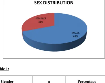
SEX DISTRIBUTION:
[image:58.595.125.497.170.463.2]Chart 1
Table 1:
Gender