Ultrasound assisted synthesis of a Zn(
II
) metal
–
organic framework with nano-plate morphology
using non-linear dicarboxylate and linear N-donor
ligands
†
Mohammad Yaser Masoomi,aAli Morsali*aand Peter C. Junkb
A 3D, porous Zn(II)-based metal–organic framework {[Zn2(oba)2(4-bpdb)]$2DMF}n(TMU-4) with double
interpenetration was prepared by using a non-linear dicarboxylate (H2oba ¼ 4,40-oxybisbenzoic acid)
and a linear N-donor (4-bpdb ¼ 1,4-bis(4-pyridyl)-2,3-diaza-1,3-butadiene) ligand. Also micro- and nano-plates of this MOF were synthesized by a sonochemical process and characterized by scanning electron microscopy, X-ray powder diffraction and IR spectroscopy. The thermal stability was studied by thermogravimetric analysis (TGA). Sonication time and concentration of initial reagents effects on the size and morphology of nano-structured MOFs, were studied. Calcination of TMU-4 at 500C under air atmosphere yields ZnO nanoparticles.
Introduction
Three-dimensional (3D) metal–organic frameworks (MOFs) are a new class of porous materials which are highly attractive because of their potential use as functional materials in structure-dependent applications, such as gas storage and separation, ion exchange, sensing, catalysis, and drug delivery.1–8The MOFs can be designed by choosing appropriate
organic ligands and inorganic secondary building units (SBUs).9
Of the many ligands that have been employed for the prep-aration of MOF structures, using a combination of functional-ized dicarboxylic acids and N-donor ligands can lead to MOFs with desired properties.5,10,11 Meanwhile, there is also an
increasing interest in MOFs withexible and dynamic frame-works with appropriate groups in their structures to exhibit high selectivity for guest inclusion and structural trans-formation upon adsorption/desorption of guest molecules.12,13
Reducing the size of MOFs to nanoscale has been extremely attractive.4 Recently using ultrasound irradiation in synthesis
and preparation of nano or microstructures of MOFs has been of interest.14–17In the research area of sonochemistry molecules
undergo a chemical reaction because of the application of powerful ultrasound radiation (20 KHz–10 MHz)18which induce
chemical or physical changes during cavitation. Cavitation
involves formation, growth, and instantaneously implosive collapse of bubbles in a liquid, which generates local hot spots having temperatures up to 5000C, 500 atm pressures with a lifetime of a few microseconds.19–21These extreme conditions
can also enhance the formation of nano-sized structures, mostly viaan increase of crystallization nuclei.22
ZnO is a polar inorganic material and an important n-type semiconductor with a wide band gap energy of 3.37 eV,23and
is an excellent material for potential applications including solar cells, luminescent materials, transparent conductors and gas sensors.24–28Up until now, various synthetic methods have
been developed to produce zinc oxide nanostructures.29,30
Thermal decomposition of MOFs under various conditions has been widely studied for the preparation of ZnO nanostructures with different sizes and morphologies.31
In this work, we have synthesized a Zn(II) metal–organic framework based on a V-shapedexible dicarboxylate ligand, 4,40-oxybis(benzoic acid) (H2oba) and the N-donor ligand 1,4-bis(4-pyridyl)-2,3-diaza-1,3-butadiene (4-bpdb) and investigated the effect of ultrasonic irradiation on shape and size. Also TMU-4 was calcined at 500C to prepare ZnO nanoparticles.
Experimental
Materials and physical techniques
All reagents for the synthesis and analysis were commercially available from Aldrich and Merck Company and used as received. The ligand 4-bpdb (1,4-bis(4-pyridyl)-2,3-diaza-1,3-butadiene) was prepared by the reported method.32 Melting
points were measured on an Electrothermal 9100 apparatus. IR spectra were recorded using Thermo Nicolet IR 100 FT-IR.
aDepartment of Chemistry, Faculty of Sciences, Tarbiat Modares University, Tehran,
Islamic Republic of Iran. E-mail: morsali_a@modares.ac.ir; Tel: +98-21-82883449
bCollege of Science, Technology & Engineering, James Cook University, Townsville,
Queensland 4811, Australia
†Electronic supplementary information (ESI) available: Other gures, full synthetic and analytical details. CCDC 996860. For ESI and crystallographic data in CIF or other electronic format see DOI: 10.1039/c4ra09186h
Cite this:RSC Adv., 2014,4, 47894
Received 24th August 2014 Accepted 22nd September 2014 DOI: 10.1039/c4ra09186h
www.rsc.org/advances
PAPER
Open Access Article. Published on 24 September 2014. Downloaded on 08/03/2015 23:56:57.
This article is licensed under a
Creative Commons Attribution 3.0 Unported Licence.
Ultrasonic generation was carried out in an ultrasonic bath SONICA-2200 EP (frequency of 40 KHz). The samples were characterized with a eld emission scanning electron micro-scope (FE-SEM) Philips XL30 and TESCAN MIRA (Czech) with gold coating.
The thermal behaviour was measured with a PL-STA 1500 apparatus with the rate of 10C min1in a static atmosphere of argon. X-ray powder diffraction (XRD) measurements were performed using a Philips X'pert diffractometer with mono chromated Cu-Karadiation.
Single crystals of TMU-4 were coated with viscous hydro-carbon oil and mounted on a loop. Data were obtained at
173C (100 K) on the MX1: Macromolecular Crystallography beamline at the Australian Synchrotron, Victoria, Australia. Data collection and integration on the MX1: Macromolecular Crystallography beamline were carried out using Blu-Ice33and
the XDS soware package.34 The structure was solved using
SHELXS35and rened by full-matrix least-squares on all F2 data
using SHELX,35in conjunction with the X-Seed graphical user
interface.36All hydrogen atoms were placed in calculated
posi-tions using the riding model. Sorption study on TMU-4 was performed using the AutosorbIQ from Quantachrome Instru-ments: CO2at 195 K. The sample was activated at 140C for 12 hours under vacuum. Dynamic light scattering measurements of particle sizes were determined by means of a Zetasizer Nano equipment.
Synthesis of {[Zn2(oba)2(4-bpdb)]$2DMF}n(TMU-4)
Conventional heating.Single crystals of TMU-4 suitable for X-ray diffraction were prepared by mixing Zn(NO3)2$6H2O (0.297 g, 1 mmol), H2oba (0.254 g, 1 mmol) and 4-bpdb (0.105 g, 0.5 mmol) in 30 mL of DMF. This mixture was sonicated until all solids were uniformly dispersed (3 min), and it was then heated at 80C. Aer 72 hours, yellow crystals of TMU-4 were collected (0.420 g, yield 71% based on oba), d.p. >300C. IR data (KBr pellet, n/cm1): selected bands: 776(s), 875(m), 1089(s), 1159(s), 1241(vs), 1412(vs-br), 1500(s), 1608(vs), 1679(vs) and 3417(w-br). Elemental analysis (%) calculated for [Zn2(C14O5 -H8)2(C12H10N4)](C3NOH7)2: C: 55.3, H: 4.0, N: 8.4; found: C: 55.8, H: 3.8, N: 8.2.
Synthesis of TMU-4 nanostructure.Ultrasonic syntheses of TMU-4 were carried out in an ultrasonic bath at ambient temperature and atmospheric pressure for 30, 60 and 90 min with different concentrations of metal and ligands, respectively. The resulting powder was isolated by centrifugation, washed with DMF three times and dried in air for characterization (Table 1). The effect of triethylamine (TEA) was investigated by adding 2 and 3 mL of TEA to adjust pH to 6 and 7, respectively.
Results and discussion
[image:2.595.308.550.230.465.2]Yellow crystals of TMU-4 were synthesized by mixing H2oba, Zn(NO3)2$6H2O and 4-bpdb in DMF solvent at 80C for 3 days.
Table 1 Experimental details for synthesis of nano TMU-4
Samples name
Molar ratio
(oba : 4-bpdb : Zn(OAc)2)
mmol in 25 mL DMF
Concentration [oba]/[4 bpdb]/
[Zn(OAc)2] (M) Time (min)
Elemental analysis,
founda(%) Yield (%) Morphology
A 1 : 1 : 0.6 [0.04]/[0.04]/[0.024] 30 C: 55.8, H: 3.7, N: 8.4 75 Micro plate B 1 : 1 : 0.6 [0.04]/[0.04]/[0.024] 60 C: 55.6, H: 3.8, N: 8.1 80 Micro plate C 1 : 1 : 0.6 [0.04]/[0.04]/[0.024] 90 C: 55.4, H: 3.9, N: 8.2 85 Micro plate D 1.5 : 1.5 : 0.9 [0.06]/[0.06]/[0.036] 90 C: 55.6, H: 3.9, N: 8.5 82 Micro plate E 0.5 : 0.5 : 0.3 [0.02]/[0.02]/[0.012] 90 C: 55.9, H: 3.8, N: 8.3 88 Micro plate F 0.5 : 0.5 : 0.3 [0.02]/[0.02]/[0.012],
TEA¼2 mL, pH¼6
90 C: 55.4, H: 3.7, N: 8.4 56 Nano plate
G 0.5 : 0.5 : 0.3 [0.02]/[0.02]/[0.012], TEA¼3 mL, pH¼7
90 C: 55.2, H: 3.6, N: 8.0 51 Nano plate
aCalculated for [Zn
2(C14O5H8)2(C12H10N4)](C3NOH7)2: C: 55.3, H: 4.0, N: 8.4.
Fig. 1 (a) Ball and stick representation of the binuclear Zn2unit (O: red;
N: blue; C: grey; and Zn: pink). (b) Layers of Zn(II)-oba (in red) pillared by 4-bpdb (in blue). (c) Representation showing the pore channels and that the network is doubly interpenetrated (in pink and blue). (d) Spacefill representation shows aperture size of pores along [101] direction. Hydrogen atoms and DMF molecules are omitted for clarity.
Open Access Article. Published on 24 September 2014. Downloaded on 08/03/2015 23:56:57.
This article is licensed under a
[image:2.595.41.564.574.710.2]Single-crystal X-ray diffraction reveals that TMU-4 crystallized in the space groupP21/cwith the formula of {[Zn2(oba)2(4-bpdb)]$ 2DMF}n.37TMU-4 is based on a binuclear Zn2unit (Zn#1 and Zn#2), both of which have tetrahedral geometry and coordi-nated to three carboxylate O atoms (for Zn#1: O2, O7, O10, and for Zn#2: O1, O5, O6) from three adjacent oba ligands and one N atom (Zn#1: N4 and Zn#2: N1) from the 4-bpdb ligand (Fig. 1a). The distance between Zn#1 and Zn#2 is 3.358 ˚A. Each non-linear (C–O–C ¼ 118.0(6)) dicarboxylate oba ligand binds three consecutive Zn(II) centers from two different units: one carboxylate group of an oba ligand adopts a bidentate mode and bridges both Zn#1 and Zn#2 centers of the unit while the other one is monodentate and binds to either Zn#1 or Zn#2, thereby forming 2D sheets (Fig. 1b). The 2D sheets are connected through the linear 4-bpdb, extending the structure in three dimensions. The V-shaped coordination of the oba ligand plays an important role in the linkage of the nodes resulting in a three-dimensional honeycomb framework with double inter-penetration (Fig. 1c). Although the double interinter-penetration is formed to avoid an extremely large void space, but still possesses one-dimensional (1D) open channels (aperture size: 5.38.8˚A, taking into account the van der Waals radii; 35.9% void space per unit cell)38running along the [101] direction and
lattice DMF molecules are accommodated in the channels (Fig. 1d). TMU-4 is porous to CO2 at 195 K and 1 bar (148.90 cm3g1at 1 bar; BET surface area calculated overp/p0¼ 0.02–0.3 : 517.9 m2 g1) (Fig. S2, ESI†). The pore volume calculated from the CO2adsorption is 0.298 cm3g1.39
The morphology and size of TMU-4 were investigated using scanning electron microscopy (SEM) by changing two parame-ters; sonication time and concentration of starting materials as well as control of nucleation.
Fig. 2 FE-SEM images of nano-plates of TMU-4 synthesized by sonochemical reaction (a) sample A, (b) sample B and (c) sample C.
Fig. 3 FE-SEM images of nano-plates of TMU-4 synthesized by sonochemical reaction (a) sample D, (b) sample E, (c) sample F and (d) sample G.
Open Access Article. Published on 24 September 2014. Downloaded on 08/03/2015 23:56:57.
This article is licensed under a
[image:3.595.309.545.174.694.2] [image:3.595.56.273.341.700.2]Firstly, sonication time as a parameter was changed at constant concentration of [0.04] M of starting materials. In all three different times (30, 60 and 90 min) micro-plates of TMU-4 were obtained in which higher sonication time (90 min) leads to uniform distribution in plate size (Fig. 2 and S3, ESI†). Hence, sonication time of 90 min is considered as the optimized value. Aer this in order to investigate the role of concentration of initial reagents on the morphology and size of product, reac-tions were performed with two different concentrations of starting materials ([0.06] M and [0.02] M). The results show that high concentrations of initial reagents increased particle size and thus lower concentration of initial reagents reduced the size of plates (Fig. 3). Aer this, increasing the speed of nucle-ation during the synthesis of TMU-4 was tested by adding tri-ethylamine (TEA). Using TEA causes fast nucleation of product due to deprotonated oba ligand and faster nucleation reduces particle size. In this mechanism nano-plates with a narrow size distribution were obtained (Fig. 3c and d).22By this, width of
nano-plate reduces to 90 nm (Fig. 3d).
The IR spectra of both crystals produced by conventional heating and nano-plate produced by the sonochemical method show the symmetric nsym (COO) and asymmetric nas (COO) vibrations of the carboxylate groups at 1409 cm1 and 1604 cm1, respectively. Also the characteristic absorption peak (nC]O¼1679 cm1) of DMF are presented in the IR spectra of TMU-4 (Fig. S4, ESI†).
A comparison between powder X-ray diffraction (PXRD) patterns of the simulated (derived from the single crystal structure of TMU-4) and experimental (resulting from the sonochemical process) conrms that the sonochemically synthesized TMU-4 is structurally identical to TMU-4 prepared through conventional heating (Fig. 4).
To examine the thermal stability of TMU-4 thermogravi-metric analysis (TGA) was carried out between 25 and 600C. The TGA curve of TMU-4 (conventional heating) shows a plateau in the range of 25 to 100C followed by a continuous loss of 14.5% (expected: 14.6%) up to 305C, which can be ascribed to removal of the guest DMF molecules (Fig. 5).
Decomposition of TMU-4's framework occurs in the temperature range of 305–500 C. Final residual mass 18.4% (expected: 19%) and the XRD pattern of thenal decomposition product show the formation of ZnO (Fig. S5, ESI†).
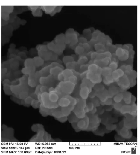
To prepare zinc oxide nanoparticles, TMU-4 was calcined at 500C for 2 h. Fig. S5†depicts the XRD patterns of the residue obtained from calcination of TMU-4. The Bragg diffraction peaks in the range of 2q¼20–80exhibit the typical patterns of hexagonal wurtzite structure of ZnO consistent with the repor-ted data by the JCPDS card number 36-1451 with lattice parameters ofa¼3.25 andc¼5.20˚A. The SEM image of the residue obtained from the direct calcination of TMU-4 at 500C shows the formation of ZnO nanoparticle (Fig. 6).
Fig. 4 XRD patterns of TMU-4 prepared by either conventional heating or sonochemical reaction (samples A–G). The data are in good agreement with the simulated pattern derived from the corresponding single-crystal structure data.
Fig. 5 Thermogravimetric profiles of TMU-4 isolated conventional heating.
Fig. 6 FE-SEM image of ZnO nanoparticles prepared by thermolysis of TMU-4 at 500C.
Open Access Article. Published on 24 September 2014. Downloaded on 08/03/2015 23:56:57.
This article is licensed under a
[image:4.595.310.547.51.204.2] [image:4.595.312.546.443.701.2] [image:4.595.45.286.519.679.2]Conclusions
A 3D, porous metal–organic framework {[Zn2(oba)2(4-bpdb)]$ 2DMF}n(TMU-4) with double interpenetration was synthesized
by conventional heating and analysed by X-ray crystallography. Using ultrasonic irradiation leads to formation of micro- and nano-plates of TMU-4 that were characterized by scanning electron microscopy, X-ray powder diffraction and IR spectros-copy. To prepare the nanostructure of TMU-4, three different times and concentrations of initial reagents, [0.06], [0.04] and [0.02] M, were tested. Also the rate of nucleation was tested by adding TEA to the reaction. Results show that best uniform distribution of nano-plates TMU-4 were obtained in 90 min with concentration of [0.02] M in the presence of TEA. These results indicate that sonochemical process can be used as an effective method for fast and readily preparation of nano-MOFs. Also calcination of TMU-4 at 500C under air atmosphere yields ZnO nanoparticles.
Acknowledgements
Support of this investigation by Tarbiat Modares University is gratefully acknowledged.
Notes and references
1 E. Lallana, A. Sousa-Herves, F. Fernandez-Trillo, R. Riguera and E. Fernandez-Megia,Pharm. Res., 2012,29, 1–34. 2 Z. Ma and B. Moulton,Coord. Chem. Rev., 2011,255, 1623–
1641.
3 M. Y. Masoomi and A. Morsali,Coord. Chem. Rev., 2012,256, 2921–2943.
4 M. Y. Masoomi and A. Morsali, RSC Adv., 2013,3, 19191– 19218.
5 A. Corma, H. Garc´ıa and F. X. Llabr´es i Xamena,Chem. Rev., 2010,110, 4606–4655.
6 M. P. Suh, H. J. Park, T. K. Prasad and D.-W. Lim,Chem. Rev., 2011,112, 782–835.
7 L. E. Kreno, K. Leong, O. K. Farha, M. Allendorf, R. P. Van Duyne and J. T. Hupp,Chem. Rev., 2011,112, 1105–1125. 8 P. Horcajada, R. Gref, T. Baati, P. K. Allan, G. Maurin,
P. Couvreur, G. F´erey, R. E. Morris and C. Serre, Chem. Rev., 2011,112, 1232–1268.
9 N. Stock and S. Biswas,Chem. Rev., 2011,112, 933–969. 10 R. Kitaura, K. Seki, G. Akiyama and S. Kitagawa, Angew.
Chem., Int. Ed., 2003,42, 428–431.
11 P. Mahata, M. Prabu and S. Natarajan,Cryst. Growth Des., 2009,9, 3683–3691.
12 R. Vaidhyanathan, S. S. Iremonger, G. K. H. Shimizu, P. G. Boyd, S. Alavi and T. K. Woo,Science, 2010,330, 650– 653.
13 K. C. Stylianou, J. E. Warren, S. Y. Chong, J. Rabone, J. Bacsa, D. Bradshaw and M. J. Rosseinsky,Chem. Commun., 2011,
47, 3389–3391.
14 N. Soltanzadeh and A. Morsali,Ultrason. Sonochem., 2010,
17, 139–144.
15 M. J. S. Fard-Jahromi and A. Morsali,Ultrason. Sonochem., 2010,17, 435–440.
16 A. Aslani and A. Morsali,Inorg. Chim. Acta, 2009,362, 5012– 5016.
17 M. Y. Masoomi, G. Mahmoudi and A. Morsali, J. Coord. Chem., 2010,63, 1186–1193.
18 K. S. Suslick, S.-B. Choe, A. A. Cichowlas and M. W. Grinstaff, Nature, 1991,353, 414–416.
19 R. Feng, Y. Zhao, C. Zhu and T. J. Mason, Ultrason. Sonochem., 2002,9, 231–236.
20 M. Strasberg,J. Acoust. Soc. Am., 1959,31, 163–176. 21 K. Negishi,J. Phys. Soc. Jpn., 1961,16, 1450–1465.
22 D. Tanaka, A. Henke, K. Albrecht, M. Moeller, K. Nakagawa, S. Kitagawa and J. Groll,Nat. Chem., 2010,2, 410–416. 23 P. X. Gao, Y. Ding and Z. L. Wang,Nano Lett., 2003,3, 1315–
1320.
24 K. Keis, L. Vayssieres, S.-E. Lindquist and A. Hagfeldt, Nanostruct. Mater., 1999,12, 487–490.
25 Y. Dai, Y. Zhang, Q. K. Li and C. W. Nan,Chem. Phys. Lett., 2002,358, 83–86.
26 M.-C. Jeong, B.-Y. Oh, W. Lee and J.-M. Myoung,Appl. Phys. Lett., 2005,86, 103105.
27 M. Bagheri, N. F. Hamedani, A. R. Mahjoub, A. A. Khodadadi and Y. Mortazavi,Sens. Actuators, B, 2014,191, 283–290. 28 M. Bagheri, A. A. Khodadadi, A. R. Mahjoub and
Y. Mortazavi,Sens. Actuators, B, 2013,188, 45–52.
29 K. T. Johnson, T. E. Gribb, E. M. Smoak and I. A. Banerjee, Chem. Commun., 2010,46, 1757–1759.
30 H. T. Ng, J. Li, M. K. Smith, P. Nguyen, A. Cassell, J. Han and M. Meyyappan,Science, 2003,300, 1249.
31 M. Y. Masoomi and A. Morsali,Coord. Chem. Rev., 2012,256, 2921–2943.
32 D. M. Ciurtin, Y.-B. Dong, M. D. Smith, T. Barclay and H.-C. zur Loye,Inorg. Chem., 2001,40, 2825–2834.
33 T. M. McPhillips, S. E. McPhillips, H.-J. Chiu, A. E. Cohen, A. M. Deacon, P. J. Ellis, E. Garman, A. Gonzalez, N. K. Sauter, R. P. Phizackerley, S. M. Soltis and P. Kuhn,J. Synchrotron Radiat., 2002,9, 401–406.
34 W. Kabsch,J. Appl. Crystallogr., 1993,26, 795–800.
35 G. Sheldrick, Acta Crystallogr., Sect. A: Cryst. Phys., Diffr., Theor. Gen. Crystallogr., 2008,64, 112–122.
36 L. J. Barbour,J. Supramol. Chem., 2001,1, 189–191. 37 Crystal data for TMU-4: C42.25H31.25N4.75O10.75Zn2, M ¼
908.21, monoclinic, space groupP21/c,a¼12.344(3)˚A,b¼ 26.323(5)A,˚ c¼ 15.663(3)˚A,b ¼97.56(3),V¼5045.2(18) ˚
A3, Z ¼ 4, crystal size (mm3): 0.12 0.08 0.01, T ¼ 173(2) K,Dcalc.¼1.196 g cm3,R1¼0.0936, wR2¼0.2706, 59 356 reections measured, 11 143 unique (Rint¼0.0560),
R1 ¼0.1156, wR2 ¼ 0.2866 (all data), GOF onF2¼ 1.049,
F(000)¼1856,m¼1.004 mm1,D
rmax¼1.171 e˚A3 38 A. L. Spek,J. Appl. Crystallogr., 2003,36, 7–13.
39 M. Y. Masoomi, K. C. Stylianou, A. Morsali, P. Retailleau and D. Maspoch,Cryst. Growth Des., 2014,14, 2092–2096.
Open Access Article. Published on 24 September 2014. Downloaded on 08/03/2015 23:56:57.
This article is licensed under a