SERUM RETINOL BINDING PROTEIN 4 (RBP4)
LEVEL IN PATIENTS WITH GESTATIONAL
DIABETES MELLITUS
Dissertation submitted for
M.D. BIOCHEMISTRY BRANCH – XIII DEGREE EXAMINATION
THE TAMILNADU DR.M.G.R.MEDICAL UNIVERSITY CHENNAI – 600 032
TAMILNADU
BONAFIDE CERTIFICATE
This is to certify that this dissertation work entitled “ SERUM RETINOL BINDING PROTEIN 4 (RBP4) LEVEL IN PATIENTS
WITH GESTATIONAL DIABETES MELLITUS" is the original bonafide work done by DR.R.GAYATHRI, Post Graduate Student, Institute of Biochemistry, Madras MedicalCollege, Chennai under our direct supervision and guidance.
Prof. Dr. V.Amuthavalli, MD., (Guide)
Professor,
Institute of Biochemistry Madras Medical College Chennai-600 003.
Prof. Dr. K.Ramadevi. MD., Ph.D. Director & Professor,
Institute of Biochemistry Madras Medical College Chennai-600 003.
Dean
Madras Medical College and
DECLARATION
I, Dr. R.GAYATHRI , Post Graduate , Institute of Biochemistry, Madras Medical College, solemnly declare that the dissertation titled “SERUM RETINOL BINDING PROTEIN 4 (RBP4) LEVEL IN
PATIENTS WITH GESTATIONAL DIABETES MELLITUS" is the bonafide work done by me at Institute of Biochemistry, Madras Medical College under the expert guidance and supervision of Prof. Dr. V.AMUTHAVALLI, M.D., Professor, Institute of Biochemistry, Madras Medical College. The dissertation is submitted to the Tamil Nadu Dr. M.G.R Medical University towards partial fulfillment of requirement for the award of M.D., Degree (Branch XIII) in Biochemistry.
Place: Chennai
SPECIAL ACKNOWLEDGEMENT
ACKNOWLEDGEMENT
The author expresses her warmest respects and profound gratitude to Dr.K.Ramadevi, M.D., Ph.D., Director and Professor, Institute of Biochemistry, Madras Medical College, Chennai, for her academic
enthusiasm and for facilitating her research work in the institute.
The author expresses her heartfelt gratitude to her guide and supervisor Dr.V.AMUTHAVALLI, M.D., Professor, Institute of Biochemistry, Madras Medical College, Chennai, for his intellectual and valuable guidance, unfailing support, encouragement and continuous inspiration throughout the period of her study.
The author in particular, is extremely thankful to
Dr.DHARMARAJAN.P.,MD,Dip(Diab), Director and Professor ,Institute of Diabetology, Rajiv Gandhi Government General Hospital, Chennai, for granting permission to obtain blood samples from the patients.
The author expresses her thanks to the Professors Dr.I.Periyandavar M.D, Dr.R.Chitraa M.D, Dr.K.Pramila M.D and Dr.Sumathy.S. M.D Institute of biochemistry, Madras Medical College, for their guidance,
encouragement, insightful comments and suggestions.
Madras Medical College for her guidance and support. The author expresses her warm respects and sincere thanks to other Assistant Professors, Dr.Karpagavalli.V.C, Dr.V.Ananthan, Dr.S.Siva, Dr.A.Veena Juliet, Dr.B.SudhaPresanna, Dr.Menaka Shanthi .Institute of biochemistry,
Madras Medical College, for their valuable suggestions regarding the practical issues of research which is something beyond the textbooks.
The author expresses warm respects to the members of the Institutional Ethical committee for approving the study.
The author expresses her special thanks to Diabetology Lab technicians, and Biochemistry Laboratory Staff, for their timely help and
cooperation during sample collection.
The author is indebted to the patients from whom blood samples were
collected for conducting the study.
The author expresses her special thanks to her co-PGs Dr.G.Chitra Siva Sankari, Dr.T.Poornima, Dr.A.K.Roopa for their cooperation and genuine support. The author expresses her thanks to all her colleagues in the institute, for their constant encouragement throughout the study period.
The author gratefully acknowledges the help rendered by Mr.Albert Joseph, for the statistical analysis of the study.
law, father in law and her parents for the moral support and encouragement extended by them which gave fulfillment to the dissertation work.
CONTENTS
SI.
NO TITLE PAGE NO.
1 INTRODUCTION 1
2 REVIEW OF LITERATURE 3
3 AIMS & OBJECTIVES 56
4 MATERIALS & METHODS 57
5 STATISTICAL ANALYSIS 70
6 RESULTS 71
7 DISCUSSION 86
8 SUMMARY AND CONCLUSIONS 97
9 LIMITATIONS OF THE STUDY 98
10 SCOPE FOR FURTHER STUDIES 99
11 BIBLIOGRAPHY 100
ABBREVIATIONS
1. GDM - Gestational diabetes mellitus. 2. RBP4 - Retinol binding protein-4. 3. PCOS - Polycystic ovarian disease 4. HLA - Human Leucocyte Antigen. 5. TNF α - Tumour necrosis factor alpha.
6. TGL - Triglycerides
7. HDL - High density lipoprotein 8. LDL - Low density lipoprotein 9. VLDL - Very low density lipoprotein. 10. T2DM - Type 2 diabetes mellitus. 11. ADA - American diabetes association. 12. IRS - Insulin receptor substrate.
13. PIP3 - Phosphatidyl inositol triphosphate. 14. MAP - Mitogen activated protein.
15. Wnt-int - Wingless family.
16. PPARG - Peroxisome proliferative activated receptor- gamma.
17. PDE3B - Phosphodiesterase 3 B
18. SREBP1 - Sterol regulatory element binding protein1 19. IGF - Insulin like growth factor.
20. ROS - Reactive oxygen species.
21. AGE - Advanced glycation end products. 22. PKC - Protein kinase C.
24. PDX - Pancreatic duodenal homeobox. 25. STRA6 - Stimulated by retinoic acid-6 26. LCAT - Lecithin choline acyl transferase. 27. LPL - Lipoprotein lipase.
28. TTR - Transthyretin. 29. PLA2 - Phospholipase A2
30. AMPK - AMP dependent protein kinase. 31. PEPCK - Phosphoenol pyruvate carboxy kinase 32. NAFLD - Non-alcoholic fatty liver disease.
33. HOMA-IR - Homeostatic model assessment for insulin resistance 34. CRBP - Cytoplasmic retinol binding protein.
35. CHE - Cholesterol esterase 36. CHD - Cholesterol oxidase 37. CAD - Coronary artery disease. 38. ABC - ATP binding cassette proteins 39. NEFA - Non-esterified fatty acids.
40. CETP - Cholesterol ester transfer protein. 41. RXR - Retinoid X receptor.
42. SNP - Single nucleotide polymorphism. 43. HNF1α - Hepatocyte nuclear factor 1α.
44. JAK-STAT - Janus kinase-single transducer and activator of transcription.
45. Mtor - Mechanistic target of rapamycin. 46. TLR4 - Toll like receptor-4
48. IL - Interleukin
49. CRP - C reactive protein.
50. SCH - Subclinical hypothyroidism. 51. NASH - Non-alcoholic steatohepatitis. 52. Apo - Apoprotein.
53. IUPAC - International union of pure and applied chemistry. 54. NGSP - National Glucohaemoglobin Standardisation
Programme.
55. GAD - Glutamic acid decarboxylase. 56. VEGF - Vascular Endothelial Growth factor. 57. IAA - Insulin auto antibodies.
58. TGF β - Transforming growth factor beta. 59. GAG - Glycosaminoglycan..
60. PDGF - Platelet derived growth factor.. 61. IGF - Insulin like Growth factor
62. VCAM - Vascular cell adhesion molecule. 63. ICAM - Intercellular adhesion molecule.
64. PG - Prostaglandin
65. GLUT - Glucose transporter.
66. NO - Nitricoxide.
67. NOS - Nitric oxide synthase.
1
INTRODUCTION
Gestational Diabetes Mellitus by definition is any degree of glucose intolerance with onset or first recognition during pregnancy1. It is characterized by insufficient insulin levels to meet the demands in later pregnancy. The significance of GDM is because of its maternal and fetal complications like polyhydramnios, preeclampsia and overt diabetes in future, fetal complications like birth trauma, macrosomia, childhood obesity and diabetes. Apart from the Asian race to be a risk factor for GDM, the other possible risk factors are
advanced maternal age, obesity, high parity, polycystic ovarian syndrome (PCOS), family history of diabetes, obstretic history of stillbirth, congenital malformation and macrosomia.
Retinol binding protein-4 is a novel marker in the pathogenesis of GDM. RBP4, a retinol transporter, plays an important role in dysregulation of insulin
sensitivity in GDM.
Thisstudy has been undertaken to find the level of serum retinol binding protein- 4 in GDM and its association with lipid profile and HbA1C has been evaluated. The raised levels also impair insulin signaling and induce gluconeogenic enzymes in the liver. Impairment of lipid metabolism is a risk factor for cardiovascular diseases.RBP4 role in lipid metabolism and metabolic
2
between RBP4 and LDL cholesterol, TGL and hepatic lipase activity in patients with Type 2 diabetes mellitus and cardiovascular disease.
EPIDEMIOLOGY
The prevalence of GDM in India is highly variable because of differences in living conditions, socio-economic status and dietary habits. A random survey done among the cities of India in 2002-2003, showed a prevalence of 16.55 per
3
REVIEW OF LITERATURE
Serum RBP4 was estimated in a group of 96 pregnant women. The values were compared between the GDM group and the control group consisting of normal pregnant women. Also RBP4 level was correlated with the patient’s lipid profile and HbA1c levels.
DIABETES MELLITUS
Diabetes mellitus is a metabolic disorder. It is characterized by chronic hyperglycemia. There is a defect in either insulin secretion or its action or both. It causes derangement of carbohydrate, lipid and protein metabolism. The chronic hyperglycemia is associated with damage and dysfunction of various organs.4
CLASSIFICATION OF DIABETES MELLITUS (According to ADA
criteria)5 TYPE 1
There is β-cell destruction leading to absolute insulin deficiency.
A. Immune mediated B. Idiopathic
TYPE 2
4 TYPE 3 - OTHER SPECIFIC TYPES
A. GENETIC DEFECTS OF Β CELL FUNCTION 1) Maturity onset diabetes of young [MODY] 2) Neonatal diabetes
B. GENETIC DEFECT IN INSULIN ACTION 1) Lipoatropic diabetes
2) Rabson-Mendelhall syndrome
C. DISEASES OF EXOCRINE PANCREAS
1) Cystic fibrosis, 2) Hemochromatosis, 3) Fibro-calculus pancreatitis, 4) Pancreatectomy etc.
D. ENDOCRINOPATHEIS
1) Cushings syndrome, 2)Pheochromocytoma,3)Acromegaly etc.
E. DRUG OR CHEMICAL INDUCED
Thiazides, adrenergic agonists, Phenytoin, Glucoocrticoids etc.
F. INFECTIONS
Like Congenital rubella and Cytomegalovirus.
G. OTHER GENETIC SYNDROME ASSOCIATED WITH DIABETES
5 TYPE 4-Gestational diabetes mellitus
GENERAL FEATURES OF DIABETES MELLITUS
Patient presents with symptom of polyuria, polydipsia, polyphagia. It may be associated with blurred vision and susceptibility to certain infections.
INTERPRETATION OF BLOOD GLUCOSE LEVELS
FASTING BLOOD GLUCOSE
POST PRANDIAL
AFTER 2 hrs DIAGNOSIS
<100 mg/dL <140 mg/dL NORMAL
100 mg/dL to 125 mg/dL 140 mg/dL to 199 mg/dL IMPAIRED GLUCOSE TOLERANCE
≥126 mg/dL ≥ 200 mg/dL DIABETES MELLITUS
It can also result in acute life threatening complications like ketoacidosis or non-ketotic hyperosmolar coma.
Long term complications include-retinopathy, nephropathy, peripheral neuropathy, autonomic neuropathy etc.
MECHANISM OF INSULIN ACTION
6
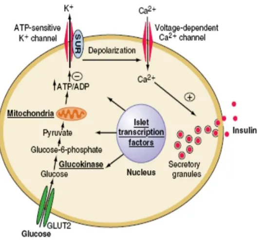
Fig-1 MECHANISM OF INSULIN SECRETION IMAGE COURTESY: RESEARCHGATE.NET
Important stimulant of insulin secretion is blood glucose. Glucose enters the β- cells. Intracellular metabolism of glucose results in increase in ATP level.
This closes the ligand gated K+ channels, leading to depolarization of the β cell. This results in opening of Ca2+ channels. Calcium entry triggers the exocytosis of the secretory vescicles that contain insulin.
The biosynthesis of insulin consists of two stages-preproinsulin which is then converted to proinsulin and insulin2. Preproinsulin is synthesized in the endoplasmic reticulum. It is cleaved by protease to form proinsulin. The proinsulin is packed in vesicles and transported to golgi apparatus where it is
7
crystals within the secretary granules. Insulin is the stored as mature secretory granules.
ACTION OF INSULIN ON TARGET CELLS
Insulin binds to its receptor on the surface of the target cells. The receptor is a transmembrane glycoprotein complex consisting of two α and two β-subunits. The α subunit is entirely extracellular and carries insulin-binding site. β subunit is transmembrane protein with tyrosine kinase activity. Binding of insulin to α subunit activates the β subunit. This activates a series of proteins the Insulin
Substrate Receptor protein (IRS 1-4). The second messenger PIP3 and tyrosine phoshorylated guanine nucleotide exchange proteins mediates the insulin sensitive translocation of GLUT4 from the cytosol to the plasma membrane especially in
skeletal muscles and adipose tissue. Over a period of time, insulin also starts to promote the expression of genes directing synthesis of GLUT4.
FIG 2 : ACTION OF INSULIN
8
Thus the stimulation of IRS is coupled to several protein kinase signal systems: 1) Signaling through PI3-kinase and phosphatidylinositol (PI-3 kinase and
protein kinase B/Akt)
2) Mitogen activated protein kinases (MAP kinases)
3) Possible interaction via kinases not coupled to IRS proteins.
MAJOR ACTIONS OF INSULIN
EFFECTS OF REDUCED INSULIN SIGNALING
Stimulates glucose transport
Increased blood glucose after meals, increased frequency of urination with loss of water
glucose and electrolytes. Control of hepatic
carbohydrate metabolism
Control over gluconeogenesis and glycogen metabolism is lost.
Control of lipid metabolism in the adipocytes
Excessive release of fatty acid due to increased lipolysis
GESTATIONAL DIABETES MELLITUS
“Gestational Diabetes Mellitus by definition is any degree of glucose intolerance with onset or first recognition during pregnancy.”
It is important to assess the trends in the prevalence of GDM, because it is associated with
Many perinatal complications
Offsprings are at an increased risk of developing diabetes later in life6. It also helps to understand the possible mechanisms for the increase of
9 PATHOPHYSIOLOGY OF GDM:
Many changes in the metabolism of mother occur in such a way as to provide sufficient energy and nutrition to the fetus. Fetus mainly depends on the maternal glucose which reaches it through the placenta, because of which the mother develops a state of “Insulin resistance” during mid-pregnancy. This state of insulin resistance progresses through the third trimester. At one stage there is
reduced state of consumption of glucose by maternal tissue and increased gluconeogenesis, ensuring sufficient supply of glucose to the fetus7.The resulting positive maternal-fetal glucose gradient, facilitates the transfer of glucose through the placenta. In a proportion of pregnancies, this state of “Insulin resistance” is greatly increased, and Gestational diabetes mellitus develops8.
GENETIC BASIS OF GDM:
Available data suggest that there is familial tendency for GDM pathogenesis. The risk of GDM is associated with family history of T2DM9. They have a 2.3 fold of increased risk. Women with diabetic siblings have 8.4 fold increased risk for GDM10.There is also a tendency for recurrence of GDM in 30%.
The defect in both insulin secretion and insulin action are explained by
genetic evidence. Genetic loci of several genes responsible for insulin secretion, insulin resistance, lipid and glucose metabolism have been associated with risk of
10
GENES AND GENETIC VARIANTS RELATED TO INSULIN
SECRETION:
1. KCJN11 GENE:
(Potassium inwardly rectifying channel, subfamily J,member 11)
This gene encodes for the ATP-sensitive potassium channel of β cells. A
variant of this, results due to substitution of lysine for glutamic acid, Glu23Lys. This variant induces over-activity of the potassium channels, thus decreasing the insulin secretion11.The risk is 6.2% for KCNJ11 Lys23/Lys23 genotype and 10.1% for KCNJ11 Glu23/Lys23 and Lys23/Lys23 combined12.
2. TCF7L2(Transcription factor 7-like 2):
It is involved in gene transcription. It is a member of the Wnt signaling
pathway. The strong association of variants of TCF7L2 and GDM is through the mechanism involving insulin secretion13,14. It is found that T allele of the rs7903146 variant15 and T allele of rs12255372 variant16 were frequently associated with GDM.
3. Mitochondrially encoded NADH dehydrogenase 1(ND1):
It is part of electron transport chain and involved in glucose metabolism. Reduced activity of the respiratory chain and decreased production of ATP result
11 INSULIN SIGNALING GENES
(1) Insulin receptor (INSR):
Insulin receptors are involved in glucose uptake. Thus insulin receptor genes are involved in glucose homeostasis. Mutations in INSR causes severe insulin resistance19.INSR polymorphism has been associated with GDM20.Polymorphism of INSR KPN1 was associated with GDM21.
(2) Insulin receptor substrate 1(IRS 1):
It is a substrate of insulin receptor tyrosine kinase. It is the key participant in insulin signaling22. The Gly972Arg polymorphism reduces tyrosine phosphorylation and inhibits insulin receptor kinase, thus resulting in insulin resistance23. The 972Arg variant has 25% greater risk of developing T2DM24.The homozygous Arg972 was associated with GDM25.
LIPID AND GLUCOSE METABOLISM GENES:
Peroxisome proliferative activated receptor-gamma(PPARG) :
PPARG is a transcription factor that regulates adipocytes differentiation and also in lipid and glucose metabolism. It binds to specific response elements in promoter regions of target genes. A variant of PPARG has been identified and
involves the substitution of alanine instead of proline at 12th amino-acid position (Pro12Ala). This alanine 12 variant decreases the promoter element affinity of
12 OTHER GENES
MANNOSE BINDING LECTIN PROTEIN C 2(MBL2):
MBL2 is a membrane of the collectin family of proteins. It is a component of the innate immune system. It also influences inflammatory response by inhibiting TNFα. Its deficiency is associated with recurrent infections and chronic
inflammatory diseases.
Two variants of MBL2 gene-Arg52Cys and Gly54Asp are associated with decreased plasma MBL2 level. Of these, the carriers of Asp54 allele of Gly54Asp polymorphism have been shown to be associated with GDM. These polymorphisms are also shown to be linked to micro and macrovascular complications associated with diabetes.
METABOLIC IMPLICATIONS OF GDM
CARBOHYDRATE METABOLISM
In early pregnancy, the blood glucose and insulin levels are almost close to the non-pregnant state. Peripheral insulin sensitivity and basal hepatic output are normal in early trimester26.
But in third trimester, though there is
Increased insulin production, the insulin action decreases by 50-70% in normal pregnancy27.
The postprandial blood glucose concentration is significantly elevated and
13
[image:26.612.119.515.138.414.2]Basal hepatic glucose output is increased by 16-30% to meet the needs of the growing fetus29.
FIG 3:PATHOPHYSIOLOGY OF GDM IMAGE COURTESY:UEDAEGYPT.ORG
Inspite of the anti-insulinogenic action of the placental hormones, carbohydrates acts as the main source of energy for the pregnant women. So the fasting glucose level remains low30.
In GDM, there is decrease in first-phase insulin response. Normally about
14
In the liver normally, hyperglycemia primarily inhibits net hepatic glycogenolysis by inhibiting glycogen phosphorylase, and hyperinsulinemia stimulates glycogen synthase. But these processes are suppressed in GDM32. The hepatic glucose output is not suppressed by insulin in later pregnancy33.This results in hyperglycemia.
LIPID METABOLISM
Increased estrogen, progesterone and insulin favor lipid deposition in early pregnancy. In later pregnancy, there is lipolysis and fat mobilization34. Thus there is a change from anabolic to catabolic state. This promotes the use of fatty acids as energy source for the mother and reserving glucose and aminoacids for the fetus.
Insulin action on mature adipocyte is to enhance glucose uptake and its
15 INSULIN
Stimulate phosphodiesterase 3B (PDE3B)
↓Intracellular cAMP
Attenuates protein kinase
Inactivation of hormone sensitive lipase
Inhibits lipolysis
In pregnancy,
Increased free fatty acid and glycerol concentration.
Decrease in glucose, alanine and β-hydroxybutyrate
Increase in triacylglycerol, fatty acids, phospholipids and cholesterol 32. Clearance of VLDL is reduced because of decreased activity of lipoprotein
lipase.
16
GDM induces a state of dyslipidemia consistent with insulin resistance. Women with GDM have higher triglyceride levels. There is reduced oxidation of exogenous triacylglycerol. The increased triacylglycerol is due to
(1) reduced fatty acid uptake and its subsequent oxidation (2) increased hepatic oxidation and esterification of fatty acid thus
increasing the synthesis of VLDL37.
INSULIN RESISTANCE IN SKELETAL MUSCLE AND ADIPOSE
TISSUE:
Muscle insulin resistance promotes hepatic steatosis by inducing SREBP1c mediated lipogenesis and inhibition of fatty acid oxidation38.
In late pregnancy, postprandial free fatty acid level increases and the disposal of blood glucose by insulin worsens by 40-60%, when compared with
pre-pregnancy status46.
Skeletal muscle is the principle site of glucose disposal in the body. But this becomes severely insulin resistant along with the adipose tissue during later half of pregnancy. Normal pregnancy itself is characterized by decrease in insulin mediated glucose disposal by 50%. In order to maintain the euglycemic state,
insulin secretion is increased by 200-250%47.Placental derived hormones, especially Human Placental Lactogen (hPL) and Human Placental Growth
17
EFFECTS OF MATERNAL HORMONES:
1) HUMAN PLACENTAL LACTOGEN( hPL):
Human placental lactogen is the product of hPL –A and hPL-B genes secreted into both maternal and fetal circulation after 6 weeks of gestation40.
Its level is decreased in hyperglycemia and elevated in hypoglycemia. Its metabolic role is to mobilize lipids and free fatty acids41. During second half of pregnancy, the hPL level increases by 10 fold. It stimulates lipolysis and increases free fatty acid level. Thus it provides a different fuel for mother so that glucose and amino acids are conserved for the fetus.
The increased fatty acid level in turn interferes with insulin directed entry of glucose into the cell. Thus hPL is considered as a potent antagonist to insulin action during pregnancy42.
PLACENTAL GROWTH HORMONE (PGH):
Produced by syncytiotrophoblast cells of human placenta. It is the product of the GH-V gene. It is predominantly found in maternal circulation. Its concentration increases progressively from mid-pregnancy43.It is an important potential regulator of maternal insulin resistance during pregnancy. It mainly
influences the fetal growth by modifying substrate availability44.It influences the p85 expression of PI 3 kinase activity in skeletal muscle, thus contributing to
18
Furthermore, hPl and PGH stimulate insulin-like growth factor (IGF) production and modulate intermediary metabolism, resulting in increase in the glucose availability to the fetus49.
MECHANISMS OF INSULIN RESISTANCE:
“When the biological effects of insulin are less than expected for glucose disposal in skeletal muscle and adipose tissue and suppression of endogenous
glucose production primarily in the liver, it is said to be insulin resistance.”
Insulin resistance may be due to a decrease in the number of insulin receptors or impairment in post-receptor signaling of insulin receptors.
GLUCOSE TOXICITY:
Glucose in chronic excess causes damage to the structure and functions of the organs. Various pathways have been suggested for metabolic consequences of excess glucose. All these pathways have in common the formation of reactive oxygen species. This causes chronic oxidative stress resulting in defective insulin gene expression and its secretion.
1) GLYCERALDEHYDE AUTOXIDATION:
Glyceraldehyde 3-phosphate is a phosphorylation product formed from
glucose during glycolysis. Autoxidation of hydroxyaldehydes generates hydrogen peroxide and ketoaldehydes50.
19
(a)ketoaldehydes- which contributes to glycosylation-related protein
development and
(b)the hydroxyl radical, which can cause mutagenic alterations in DNA.
Excess glyceraldehydes inhibits insulin51.
Long term high glucose level decreases Glyceraldehyde 3-phosphate
dehydrogenase(GAPDH) activity,through the ROS-activated poly ADP ribosylation52. This in turn is associated with intracellular advanced glycation end product (AGE) .
2) PKC ACTIVATION:
Dihydroxyacetone undergoes reduction to glyceraldehydes 3-phosphate and acylation, thus increases de novo synthesis of diacylglycerol, which activates
protein kinase C. Activation of PKC is associated with increase in TGF-1, vascular endothelial growth factor, NADPH-oxidase, NF-B and ROS53,54.
3) METHYLGLYOXAL AND GLYCATION:
These 3 molecules are reactive intracellular dicarbonyls. They form Advanced Glycation End products by reacting with amino group of intra and
20
High blood glucose
Impaired GADPH activity
Accumulation of glyceraldehydes 3-P & DHAP
↑ Methylglyoxal
Hyperglycemia leads to the formation of Advanced glycosylation End (AGE) products by Non-enzymatic glycosylation of intracellular proteins amino group with these dicarbonyl molecules. These glycated proteins undergo progressive dehydration, cyclization and oxidation.
4) POLYOL PATHWAY:
Hyperglycemia increases glucose metabolism via sorbitol pathway. Glucose is converted to sorbitol by the enzyme aldose reductase.
It causes, reduced cytosolic NADPH which in turn increases oxidative
stress in the cell.
Increase cytosolic ratio of NADH/NAD+ causes inhibition of the enzyme
glyceraldehydes 3-phosphate dehydrogenase
Formation of methlglyoxal.
21
This pathway is normally inactive and gets activated only when intracellular glucose level increases.
5) INCREASED HEXOSAMINE PATHWAY:
Fructose 6-phosphate is diverted from glycolysis to form glucosamine 6
phosphate, which in turn is converted to UDP-N-Acetyl glucosamine. N-Acetyl glucosamine supports proteoglycan synthesis and also formation of O-linked glycoproteins. They can glycate transcription factors and thus enhance transcription of gene including plasminogen activator inhibitor.
EFFECTS OF CHRONIC OXIDATIVE STRESS ON INSULIN GENE EXPRESSION:
Chronic exposure of the beta cell to high concentrations of glucose causes
defective insulin gene expression as well as marked decrease in insulin secretion57.
The defect in insulin gene expression is due to loss of atleast two critical proteins that activate the insulin promoter. One is Pancreatic duodenal homeobox-1(PDX-1)58,59and the other is MafA60,61. MafA is RIPE -3b1 activator.
Due to chronic oxidative stress, the DNA binding capacity of PDX-1 is
22
RELATIONSHIP BETWEEN GLUCOSE TOXICITY AND
LIPOTOXICITY:
Diabetes mellitus is often accompanied by raised levels of cholesterol, free fatty acids and triglycerides65.
It has been reported that prolonged exposure of pancreatic β-cells to fatty acids, inhibit insulin gene expression66.Simultaneous presence of hyperglycemia and elevated fatty acids cause accumulation of cytosolic citrate, the precursor of malonyl-CoA, which in-turn inhibits carnitine palmitoyl-transferase 1(the enzyme required for fatty acid transport into the mitochondria)67.
In the presence of high glucose concentrations, elevated fatty acids are not
readily oxidized in mitochondria but are shunted towards esterification pathways. It has been observed that adverse effects of palmitate on insulin gene expression and secretion was seen only when β-cells were simultaneously exposed to high concentrations of glucose. Palmitate induced accumulation of beta cell triglycerides occurred only in the presence of high glucose68. Thus lipotoxicity requires antecedent hyperglycemia69.
DIAGNOSIS OF GDM.
SCREENING TESTS:
23
Generally, screening and diagnostic tests for GDM are performed between 24 to 28 weeks of gestation, because at this point in gestation the diabetogenic effect of pregnancy manifests and there is sufficient time to intervene70. A good screening test has a positive likelihood ratio of atleast 6. Screening on the basis of risk factors seems to be inefficient71. In other words, a diagnostic test only in women with risk factors will miss many women with GDM.
GLUCOSE CHALLENGE TEST (GCT):
Patient is given 50 grams glucose solution irrespective of food intake72,73. Blood glucose measurement is taken either after 1 or 2 hours, each having different cutoff values.
The American Diabetes Association (ADA) recommends a cutoff value of 140mg/dL after 1 hr which identifies 80% of women with GDM or 130 mg/dL which identifies 90% cases74.
The 2 hours cutoff is 118 mg/dL.
Patients with positive GCT are subjected to diagnostic test.
The limitation of this test is that it has many false-positives and sensitivity
24 OTHER TESTS:
Capillary blood glucose measurement with hemocue
Mainly depends on the meter and not reliable.
Breakfast test and lunch test Needs to use standard diet Not commonly used
Glycosuria Not reliable in GDM
Fructosamine Less sensitive
Glycated hemoglobin Less sensitive in pregnancy Not reliable.
Fetal abdominal circumference (AC) Misses 43% of GDM cases.
DIAGNOSTIC TEST :
“The gold standard for diagnosis of GDM is the 100 gm 3 hour Oral Glucose Tolerance test (OGTT)”.
Patient preparation includes
1) Patient must take atleast 150 gm of carbohydrate per day for the preceding 3 days.
2) Fasting between 10 to 16 hrs of fasting
3) Patient must be at rest for 30 minutes before blood collection. 4) No smoking is allowed.
PROCEDURE:
Fasting blood and urine samples are collected.
After the first measurement, the patient drinks 100 gm glucose solution with 300 ml water within 5 minutes.
25
During the procedure, the patient is not allowed to smoke or walk.
But the WHO recommends to use a low glucose load of only 75 gms. Only
fasting blood glucose measurement and one measurement 2 hrs after the glucose load is taken. The interpretation of these test results are shown in
table.
Carpenter and Coustan
/ADA (2004) WHO (2008)
IASPSG / ADA (2012)
75 gm OGTT 100 gm
OGTT 75 gm OGTT 75 gm OGTT
Fasting 95 mg/dL 95 mg/dL 126 mg/dL 92 mg/dL
1 hr 180 mg/dL 180 mg/dL 180 mg/dL
2 hr 155 mg/dL 155 mg/dL 140 mg/dL 153 mg/dL
3 hr 140 mg/dL
Citation : Cheung KW, Wong SF (2012) Gestational Diabetes Mellitus update and review of literature, Reproductive Sys Sexual Disorder S2:003.
COMPLICATIONS OF GDM:
It includes both maternal and fetal complications. 1) FETAL MACROSOMIA:
26
Mother’s age, pre-gestational weight, weight gain during pregnancy, fetal insulin secretion and substrate concentration in maternal circulation are the main factors which influence the fetal weight81.
Maternal glucose passes through the placenta to reach the fetus, which stimulates fetal insulin secretion. Insulin, being an anabolic hormone increases fetal dimension causing macrosomia82. Significantly elevated levels of triglycerides in cord blood of obese GDM women fetuses with macrosomic babies suggest that triglyceride plays an important role in pathogenesis of macrosomia83,84 . There is reduced activity of lipoprotein lipase in GDM due to insulin resistance. So there is reduced lipolysis of TGs and decreased suppression of endogenous TGs synthesis85.
2) SHOULDER DYSTOCIA AND BIRTH TRAUMA:
The increase in birth trauma in the offsprings of mother with GDM is due to higher rate of macrosomia, which predisposes to shoulder dystocia.
Shoulder dystocia leads to consequences like clavicular fracture and brachial plexus injury72. A planned caesarean could avoid such complications86.
3) CAESAREAN DELIVERY:
27
brachial plexus injury or neonatal hypoglycemia. An early diagnosis and treatment of GDM can avoid such consequences.
4) NEONATAL METABOLIC PROBLEMS: Due to GDM, anincreased incidence of 87
neonatal hypoglycemia,
hypocalcemia,
hyperbilirubinemia and
polycythemia has been reported.
This causes unnecessary admissions in Neonatal Intensive Care unit (NICU). Neonatal hypoglycemia is much more related to macrosomia, than to
maternal GDM88. Also the long term damage due to neonatal hypoglycemia is seen only in those with hypoglycemic seizures or recurrent hypoglycemia89. Adequately treated hyperbilirubinemia has no lasting effects on the infant90.The other outcomes include, inconvenience for the baby being subjected to repeated blood tests, costs of these tests, separation of mother and the neonate due to NICU
admission.
5) HYPERTENSION/PREECLAMPSIA:
28
6) LATE EFFECTS OF GDM ON MOTHER AND BABY:
The prevalence of T2DM is higher, when compared to those without GDM in earlier pregnancies92. The prevalence of T2DM later in life for women with GDM varies from 9% in Causcasians to 25% in Asian women93,94.Children of mother with GDM are also at increased risk of developing T2DM and obesity in
later life95. They are also at a risk of developing neuropsychological problems96.
RETNOL BINDING PROTEIN-4
Serum Retinol binding protein-4 is a specific carrier protein which belongs to lipocalin family, of kernel type, calycin superfamily.
It is coded by chromosome no: 10. Its location is 10q23.33 in cytosol.
The protein weighs 23 kDa and has 201 amino acids.
It consists of 4 chains which are linked by 3 disulphide bonds.
It is a monomer.
It undergoes methylation as a post translational modification.
It is secreted by hepatocytes and adipocytes.
It is the transporter of Retinol (Active form of vitamin A) from the liver to the
peripheral tissues.
It is bound to transthyretin (prealbumin), which is a carrier of thyroid
hormone.
Lipocalin protein family are small secreted proteins, which are characterized by
29 binding to specific cell surface receptors.
formation of macromolecular complexes.
They exhibit great structural and functional diversity. They have been
shown to be involved in
retinol transport
pheromone transport
prostaglandin synthesis
cell growth and metabolism modulation
immune response regulation
tissue development
STRUCTURE97:
The members of this family share about 20% sequence homology, but mainly confined to a common tertiary structure determined by conserved
segments of the individual lipocalin protein, called the lipocalin folds.
These folds organize the lipocalins in eight anti-parallel β- sheets surrounding a hydrophobic pocket. This hydrophobic pocket is essential for functioning as carrier proteins.
30
FIG 4: STRUCTURE OF RBP4 PROTEIN WITH TRANSTHYRETIN
IMAGE COURTESY:en.wikipedia.org
Additionally, there are 3 to 10 helices at the amino terminus, outside the β
-barrel and an α-helix at the carboxy terminus.
RBP4-TRANSTHYRETIN COMPLEX97:
In plasma, RBP4 is complexed to transthyretin by a non covalent linkage. It combines with transthyretin in equimolar concentration in 1: 1 ratio. It is bound
to transthyretin to avoid excretion by the renal glomeruli.
Only 4% of RBP is free, the rest is bound to transthyretin98. Transthyretin has greater affinity for holo-RBP than apoRBP. The complex is stable between pH 5.0 and 9.0 and dissociates at low ionic strength.
31
molecule. The variation in the length of the loop, its conformation and amino acid composition is specific, which binds with high affinity to transthyretin.
RBP4 RECEPTOR100,101: RBP4 has two receptors namely
1) Stimulated by Retinoic Acid 6(STRA6) 2) RBP4 Receptor 2(RBPR2)
The cellular receptor for RBP was identified and biochemically characterized as the protein named STRA6 (Stimulated by Retinoic acid 6)99. It is a 75-kDa multipass transmembrane protein. It has 9 transmembrane segments.
STRA6 receptors are expressed in several tissues, including retina,brain,
testis, muscle and placental endothelial cells. It is not found in the liver and adipose tissue.
RBPR2 (RBP Receptor 2) is another receptor found in the liver and adipose tissue. It is a 70.1kDa protein. It is also a transmembrane receptor with more than 50% similarity in its amino acid sequence with the STRA6 receptors. Both the receptors have similar intracellular C-terminal soluble domains containing 74-75 amino acids. But RBPR2 lacks the conical Src homology 2
32
STRA6 catalyses the release of retinol from RBP, where it is tightly bound. This facilitates the translocation of retinol across the membrane and its association with the cytoplasmic CRBP1 receptors.
Conversion of retinol to retinyl esters for storage by the enzyme lecithin retinol acyl transferase (LRAT), increases the amount of CRBP1 protein, thereby enhancing the action of STRA6 receptors99.
BIOCHEMICAL FUNCTIONS OF RBP4:
It is an extracellular transport protein. RBP is the sole retinol transporter in the plasma. It binds to a single all-trans-retinol molecule. The apo-protein is saturated with retinol, which triggers its secretion into the plasma.
Its physiological functions are97
1) It facilitates transport of insoluble retinol from storage site to the peripheral tissues
2) RBP protects bound retinol from oxidation.
3) The synthesis of RBP regulates release of retinol from the liver and mediates specific uptake by target tissue.
MECHANISM OF ACTION ON TARGET CELLS101:
33
to the receptor weakens its affinity for retinol leading to its delivery to the outer cleft of the receptor.
Inside the cell, the retinol is delivered to the CRBP-1(Cytoplasmic retinol binding protein 1). There is unidirectional movement of retinol from RBP to CRBP1. As per the body requirement, retinol is converted to retinoic acid which is carried by cytoplasmic retinoic acid binding protein (CRABP). It is then
transported to the nuclear receptors, RXR and RAR resulting in transcription activity and gene regulation.
After the release of retinol, RBP loses its affinity for transthyretin forming apo-RBP. This gets altered by the kidney- reabsorbed and catabolised. Thus, one RBP carries only one retinol molecule before being degraded.
RBP4 GENE POLYMORPHISM108:
It has been shown that single nucleotide polymorphism (SNP) in the RBP4 promoter region is associated with 2 fold increase in risk of T2DM. The increase in RBP4 promoter activity is positively associated with its expression in the adipose tissue and also with BMI.
The SNP in the regulatory region of the RBP4 gene is identified as 803 GA
polymorphism of rs3758539. This 803GA polymorphism is a functional variant that affects HNF1α binding, RBP4 transcription efficiency and plasma level of
34
Dietary retinol is not associated with RBP4 polymorphism or risk of T2DM. RBP4 polymorphism is not associated with circulating levels of retinoids.
POLYMORPHISM OF RECEPTORS OF RBP4107:
STRA6 (Stimulated by retinoic acid gene homolog 6) is a cell surface receptor. It also acts as a cytokine receptor by activating JAK/STAT signal cascade by combining with RBP4.
STRA6 gene is located on chromosome 15q24.1 region. It has 20 exons and 19 introns. The association between STRA6 rs974456 and rs736118 polymorphism and T2DM in south Indian population has been established. They all are C/T dimorphic. The risk variants are CT heterozygous and TT homozygous.
Because of this SNP, that is change from C to T allele is due change of amino acid from methionine to tyrosine. This takes place in the c- terminal of STRA6, which is involved in signal transduction by phosphorylation. Tyrosine conversion has a potential impact in this signal cascading.
ROLE OF RBP4 IN PATHOGENESIS OF GESTATIONAL DIABETES
MELLITUS:
INSULIN RESISTANCE IN SKELETAL MUSCLE105,106
35
[image:48.612.139.496.79.304.2]MECANISHM OF ACTION IN CAUSING INSULIN RESISTANCE
FIG 5: MECHANISM OF RBP4 IN CAUSING INSULIN RESISTANCE
IMAGE COURTESY: researchgate.net
Normally, the attachment of insulin to its receptor stimulates tyrosine phosphorylation of these receptors. In GDM, there is defective tyrosine phosphorylation due to instrinsic defect in the insulin signaling pathway. This is
the major role of RBP4 in the pathogenesis of GDM. This is through stimulation of alternate JAK-STAT cascade.
The cytosolic domain of STRA6 receptor of RBP4, contains a stretch of
residues that conform to a consensus phosphotyrosine motif.
Phosphotyrosines are often found on surface receptors, that transduces
extracellular signals by activating JAK-STAT cascades.
Thus the presence of phosphotyrosine motif in STRA6 suggests the
36
function as a signaling receptor which is activated by large amount of RBP4 in GDM.
Thus RBP4 acts as an extracellular ligand that activates STRA6, which in
turn modulates cellular response by triggering JAK/STAT signaling.
The resultant action is abnormal phosphorylation of serine/threonine
residues in the insulin receptor (IR) and insulin receptor substrate (IRS).
The intracellular substrate 1 (IRS 1) is a major docking protein in muscles. It regulates insulin uptake in insulin sensitive tissues. It has been shown that, the IRS-1 protein level is reduced by 30-50% in GDM. Also, there is increased 312-serine phosphorylation of these proteins, which inhibits PI-3 kinase activity.
Inhibition of PI-3 kinase activity, affects the phosphrylation of Akt, which is necessary for translocation of GLUT4 receptors.
Alternatively, increased serine phosphorylation of IRS-1 can be due to activation of mTOR-p 70S6 pathway.
mTOR-p 70S6k1 and AMPK act as nutrient and energy sensors in the cellular level. AMPK is a negative regulator of mTOR. Its level is elevated in GDM.
RBP4 AND INSULIN RESISTANCE IN ADIPOSE TISSUE109:
37
Recent evidences suggest the possibility of pro-inflammatory pathway in RBP4- induced insulin resistance102 in adipose tissue. The proportion of RBP4 that is apo-RBP4 is increased in obese persons, and also the ratio of RBP4 to retinol is increased in people with T2DM.
Similar to the action in the muscle, RBP4 can act through the JAK/STAT pathway through the RBPR2 receptor. The other way of action is mediated
through stimulation of macrophages.
It has been shown that RBP4 acts independent of retinol to impair insulin signaling in adipocytes. It acts indirectly by inducing proinflammatory cytokine release from the macrophages. These actions by the Toll-like receptor 4(TLR4) cell surface receptor, involving the c-Jun N-terminal protein kinase (JNK) signaling pathway. The effect of RBP4 in inducing IL-6 synthesis is greater than
in stimulating TNF.
“Peroxisome proliferator- activated receptor (PPAR) is a negative regulator of proinflammatory pathways in macrophages.” The expression of PPAR was reduced by 50 to 70% by RBP4. Thus reduced PPAR expression is involved in the induction of proinflammatory state by RBP4.
38
RBP4 causes suppression of Akt phosphorylation in 3T3L1 adipocytes intermingled with macrophages, thus resulting in insulin resistance in adipose tissue. This shows that macrophage- derived proinflammatory cytokines induced by RBP4 mediated insulin resistance in adipose tissue.
This effect of RBP4 is mediated through the TLR4 and JNK pathway, independent of retinol binding to RBP4. Thus RBP4 is not the only adipose tissue
derived factor, that is important for its inflammation and T2DM.
Others like leptin and FFA also induce proinflammatory cytokine production. But it has been identified that RBP4 is a key endogenous protein
that contributes to adipose tissue inflammation and insulin resistance by triggering interaction between innate and adaptive immune system. These understanding of integration of inflammatory pathway and insulin resistance have
therapeutic implications for T2DM.
RBP4 AND CORONARY ARTERY DISEASE110
Type II DM is characterized by inadequate insulin secretion as well as insulin deterioration and resistance. It is described as “silent disease”. T2DM is an independent risk factor for CAD. Risk factors including hyperglycemia,
39
Insulin resistance acts as a strong linking factor between T2DM and CAD. It is characterized by malfunctioning adipocytes and greater amount of proinflammatory cytokines. There is
low-grade elevation of acute phase reactants
proinflammatory cytokine secretion and
cell adhesion molecules.
The resulting subclinical inflammation causing insulin resistance correlates with pathogenesis of all phases of atherosclerosis.
Persistent hyperinsulinemia causes raised levels of TGL, FFA and LDL as
well as reduced HDL.
Increased FFA activates innate immune system and there is resultant
release of proinflammatory cytokines like TNF-α, IL6 etc.
Cytokines mediate abnormal insulin signaling pathway in adipocytes and
muscle causing insulin resistance.
They disable the liver X receptors(LXRs) and increased accumulation of
cholesterol.
There is resultant release of inflammatory markers including
CRP(C-Reactive protein), plasminogen inhibitor- 1 etc.
Cytokines stimulate fibrinogen leading to CAD.
Increased VLDL and FFA, causes characteristic diabetic dyslipidemia and
40
Increased Serum RBP4 level corresponds to the number of diseased arteries.
These striking features suggest a strong correlation between RBP4 and
pathogenesis of CAD.
[image:53.612.131.498.273.544.2]These findings are further supported by the higher RBP4 expression by the epicardial fat in coronary artery disease patients.
FIG 6: SECRETION OF INFLAMMTORY ADIPOKINES FROM
ADIPOSE TISSUE
41 RBP4 IN METABOLIC SYNDROME:
Metabolic syndrome is a cluster of metabolic abnormalities, which includes
Waist circumference(WC) >102 cm for men,>88 cm for women
Triglycerides (TGs) >150 mg/dL
High density lipoprotein (HDL) <40 mg/Dl for men and <50 mg/dL for
women
Blood pressure(BP) >130/85 mmHg
FBS >100 mg/dL
It increases the risk of many chronic diseases like T2DM and CAD.
The prevalence of metabolic syndrome based on WHO criteria was 24% after 6 to 12 weeks postpartum in women with GDM. RBP4 level positively correlates with systolic hypertension, abdominal fat, fasting insulin concentration and insulin resistance.
RBP4 AND THYROID DISORDERS:111
Prevalence of subclinical hypothyroidism (SCH) is high in elderly
population. Cardiac and metabolic risk factors are important in older individuals
with abnormal thyroid function. Subclinical hypothyroidism (SCH) is an
independent risk factor for CVD.
RBP4 level was found to be higher in SCH than in euthyroid state.It was
significantly and positively correlated with TSH levels. Altered adipokine
42
RBP4 in Non-alcoholic Fatty Liver Disease(NAFLD):112
Nonalcoholic fatty liver disease (NAFLD) is the most common chronic liver disease. It is considered as the hepatic manifestation of metabolic syndrome. Insulin resistance (IR) is the pathophysiological basis of NAFLD. Non alcoholic steatohepatitis (NASH) is the most severe form of NAFLD resulting in fibrosis.
Significant inverse correlation was found between the stage of fibrosis and
RBP4 levels. For each stage of increase in fibrosis, the mean RBP4 levels decreases by 3.06 mg/L.
Serum RBP4 can be used as a potential marker to assess fibrosis
progression in NAFLD.
This noninvasive test also helps to distinguish NASH and simple steatosis.
It helps to assess histological severity in patients with NAFLD.
The inverse relationship between RBP4 level and fibrosis is due to the fact that, retinoic acid is a supressor of type I collagen, expressed by the hepatic stellate cells. This is important in fibrinogenesis. Lower level of RBP4 is involved in activating stellate cells to overexpress and deposit type I collagen in liver.
Thus lower levels of RBP4 in patients with NAFLD can be used as a
43
RETINA DYSTROPHY, IRIS COLOBOMA, COMEDOGENIC ACNE
SYNDROME:
There is deficiency of RBP4 levels in this syndrome. It is due to the mutations affecting the gene represented in its attachment to the target cells. Loss of functional RBP4 levels results in serum retinol deficiency. Reduced levels of
retinol results in dry skin, increased susceptibility to infections and acne.
There is also degeneration of retina, ocular coloboma, impaired scotopic vision and loss of visual acquity.
Heterozygous mutations of Ile41Asn(rs121918584) and Gly75Asp (rs1218585) in RBP4 gene are associated with retinal degeneration.
MICROPHTHALMIA SYNDROMIC 9
Mutation of STRA6 receptors results in decreased uptake of retinol by the cells. This results in decreased photoreceptor formation within the cell. The defect results in various ocular abnormalities. It also causes various systemic effects in the cells which require retinoic acid for its differentiation and development.
STRA6 receptors are present in a variety of embryonic as well as adult cells and tissues. The mutations of STRA6 receptors can result in fatal
Matthew-Wood syndrome.
This is characterized by
- multisystem malformations
44
LIPOPROTEINS AND TRANSPORTATION OF LIPID:113
Lipoproteins are micro-emulsions containing lipids and proteins. They are held together by non-covalent forces. Lipoproteins consists of a
- hydrophobic core made of mainly triglycerides and cholesteryl esters
- amphipathic coating composed of phospholipids, cholesterol and apoproteins.
FIG 7:LIPOPROTEIN PARTICLE
IMAGE COURTESY: www.studentconsult.com
Lipoproteins differ in their lipid and protein composition. Based on their density, lipoproteins are classified as:
Chylomicrons(<0.95g/mL)
[image:57.612.134.497.260.492.2]45
Intermediate density lipoprotein (1.006-1.019 g/mL)
Low density lipoprotein (1.019-1.063 g/mL)
High density lipoproteins (1.063-1.210g/mL)
CHYLOMICRONS:
It is the largest lipoprotein
It is formed in the intestine
Major apoprotein in apo B-48
It also contains apo A-I, A-II and A-IV
It gets its apo C and apo E from HDL
Mainly transports triglycerides of dietary origin
VERY LOW DENSITY LIPOPROTEINS
Contains endogenously synthesized triglyceride.
Transport TGL from liver to extra hepatic tissues.
major apoprotein is apo B-100.
It also contains apo C-I, apo C-II, apo C-III ,apo-E
INTERMEDIATE DENSITY LIPOPROTEIN
Formed as an intermediate during conversion of VLDL to LDL.
Its apoporotein copmposition is similar to VLDL.
It transports TGL and cholesteryl esters.
LOW DENSITY LIPOPROTEIN
46
Its major apoprotein is B-100
Carries mainly cholesteryl esters.
HIGH DENSITY LIPOPROTEIN
Smallest and most dense
Nascent HDL is discoidal in shape. It accepts cholesterol and becomes
spherical.
Its apoproteins include apo A-I, apo A-II, apo A-IV, apo C-I, apo C-II.C-III
and E.
It is the donor of apo C and E to other lipoproteins.
Apo C is a co-factor for lipoprotein lipase enzyme and apo-E is required
for hepatic uptake of lipoproteins.
CHYLOMICRONS AND VLDL METABOLISM:
Lipoprotein lipase enzyme is found attached to the endothelium of capillaries by negatively charged heparin sulphate. It plays a major role in TGL metabolism.
The Lipoprotein lipase isoform in the heart has a low Km than the isoform in the adipose tissue. This allows the heart to make a better use of TGL during
starvation. The enzyme requires cofactors like phospholipids and apo C-II. Its action is inhibited by apo C-III and apo A-II. Triglycerides are progressively
47
Insulin enhances lipoprotein lipase synthesis and its translocation to the luminal surface of capillaries. The action of lipoprotein lipase is to decrease the lipid content of chylomicrons and VLDL, which results in the formation of chylomicron remnant and VLDL remnant. They contain cholesterol and cholesteryl esters. VLDL remnant is called IDL.
Chylomicron remnant is taken up by the liver through receptor mediated
endocytosis. The cholesteryl esters and left over TGL are metabolized by hepatic lipase. This uptake is enhanced by apo-E by 2 apo-E dependent receptors
(1)LDL (apo B-100, apo E) receptor (2)LDL receptor related protein (LRP)
IDL can be taken up by liver via LDL receptor or it can be converted into LDL. LDL mainly contains cholesterol and cholesteryl esters.
LDL METABOLISM:
LDL is metabolized in liver as well as extrahepatic tissues. The LDL receptors are specific for apo 100, not for 48. The carboxy terminal of apo B-100 acts as a ligand for LDL receptor. It also takes up lipoproteins rich in apo- E. After binding to the receptor, LDL is endocytosed. The endocytosed vescicles
48
Fig 8: LDL RECEPTOR MEDIATED ENDOCYTOSIS
IMAGE COURTESY: McGrawhill. companies.Inc
HDL METABOLISM:
HDL is synthesized and secreted by liver and intestine. Apo-C and apo-E are synthesized in the liver. They are secreted along with HDL from which it is transferred to other lipoproteins.
Nascent HDL (pre-β HDL) is discoidal in shape. It has a phospholipid bilayer with apo-A and cholesterol. Apo-A1 acts as an activator of LCAT enzyme.
LCAT binds to nascent HDL and converts cholesterol to cholesteryl ester by transfer of acyl group from the membrane phospholipid. Lysolecithin is formed as a by-product.
As HDL becomes loaded with cholesteryl ester, it acquires a spherical shape converting into HDL3 and finally HDL2. HDL2 is taken up by class B scavenger receptor B1 (SR-B1) present in the cells. In liver and steroidogenic tissues, SR-B1 binds to HDL through apo A-I and cholesteryl ester is delivered
49
In other tissues, the receptor mediates efflux of cholesterol from the tissues and its acceptance by HDL. Then HDL transports cholesterol to the liver where it is excreted in bile. This is called reverse cholesterol transport.
HDL3 accepts cholesterol from peripheral tissues and gets esterified by LCAT to form cholesteryl ester. This causes increase in size of HDL, which is now called the HDL2. This delivers cholesteryl ester to liver through the SR-B1or
undergoes hydrolysis of HDL by hepatic lipase and endothelial lipase reforming HDL3. The interchange between HDL2 and HDL3 is called HDL cycle.
Another mechanism of reverse cholesterol transport occurs through the ABCA1 and G1 transporters. These transporters, couple the hydrolysis of ATP to binding of substrate, and facilitates its transport across membrane. ABCG1 transports cholesterol from cells to HDL. ABCA1transports cholesterol from cell
to pre-β HDL or apo A-I containing nascent HDL. Pre-β form of HDL is the most potent form of HDL facilitating cholesterol efflux from the cells.
Plasma HDL level usually varies reciprocally with TGL levels and directly with lipoprotein lipase activity. Also HDL concentration is inversely related to atherosclerosis.
FREE FATTY ACID METABOLISM:
50
oxidized in the mitochondria or esterified to form TGL. The FFA uptake by the cells is directly proportional to its concentration in the plasma, that is, it indicates the rate of lipolysis.
On reaching the plasma membrane, FFA dissociates from albumin and transported into cell by membrane fatty acid transport protein. It is a cotransporter of Na+. Inside the cytosol it is bound by intracellular fatty acid binding protein.
REGULATION OF VLDL SYNTHESIS:
Hepatic triglyceride synthesis acts as a stimulator for VLDL synthesis. Fatty acids essential for TGL synthesis is derived from
1. From acetyl CoA, in well fed state 2. High fat diet
3. High insulin level and low glucagon
4. Ethanol intake 5. Enhanced lipolysis
Triglyceride is normally not stored in the liver. Instead, it is packed into VLDL and secreted immediately. VLDL is synthesized in endoplasmic reticulum by the enzyme microsomal triacylglycerol transfer protein. The enzyme transfers
TGL from cytosol to endoplasmic reticulum, where it is packed with cholesterol, apo B-100 and phospholipids. Imbalance in the synthesis and secretion of TGL
51 TRIGLYCERIDE STORAGE:
Triacylglycerol is stored in the adipose tissue as lipid droplets. It is continuously hydrolysed and re-esterified. The balance between these two cycles determines the circulating non-esterified FFA level in the blood.
REGULATION OF ESTERIFICATION OF FATTY ACID:
Acyl CoA and glycerol-3-phosphate combine to form triacylglycerol. The
enzyme glycerol kinase is absent in adipose tissue. So, adipose tissue cannot use glycerol to synthesize TGL.
Glycerol-3-phophate is obtained from glycolysis. Dihydroxyacetone phosphate, an intermediate in glycolysis, is reduced to glycerol-3-phosphate. This
requires NADH. Thus, the availability of glycerol-3-phosphate regulates synthesis of TGL.
REGULATION OF LIPOLYSIS:
TGL is hydrolyzed to FFA and glycerol by hormone sensitive lipase in adipose tissue. As glycerol cannot be utilized by adipose tissue, it is transported to the liver and kidney for further phosphorylation and utilization.
The FFA is converted to acyl CoA by acyl CoA synthetase and re-esterified with glycerol-3-phosphate to form TGL.
52
circulates. When there is excess of glycolysis, much of glycerol-3-phosphate is available for esterification, promoting TGL synthesis. Increased utilization of glucose prevents efflux of FFA from adipose tissue.
REGULATION BY INSULIN:
Adipose tissue is one of the major site of action of insulin.
Insulin inhibits lipolysis and release of FFA
Promotes uptake of glucose via GLUT4 receptors
Enhances the utilization of glucose as substrate for fatty acid synthesis
Promotes TGL synthesis.
Inhibits synthesis of cAMP at adenylyl cyclase level.
Stimulates phophodiesterase by decreasing cAMP concentration.
Enhances protein phosphatase ,dephosphorylates hormone sensitive lipase,
making it inactive.
It increases the activity of the following enzymes,which enhance fatty acid and TGL synthesis.
1. Pyruvate dehydrogenase
2. Acetyl CoA carboxylase
3. Glycerol phosphate acyl transferase
GLYCATED HEMOGLOBIN114,115
“Glycation is the nonnzymatic addition of a sugar residue to amino groups of proteins”. Adult hemoglobin is made of four polypeptide chain (2α,2β). It
53
Glycosylation of specific proteins occur under precise enzymatic control
which makes the protein functional, such as those proteins involved in integrity of plasma membrane and secretory proteins. On the other hand, in conditions of high level of blood sugar levels, there is non-physiological ‘browning’ reactions of proteins. But formation of HbA1c is physiological and non-enzymatic.
Chromatographic separation shows several minor types, which includes-
HbA1a, HbA1b, HbA1c which are collectively known as HbA1. They are also known as fast hemoglobin, glycohemoglobis or glycated hemoglobin. IUPAC recommends the use of the term neoglycoprotein.
HbA1c is formed by the condensation of glucose with the N-terminal valine residue of both the β chain HbA.This first forms an unstable Schiff base. Later the Schiff base dissociates or undergoes an amadori rearrangement thus forming a stable ketoamine HbA1c. It forms 80% of HbA1.
Others,
HbA1a1 has fructose 1,6-diphosphate
HbA1a2 has glucose-6-phosphate
HbA1b has pyruvic acid
Formation of glycated hemoglobin is an irreversible process. Its
54 CLINICAL SIGNIFICANCE:
Alteration in tissue structure and function due to hyperglycemia is one of the pathogenic mechanisms resulting in complications of diabetes mellitus.
Of the many pathogenic mechanisms due to hyperglycemia, alteration in tissue structure and function is one involved in complications of diabetes. Changes in the matrix components by glycation causes alteration in cell behavior.
These changes includes
changes in cell spread
phosphorylation of several intracellular signaling molecules
expression of extensively glycated extracellular matrix.
The glycation also affects activity of enzymes like glutathione
S-transferase, Cu-Zn superoxide dismutase. This also further contributes to the pathogenesis of diabetes.
The changes in the structure of the tissue as well as its function are slow but cumulative. This gives a long lag time between the disease onset and progress to complications.
Rate of formation of glycated Hb is directly proportional to the blood
55
As the interpretation of values depends on the lifespan of RBCs it is
difficult to infer the results directly in patients with hemolytic disorder or with recent history of severe blood loss, so it is compared with the previous results.
In the year 2009, HbA1c was recommended by the International Expert
committee as a diagnostic test for diabetes with a threshold of >6.5%. The diagnostic test should be standardized to DCCT(Diabetes Control and
Complication Trial) reference assay or method certified by “National Glucohaemoglobin Standardisation Programme”(NGSP).
ADVANTAGES OF USING HbA1c:
1) As a test for glycemia- less intraindividual variation.
56
AIM
1. To estimate the level of serum retinol binding protein 4 (RBP4) in women with gestational diabetes mellitus.
2. To estimate the lipid profile in patients with gestational mellitus.
OBJECTIVES
1. To correlate the level of serum retinol binding protein protein 4 (RBP4) and lipid profile in patients with gestational diabetes mellitus.