RECESSION USING NEXT GENERATION
SEQUENCING TECHNOLOGY
Dissertation submitted to
THE TAMILNADU Dr. M.G.R. MEDICAL UNIVERSITY
In partial fulfillment for the Degree of
MASTER OF DENTAL SURGERY
BRANCH II
PERIODONTOLOGY
ACKNOWLEDGEMENT
First of all I would like to thank LORD JESUS CHRIST, for
bestowing His everlasting Grace and Mercy upon me, without which pursuing
this degree would have been an impossible uphill task in my life.
I take this opportunity to thank Dr. N.S. Azhagarasan, MDS,
Principal, Ragas Dental College and Hospital for his support and guidance
during my postgraduate course at Ragas Dental College and Hospital.
I express my sincere thanks to my respected and beloved Professor
Dr. K.V. Arun, MDS, Professor and Head of the Department of Periodontics,
Ragas Dental College Chennai, for his valuable advice, guidance and
encouragement during my postgraduate course. I am deeply grateful to him for
his patience and guidance during the study process.
I express my sincere gratitude to my beloved guide,
Dr. Ramya Arun , MDS, Reader, Department of Periodontics, Ragas Dental
College and Hospital for guiding me through the strenuous path of dissertation
and providing me with her valuable advice, guidance, support and
encouragement during the entire period of my postgraduation.
I also extend my gratitude to Dr. G. Sivaram, MDS, Professor,
Dr. B. Shiva Kumar, MDS, Professor, for their continuous guidance and
Iwould like to express my profound sense of gratitude to Dr. Swarna
Alamelu, MDS, Reader and Dr. Archana Meenakshi, MDS, for their
constant support and encouragement throughout my tenure.
I would like to thank Dr. Radhabharathi, MDS, Senior Lecturer,
Dr. Deepavalli, MDS, Senior Lecturer and Dr. A.R. Akbar, MDS, Senior
Lecturer for their continuous support and guidance. I would also like to thank
Dr. R.S. Pavithra, MDS, Senior Lecturer, Dr. J. Velkumar, MDS, Senior
Lecturer and Dr. M. Divya, MDS, Senior Lecturer for their constant support.
I remain ever grateful to my batch mates Dr.Arvinth,
Dr.Gayathri, Dr. Latha, Dr. Manimalla and Dr. Sakthi Ganesh, for their
constant support and encouragement. I thank all my seniors, especially
Dr.Guhanathan and Dr.Kalaivani, and my juniors, especially Dr.Cynthia
for their help, support and encouragement.
I extend my gratitude to Mrs. Parvathi, Mrs. Rosamma,
Mr. Chellapan, Mrs. Mala and Ms. Sheela for their timely help during the
tenure.
I would like to thank Dr. Aarthy M.D.S., Dr. Anusha M.D.S., and
Mr. Mythreya, for helping me with the statistical part of this dissertation.
I would like to thank my parents Cdr. Douglas Jeyakumar and
encouragement throughout these years. I would also like to thank my in-laws,
Dr. Asirvatham Jayaseelan and Mrs. Malathi Jayaseelan for supporting me
emotionally throughout my postgraduation.
I would like to express my heartfelt love and gratitude to my husband
Dr. Jason Paul Danny, for his belief in me and constant support and
encouragement throughout my course. Without his support this course and this
PSD Polymicrobial Synergy and Dysbiosis
DNA Deoxyribonucleic acid
rRNA Ribosomal Ribonucleic acid
HOMIM Human Oral Microbiome Identification Microarray
NGS Next Generation Sequencing
MSR MiSeq Reporter software
HOMD Human Oral Microbiome Database
OTU Operational Taxonomic Unit
PCR Polymerase Chain Reaction
SoLiD Supported Oligonucleotide Ligation and Detection
BLAST Basic Local Alignment Search Tool
S.No.
INDEX
Page No.
1. INTRODUCTION 1
2. AIMS AND OBJECTIVES 5
3. REVIEW OF LITERATURE 6
4. MATERIALS & METHODS 41
5. RESULTS 47
6. DISCUSSION 51
7. SUMMARY & CONCLUSION 65
8. BIBLIOGRAPHY 66
TABLE NO. TITLE
1
Distribution of bacteria in healthy and gingival recession
sites
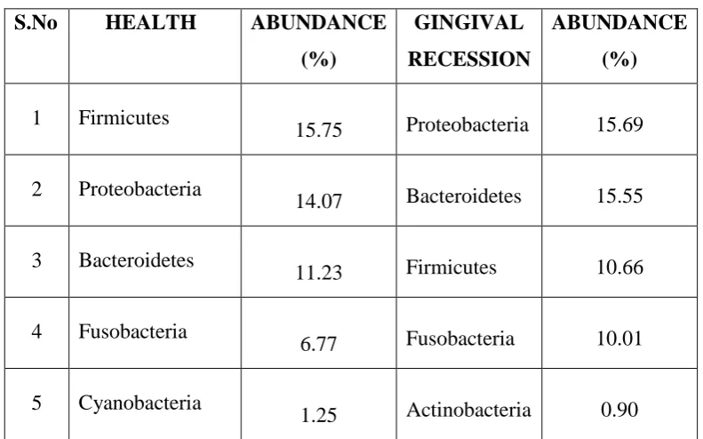
2 Top 5 phyla in healthy and gingival recession sites
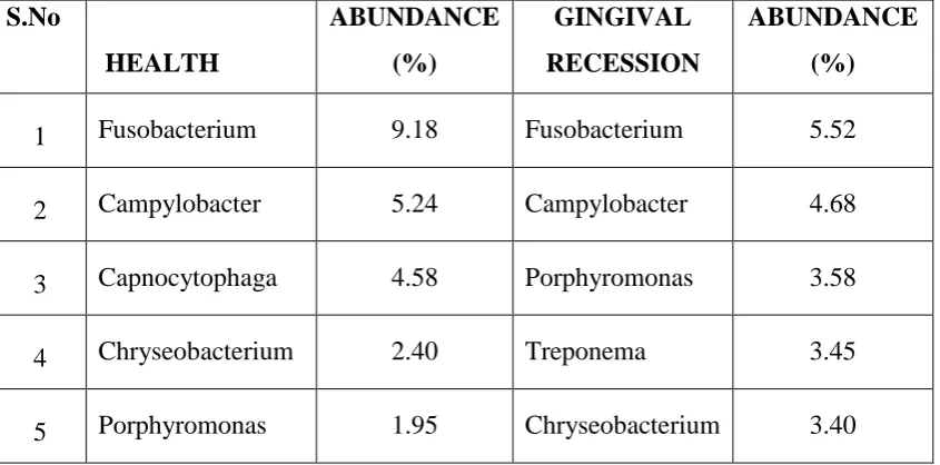
3 Top 5 genera in healthy and gingival recession sites
4 Periodontal health associated microbiome
5 Disease associated microbiome
6
Relative abundance of top 20 bacterial species in healthy
versus gingival recession sites
7
Relative abundance of top 20 species in gingival recession
NO.
1-A Bar graph demonstrating distribution of bacterial phylum in health and gingival recession
1-B Bar graph demonstrating distribution of bacterial genus in health and gingival recession
1-C Bar graph demonstrating distribution of bacterial species in health and gingival recession
2-A Pie chart depicting top 5 phyla in health
2-B Pie chart depicting top 5 phyla in gingival recession
3-A Pie chart depicting top 5 genera in health
3-B Pie chart depicting top 5 genera in recession
3-C Phylogenetic tree at genus level
4 Bar graph showing percentage abundance of top 20 bacterial species in health and corresponding abundance in gingival recession
NO. TITLE
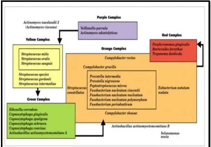
1 Bacterial Complexes
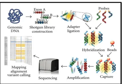
2 Roche 454 Life Sciences System
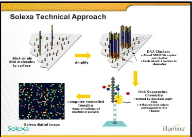
3 Illumina Solexa Genome Analyzer
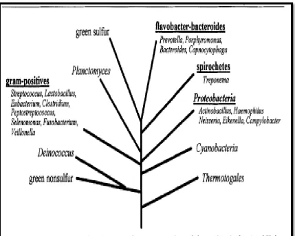
4 Taxonomy Of Oral Bacteria (10 Major Phyla)
1
INTRODUCTION
Human body is estimated to be composed of more than 1014 cells of which only 10% are mammalian.98 Microorganisms contribute to majority of cells found in our body, forming resident microflora, surviving in symbiotic
relationship with the host and thus maintaining homeostasis.
Oral microbiome exhibits the largest core of commonly related
microbes among unrelated subjects when compared to gut or skin
microflora.18,59,136 The relationship between periodontal microflora and host is known to be benign, but changes in subgingival microbiome and bacterial
community structures can contribute to pathogenesis of periodontal disease.49 Periodontal diseases are a heterogeneous group of chronic conditions
affecting tooth supporting structures.126 It is described as a dysbiotic disease characterized as being polymicrobial and multifactorial in nature. Dysbiosis is
a symbiotic relationship which has gone astray caused by a decrease in
number of beneficial symbionts and/or an increase in number of pathobionts.12 Research over the past decade has led to recognition of microbes
residing in various oral ecological niches as a part of dental plaque. Plaque
biofilm is a highly organized accumulation of microbial communities adhering
to favourable environmental surfaces, which functions to maximize energy,
spatial arrangements, communication, and continuity of bacterial
communities. The origin, development and structural adaptation of dental
2
microbiota and multiple factors that differentially promote or inhibit survival
of its microbial constituents.62
According to previous studies by Socransky et al,105 the key bacterial species which play a role in the disease process have been segregated into
microbial complexes based on their correlation with clinical parameters and
severity of periodontal disease. Over the past 50 years, understanding and
characterization of dental plaque has undergone significant evolution from
nonspecific plaque hypothesis13,76 and specific plaque hypothesis66 to Ecological Plaque Hypothesis.72 Currently, the pathogenesis of periodontal diseases is explained by “Polymicrobial Synergy and Dysbiosis (PSD) Model”
proposed by Hajishengallis et al.35 This model states that dysbiotic environment and polymicrobial synergy are the key events that lead to
development of periodontitis rather than individual bacterial species. Thus
biofilm community as a whole was thought to be either health-associated or
disease-associated.
Abnormal pocket depth and pocket-free gingival recession have been
recognized as two separate periodontal phenotypes at least since the 18th
century.43 Gingival recession refers to exposure of root surfaces caused by apical displacement of gingival margin beyond cemento-enamel junction.125 The three primary etiologic agents of gingival recession are plaque-induced
inflammation, mechanical abrasion/removal due to faulty tooth brushing, and
developmental/mucogingival deformities. In the Indian subcontinent an
3
inadequate oral hygiene practices. Several studies on subgingival microbiome
have documented data regarding periodontal pocket,54,79,87,105 but there is paucity in literature regarding subgingival microbiome in gingival recession
sites.
Traditional culturing methods for bacterial identification are often
insufficient for biofilm analysis due to unknown culture characteristics of
several bacterial species; hence a vast majority of oral bacteria evade standard
microbiological detection methods. This has led to development of alternative
methods to assess dental biofilms based on DNA analysis or other molecular
techniques.41 Studies of subgingival microbial community identified a link between taxonomic composition and disease pathogenesis. Current trend in
sequencing of microbiome is based on Next-Generation Sequencing (NGS),
that uses parallel sequencing of multiple small fragments of DNA to determine
genetic sequences.93 The speed of sequencing and amounts of DNA sequence data generated with NGS, which is considered a “high-throughput
technology”, are exponentially greater and are produced at significantly
reduced costs.7 NGS technology was utilized earlier in our department (unpublished data) to identify and characterize subgingival microbiome from
periodontal pockets.
Subgingival community has been extensively studied earlier to
4
perplexing and identifying these changes offer a promising potential for
research in pathogenesis of periodontal disease.102
For a better understanding of the etiology and polymicrobial
mechanisms of localized chronic periodontitis presenting as gingival recession
due to plaque-induced bacterial inflammation, the current study was
undertaken to identify, characterize and analyse species of subgingival
microbiome and characterize novel micro-organisms among periodontal health
5
AIM AND OBJECTIVES
Aim:
To identify and characterize subgingival microbiome using Next
Generation Sequencing Technology in periodontal health and chronic
periodontitis presenting as gingival recession.
Objectives:
1. To identify and characterize the subgingival microbiome in periodontal
health and in gingival recession sites using Next Generation
Sequencing Technology.
2. To compare and analyze subgingival microbiome in sites of gingival
recession with those of periodontally healthy controls.
3. To study species diversity in subgingival microbiome, and characterize
novel micro-organisms in gingival recession sites for a better
6
REVIEW OF LITERATURE
Mouth as a microbial habitat:
The human body is estimated to be composed of more than 1014 cells, of which only 10% are mammalian.98 The majority of these cells are the microorganisms that make up the resident microflora found on all
environmentally exposed surfaces of the body, and this human ―microbiome‖
is reported to have a metabolic capacity equivalent to that of the human liver.71 It is estimated that between 300 and 400 different species are capable of
colonizing the mouth, and any individual may typically harbor 150 to 200
different species. Counts in subgingival sites range from about l03 in healthy, shallow sulci to more than 108 in deep periodontal pockets.106 The oral microflora is distinctive because of the characteristic biological and physical
properties of each site despite potential movement of microorganisms between
sites. Bacteria may attach to tooth itself, to epithelial surfaces of gingiva or
periodontal pocket, to underlying connective tissues, if exposed, and to other
bacteria attached to these surfaces. These observations illustrate a key concept
that properties of the habitat are selective and dictate which organisms are able
to colonize, grow and be minor or major members of the community.71 HUMAN ORAL MICROBIOME:
Concept of the human oral microbiome
The microorganisms found in human oral cavity have been referred to
7
term microbiome was coined by Joshua Lederberg ―to signify the ecological
community of commensal, symbiotic, and pathogenic microorganisms that
literally share our body space and have been all but ignored as determinants of
health and disease‖. Turnbaugh et al (2007)118 described human microbiome to be classified into a core microbiome and a variable microbiome. Core
microbiome comprises of the predominant species that exist under healthy
conditions at different sites of the body, and it is shared by all
individuals.109,118,132 Variable microbiome is one that has evolved in response to unique lifestyle, phenotypic and genotypic determinants; it is exclusive for
an individual. Even though individuals share microbiota at similar sites of the
body, varying differences are observed at species and strain level of the
microbiome which may be as unique as a fingerprint to an individual.20
The oral cavity is highly populated with numerous polymicrobial
communities, each occupying highly specific niches that differ in both
anatomic location and nutrient availability. Oral host colonization is a
reflection of bacterial proficiency to adapt to a variety of different niches
through high rates of genetic recombination.94 Oral microbiota needs to cope with daily physical and chemical perturbations from intake of food and
personal hygiene measures, and these include fluctuations in temperature, pH,
antimicrobial and dietary components, and mechanical sheer forces from
8
microbes among unrelated individuals compared to other habitats such as gut
or skin.18,59,136
Dysbiosis is defined as change/perturbations in the structure and
composition of resident commensal bacterial communities relative to the
community found in healthy individuals.89 Dysbiosis, as the term implies, is a symbiotic relationship which has gone astray caused by a decrease in the
number of beneficial symbionts and/or an increase in the number of
pathobionts.12 Biological properties that help to maintain stability in the microbiome are important for sustaining symbiotic environment and for
prevention of dysbiotic state.
Constitution, acquisition and maintenance of the normal oral microbiome
Human oral cavity is colonized by a wide range of microorganisms.
Besides bacteria and fungi, Archaea, viruses and protozoa form a part of
normal microbiome.122 The microbiome has been studied in great detail and phylogenetic information of oral bacteria is gathered in databases dedicated to
oral cavity.86 The HMP assessed microbiome composition of nine intraoral sites (buccal mucosa, hard palate, keratinized gingiva, palatine tonsils, saliva,
subgingival and supragingival plaque, throat and tongue dorsum) from about
200 subjects and found 185 to 355 genera, belonging to 13 to 19 bacterial
phyla.136 The microbiome has evolved through hundreds of thousands of years of co-habiting into a microbe-human symbiosis with mutual benefits. Costello
et al18 reported that oral microbiome in newborns was shown to seed the gut
9
2 weeks’ time to gut-specific communities. Based on current knowledge it is
apparent that acquisition of such normal, beneficiary microbiome by newborns
is an essential process. Infants are colonized rapidly after birth by bacteria
present in their direct environment, through bacterial transfer from their
mother but also from other sources.
Zaura et al133 has suggested that development of fetal tolerance towards microbiome of the mother during pregnancy is a major factor for
successful acquisition of a normal microbiome. Although the first encounter of
a newborn with microbiota is considered to be postnatal, several studies have
shown clinical evidence for microbial presence in placenta, umbilical cord
blood, amniotic fluid, and meconium in full-term pregnancies without overt
infection.1,10,45,111 Vertical transmission from mother to child starts at birth. The delivery mode (vaginal or Caesarian), to a large extent, determines which
microorganisms—vagina or skin-derived— will be encountered first by the
new born. This affects diversity of oral microbiome: vaginally born infants
showed higher taxonomic diversity at 3 months of age.61 Li et al59 reported that birth mode may have a lasting impact as infants born with Caesarian
section acquired Streptococcus mutans almost 1 year earlier (at 17.1 months of age) than vaginally born infants (28.8 months). The method of feeding
(breast-feeding or infant formula) affects the infant’s microbiome as well. Breast-fed
infants of 3 months of age carried oral lactobacilli with antimicrobial
10
Once established, the microbiome should be sustained. This is reported
to be achieved through host-derived and microbe-derived microbiome
maintenance factors. The interactions between the microbiome and the host
are bidirectional, and the factors that determine the continued communication
routes include innate and adaptive immune responses, host pattern recognition
receptors (Toll-Like Receptors play a key role), chemosensory activation
mechanisms, crevicular and salivary glycoproteins.
Co-evolution of the microbiome with host has resulted in
host-associated microbial communities that are equipped with mechanisms that
allow them to prevent colonization and establishment of foreign microbes, so
called ―colonization resistance‖.40
Integrity of the microbial community is
maintained by specific inter-microbial adhesion, cell signaling through
cell-to-cell contact, metabolic interactions and quorum sensing. Besides bacterial
inter-species communication, inter- kingdom communication also plays a role
in oral microbial ecosystem.44,80
PERIODONTAL DISEASE- ETIOLOGY AND PATHOGENESIS
Periodontal diseases are a heterogeneous group of chronic conditions
that reflect a cellular inflammatory response of supporting periodontal tissues
of the teeth against bacterial challenges.90 It is a dysbiotic disease characterized as being polymicrobial and multifactorial in nature exhibiting a
shift from predominantly gram-positive bacteria found in healthy sites to
mostly gram-negative bacteria found in clinically diseased sites. The initiation
11
related to the lack or minimal proportions of beneficial microorganisms in a
susceptible host.90 The end outcome of untreated periodontal disease is loss of attachment apparatus and subsequent loss of teeth often leaving patients
unable to eat and function properly.
Löe et al (1978)65 demonstrated the natural progression of periodontal disease through a series of studies over several years following a population of
Sri Lankan tea workers which represented a relatively uniform population that
had little to no dental care and also had extremely poor oral hygiene. Several
lines of evidence indicate that bacteria are necessary for the development of
inflammation in the periodontal tissues. In a study by Mitchell and Johnson77 bacteria were implicated in periodontal disease with the observation that
administration of penicillin inhibited periodontitis in laboratory animals, and
Keyes and Jordan48demonstrated the infectious nature of periodontitis by its transmissibility in animal models.
The current concept concerning the etiology of periodontal disease
considers three groups of factors which determine whether active periodontal
disease will occur: a susceptible host, presence of pathogenic species, and
absence of so-called "beneficial bacteria".47 It has been demonstrated that initiation and progression of inflammatory and destructive periodontal lesions
is related not only to presence of bacterial strains pathogenic for the
periodontium, but also to lack or minimal proportions of beneficial
microorganisms in a susceptible host. The unequivocal role of dental bacterial
12
40 years ago. Gingivitis is a reversible inflammatory reaction of marginal
gingiva to plaque accumulation, whereas periodontitis is a destructive,
nonreversible condition resulting in loss of tooth connective-tissue attachment
to bone, which ultimately leads to loss of the involved teeth. Existing evidence
indicates that gingivitis precedes onset of periodontitis; however, not all
gingivitis cases develop into periodontitis. The reason for this is that
accumulation of plaque bacteria is necessary but not sufficient by itself for
development of periodontitis: a susceptible host is necessary.114 GINGIVAL RECESSION
According to Wensstrom JL (1996)125 gingival recession refers to exposure of root surface caused by apical displacement of gingival margin
beyond cementoenamel junction. Gingival recession, either localized or
generalized, is one of the clinical features of periodontal disease and is
frequently associated with clinical problems such as root surface
hypersensitivity, root caries, cervical root abrasions, erosions, plaque retention
and aesthetic dissatisfaction.15
Abnormal pocket depth and pocket-free gingival recession have been
recognized as two separate periodontal phenotypes at least since the 18th century.43 The distinction as two different phenotypes was summarized by Page & Sturdivant85 as:
1. Periodontal atrophy, where the gingiva retain a very healthy aspect and
13
2. Destructive periodontal disease with presence of deepened periodontal
pockets and underlying bone loss
Etiology of gingival recession:
The primary causes of gingival recession are plaque-induced
inflammation and mechanical (physical) abrasion/removal. Occasional causes
of recession include thermal and chemical injury. Three major factors are
associated with increased susceptibility to gingival recession: (i) thin gingival
tissue; (ii)mucogingival conditions; and/or (iii) a positive history of
progressive gingival recession and/or inflammatory periodontal diseases in
teeth presenting with either or both of the first two factors.74 Mucogingival conditions are defined as deviations from the normal anatomic relationship
between the gingival margin and the mucogingival junction.
The etiology is multifactorial and includes excessive or inadequate
teeth brushing, destructive periodontal disease, tooth malposition, alveolar
bone dehiscence, high muscle attachment, aberrant frenal pull, occlusal
trauma, iatrogenic factors (such as orthodontic, or prosthetic treatment) and
smoking.55
Pathogenesis and severity of recession:
The mechanism of gingival recession due to localized inflammatory
processes in connective tissues with the accumulation of mononuclear cells
14
clinically and histologically where there is proliferation of epithelial rete pegs.
Stage three shows increased epithelial proliferation resulting in loss of
connective tissue core and finally there is merging of oral and sulcular
epithelium resulting in separation and recession of the gingival tissues due to
loss of nutritional supply.
Waerhaug J (1952)123 proposed that the distance between the periphery of plaque on the tooth surface and the labial, apical extension of the
inflammatory infiltrate hardly ever exceeds 1-2 mm. Thus if the free gingiva is
voluminous the infiltrate will occupy only a small portion of the connective
tissue; however if it is thin the entire connective tissue portion may be
involved, consequently there is proliferation of epithelial cells from the oral
and dentogingival epithelium, the zone of connective tissue decreases and
finally recession ensues.
Sarfati et al (2010)100 reported that gingival bleeding was significantly associated with gingival recession severity and concluded that inflammatory
reaction to dental biofilms is the predominant biologic feature shared by
gingival recession and periodontitis. In his study, he found that gingival
bleeding was significantly associated with severity of gingival recessions
(P value = 0.010) and not with extent of gingival recessions, which suggested
that extent of recession is related to plaque accumulation and that host
response, i.e., inflammation conditions, is a key factor for recession severity.
Similarly, number of missing teeth was associated with severity of
15
more tooth loss, it has been stressed that there is more periodontitis,
attachment loss, and consequently, gingival recession. This finding illustrates
that periodontitis is a site-specific disease, which means that number of
missing teeth does not depend on extent of the disease but rather on defect
severity. Thus the number of missing teeth could be a better surrogate criterion
of recession severity than extent of recession. Van der Velden et al119 showed an association between gingival recession and periodontitis severity in a Java
population. This outcome, together with results from the study by Sarfati in
2010, may indicate a bi-directional association between gingival recession
severity and periodontitis severity, rather than a causal relationship. It may be
assumed that inflammatory reaction to dental biofilm is the predominant
biologic feature shared by gingival recessions and periodontitis.
Keratinized tissue width as a parameter for gingival recession:
Lang and Loe (1972)58 demonstrated that although tooth surfaces may be kept free of clinically detectable plaque, areas with less than 2 mm of
keratinized gingiva tend to remain inflamed. However, clinicians like
Dorfman HS24, Freedman AL et al29, Kisch et al50 and Miyasato78 have evaluated sites with less than 2 mm of keratinized tissue and concluded that
these sites do not necessarily develop gingival recession solely as a result of a
narrow width/band of keratinized tissue. Serino et al101 reported that teeth with a positive history of progressive gingival recession have increased
16
In 1999, Albandar and Kingman5 suggested that gingival recession occurs primarily as a consequence of periodontal diseases and aggressive use
of mechanical oral hygiene measures. A positive history of inflammatory
periodontal disease (e.g. plaque-induced gingivitis and localized chronic
periodontitis) can be considered an important factor associated with gingival
recession, especially for teeth with thin gingival tissues and/or mucogingival
conditions.
ETIOLOGIC ROLE OF PLAQUE IN PERIODONTITIS
Plaque biofilm as an etiological agent for periodontitis
The microbial etiology of periodontitis has been extensively researched
for the past few decades, and it is now well known that periodontitis is not
associated with a single micro-organism but is a consortium of bacteria
participating in initiation and progression of disease process.96 The most significant consequence of biofilm formation on tooth surfaces is continuous
release of bacterial toxins into the local microenvironment. The composition
of subgingival microbial flora and level of pathogenic species differ from
subject to subject as well as from site to site. Marsh (2011)71described dental plaque as the microbial community that develops on the tooth surface,
embedded in a matrix of polymers of bacterial and salivary origin. Dental
plaque forms via an ordered sequence of events resulting in a structurally and
17
Changing views on etiologic role of dental plaque
According to an experimental gingivitis study by Loe (1965)65, etiologic role of plaque in periodontal disease and its involvement in initiation
and progression of periodontal diseases was firmly established. It has been
generally accepted that periodontal diseases appear to be initiated by a
relatively limited number of periodontal pathogens in the complex dental
biofilm, and they represent a small part of approximately 600 bacterial species
that have been found to colonize dental surfaces over and below the gingival
margin and oral mucous membranes.90 Clinical and experimental evidences in last three decades confirm that certain bacterial strains in the periodontal
environment can induce gingival tissue inflammation and bone destruction,
and were termed as periodontal pathogens.83 Only a small percentage of dental biofilm bacteria are defined as pathogenic for periodontal tissues and these
bacteria, even when present in very small quantities, possess the ability to
damage periodontal structures.
According to the criteria proposed by Socransky and Haffajee108, a microorganism must meet certain conditions to be considered a potential
periodontal pathogen: association with disease by means of increased number
in diseased patients and sites; reduction or elimination after treatment; capable
of provoking destructive host responses; capacity to cause disease in
experimental animal models; ability to produce virulence factors known to
cause periodontal destruction. On the basis of the proposed criteria by
18
examined and shown to be periodontopathogens and disease initiators of
periodontitis.83 Over the past 50 years, ideas about changes in plaque relating to shift from oral health to disease, and understanding and characterization of
dental plaque have undergone significant evolution.
Non-specific plaque hypothesis
Based on work of researchers Black13 and Miller76 the non-specific plaque hypothesis was proposed which stated that periodontal disease is due
to bacterial accumulation, irrespective of its composition. This implies that no one specific bacterial species is any more significant than another in its ability
to cause periodontal disease.39 Increase in quantity of plaque, as opposed to specific pathogenic microorganisms (quality) found in plaque, were viewed as
being primarily responsible for inducing disease and disease progression.33 Although the amount of plaque present may correlate well with disease
severity in cross-sectional studies, it correlates poorly in longitudinal studies.
Non-specific plaque hypothesis is valid for development of gingivitis but not
for periodontitis, which is a multifactorial disease.84 This concept failed to explain why all gingivitis do not progress to periodontitis, why some
individuals with increased plaque showed little overt periodontitis and why
some individuals with very little plaque manifested with aggressive and
advanced forms of periodontitis.108 This hypothesis did not consider variations in dental biofilm that may affect its pathogenicity or, most importantly, host
19 Specific plaque hypothesis
Specific plaque hypothesis proposed by Walter J. Loesche66 stated that periodontal disease is the result of an infection with a single specific pathogen. Studies on microbial etiology of various forms of periodontitis
support this hypothesis, which proposes that only certain microorganisms
within the plaque complex are pathogenic. Newman MG82 and Slots104 identified Aggregatibacter actinomycetemcomitans as a specific pathogen in
localized aggressive periodontitis. Despite the presence of hundreds of species
of microorganisms in periodontal pockets, fewer than 20 are routinely found in
increased proportions at periodontally diseased sites. These specific virulent
bacterial species activate the host's immune and inflammatory responses that
then cause bone and soft tissue destruction. This hypothesis failed to explain
why putative periodontal pathogens like Porphyromonas gingivalis, Tannerella forsythia are frequently found in healthy periodontal sites.
Based on this concept, it was recognized that early plaque consists
predominantly of gram-positive organisms and if left undisturbed it undergoes
a process of maturation resulting in a more complex and predominantly
gram-negative flora. Sockransky et al105 assigned organisms of subgingival microbiota into groups, or complexes, based on their association with health
and various disease severities.33 The yellow, green and purple complexes were early colonizers that favour colonization of orange and red complexes. Red
20
forsythia), Porphyromonas gingivalis and Treponema denticola and they were significantly associated with periodontitis. (Figure 1)
Ecological plaque hypothesis
Ecological plaque hypothesis, proposed by Philip D. Marsh (1994)72, described and explained the dynamic relationship between resident microflora
and host in health and disease in ecological terms. The theory underpinning
this hypothesis in the context of periodontal disease is that changes in the
environment increase competitiveness of putative pathogens at the expense of
species associated with oral health and upregulate the expression of virulence
factors. Marsh et al71 reported a clear link between local environmental conditions and activity and composition of the biofilm community; any change
to the environment induces a response in the microflora and vice versa.
Keystone Pathogen Hypothesis
Socransky et al105 reported that when disease develops and advanced stages are reached, the keystone pathogens are detected in higher numbers.
Keystone Pathogen Hypothesis, proposed by Hajishengallis et al (2012)35, indicates that certain low-abundance microbial pathogens can cause
inflammatory disease by increasing the quantity of normal microbiota and by
changing its composition.
Polymicrobial Synergy and Dysbiosis Model (PSD):
PSD model of pathogenesis described by Hajishengallis et al35 states that periodontitis is initiated by a broadly based dysbiotic, synergistic
21
several periopathogens like red complex bacteria. This dysbiotic, synergistic
microbiota alters host-microbe homeostasis and facilitate its transition to a
chronic inflammatory state. Lamont and Hajishengalis57 observed that the whole microbial community drives disease progression, representing the
interplay between subgingival community of microorganisms and local
immune responses which ultimately drives bone and connective tissue
attachment loss.
SUBGINGIVAL MICROBIOME
Members of the human oral microbiome were among the first bacteria
ever to be observed. In 1683, Antonie van Leeuwenhoek used his microscope
to observe a large number of what he named ―animalcules‖ in scrapings taken
from his teeth. Over 200 years later, the seminal work of Koch, Pasteur and
their contemporaries identified the animalcules as microorganisms and the
first isolates of cultivable members of the oral microbiome were studied in the
laboratory.
The subgingival microbiome is the community of microorganisms
inhabiting the subgingival environment. Haffajee and Socransky34 and Zambon131have extensively studied the microbial composition of subgingival plaque at periodontally diseased sites. In a landmark study by Socransky and
Haffajee105, they attempted to define bacterial communities existing as different complexes in subgingival plaque by studying 13,261 plaque samples
from 185 subjects using whole genomic DNA probes and checkerboard
22
different clustering and ordination techniques. The complex most significantly
associated with periodontitis and to clinical measures like probing depth and
bleeding on probing was the red complex, comprising of Tanerella forsythia, Porphyromonas gingivalis and Treponema denticola.105 A series of culture studies by Moore and Moore79involving analysis of subgingival plaque taken from subjects with different forms of periodontal disease and health reported a
shift in the subgingival microbiota as the periodontium progressed from health
through gingivitis to periodontitis. Liu et al63 and Chen et al16 investigated bacterial diversity between periodontal health and disease status using 16S
rRNA amplicon sequencing and showed that there is a shift in the composition
of the oral microbiota between healthy and diseased samples.
Kumar et al (2003)54 investigated subgingival microbiome based on 16S rDNA cloning and sequencing and showed that 40% of bacterial species
present were either novel species or phylotypes. Paster BJ and Dewhirst
(2006)87 developed a molecular technique to detect oral biofilms using a 16S rRNA-based microarray technology called Human Oral Microbiome
Identification Microarray (HOMIM). This system provides information on the
9 most commonly found oral bacterial flora namely Bacteroidetes, Firmicutes, Proteobacteria, Synergistetes, Fusobacteria, Spirochaetes, Actinobacteria,
23
DETECTION AND ENNUMERATION OF BACTERIAL SPECIES
FROM PERIODONTAL SAMPLES:
Microscopic studies
In 1683 Antonio van Leeuwenhoek studied scrapings from human
teeth using microscope and described 5 types of animalcules. Coyler had
proposed use of dark field microscopy for evaluation of pocket
microorganisms.91 Theodore Rosebury conducted a series of experiments to isolate bacteria of etiological importance in periodontal disease. Keyes
(1965)48 proposed use of phase contrast microscopy in periodontal diagnosis to identify bacterial morphotypes.
Microscope techniques were reasonably rapid, but limited in the
precision of identification of individual bacterial species. The strength of
microscopy techniques is delineation of spatial arrangements of organisms;
whereas its weakness from an ecologic perspective is that they are slow, labor
intensive and precise speciation using immunologic or hybridization
techniques can only be performed for a very limited number of species in any
given sample.107
Culture based methods
Cultivation of organisms and identification of species by their
phenotypic traits was one of the major techniques used by researchers to
identify plaque bacteria. The classic studies of Moore & Moore79 in which they examined composition of subgingival plaque samples in periodontal
24
examine over 17,000 isolates from over 600 periodontal sites. Although this
was a massive amount of work, it was considered a limited number of samples
by current standards. The major strength of culture is that majority of bacterial
species sampled can be grown and identified in lab conditions. The main
drawback of culture method is its narrow spectrum, and it is regarded as a
time-consuming, labor-intensive, and expensive undertaking because only few
plaque samples in small numbers of subjects can be examined. Studies by
Kolenbrander (2000)51, Vartoukian et al (2007)120 and Siqueria et al (2010)103 have estimated that 50% to 60% of distinct bacterial phyla in oral cavity still have no cultivable representatives. However, cell culture is still
essential to assess bacterial sensitivity to antibiotics and for verifying presence
of known species.
Immunologic and enzymatic assays
Immunofluorescence techniques and enzyme-linked immunosorbent
assay (ELISA) techniques are antibody-based methods used to enumerate
specific species of microorganisms without their cultivation; it is dependent on
specificity of developed antibodies to specific taxa. These techniques have the
advantage that samples do not have to be cultured for enumeration; they are
rapid and less expensive than culture. However, they are limited to species for
which reagents have been developed; it is difficult to use these techniques to
evaluate species in large numbers of plaque samples and it is time-consuming
25 DNA – DNA hybridization or checkerboard
DNA-DNA hybridization is a molecular approach used to detect
bacteria based on hybridization of target species to labeled genomic DNA that
has been attached to nylon membranes. Studies by LoescheWJ et al66, Haffajee34, Ximenez-Fyvie LA129, Feres M et al27 and Socransky107 have reported on levels of limited number of species with this method in adult
periodontitis, periodontal health, refractory periodontitis and response to
therapy. This method provides a major benefit for studies of oral microbial
ecology due to advantages like detection of multiple species from each sample
simultaneously, and study of large sample size for large numbers of species.
Checkerboard technique is rapid, sensitive, and relatively inexpensive but is
also dependent on culture technique to cultivate the target species for creating
genomic probes. Like antibody-based assays, cross reactivity can be verified
only with cultivated species hence specificity of the probe is an unknown
variable.
Polymerase chain reaction
Kary Mullis first developed polymerase chain reaction (PCR)
technique to amplify specific genes or parts of genes which are then used to
identify bacterial species from which they originated.53 In a study by Kumar PS et al (2005)54 species-specific PCR primers were designed and used in individual PCR reactions to detect prevalence of target species in plaque
samples of healthy and diseased subjects. These studies confirmed that several
26
periodontitis. Given the appropriate primers, this method is rapid, simple, can
detect small numbers of cells of a given species, and indicates the presence or
absence of a species in the sample. It has certain disadvantages of not
providing quantitative data, may not be cost effective for large sample sizes,
and for applications where relative levels of species are important, PCR may
not be ideal.107 DNA Probes
Oligonucleotide probes are short probes designed to identify unique
regions of DNA within cells of a given bacterial species. These probes are
highly specific and likelihood of cross-reactions with other species is very
low. Because they target a limited segment of DNA of an organism,
oligonucleotide probes tend to be less sensitive for detection of low numbers
of bacteria than whole genomic probes.107
Whole genomic DNA probes are constructed using the entire genome
of a bacterial species as the target and thus can be quite sensitive. The use of
the entire genome may increase the probability of cross-reactions between
species because of common regions of DNA among closely related species.
The technique can detect only species for which DNA probes have been
prepared, thus novel pathogens or environmentally important species that
might be detected in culture or by other molecular techniques may not be
27
OPEN ENDED APPROACHES- 16S rRNA sequencing analysis:
Open ended approaches allow identification of even uncultivated and
previously unknown species. According to Spratt (2004)110 these approaches are based on 16 S rRNA sequencing, amplification and analysis of the 16S
rRNA genes in a microbiome sample. 16S rRNA has proven to be the most
useful phylogenetic marker to identify bacteria and to determine their
evolutionary relationships. Ribosomal RNA gene is essential for life and
present in all prokaryotes. It contains nucleic acid sequences with highly
conserved and variable regions; conserved regions are used to design universal
PCR primers capable of recognizing segments of 16S rRNA gene sequence of
all bacterial species and hypervariable regions are used as signatures to
discriminate one species from another. 16S rRNA gene is large enough (about
1500 bases) to provide sufficient sequence variability among bacteria, thereby
making comparisons possible at different taxonomic levels.
DNA and protein sequencing started in the 1970s when the virus
Lambda (50,000 nucleotides) was sequenced by Sanger et al.97 Frederick Sanger and colleagues described the use of chain-terminating
dideoxynucleotide analogs that caused base-specific termination of primed
DNA synthesis and this came to be popularly known as Sanger sequencing
method.107 This method of sequencing was considered the gold standard, and over the years, whole genome sequencing of many bacteria has been carried
out using this method. Sanger technology was used in the sequencing of the
28
Genome Project, a 13-year effort with an estimated cost of 2.7 billion
dollars.121
Over the past decade, next generation sequencing technologies have
emerged, which are high throughput and able to generate three to four orders
of magnitude more sequences and are also relatively less expensive.23
NEXT GENERATION SEQUENCING TECHNOLOGY (NGS):
Next generation sequencing methods employ a wide spectrum of
technologies such as sequencing by synthesis, sequencing by ligation, single
molecule DNA sequencing and colony sequencing. NGS is performed by
repeated cycles of polymerase-mediated nucleotide extensions or by
machinery automated cyclical ligation of oligonucleotides.69,121 Fundamentals of NGS platforms:
NGS platforms share a common technological feature—massive
parallel sequencing of clonally amplified or single DNA molecules that are
spatially separated in a flow cell. This design is a paradigm shift from that of
Sanger sequencing, which is based on electrophoretic separation of
chain-termination products produced in individual sequencing reactions. In NGS,
sequencing is performed by repeated cycles of polymerase-mediated
nucleotide extensions or, in one format, by iterative cycles of oligonucleotide
ligation.121 As a massively parallel process, NGS generates hundreds of megabases to gigabases of nucleotide sequence output in a single instrument
29
fragments with oligonucleotide adaptors and fragment immobilization to a
solid surface, such as a bead.
The three commonly used platforms for massive parallel DNA
sequencing at present are the Roche/454 FLX (Life Sciences, Branford, CT,
Margulies et al., 2005)70, the Illumina/ Solexa Genome Analyzer (Illumina, San Diego, CA, Bentley DR, 2006, Korbel et al., 2007)52 and the Applied Biosystems ⁄ SOLiD (Life Technologies, Carlsbad, CA).69,121
The most recent
powerful NGS platforms have significant reductions in the run time and
remarkable data output, they include HiSeq and the Ion Torrent Personal
Genome Machine (PGM).95
ROCHE 454 LIFE SCIENCES SYSTEM
The 454 technology is derived from technological convergence of
pyrosequencing and emulsion PCR. In 2000, Jonathan Rothberg founded 454
Life Sciences, which developed the first commercially available NGS
platform, GS 20, launched in 2005.121 One of the major drawbacks of this system is that sometimes more than one nucleotide is incorporated in DNA
template during a cycle, making it difficult to resolve homopolymeric
stretches of sequence (e.g. CCCCC or AAAAA). (Figure 2)
ILLUMINA/SOLEXA GENOME ANALYZER
In 1997, British chemists Shankar Balasubramanian and David
Klenerman conceptualized an approach for sequencing single DNA molecules
attached to microspheres and founded Solexa in 1998. The Solexa Genome
30
launched and acquired by Illumina in 2006. Genome Analyzer uses a flow cell
with bound oligonucleotide anchors wherein template DNA is fragmented into
several hundred base pairs and end-repaired (Figure 3).121
The newest platform, the Genome Analyzer II, has optical
modifications enabling analysis of higher cluster densities. Illumina and other
NGS technologies have devised strategies to sequence both ends of template
molecules. Such ―paired-end‖ sequencing provides positional information that
facilitates alignment and assembly, especially for short reads.
The advantage of Solexa system is that it can generate 1.5 GB of
sequence per run with read lengths that range from 35 to 100 bases and each
run requires 3–5 days to complete.95 A technical concern of Illumina sequencing is that base-call accuracy decreases with increasing read length
primarily due to ―dephasing noise‖ which occurs when a complementary
nucleotide is not incorporated or when fluorophore is not properly cleaved at
the end of cycle, thus blocking incorporation of next nucleotide base.121 As a consequence, the sequence is out-of-phase for remainder of the template.22 Another shortcoming is that short read lengths tend to produce biased
sequence coverage that occurs in AT-rich repetitive sequences.38 APPLIED BIOSYSTEMS SOLiD:
The SOLiD (Supported Oligonucleotide Ligation and Detection)
System 2.0 platform is a short-read sequencing technology based on ligation.
This approach was developed in the laboratory of George Church and reported
31
can generate 4 GB of sequence but the reads are only 35 nucleotides.121 The weakness of SOLiD system is that it yields biased sequence coverage in
AT-rich repetitive sequences38 and only 35% of the raw reads are useable, compared with 95% for the 454 system. Another disadvantage is that it
requires long run times.
THE HUMAN ORAL MICROBIOME DATABASE
Research over the past 20 years has focused on defining breadth and
diversity of oral microbiome by obtaining 16S rRNA gene sequence
information for both cultivable and as yet uncultivated oral bacteria.113 The majority of bacterial species isolated from the oral cavity are included in 4 of
the 10 bacterial phyla; Phylum 1 (Proteobacteria), Phylum 2 (the
gram-positives), Phylum 5 (the spirochetes) and Phylum 6 (the
flavobacter-bacteroides group).There are no known human oral representatives from the
other 6 phyla. Though human oral microbiome is the most studied human
microflora, 53% of species have not been named yet and 35% of species are
uncultivated. The uncultivated taxa are identified mainly by 16S rRNA
sequence information. (Figure 4; Figure 5)
The Human Oral Microbiome Database (HOMD) is a specifically
designed database to provide a provisional naming scheme where each oral
taxon is given a human oral taxon (HOT) number linked to comprehensive
information and tools for examining and analyzing each taxon in the human
32
full-length 16S rRNA reference sequences, and BLAST tools that allow
identification of unknown isolates or clones based on their 16S rRNA
sequence; additionally phenotypic, bibliographic, clinical and genomic
information are linked for each taxa. Organisms of the human oral cavity are
organized in a taxonomy hierarchy, which leads to individual pages for every
oral taxon with comprehensive information and links. The genomic
component of HOMD contains both static and dynamically updated
annotations as well as bioinformatics analysis tools for all the genomic
sequences, and curated 16S rRNA gene reference sequences for all human oral
microbes. HOMD may serve as an example of a body site-specific tool for
other communities.
More recently, a similar database was set up by Griffen A et al31 known as CORE, a phylogenetically curated 16S rDNA database of the core
oral microbiome, which offers improved and more robust identification of
human oral bacterial 16S rRNA gene sequences compared with other methods.
Its main goal is to provide a comprehensive and minimally redundant
collection of oral bacteria at the genus and species level, as well as providing
support for inferring community divergence and analysis of large datasets.
The basic list of oral bacteria came from the literature works of Dzink JL25,26, Sockransky106, Tanner112,113 and Moore WE79. In 2010, Dewhirst identified 1,179 taxa of which 24% were named, 8% were cultivated but unnamed, and
33
metagenomic approach by applying next-generation sequencing to sequence
entire microbial DNA within a sample directly, and reconstructing genomes of
microbiota via de novo assembly or mapping against a reference genome
database. According to Blainey P14, the emerging field of single-cell genomics is also currently being implemented for bacteria and Archaea. The issue with
these commercially available tests is the question of their true value in terms
of reliability for detecting causative agents of disease, given our limited
knowledge of the complex ecosystem involved. The other major concern lies
in the ability of clinician or diagnostic company to interpret results correctly
and in such a way as to provide benefit for patients.21
STUDIES ON THE ANALYSIS OF PLAQUE MICROBIOME
The proven microbiological etiology of periodontal diseases is the
rationale for application of various methods for microbiological identification,
in order to accomplish better diagnosis and for targeted anti-infectious
periodontal treatment.53 Various microorganisms inhabiting the periodontal environment and their complex relationships were presented by Socransky and
Haffajee and were united in periodontal complexes.
The first complete sequence of a microbial genome was published in
1995 by Fleischmann RD et al.28 In the past 50 years, numerous studies by Paster et al87, Baker et al9, Kumar et al54 and Aas et al2 have characterized the community composition of oral microbiota.. Using culture-dependent and
independent methods, estimates of oral biodiversity have implicated more than
34
microbiota of chronic periodontitis in adults has been described by
culture79,112, immunological and molecular methods.54,105 Culture analysis of subgingival plaque samples of early periodontitis by Tanner et al112 detected
Tannerella forsythia, Campylobacter rectus, and Selenomonas noxia
associated with progressing disease pattern compared with non-progressing
disease subjects, whereas Porphyromonas gingivalis was associated, by whole genomic DNA probes, with progressing periodontitis. These species have also
been associated with moderate and advanced periodontitis.79,105 Molecular PCR cloning and sequencing methods have identified several species that are
rarely or not detected by culture methods,87 some of which show strong associations with adult periodontitis.54 Aas J et al2 in a study in 2005 established that there is a distinctive predominant bacterial flora of the healthy
oral cavity that is highly diverse, as well as site and subject specific. They
found 141 predominant species of which 60% have not been cultivated. 13
new phylotypes were identified, and species typically associated with
periodontitis were not detected.
Tanner AC et al113 conducted a cross-sectional evaluation of 141 healthy and periodontitis individuals to compare microbiota of subgingival
and tongue samples between early periodontitis and health using
oligonucleotide probes and PCR. Porphyromonas gingivalis and Tannerella forsythia were associated with early periodontitis by direct PCR, and they found that microbiota of tongue samples was less sensitive than that of
35
Several studies have employed next-generation sequencing
technologies to analyze the species richness of the oral microbiota.46,132. Estimates from one of these studies by Keijser et al (2008)46 suggested that up to 19,000 phylotypes may exist in human oral cavity. Keijser conducted a
study for pyrosequencing analysis of oral microflora from saliva and
supragingival plaque in 71 and 98 healthy individuals respectively using 454
Life sciences and Genome Sequencer 20 system. His results generated 19,000
phylotypes representing 22 taxonomic phyla and 3621 and 6888 species-level
phylotypes in saliva and plaque respectively. He showed that the vast majority
(namely 99.6%) of sequences in saliva and subgingival plaque samples of
adults belong to one of the seven major phyla: Actinobacteria, Bacteroides, Firmicutes, Fusobacteria, Proteobacteria, Spirochetes, or candidate division
TM746.
G Xie et al128 reported a metagenomic analysis of a healthy human plaque sample using a combination of second generation sequencing
platforms, and revealed the presence of 12 well-characterized phyla, members
of the TM-7 and BRC 1 clade, and unclassified sequences. 73% of the total
assembled contig sequences were predicted to code for proteins, 2.8% of the
predicted genes coded for proteins involved in resistance to antibiotics and
toxic compounds.
Liu B et al63 performed a pilot study in 2012 to analyze the global genetic, metabolic and ecological changes associated with periodontitis in 15
36
individuals using metagenomics. They found that the disease samples shared a
similar bacterial species cluster that was different from the completely healthy
samples suggesting that the disease state occupied a narrow region within the
space of possible configurations of the oral microbiome. They observed a shift
in the oral bacterial composition from a gram-positive dominated community
in the healthy subject to a gram-negative dominated community in periodontal
disease. The shift in bacterial species from gram-positive to gram-negative
confirmed previous findings using different molecular biological methods. Liu
and colleagues also observed higher bacterial diversity in the diseased samples
than in the healthy samples, which confirmed results obtained using 16S
rRNA sequence analysis.
Griffen et al32 conducted a study using 454 pyrosequencing of 16S rRNA genes and identified and reported 16 phyla, 106 genera and 596 species.
Community diversity was higher in disease, 123 species were significantly
abundant in disease and 53 species in health. Spirochaetes, Synergistetes and Bacteroidetes were more abundant in disease whereas Proteobacteria were found in higher levels in healthy controls. Within the phylum Firmicutes, the
class Bacilli was health associated whereas Clostridia, Negativicutes and Erysipelotrichia were associated with disease.
Abusleme et al3 conducted a study in 2013 to and found a higher biodiversity and biomass in periodontitis compared to health, with
periodontitis having higher proportions of Spirochetes, Synergistetes,
37
Actinomyces were increased in health. They also showed an association
between biomass and community structure in periodontitis with proportions of
specific taxa correlating with bacterial load.
Trajanoski et al117 conducted a study to demonstrate pyrosequencing data processing for the characterization of the subgingival microbiome in
healthy children and reported 2617 operational taxonomic units (OTU) that
were classified into 11 phyla with the majority accounted for by Bacteroidetes
(27.24%), Actinobacteria (14.21%), Firmicutes (17.92%), Proteobacteria
(10.85%), Spirochaetes (4.09%) and Fusobacteria (3.59%) in the subgingival
samples.
Wang J and colleagues124 they analyzed periodontal samples and found a strong correlation between bacterial community structure and disease
status, and identified numerous novel microbial inhabitants. The 4 most
abundant phyla were Bacteroidetes (41-59.2%), Actinobacteria (9.3-41%),
Proteobacteria (5.2-40.1%) and Firmicutes (14.8-58.3%). They also examined FimA type, an important biofilm gene involved in interactions of
Porphyromonas gingivalis with other microorganisms. They found that the
most prevalent P. gingivalis FimA was type II, which is consistent with
previous studies.
Xiuchun Ge et al30 examined the subgingival bacterial biodiversity in 88 untreated chronic periodontitis patients by comparing the oral microbiome
in deep (diseased) and shallow (healthy) sites. 51 of 170 genera and 200 of
38
sites. They also reported that this difference was influenced by patient level
effects such as clinic location, race and smoking.
In a study by Yan Li et al60 they examined the phylogenetic and functional gene differences between 25 periodontal and 12 healthy subjects
using MiSeq sequencing. 39 genera were significantly different between
healthy and periodontitis group and Fusobacterium, Porphyromonas, Treponema, Filifactor, Eubacterium, Tannerella, Hallella, Parvimonas,
Peptostreptococcus and Catonella showed higher relative abundances in the periodontitis groups. A variety of genes involved in virulence factors, amino
acid metabolism and glycosaminoglycan and pyrimidine degradation were
enriched in periodontitis whereas genes involved in amino acid synthesis and
pyrimidine synthesis exhibited a significantly lower relative abundance
compared with healthy group.
Shi B et al102 aimed to determine whether dynamic changes in the subgingival microbiome in periodontitis patients before and after treatment at
the same tooth sites can serve as a diagnostic and prognostic indicator. 38
genera that had an abundance of more than 1% were identified, Prevotella and
Fusobacterium being the most abundant genera. Their results suggested that
Synergistetes, Filifactor and Mycoplasma should be considered expanded
members of the red complex.
Hong BY et al42 conducted a study to explore the existence of different community types in periodontitis and their relationship with host
39
suggested 2 types of communities (A and B) existed in periodontitis. Type B
communities harbored greater proportions of certain periodontitis associated
taxa like P. gingivalis, T. forsythia and T. denticola and other recently linked periodontitis associated ones. In contrast, type A communities had increased
proportions of different species and were also enriched for health associated
species and core taxa.
Zheng et al135 performed a study to analyze the microbial
characteristics of oral plaque around implants using pyrosequencing of
16S rRNA gene, and reported an increase in microbial diversity in subgingival
sites of ailing implants compared with healthy implants. Periodontal pathogens
like P. gingivalis, T. forsythia and P. intermedia were clustered into modules in the peri-implant mucositis network (Zheng H et al, 2015).
Kotsilkov (2015)53 compared the diagnostic potential of microbiologic culture and real-time PCR identification for detection of putative
periodontopathic bacteria of 60 patients from deep periodontal pockets with
probing depth of more than 7mm. Their results yielded a statistically
significant higher detection levels and better diagnostic capability of the
RT-PCR, whereas the culture analysis detected only 3 pathogens (Kotsilkov K et
al, 2015).
Mason MR et al (2015)73 studied the subgingival microbiome of clinically healthy current and never smokers to assess the extent to which
smoking can increase risk for periodontal disease by influencing subgingival