0022-538X/10/$12.00 doi:10.1128/JVI.02425-09
Copyright © 2010, American Society for Microbiology. All Rights Reserved.
Development of a Targeted Gene Vector Platform Based
on Simian Adenovirus Serotype 24
䌤
Natalya Belousova,
1§ Galina Mikheeva,
1§ Chiyi Xiong,
1Suren Soghomonian,
1Daniel Young,
1Lucia Le Roux,
1Katherine Naff,
2Luc Bidaut,
3Wei Wei,
4Chun Li,
1Juri Gelovani,
1and Victor Krasnykh
1*
Departments of Experimental Diagnostic Imaging,1Veterinary Medicine and Surgery,2Imaging Physics,3and Biostatistics,4
the University of Texas M. D. Anderson Cancer Center, Houston, Texas 77030
Received 17 November 2009/Accepted 22 June 2010
Efforts to develop adenovirus vectors suitable for genetic interventions in humans have identified three major limitations of the most frequently used vector prototype, human adenovirus serotype 5 (Ad5). These limitations—widespread preexisting anti-Ad5 immunity in humans, the high rate of transduction of normal nontarget tissues, and the lack of target-specific gene delivery—justify the exploration of other Ad serotypes as vector prototypes. In this paper, we describe the development of an alternative vector platform using simian Ad serotype 24 (sAd24). We found that sAd24 virions formed unstable complexes with blood coagulation factor X and, because of that, transduced the liver and other organs at low levels when administered intravenously. The overall pattern of biodistribution of sAd24 particles was similar, however, to that of Ad5, and the intravenously injected sAd24 was cleared by Kupffer cells, leading to their depletion. We modified the virus’s fiber protein to design a Her2-specific derivative of sAd24 capable of
infecting target human tumor cells in vitro. In the presence of neutralizing anti-Ad5 antibodies,
Her2-mediated infection with targeted sAd24 compared favorably to that with the Ad5-derived vector. When used to target Her2-expressing tumors in animals, this fiber-modified vector achieved a higher level of gene transfer to metastasis-containing murine lungs than to tumor-free lungs. In aggregate, these studies provide important insights into sAd24 biology, identify its advantages and limitations as a vector proto-type, and are thus essential for further development of an sAd24-based gene delivery platform.
Ads are a highly diverse family of nonenveloped, DNA-containing viruses isolated from humans, various mammals, and other vertebrates. Despite this diversity, the structure of a typical Ad particle is highly conserved among various Ad se-rotypes. The double-stranded DNA genome of Ad associated with the core proteins is encapsidated in an icosahedral protein shell. The most abundant structural component of an Ad par-ticle is the hexon protein that forms the facets of the capsid. The other two major capsid proteins, the penton base and the fiber, associate at a 5-to-3 ratio to form the penton capsomers located at each of the apexes of the virion.
Efforts to develop efficient Ad vectors for gene delivery in humans have centered primarily on human Ad serotype 5, owing to its well-elucidated biology. However, testing of Ad5 vectors in preclinical studies and gene therapy clinical trials identified several important drawbacks: widespread preexisting anti-Ad5 immunity in humans (12, 55), an unacceptably large amount of damage to normal nontarget tissues, which causes severe toxicity (28, 37), and low levels of target-specific gene delivery (15, 18, 19). Collectively, these findings warranted the search for Ad vector prototypes better suited for gene transfer in humans.
Recent improvements in vector technology have exploited the unique biological properties of Ad serotypes other than Ad5. For instance, the low seroprevalence of certain Ads in humans has justified the use of these viruses as alternative vector prototypes to overcome preexisting anti-Ad5 immunity. Indeed, it has been shown that the efficacy of in vivo gene delivery by vectors derived from simian Ads is minimally af-fected by Ad5-neutralizing antibodies (16, 45, 55), making these viruses promising vector prototypes.
The use of vectors based on Ads other than Ad5 may also offer the solution to the problem of the undesiredin vivo
transduction of nontarget tissues. Recent studies showed that the high-affinity association of FX with the hexon of systemically injected Ad5 directs the virus to FX receptors abundantly expressed in the liver, causing massive hepatic transduction (22, 56). Importantly, some Ad serotypes, such as Ad26 and Ad48, do not bind FX and cause no detectable transduction of the liver on vascular delivery (56). These findings provided the rationale for the recently reported improvements of Ad5 vectors through genetic engrafting into their hexons of either the hypervariable regions or the individual amino acids from the hexons of these FX-non-binding serotypes (2, 42). They also provided an additional justification for developing such Ad serotypes as alternative gene vector platforms.
While the use of alternative Ad serotypes promises to solve two of the main drawbacks of Ad5 vectors, it does not address the problem of inefficient gene delivery to target tissues. Thus, development of an Ad vector targeting strategy applicable to
* Corresponding author. Mailing address: Department of Experi-mental Diagnostic Imaging, The University of Texas M. D. Anderson Cancer Center, 1515 Holcombe Boulevard, Unit 59, Houston, TX 77030. Phone: (713) 563-4873. Fax: (713) 563-4894. E-mail: vkrasnykh @mdanderson.org.
§ These authors contributed equally to this work.
䌤Published ahead of print on 14 July 2010.
10087
on November 8, 2019 by guest
http://jvi.asm.org/
these promising vector prototypes would constitute an impor-tant step toward the use of Ads in humans.
Most attempts to design gene delivery vectors with selectiv-ity for specific markers on target cells have involved genetic alteration of the Ad5 fiber protein. The fiber protein is the mediator of the initial high-affinity interactionin vitrobetween the Ad virion and the primary receptor (14, 41) that is followed by secondary contact between the Ad penton base protein and cellular integrins (58, 59), which triggers internalization of Ad by the cell. The primary receptor binding is facilitated by the globular, carboxy-terminal knob domain of the fiber (20, 29). The knob also initiates the fiber trimerization (36) that is essential for its encapsidation (38) through anchoring of the amino-terminal tail of the protein within the penton base pen-tamer. The knob is extended away from the capsid surface by the fiber’s central shaft domain, which is structured as a -spi-ral (54). The primary Ad receptors identified to date, such as CAR (8, 51), CD46 (17), sialic acid (4), and CD80 and CD86 (50), are suboptimal targets for gene therapeutics because their expression is not correlated with disease.
The approach to Ad targeting initially developed for the Ad5 fiber protein includes identification of the receptor-bind-ing site within the knob (9, 23, 43), its inactivation by mutagen-esis (24, 43), and introduction of a target-specific ligand within the structure of the modified knob (6, 15, 31, 33, 60, 61). The unique structures of Ad knobs and their receptor-binding sites, together with the limited tolerance of the knob structure for ligand incorporation, made it difficult to apply this strategy to underexplored fibers, including those of the Ads that are being developed as alternative vector prototypes.
To overcome these challenges, alternative strategies based on fiber knob replacement rather than modification have been proposed. In these approaches, the knob of the Ad5 fiber is deleted and trimerization of the knobless protein restored by fusing it with a trimerization moiety, such as the coiled-coil domain of the retrovirus envelope glycoprotein (53), the neck region peptide of human lung surfactant D (32, 48), or reovirus sigma-1 protein (35, 47, 52).
We previously reported successful targeting of the Ad5 fiber through replacement of its knob with trimeric fragments of the fibritin protein of phage T4. Fusing the peptide and polypep-tide ligands to the fiber-fibritin chimeras allowed successful targeting of Ad5 to a designed artificial receptor (26) and to natural receptors such as CD40 (5) and Her2 (7).
Given the promise of non-Ad5 serotypes as gene vector prototypes, we sought to test whether the fiber of one of these Ads could be targeted by knob replacement. We applied this strategy to the fiber of sAd24 (also known as Pan7), which is being developed as a vector for genetic immunization owing to its low seroprevalence in humans (45, 64) and its antigenic distinction from Ad5 (44–46, 64). The sAd24 fiber is an un-characterized protein, and neither its native receptor nor its receptor-binding site have been identified. Nevertheless, as we report here, it has been possible to target this protein entirely on the basis of its predicted domain structure. The designed sAd24 fiber chimera efficiently incorporated in sAd24 particles and made viral infection dependent on the presence of the intended target receptor, Her2, which was chosen on the basis of the established correlation of its expression by human tu-mors, the aggressiveness of those tutu-mors, and the poor
prog-nosis for patients. We also demonstrated in this study the poor stability of FX-sAd24 complexes, which resulted in greatly diminished liver transduction by the sAd24-derived vector compared to that of the Ad5-derived vectors. In addition, we studied aspects of sAd24 biology directly relevant to thein vivo
use of sAd24-derived vectors and tested an Her2-targeted ver-sion of sAd24 in two animal models.
Collectively, these studies provide important insights into sAd24 biology and identify its advantages and limitations as a vector prototype and are therefore essential for further devel-opment of an sAd24-based gene delivery platform.
MATERIALS AND METHODS
Abbreviations.aa, amino acid; Ab, antibody; Ad, adenovirus; Ad5, adenovirus serotype 5; ANOVA, analysis of variance; CAR, coxsackievirus and Ad receptor; CT, computed tomography; EGFP, enhanced green fluorescent protein; FEAU,
fluoro-5-ethyl-1--D-arabinofuranosyluracil; Fluc, firefly luciferase; FX, blood
coagulation factor X; Her2, human epidermal growth factor receptor type 2;
hRluc, codon-optimizedRenillaluciferase; HVR, hypervariable region; IL,
in-terleukin; IFN, interferon;kd, dissociation rate constant;KD, equilibrium
disso-ciation constant; KCs, Kupffer cells; MAb, monoclonal antibody; MCP, mono-cyte chemotactic protein; MOI, multiplicity of infection; ORF, open reading frame; PBS, phosphate-buffered saline solution; PET, positron emission
tomog-raphy; qPCR, quantitative PCR; Rluc,Renillaluciferase; ROI, region of interest;
sAd24, simian adenovirus serotype 24; SDS-PAGE, sodium dodecyl sulfate poly-acrylamide gel electrophoresis; TNF, tumor necrosis factor; TL, genetic fusion of HSV tk and Fluc; HSV tk, herpes simplex virus thymidine kinase; VP, viral particles; wt, wild type.
Cells and antibodies.Cell lines 293, 293T, HCC1954, SKOV3, BT474, MDA-MB-361, and SK-BR-3 (all from American Type Culture Collection, Manassas, VA), human breast carcinoma cell line MDA-MB-231 (provided by Janet Price, the University of Texas M. D. Anderson Cancer Center), 293/Her2 cells (7), and 293/F28 cells (5) were maintained as described previously.
Cell line MDA-MB-231/hRluc-EGFP was made to express a hRluc-EGFP fusion protein by transduction of MDA-MB-231 cells with a retroviral vector encoding this protein. To generate this cell line, MDA-MB-231 cells were incu-bated overnight with medium containing the hRluc/EGFP-expressing retroviral vector LhRluc/EGFP (described below) and polybrene at a final concentration of
8g/ml. The transduced cells were selected by adding G418 at a final
concen-tration of 1 mg/ml, and the GFP-positive MDA-MB-231/hRluc-EGFP cells were selected from the G418-resistant pool by four sequential rounds of cell sorting. Cell line MDA-MB-231/Her2, a Her2-expressing derivative of MDA-MB-231/ hRluc-EGFP cells, was made by transduction of MDA-MB-231/hRluc-EGFP cells with a Her2-expressing retrovirus. To generate this cell line, MDA-MB-231/hRluc-EGFP cells were incubated overnight with medium containing the Her2-expressing retroviral vector QCXIP.Her2 and polybrene. The transduced cells were selected in medium containing puromycin at a final concentration of
0.7g/ml. Her2 expression in individual puromycin-resistant clones was
con-firmed by fluorescence-activated cell sorting.
293/FsAd24
cells constitutively expressing wt sAd24 fiber were derived from 293 cells through transfection with the pFXsAd24 plasmid (described below) con-taining the wt sAd24 fiber ORF. Hybridoma line 20C1, expressing anti-sAd24 fiber MAb, was generated using recombinant N100sAd24 protein (described below) in a standard protocol established at the Monoclonal Antibody Facility of the M. D. Anderson Cancer Center.
Anti-fiber tail mouse MAb 4D2 (21) was a kind gift from Jeff Engler (Uni-versity of Alabama, Birmingham). Anti-Her2 mouse MAb 3B5 was purchased from Merck KGaA (Darmstadt, Germany). Anti-fibritin mouse MAb 5E1 was described in our previous report (7).
Genetic engineering. (i) Plasmid for N100sAd24 protein expression.To ex-press the amino-terminal segment of the sAd24 fiber protein for the subsequent generation of MAbs, a fragment of this fiber ORF containing codons 2 through 102 was PCR amplified and cloned in pET20b (EMD Biosciences, Gibbstown, NJ). In the resultant plasmid, pET20b.6H.N100sAd24, this ORF was fused at its
5⬘end with the six-histidine-tag-encoding sequence.
(ii) Plasmids for expression of wt and modified sAd24 fiber proteins.These
plasmids were designed for the subsequent generation of cell line 293/FsAd24and
for transient expression of the fiber chimera. To this end, the ORF of the wt Ad5 fiber was deleted from the previously described pVSII (25) by using inverse PCR and was replaced with either the ORF of the wt sAd24 or the ORF of the sAd24
on November 8, 2019 by guest
http://jvi.asm.org/
fiber chimera, FsAd2411FHer2:7, yielding pFXsAd24 and pFusion2724,
respec-tively. FsAd2411FHer2:7was designed to contain the tail and the shaft domains of
the sAd24 fiber (aa 1 to 264 [GenBank accession number AY530878]) followed by the carboxy-terminal fragment of the phage T4 fibritin protein comprising the
last twoa-helical repeats of the stalk and the foldon domain (aa 265 to 359 of the
chimera, corresponding to aa 393 to 487 of the fibritin [GenBank accession
number L43611]), the (Gly4Ser)3linker (aa 360 to 374), and the Her2-specific
affibody Zher2:7(aa 375 to 432 [62]).
(iii) sAd24 shuttle plasmids.The shuttle plasmid for replacement of the E1 region, pE1sAd24002, is a derivative of pZeRo2 (Invitrogen, Carlsbad, CA), which contains the previously described TL-expressing gene cassette (7) driven by the immediate-early cytomegalovirus promoter, which is flanked by the seg-ments of the wt sAd24 genome that are immediately adjacent to the E1 region. The left and right flanks correspond to positions 1 to 474 and 3420 to 5893 in the wt sAd24 genome, respectively. The fiber gene shuttle plasmid, pFs24027, also a
derivative of pZeRo2, contains the FsAd2411FHer2:7-encoding sequence flanked
by the sequences of the wt sAd24 genome that surround the wt fiber gene (positions 29999 to 32111 and 33423 to 34306 in the wt sAd24 genome, respec-tively).
(iv) sAd24 rescue plasmids.The master rescue plasmid containing the unmod-ified sAd24 genome was designed by cloning full-sized sAd24 genomic DNA in place of the Ad5 genome in the previously made pVK50 (27), yielding pRs24000. To design pRs24002, the TL-expressing cassette was transferred into the sAd24 genome by recombination between pRs24000 linearized with SnaBI and the pE1sAd24002 shuttle. Similarly, the wt fiber gene in pRs24002 was replaced with
the FsAd2411FHer2:7gene, using the pFs24027 shuttle and SwaI-cut pRs24002,
thus yielding pRs24004. Ad genomes in all rescue vectors are flanked with PacI sites that are used to release these genomes for subsequent virus rescue by transfection.
(v) Retrovirus plasmid vectors. The genome of retroviral vector LhRluc/ EGFP, which expresses the fusion of hRluc and EGFP, was obtained by trans-ferring the hRluc coding sequence from phRL null (Promega, Madison, WI) into the pLEGFP-N1 plasmid (BD Biosciences, San Jose, CA). The genome of retroviral vector QCXIP.Her2, which expresses Her2, was obtained by transfer-ring the ORF of Her2 from the pIRES.neo3-c-erbB2 plasmid vector (7) into pQCXIP (Clontech, Mountain View, CA).
Additional experimental details, maps, and sequences of the designed DNA molecules are available upon request.
Viruses.Stock of sAd24 was obtained from the American Type Culture Col-lection (VR-593) and expanded in 293 cells. The genomes of recombinant sAd24 vectors were assembled in plasmid vectors using homologous recombination in
Escherichia colistrain BJ5183 between the corresponding shuttle and rescue plasmids as previously described (11). The genes of the E1 region (positions 475 to 3419 in the sAd24 genome) within these genomes were replaced with a cytomegalovirus promoter-driven expression cassette containing the TL
trans-gene. The viruses were rescued by transfecting either 293 or 293/FsAd24cells with
the Ad genomes released from the corresponding rescue plasmids by restriction endonuclease digestion. All Ads were purified by double banding in CsCl gra-dients, subjected to dialysis against a buffer containing 10 mM Tris (pH 8.0), 50
mM NaCl, 2 mM MgCl, and 10% glycerol, and stored at⫺80°C. The titers of the
viral preparations were determined by using the total protein concentration as previously described (7).
hRluc/EGFP- and Her2-expressing retroviruses were rescued by transfecting the packaging 293GPG cells (40) with pLhRluc/EGFP and pQCXIP.Her2 ret-roviral plasmids, respectively, using Lipofectamine reagent (Invitrogen). The media containing retroviruses was collected at 72 to 96 h after transfection,
filtered through a 0.45-m-pore-size filter, and stored at⫺80°C.
Surface plasmon resonance experiments.Biosensor studies were done essen-tially as described by Kalyuzhniy et al. (22). In brief, purified Ad virions were immobilized on a CM5 sensor chip (GE Healthcare, Piscataway, NJ) by using an amine-coupling reaction as described by the manufacturer. Affinity measure-ments were done using a Biacore 3000 instrument (GE Healthcare). The chip was probed with murine factor X (Haematologic Technologies, Essex Junction, VT) in an HBSP running buffer (0.01 M HEPES [pH 7.4], 0.15 M NaCl, and
0.005% [vol/vol] Surfactant P20) supplemented with 1 mM CaCl2, 0.5 mM
MgCl2, and 0.1 mg/ml of bovine serum albumin. The 5-min injection of FX was
followed by 40 min of dissociation. The chips were regenerated after each binding cycle, using 30-s pulses of buffer containing 3 mM EDTA. The data were evaluated with BIAevaluation software version 3.1 (GE Healthcare) with the application of a simple 1:1 binding mass transfer model. The obtained sensor-grams were fitted globally over the whole range of injected concentrations and simultaneously over the association and dissociation phases. Equilibrium disso-ciation constants were calculated from the measured rate constants.
Expression and purification of recombinant proteins.The N100sAd24 protein
was expressed inE. coliRosetta2(DE3)pLysS cells (EMD Biosciences)
trans-formed with pET20b.6H.N100sAd24. Protein expression was induced by growing the culture in Overnight Express medium (EMD Biosciences) per the vendor’s recommendations. The bacteria were collected by centrifugation and subjected to lysis by sonication, and the six-histamine-tagged N100sAd24 was purified from the lysate by immobilized metal ion affinity chromatography on a HisTrap col-umn (GE Healthcare).
Methods used for bacterial expression and purification of Her2-specific affi-bodies were described elsewhere (7).
In vitro gene transfer.Transduction of cells was done as previously described (7). Briefly, cells grown in 24-well plates were rinsed with fresh medium and incubated for 10 min with either medium alone or medium containing the
Her2-binding Zher2:4affibody. Next, the cells were infected for 30 min with one
of the Ad vectors. The medium was replaced with fresh medium, and the incubation was continued for 24 h, after which the cells were subjected to lysis, using reporter lysis buffer (Promega). The Fluc activity in the lysate was mea-sured in a tube luminometer (Sirius; Berthold, Pforzheim, Germany) using a luciferase assay system (Promega).
In the virus neutralization assay, the viruses were mixed with the diluted anti-Ad5 sera prior to being added to cells.
Flow cytometry.Cell attachment by the targeted fiber was detected according to a previously described protocol (7). In brief, the cells, suspended in PBS with
0.1% bovine serum albumin and 0.01% NaN3were incubated with aliquots of
cleared lysates of 293T cells either mock transfected or transfected with the chimera-expressing pFXsAd24 plasmid. The cell-bound chimera was detected by anti-fibritin stalk MAb 5E1 (7) and secondary goat anti-mouse IgG Ab labeled with Alexa Fluor 488 (Invitrogen).
Western blotting.Fibers, either transiently expressed in 293T cells or con-tained in purified viruses, were detected by Western blotting essentially as pre-viously reported (7). However, because the anti-sAd24 fiber MAb 20C1 recog-nizes its epitope only in a fully denatured protein, detection of the bands
corresponding to nondenatured wt sAd24 fiber and the FsAd2411FHer2:7fiber
chimera required a modified, two-step procedure. First, these proteins were separated by electrophoresis without prior denaturation; second, after elec-trotransfer of the proteins on a membrane, they were denatured by boiling the membrane in the transfer buffer for 10 min and were then probed with antibodies.
Studies with mice and experiments using murine tissues. All experiments involving animals were done according to protocols approved by the Institutional Animal Care and Use Committee of the M. D. Anderson Cancer Center. Only replication-deficient Ad vectors with E1 deleted were used in these studies.
In vivogene expression.To study thein vivopatterns of transgene expression by Ad vectors, 6- to 8-week-old female NCr nu/nu mice (Taconic, Hudson, NY) were injected intravenously (via the tail vein) with either PBS or the tested Ad
vector diluted in 100l of PBS. Forty-eight hours later, the mice were
anesthe-tized by isoflurane inhalation and injected in the tail vein with a solution of the
Fluc substrateD-luciferin (Caliper, Hopkinton, MA) in PBS (30 mg/ml, 100l
per injection). The animals were transferred into the chamber of an IVIS 200 imaging system (Caliper) and imaged while under continuous anesthesia. The region of interest was drawn over the body area corresponding to the liver. The bioluminescence signals measured within the region of interest were then ana-lyzed using Living Image software version 3.0 (Caliper). Twenty-four hours later,
the mice were euthanized by CO2inhalation, and individual organs were
col-lected.
Pieces of the tissue samples were weighed and homogenized in PBS, using TissueLyser (Qiagen, Valencia, CA) at 30 Hz for 1 min. Aliquots of homoge-nized tissues were subjected to lysis in reporter lysis buffer (Promega); the samples were cleared of debris by centrifugation, and the Fluc activity in the
supernatants was measured as already described under “In vitrogene transfer.”
The Ad-treated mice were also subjected to PET scans 48 h after being injected with the virus. The animals were anesthetized and injected via the tail
vein with18F-labeled FEAU (100Ci in 100l of PBS) prepared as previously
described (1). Two hours later, anesthetized mice were secured on custom holders to minimize motion across modalities and underwent a 10-min session of static PET imaging done in two-dimensional mode using an R4 microPET scanner (Concorde Microsystems, Knoxville, TN). The images were recon-structed using an ordered subset expectation maximization algorithm. Immedi-ately after the PET scan, each animal underwent CT scanning in an RS-9 Micro-CT instrument (General Electric, London, Ontario) using the following parameters: 80 kVp, 450 A, 100 ms per frame, and calibration images (bright and dark fields). Next, PET data were registered to the reference anatomical CT data through a combination of interactive translations and rotations to match major
on November 8, 2019 by guest
http://jvi.asm.org/
structures such as body outlines that are visible on both modalities. This initial registration was then further refined through the multiresolution iterative max-imization of a normalized mutual-information cost function (30). Once regis-tered, the PET and CT data sets were fused together, processed, and visualized as described elsewhere (10) to localize tracer signals in relation to the anatomy.
Ad biodistribution experiments.To establish patterns of biodistribution of Ad
particles, the mice were injected intravenously (tail vein) with 1011
VP of one of the tested Ads and were killed at either 1 h or 24 h after injection. The organs were collected, weighed, and homogenized in PBS as described above. Protein-ase K, SDS, and EDTA were added to aliquots of these homogenates to final
concentrations of 200g/ml, 5%, and 100 mM, respectively, and the samples
were incubated overnight at 56°C. Proteinase K was inactivated by incubating the lysates at 95°C for 10 min; 100-fold dilutions of these lysates in water were used
as templates for qPCR (3l per 25l of reaction mixture). Ad DNA was
detected using primers and probes complementary to the E4 regions of the respective genomes. Ad5 genomes were detected with primers Ad5.F (TGG CTT CGG GTT CTA TGT AAA CTC) and Ad5.R (TTC TGC GGT GGT GGA TGT TA) and probe Ad5.Pr (TTC ATG CGC CGC TGC CCT G), which was labeled with 6-carboxyfluorescein (6FAM); sAd24 genomes were detected with primers sAd24.F (TGG CCG GCG TGA ATG) and sAd24.R (TCG CGG ACG CCA ATG) and a 6FAM-labeled probe, sAd24.Pr (TTC GAC AAG ATG AAT ACA CCC CCG GA). Reactions were run in an ABI 7500 instrument, and the data were processed using ABI 7500 software. Statistical comparison of Ad DNA copy numbers per organ for the different vectors was performed separately for each organ at each time point. No multiple-comparison adjustment was imple-mented for these analyses. DNA copy numbers were analyzed on the logarithmic
scale by two-samplettests. The tests were two sided, andPvalues of 0.05 or less
were considered statistically significant. Statistical analysis was carried out using SAS version 9 software (SAS Institute, Cary, NC).
Sequestration of Ad by KCs.For immunofluorescent detection of Ad virions
and KCs, livers were collected from mice injected with 4⫻1010VP of Ads (3
mice per group) at either 10 min or 16 h after intravenous administration of the
vectors. The livers were fixed and embedded in paraffin. The 5-m sections were
prepared using a rotary microtome Leica RM2255 instrument (Leica Microsys-tems, Bannockburn, IL). To detect KCs, the sections were incubated with rat anti-mouse IgG Ab (BD Biosciences) to F4/80, a recognized biomarker of murine macrophages, followed by the secondary goat anti-rat IgG Ab labeled with Alexa Fluor 488 (Invitrogen). The cells containing Ad virions of both serotypes were detected with rabbit polyclonal anti-Ad5 Ab (Abcam, Cambridge, MA) and stained with Alexa Fluor 546-labeled goat anti-rabbit IgG Ab (Invitrogen). Nu-clei were stained with Hoechst 33342. Images of liver sections were acquired using an Olympus BX51 microscope (Olympus America, Center Valley, PA) and were processed with Olympus Micro software. To quantify KCs, they were counted by two readers in five randomly selected field images (per animal)
acquired at⫻40. A linear mixed model was used to estimate the number of KCs
for each vector and to compare between vectors. The linear mixed model took into account that KCs in each field were measured twice. A Tukey-Kramer adjustment was used for all pairwise comparisons between vectors to control the
overall type I error rate at 5%. The tests were two sided, andPvalues of 0.05 or
less were considered statistically significant. Statistical analysis was carried out using SAS version 9 software.
Cytokine release study.For the cytokine response study, C57BL/6 mice were
injected intravenously with Ads (4⫻1010
VP per injection), and blood samples were collected in heparinized tubes (Multivette 600; Sarstedt, Newton, NC).
Blood cells were pelleted by centrifugation (2,000⫻g; 10 min), and plasma
samples were frozen. Plasma samples diluted 1:4 in assay diluent were analyzed in a FACSCalibur flow cytometer using a mouse inflammation cytometric bead array kit (all from BD Biosciences) according to the protocol provided with the kit. Concentrations of cytokines and chemokines in samples were determined by processing the raw data with FCAP Array software. The duplicate concentration measurements obtained from the same mouse were averaged to obtain a single
measurement for each animal. A two-samplettest with unequal variance was
used to compare the average final concentrations of Ad5 and sAd24 by cytokine
and time point. All tests were two sided, andP values of 0.05 or less were
considered statistically significant.
Generation of neutralizing anti-Ad5 Ab.To raise anti-Ad5 antibodies, a group
of six 6-week-old C57BL/6 mice were injected in the tail veins with 1010VP of
Ad5TL (7). On postinjection day 26, blood samples were collected by cardiac puncture and pooled. The sera obtained from these pooled samples were used as a source of polyclonal anti-Ad5 antibodies.
In vivotransduction studies.To target tumor cells in circulation, nude mice
were injected intravenously with MDA-MB-231/Her2 cells (106cells/injection)
and, 5 min later, with either nontargeted sAd24 vector, sAd24TL, or its
Her2-targeted counterpart, sAd24TL.11FHer2:7, (4⫻1010VP/injection). The patterns
of the Rluc reporter activities in these animals were established by using whole-body bioluminescence imaging as described above. Mice were killed and their organs were collected at 26 h after the administration of the virus.
To establish the target metastases in the lungs, the animals were injected with MDA-MB-231/Her2 cells as outlined in the previous paragraph. The develop-ment of metastases was monitored by the whole-body imaging of Rluc activity and was also corroborated by visual examination of lungs isolated from randomly selected mice showing bioluminescence in the thoracic area. Viral injections in tumor-positive mice started on day 66 after administration of the cells, at which
point each animal was given 1011
VP intravenously. A second dosing of 1011
VP was given 24 h later. The same two-injection regimen was used in control, tumor-free mice. Animals in both groups were imaged and killed 48 h after the second administration of the virus. Homogenates of isolated lungs were
pre-pared, and measurements of Fluc activity were done as described under “In vitro
gene transfer.”
For statistical analysis, the measurements of Fluc activity were transformed to the logarithmic scale and a one-way ANOVA was carried out to compare
mea-surements between treatment groups. All tests were two sided, andPvalues of
0.05 or less were considered statistically significant. Pairwise comparisons were performed only if the overall likelihood ratio test for groups was significant. Statistical analysis was carried out using SAS software.
RESULTS
Complexes formed through association of FX with virions of
sAd24 are unstable.Two recent studies have demonstrated a
direct correlation between the efficacy of binding of blood coagulation factor FX to particles of a particular Ad serotype and that serotype’s ability to transduce the liver (22, 56). Given these findings, we wished to assess the stability of FX in a complex with the virions of sAd24. We conducted a surface plasmon resonance study on immobilized sAd24 particles using purified FX as an analyte. Similarly prepared, biosensor-bound virions of Ad5 were used as controls.
This experiment showed that compared to the expected sta-ble complexes of FX with Ad5 particles, the FX-sAd24 asso-ciation is significantly weak (as evidenced by rapid dissoasso-ciation of the FX-sAd24 complex [Fig. 1A]). In particular, while the equilibrium dissociation constants,KDs, measured in this ex-periment were not dramatically different (6.95⫻10⫺10M for FX-Ad5 and 3.08⫻10⫺9M for FX-sAd24), the dissociation rate constant, kd, whose value inversely correlates with the stability of the complex, was much higher for the FX-sAd24 complex (1.17 ⫻ 10⫺3 s⫺1) than for the FX-Ad5 complex (7.16⫻10⫺5s⫺1).
The relative instability of the FX-sAd24 complex suggested that systemically delivered sAd24 may transduce the liver to a lesser extent than does Ad5.
Systemically administered sAd24-based gene vectors greatly
reduced transduction of liver.Liver transduction by Ad5 and
sAd24 was comparedin vivoby monitoring reporter trans-gene expression in mice that were intravenously injected with either the Ad5-derived vector (Ad5TL) or the sAd24-derived vector (sAd24TL). These vectors, designed to ex-press Fluc, were administered to nude mice through tail vein injection, and the pattern ofin vivoreporter expression was established 48 h later by noninvasive bioluminescence im-aging. Comparison of the reporter expression patterns for Ad5TL and sAd24TL revealed a dramatic serotype-related difference in the strength of bioluminescence signals in the livers of the vector-injected mice. On average, those signals were 1.9⫻103-fold weaker in mice that were given 2⫻1010
VP of sAd24TL (7.7⫻ 106photon/s/ROI) than in animals
on November 8, 2019 by guest
http://jvi.asm.org/
injected with the same dose of Ad5TL (1.4⫻ 1010photon/
s/ROI) (Fig. 1B, upper row of images). When the viral dose was elevated to 1011VP, the average liver-localized signals
increased to 2.5⫻107photon/s/ROI and 6.5⫻1011photon/
s/ROI in sAd24TL- and Ad5TL-injected mice, respectively (Fig. 1B, lower row of images). These bioluminescence data were further corroborated by [18F]-FEAU-enabled PET
im-aging, which was facilitated by the HSV tk activity of the vector-encoded TL reporter (Fig. 1C).
[image:5.585.90.491.67.451.2]The results of these imaging studies were then confirmed by measurements of luciferase activity in homogenates of organs isolated from the vector-treated animals after image acquisi-tion. These measurements demonstrated that the levels of bio-luminescence were significantly higher in all tested organs
FIG. 1. Instability of FX-sAd24 complexes results in reduced liver transduction by sAd24-derived vector. (A) The interactions of Ad5 and sAd24 virions with FX were analyzed using surface plasmon resonance. Viral particles bound to the biosensor chip (Ad5 at a coating level of 1,387 resonance units [ru] and sAd24 at 1,699 ru) were probed with soluble FX. Colored lines correspond to the signals obtained in duplicate runs at each tested concentration of FX (blue, 12.5 nM; red, 25 nM; brown, 50 nM; green, 100 nM; and black, 200 nM). As evidenced by the dissociation part of the sensorgrams, the FX-sAd24 complexes were much less stable than the FX-Ad5 complexes. (B) Whole-body images of female nude mice that were injected intravenously (tail vein) with an Ad5- or an sAd24-derived vector, each expressing a dual-modality TL reporter (HSV tk fused with Fluc). Mice injected with 2⫻1010VP (upper panels) and 1011VP (lower panels) of either Ad5TL (left panels) or sAd24TL (right panels)
are shown. The intensities of the light signals in the selected regions of interest (red squares) are shown as pseudocolor overlays. Here and in Fig. 8 and 9, the pseudocolor scale shows the signal intensity range. Notably, the relative infectivities of Ad5TL and sAd24TL, which had been determined by the spot assay in 293/Her2 cellsin vitro, were 10 VP/infectious units and 53 VP/infectious units, respectively. p/s/cm2/sr, photons/
s/cm2/steradian. (C) On the left on each panel, PET images (coronal slices) of Ad-injected animals enabled by18F-labeled FEAU are shown. On
the right, these PET images are superimposed on the CT images that provide anatomic landmarks. The white-dotted contours show the tracer uptake by the livers (L) and the spleens (S) of the animals. The signals in the bladders (B) are due to physiologic accumulation of the small-molecule tracer in the urine rather than vector-expressed HSV tk activity. The pseudocolor scale between the panels shows the PET signal intensity range, from 0 (black) to 6 times the mean muscle uptake value (white). (D) Activities of Fluc reporter in homogenates of tissue samples collected from mice injected with 2⫻1010VP or 1011VP of either Ad5TL (black bars) or sAd24TL (white bars) is shown. Each bar represents
an average signal intensity (shown in relative light units [rlu] per entire organ) measured in organ samples obtained from five animals. Error bars indicate the standard deviations calculated for duplicate data points.
on November 8, 2019 by guest
http://jvi.asm.org/
from the Ad5TL-injected mice than in organs from sAd24TL-injected animals (Fig. 1D and Table 1).
The pattern of in vivo transgene expression by an sAd24
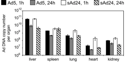
vector does not correlate with the virus biodistribution profile. Prior study of thein vivodistribution of systemically adminis-tered Ads representing groups A through F identified signifi-cant interserotype differences (3). Because the biodistribution of sAd24 particles has never been explored, we wished to establish it by injecting sAd24TL intravenously in mice and measuring the amount of viral DNA in organs by qPCR at 1 h and 24 h after injection. In parallel, a control group of animals was injected with Ad5TL, and this vector DNA was quantified using Ad5-specific primers and an Ad5-specific probe. This experiment showed significantly greater sAd24 DNA content at both time points in the heart, the lungs, and the spleen (Fig. 2). These organs contained 1.7 (heart, 24 h after injection) to 10 times (heart, 1 h after injection) more of the sAd24 ge-nomes than of the Ad5 gege-nomes (allPvalues are below 0.02). At the 1-h time point, the kidneys of sAd24-injected mice contained 2 times the amount of viral DNA seen in the kidneys of mice that received the Ad5 vector (P⫽0.014). No statisti-cally significant difference in DNA content was seen in kidneys 23 h later. The liver was the main site of viral DNA accumu-lation. At 1 h after injection, the livers contained 79% of injected Ad5 virions and 85% of injected sAd24 virions; at the 24-h time point, these levels had decreased to 9.9% and 4.5%, respectively.
This noted discrepancy between the low level of reporter
expression by the sAd24 vector in the liver and the high con-centration of its virions in the liver strongly suggested an in-volvement of virus retention mechanisms that do not yield gene transfer.
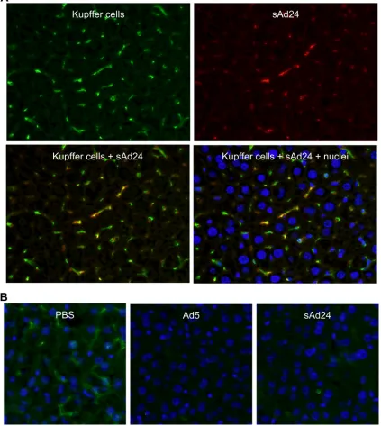
Sequestration of intravenously injected sAd24 virions leads
to rapid depletion of KCs in the liver. Sequestration of
sys-temically delivered Ad particles by KCs, macrophages residing in the liver sinusoids, has been reported for several Ads, in-cluding human serotypes 3, 5, 31, 37, and 41 and simian Ad23 (3). The uptake of Ad5 virions by KCs is mediated primarily by the scavenger receptors, with the natural Abs and complement playing secondary roles (63). Accumulation of Ad5 virions in KCs very shortly after virus administration rapidly kills KCs (34) through as-yet-unknown molecular mechanisms. Given these findings, we wished to examine whether KCs play any significant role in the clearance of intravenously delivered sAd24 and, in doing so, contribute to the elimination of sAd24 vector particles from the gene delivery process.
To this end, we injected mice with sAd24TL and analyzed the livers collected from these animals 10 min and 16 h later by fluorescence microscopy, using an Ab to an established mac-rophage biomarker, F4/80, and anti-Ad Ab, each labeled with a different fluorophore. As shown in Fig. 3A, 10 min after vector administration all Ad-associated fluorescence signals colocalized with the KCs, thus confirming efficient clearance of the virus by these liver macrophages. In concordance with the data previously reported for other Ad serotypes, we also saw massive depletion of KCs by sAd24 (Fig. 3B). Notably, 16 h after virus injections, the rate of KC depletion caused by sAd24 (89%) was somewhat lower than that caused by the control Ad5 vectors (94%) (P⫽0.018).
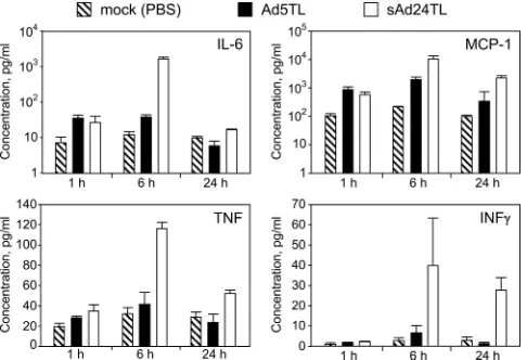
The profile of the acute inflammatory response to sAd24 is
different from that caused by Ad5. To further characterize
[image:6.585.42.543.80.202.2]sAd24 interaction with the host, we established both the tem-poral profile and the magnitude of the acute inflammatory response to sAd24 virions, using cytometric bead array tech-nology. In particular, concentrations of six cytokines (IL-6, IL-10, IL-12p70, IFN-␥, TNF, and MCP-1) were measured in plasma samples collected from sAd24-injected mice at 1 h, 6 h, or 24 h after injection. Comparison of these data with the similarly measured cytokine concentrations in plasma of Ad5-injected mice showed significant differences for IL-6, MCP-1, TNF, IFN-␥ (Fig. 4), and IL-12p70 (data not shown). The
TABLE 1. Reporter activities in homogenates of tissues isolated from Ad-injected micea
Vector (dose)
Reporter activities in indicated organs per organ or gram of tissue (rlu)a
Liver Spleen Lung Heart Kidney
Per organ Per gram
of tissue Per organ
Per gram
of tissue Per organ
Per gram
of tissue Per organ
Per gram
of tissue Per organ
Per gram of tissue
Ad5TL (2⫻1010VP) 3.6⫻1012 2.8⫻1012 8.1⫻109 6.0⫻1010 3.8⫻108 1.2⫻109 3.4⫻108 2.5⫻109 3.5⫻108 1.0⫻109
sAd24TL (2⫻1010VP) 3.1⫻108 2.2⫻108 5.0⫻106 5.3⫻107 5.5⫻106 1.6⫻107 2.3⫻106 1.8⫻107 7.4⫻106 2.4⫻107
Signal ratiob 1.2⫻104 1.3⫻104 1.6⫻103 1.1⫻103 6.9⫻10 7.4⫻10 1.5⫻102 1.3⫻102 4.7⫻10 4.2⫻10
Ad5TL (1011VP) 2.8⫻1013 1.6⫻1013 7.0⫻109 6.3⫻1010 1.1⫻109 2.9⫻109 6.2⫻108 3.3⫻109 2.4⫻109 6.8⫻109
sAd24TL (1011VP) 2.0⫻109 1.2⫻109 3.0⫻107 3.4⫻108 1.1⫻108 4.1⫻108 7.0⫻106 4.7⫻107 5.9⫻107 1.7⫻108
Signal ratiob 1.4⫻104 1.3⫻104 2.4⫻102 1.8⫻102 9.9 7.0 8.8⫻10 7.1⫻10 4.1⫻10 3.1⫻10
PBS 1.1⫻107 8.5⫻106 7.3⫻105 8.4⫻106 3.1⫻106 9.3⫻106 1.4⫻106 9.6⫻106 3.3⫻106 1.0⫻107
a
The activities of the reporter averaged for groups of five animals is shown in relative light units (rlu) per entire organ and per gram of tissue.
b
Ratios of bioluminescence signals measured in Ad5TL-injected mice to those measured in sAd24TL-injected mice.
FIG. 2. Ad5 and sAd24 virions show similar patterns ofin vivo
distribution. Shown are the copy numbers of Ad5 and sAd24 viral genomes per organ detected by qPCR in isolated murine organs at 1 h and 24 h after systemic administration of the vectors. Error bars indi-cate the standard deviations calculated for dupliindi-cate data points.
on November 8, 2019 by guest
http://jvi.asm.org/
[image:6.585.59.267.575.678.2]differences in concentrations of IL-6, MCP-1, and TNF in-duced by sAd24 and Ad5 vectors were most significant at the 6-h time point and, to a lesser extent, at the 24-h time point; IFN-␥and IL-12p70 concentrations were different only at the 24-h time point (P values of 0.02 and 0.047, respectively). Importantly, cytokine release by the sAd24-injected mice was invariably more robust than that by the animals that received Ad5. Compared to those for mock-injected animals, no statis-tically significant changes in the concentrations of IL-10 were
seen in mice injected with either serotype of Ad (data not shown).
The modification of sAd24 fiber by knob replacement
strat-egy yields protein chimera suitable for Ad vector targeting.We
[image:7.585.81.504.78.551.2]previously succeeded in altering the receptor specificity of Ad5 through genetic replacement of the CAR-binding knob do-main of its fiber with a trimerization dodo-main and a targeting ligand (5, 7, 26). We then reasoned that because of evolution-ary conservation of the overall Ad fiber structure, which is very
FIG. 3. Uptake of sAd24 virions by KCs in the liver leads to KC depletion. (A) Immunofluorescence staining of the liver collected from a mouse injected with sAd24 vector 10 min after injection. The upper panels show the liver section stained with either anti-F4/80 (left, green fluorescence) or anti-Ad Ab (right, red fluorescence). The lower panels show the overlay of the upper images, either alone (left) or merged with the staining of nuclei (right, blue staining). KCs containing sAd24 particles are orange. (B) Immunofluorescence staining of the livers collected from mice injected with PBS (left), Ad5 (center), or sAd24 (right) 16 h after injection. All images show Hoechst-stained nuclei (blue).
on November 8, 2019 by guest
http://jvi.asm.org/
similar in most known fiber proteins, the same approach should also be applicable to the sAd24 fiber. Importantly, application of this strategy to sAd24 did not require identifi-cation of its natural fiber receptor, which is not yet known, and, if successful, would further support the applicability of this strategy to fibers of other Ads.
To test this assumption, we designed a recombinant gene encoding the tail and shaft domains of the sAd24 fiber protein fused to the 12th␣-helical coiled-coil of the phage T4 fibritin protein, a peptide linker, and the Her2-specific affibody Zher2:7 and transiently expressed it in 293T cells. The product of this expression (designated FsAd2411FHer2:7) was analyzed by West-ern blotting to test whether the designed chimera assumes the trimeric configuration that is both characteristic of Ad fibers and essential for their encapsidation.
The results of this analysis, however, were unconvincing: in contrast to the well-defined bands corresponding to the tri-meric wt Ad5 fiber (Fig. 5A, lane 2), the sAd24 fiber chimera failed to produce such a pattern. Instead, the major band seen in the nondenatured sample of FsAd2411FHer2:7migrated con-siderably faster than had been expected (Fig. 5A, lane 4) and corresponded to a protein with an apparent molecular mass of approximately 105 kDa, not the 136 kDa predicted for the trimeric FsAd2411FHer2:7. The fact that the same sample, when fully denatured, clearly showed the monomeric chimera of the expected size (Fig. 5A, lane 3) ruled out the possibility that the smaller-than-expected size of the nondenatured protein was due to degradation or truncation of its subunits. Also, com-parison of the electrophoretic mobilities of the FsAd2411FHer2:7
chimera and the wt sAd24 fiber showed that the nonboiled samples of both proteins produced very similar patterns of gel migration (Fig. 5A, lanes 4 and 6). On the basis of this com-parison, we concluded that the trimer of FsAd2411FHer2:7was
sufficiently stable to be incorporated into sAd24 virions. The FsAd2411FHer2:7 protein was designed to mediate
effi-cient attachment of sAd24 viral particles to Her2 and, as a prerequisite to this, was expected to bind the cell-associated Her2. To test this function of FsAd2411FHer2:7, the transiently expressed protein was used in a flow cytometry experiment to probe the surfaces of cells either expressing or not expressing Her2. As shown in Fig. 5F, the strength of the fluorescence signal measured in Her2-positive cell targets was significantly higher than that measured in cells lacking the target receptor. Collectively, our data confirm both the expected trimeric configuration of the FsAd2411FHer2:7protein and the ability of
this molecule to recognize its intended target receptor on the cell surface, thereby providing a rationale for development of an sAd24 vector containing this chimera.
sAd24 virions incorporating designed fiber chimeras infect
cells in a Her2-dependent manner. The final proof of the
FsAd2411FHer2:7chimera’s functionality was obtained by using
this protein to replace the wt fiber in sAd24 virions. To this end, we constructed a replication-deficient genome of sAd24 with the E1 region deleted and containing the ORF of FsAd2411FHer2:7 in place of the wt fiber-coding sequence; the
virus was rescued and propagated using the two-step approach described in Materials and Methods. The efficacy of incorpo-ration of FsAd2411FHer2:7 into sAd24 virions was gauged and compared with that of the wt sAd24 fiber by using Western blotting of purified viral particles. As Fig. 6A illustrates, the outcome of the trimerization assays of the transiently ex-pressed FsAd2411FHer2:7 correctly predicted efficient encapsi-dation of the designed protein. This efficient encapsiencapsi-dation was in agreement with the high yield of the modified sAd24 vector (5,500 VP/infected cell), which was comparable to the yield for sAd24 containing the wt fibers (5,000 VP/infected cell). Com-parison of the protein compositions of the virions of both types showed no differences other than the presence of the expected fiber proteins, the wild type or the fiber chimera, in the
[image:8.585.43.286.69.235.2]respec-FIG. 4. Comparison of cytokine release in response to intravenous injections of Ad5 and sAd24. Concentrations of IL-6, MCP-1, TNF, and IFN-␥in plasma samples collected from mice at 1 h, 6 h, or 24 h after injection with either Ad5- or sAd24-derived vectors are shown in pg/ml. Three mice were injected with a given Ad to generate data for each time point shown. Error bars indicate the standard deviation calculated for duplicate data points corresponding to each animal in the group.
FIG. 5. sAd24 fiber-derived targeting chimera forms stable trimers and binds to cell-associated Her2. (A) Western blotting of lysates of 293T cells transiently expressing the fiber constructs. Lanes 1 and 2, wt Ad5 fiber; lanes 3 and 4, FsAd2411FHer2:7, lanes 5 and 6, wt sAd24 fiber;
lane S, protein standards with the molecular masses shown in kDa (here and in Fig. 6). Samples in lanes 1, 3, and 5 were boiled prior to being loaded onto the gel and thus show proteins in their fast-migrat-ing monomeric form; samples in lanes 2, 4, and 6 were not boiled. Fibers were detected with either the anti-Ad5 fiber tail MAb 4D2 (wt Ad5 fiber), the anti-fibritin MAb 5E1 (FsAd2411FHer2:7), or the
anti-sAd24 fiber MAb 20C1 (wt sAd24 fiber) followed by a fluores-cence-labeled secondary antibody. Predicted molecular masses for tri-meric wt Ad5, FsAd2411FHer2:7, and wt sAd24 fiber are 185 kDa, 136
kDa, and 142 kDa, respectively. (B) Flow cytometry detected binding of the transiently expressed FsAd2411FHer2:7chimera (shown by solid
black line) to Her2-expressing 293/Her2 cells but no binding above the background to Her2-negative 293 cells. The background signals gen-erated in both cell lines by the lysate of mock-transfected 293T cells are shown by the dotted lines.
on November 8, 2019 by guest
http://jvi.asm.org/
[image:8.585.302.541.69.150.2]tive vector particles (Fig. 6B), thus confirming the proper as-sembly and maturation of the modified sAd24 particles.
Further, in good agreement with the cell-binding data shown in Fig. 5B, our subsequent gene transfer experiments demon-strated that the FsAd2411FHer2:7 chimera contained in sAd24TL.11FHer2:7 virions enables Her2-dependent
trans-duction of target cells (Fig. 7A). The Her2 specificity of sAd24TL.11FHer2:7infection was confirmed by transducing two
isogenic cell lines, MDA-MB-231 and MDA-MB-231/Her2, that differ from each other by their Her2 phenotypes. In addi-tion, augmentation of gene transfer to the target receptor-expressing cells by the tropism-modified sAd24 was demon-strated through transduction of a panel of Her2-positive
human tumor cell lines. In this experiment, gene delivery by the targeted sAd24 vector was 3.7 to 14 times more efficient than delivery by the vector bearing unmodified wt fibers. Importantly, by using the free Her2-binding affibody com-petitor we showed selective inhibition of transduction by sAd24TL.11FHer2:7 but not by the nontargeted control Ad (Fig. 7A).
The efficacy of this gene transfer was then compared with that by the previously designed Her2-targeted derivative of Ad5 (7) (Fig. 7B). In this experiment, two pairs of cell lines in which the parental line was Her2 negative (293 and MDA-MB-231) and the derivative line was Her2 expressing (293/Her2 and MDA-MB-231/Her2) were transduced with either Ad5TL.11FHer2:7or sAd24TL.11FHer2:7at various MOIs. With the exception of the transduction of MDA-MB-231/Her2 at an MOI of 10, at which both Ads were equally efficient, the Ad5-derived vector was more efficient in transducing both Her2-expressing cell lines. The differences between the levels of gene expression directed by the vectors depended on the vectors’ replication. Specifically, in 293/Her2 cells, which complement deletion of the E1 genes in both the Ad5- and the sAd24-derived vectors, reporter expression by Ad5TL.11FHer2:7was 3 to 6 times more efficient. In comparison, in MDA-MB-231/ Her2 cells, which do not complement deletion of the E1 genes, reporter expression by Ad5TL.11FHer2:7 was only 1.3 to 1.6 times that by sAd24TL.11FHer2:7. Notably, the relative infec-tivities of the tested vectors, which had been determined by the spot assay in 293/Her2 cells, were 51 VP/infectious unit for sAd24TL.11FHer2:7 and 33 VP/infectious unit for
Ad5TL.11FHer2:7.
In light of the potential use of sAd24TL.11FHer2:7-derived vectors for gene delivery in humans with preexisting anti-Ad5 antibodies, moreover, we used the established virus
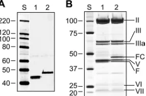
neutraliza-FIG. 6. Efficient encapsidation of FsAd2411FHer2:7 protein yields
fully matured sAd24 virions. (A) Western blot of sAd24TL (1010VP,
lane 1) and sAd24TL.11FHer2:7(10
10 VP, lane 2) virions. The fully
denatured fibers were detected with the anti-sAd24 fiber tail MAb 20C1. (B) SDS-PAGE-resolved proteins of sAd24TL (2⫻1010VP,
lane 1) and sAd24TL.11FHer2:7(2⫻1010VP, lane 2) virions stained
with silver using a PageSilver kit (Fermentas, Glen Burnie, MD). Indi-cated are the migration positions of the following sAd24 proteins: hexon (II), penton base (III), peripentonal protein (IIIa), FsAd2411FHer2:7fiber
[image:9.585.93.236.70.164.2]chimera (FC), major core protein (V), fiber (F), hexon-associated protein (VI), and minor core protein (VII).
FIG. 7. Gene transfer by the targeted sAd24TL.11FHer2:7vector is Her2 dependent. (A)In vitrotransduction of human tumor cell lines by
sAd24TL and sAd24TL.11FHer2:7vectors. Viral infections were done in either standard medium (white and black bars) or in medium containing
free affibody Zher2:4used at a concentration of 100g/ml (gray and striped bars). Shown are the activities of Fluc in lysates of infected cells. Inserts
below the graph show the signals detected in each of the tested cell lines (7⫻103cells/lane) by Western blotting with anti-Her2 antibody.
(B) Comparison of the efficacies of gene transfer by Her2-targeted vectors derived from Ad5 (Ad5TL.11FHer2:7, white bars) and sAd24
(sAd24TL.11FHer2:7, black bars). Levels of transgene expression in the lysates of cells infected with Ads at the MOIs indicated below the graph
(in VP/cell) are shown. Error bars indicate the standard deviations calculated for triplicate data points.
on November 8, 2019 by guest
http://jvi.asm.org/
[image:9.585.135.450.459.658.2]tion assay to test whether gene delivery by this vector would be affected by such antibodies. This test showed that sAd24TL.11FHer2:7largely evaded inactivation by Ad5-specific antibodies and retained most of its infectivity in their presence (data not shown).
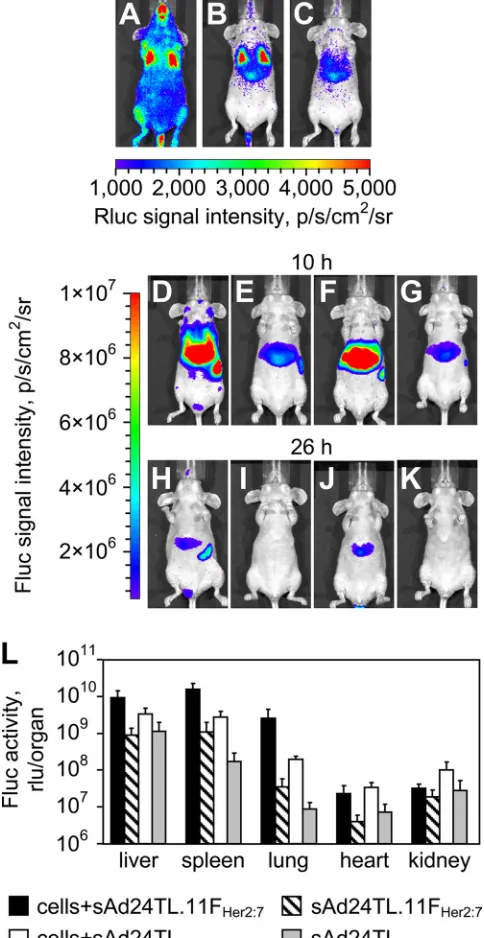
In vivo testing of the fiber-modified sAd24 vector.Having established the Her2 specificity of sAd24TL.11FHer2:7, we next wished to test this vector’s ability to accomplish transductionin vivo. To this end, we injected mice intravenously with MDA-MB-231/Her2 cells, allowed them to distribute in the blood-stream (Fig. 8A), and targeted these cells with the vector, which was also administered intravenously. Another group of cell-injected animals was given the nontargeted control vector, sAd24TL. Two groups of mice that had received one of the vectors, but no tumor cells, were used as additional controls. To monitor the spread of cells and the patterns of Ad-medi-ated transduction, we took advantage of luciferase reporters expressed by the MDA-MB-231/Her2 cells (Rluc) and the vi-rus (Fluc).
As expected, the whole-body imaging of Rluc activity in the control animals, which did not receive tumor cells, did not reveal any bioluminescence above background throughout the duration of the study (data not shown). Imaging of Rluc activ-ity in mice that had received both the cells and the viruses, done at 8 h after injections, showed that by that time point most tumor cells had cleared the circulation and were localized to the liver and the lungs (Fig. 8B). Imaging of the sAd24TL.11FHer2:7-expressed Fluc activity shortly thereafter revealed a 10.7-fold difference in signal intensity between the MDA-MB-231/Her2-injected mice (Fig. 8D) and the control mice (Fig. 8E) (P ⬍ 0.0001). Importantly, the Fluc-enabled signals largely localized within the regions of Rluc-caused lu-minescence, thereby linking the observed increase in vector-expressed reporter activity with the presence of tumor cells in a particular locale. However, a similar 9.6-fold difference was seen between the groups of mice that received the nontargeted control vector sAd24TL in combination with the tumor cells (Fig. 8F) or alone (Fig. 8G) (P⫽0.0001). Comparison of Fluc signals measured in cell-injected animals showed a 1.7-fold-higher level of activity in mice that received sAd24TL.11FHer2:7than in mice injected with sAd24TL. This difference was, however, statistically insignificant (P⫽0.22).
Sixteen hours later, the strength of the Rluc signals in mice injected with MDA-MB-231/Her2 cells decreased substan-tially, suggesting their continuous clearance (Fig. 8C). As ex-pected, this depletion of tumor cells resulted in a decrease of the vector-expressed Fluc activity. At this time point, the ratios of signal intensities measured during the whole-body imaging in the experimental and control groups of mice were 23.1 for groups of mice injected with sAd24TL.11FHer2:7(Fig. 8H and I) (P ⫽ 0.0007) and 14.1 for groups of mice injected with sAd24TL (Fig. 8J and K) (P⫽0.0008). While the ratio of Fluc activity in the group of mice injected with the cells and targeted Ad to that in the group that received the cells and sAd24TL increased to 2.9, it remained statistically insignificant (P ⫽
0.28).
[image:10.585.300.542.85.554.2]To establish a more detailed pattern of Ad vector reporter activity at this time point, we measured Fluc luminescence in the homogenates of organs collected from mice in all groups (Fig. 8L). In agreement with the whole-body imaging of Fluc
FIG. 8. Transduction of circulating tumor cells by a Her2-targeted sAd24 vector. (A, B, and C) Patterns of Rluc expression at 5 min (A), 8 h (B), and 24 h (C) after intravenous injection of MDA-MB-231/Her2 cells into mice. (D to K) Fluc-enabled luminescence in Ad-injected mice at 10 h (D to G) and 26 h (H to K) after vector administration. The 2-h intervals between imaging of Rluc and Fluc activities were allowed for the Rluc signals to decay to background level. Mice shown in panels D and H were injected with MDA-MB-231/Her2 and with sAd24TL.11FHer2:75
min after administration of cells. Control mice shown in panels E and I were injected with sAd24TL.11FHer2:7but did not receive tumor cells.
Panels F and J show mice that were injected with the cells and with sAd24TL; the corresponding control mice, which were injected with sAd24TL alone, are shown in panels G and K. Each experimental group contained four animals. (L) Activities of the vector-encoded Fluc reporter in individual organs isolated from experimental mice that were injected with both the cells and the virus and from control mice injected with the virus alone. Error bars indicate the standard deviations calculated for duplicate data points.
on November 8, 2019 by guest
http://jvi.asm.org/
activity, these measurements confirmed invariably higher levels of transduction in animals injected with the cells and the vi-ruses than in animals treated with the Ads only. They also showed that the livers, spleens, and lungs were the primary sites of viral transduction. In these organs, sAd24TL.11FHer2:7
-caused transduction was 2.8-fold (livers; P ⫽ 0.07), 5.7-fold (spleens;P⫽0.003), and 13.5-fold (lungs;P⫽0.003) higher than transduction caused by sAd24TL. This difference in the liver was, however, statistically insignificant, as was the differ-ence in reporter expression in the hearts (P ⫽ 0.25). The kidneys of cell-injected animals that received sAd24TL con-tained a 3.3-fold-higher level of Fluc activity than kidneys in mice injected with the cells and sAd24TL.11FHer2:7(P⫽0.04).
We also tested the performance of sAd24TL.11FHer2:7in an
in vivomodel of metastatic Her2-expressing cancer. For this study, we established target metastases in the lungs of mice by systemically injecting the animals with MDA-MB-231/Her2 cells (Fig. 9A). Next, the tumor-bearing animals and the tu-mor-free control animals were injected intravenously with sAd24TL.11FHer2:7 at a dose of 1011VP/injection, using the
two-injection regimen described in Materials and Methods. Bioluminescence imaging of Fluc activity done 48 h after the second vector administration detected weak signals in the tho-racic areas of mice in both groups (Fig. 9B and C). Measure-ments in homogenates of the lungs isolated from animals in both groups showed that, on average, Fluc activity was 2.5 times higher in mice with metastases than in tumor-free mice (Fig. 9D). This difference was statistically significant (P ⫽
0.005).
DISCUSSION
Prior work on modifying the tropism of Ad vectors was focused mostly on Ad5-derived vectors and was based primar-ily on modification of the fiber knob domain with ligands. The natural diversity of Ad serotypes and the advantages offered by some of those Ads as targeted vector prototypes remain un-derexplored. In this study, we sought to test whether the Ad tropism alteration approach based on fiber knob domain re-placement, which we had previously developed for Ad5, could be applied to another Ad serotype. The rationale for this work was that its success would support the feasibility of developing a versatile targeting approach that would be applicable to many Ads, thus making it possible to fully exploit the potential benefits offered by underexplored Ad serotypes.
Toward these goals, we sought to make a tropism-modified
vector based on sAd24, which was chosen for this study for the following reasons. First, owing to the widespread preexisting immunity to Ad5 in humans (12, 55), the serological distinction of sAd24 from Ad5 makes this virus a preferred alternative to Ad5 as a vector platform. Second, the fiber of sAd24 is an unexplored molecule and is thus a reasonable starting point to test an approach that is expected to be applicable to many fibers, including the uncharacterized ones. This protein has never been studied in detail; at the level of the primary struc-ture it shares little homology with the Ad5 fiber and other well-characterized Ad fibers. Neither its primary receptor nor the receptor-binding site within its knob has been identified. Against this background, successful targeting of the sAd24 fiber through the knob replacement strategy would yield a vector prototype of potential clinical utility and would also suggest the feasibility of targeting of other Ads.
sAd24 would be an even stronger candidate as a vector for gene delivery in humans should it avoid liver tissue on systemic delivery. The massive transduction of the liver by the Ad5 vectors causes severe systemic toxicity and raises serious safety concerns. Recent studies have identified the high-affinity bind-ing of FX to the Ad5 hexon protein as the molecular basis for hepatic transduction (22, 56). These studies also showed that the particles of several Ad serotypes bind FX poorly or do not bind it at all and that the stability of the resultant complexes correlates with the degree of liver transduction by the Ads. Given these findings, we studied the association of FX with the virions of sAd24 and showed that FX-sAd24 complexes are rather unstable. In particular, while the equilibrium dissocia-tion constant, KD, for the FX-sAd24 complexes was only slightly higher than theKDfor the control FX-Ad5 complex, the FX-sAd24 dissociation rate constant,kd, was 16 times the FX-Ad5kd. To put these findings in perspective, recent
suc-cesses in drug development have more often correlated with improvements of thekdvalues of the most efficacious drugs rather than with improvements of theirKDvalues. In particu-lar, thekdvalues correlated much better with the agent-target complex residence time, its dissociative half-life, and target selectivity (13). On this basis, the dissociation rate constant is now seen as a more meaningful parameter in assessing impor-tant biological interactions.
[image:11.585.52.533.70.155.2]The observed instability of the FX-sAd24 complexes could be explained by variations in the primary sequences of the HVRs of the Ad5 and sAd24 hexons. Notably, recent studies have collectively suggested that FX binding to the Ad5 hexon
FIG. 9. Gene delivery by sAd24TL.11FHer2:7 in mice with metastatic tumors. (A) Whole-body images of mice bearing Her2- and
Rluc-expressing breast cancer metastases in the lungs. (B and C) Fluc bioluminescence in the mice shown in panel A (B) and in control tumor-free mice (C) 48 h after animals in both groups were injected intravenously with sAd24TL.11FHer2:7. (D) Activity of Fluc reporter in the lungs isolated from
Ad-injected mice after image acquisition. The black and white bars show the averaged values of signals in the lungs of tumor-bearing and control mice, respectively. Each experimental group contained six animals. Error bars indicate the standard deviations calculated for duplicate data points.