0022-538X/10/$12.00 doi:10.1128/JVI.01053-10
Copyright © 2010, American Society for Microbiology. All Rights Reserved.
CD1d, a Sentinel Molecule Bridging Innate and Adaptive Immunity, Is
Downregulated by the Human Papillomavirus (HPV) E5 Protein: a
Possible Mechanism for Immune Evasion by HPV
䌤
Shiho Miura,
1Kei Kawana,
1* Danny J. Schust,
2Tomoyuki Fujii,
1Terufumi Yokoyama,
3Yuki Iwasawa,
1Takeshi Nagamatsu,
1Katsuyuki Adachi,
1Ayako Tomio,
1Kensuke Tomio,
1Satoko Kojima,
1Toshiharu Yasugi,
1Shiro Kozuma,
1and Yuji Taketani
1Department of Obstetrics and Gynecology, Faculty of Medicine, University of Tokyo, 7-3-1 Hongo, Bunkyo-ku, Tokyo 113-8655,
Japan1; Division of Reproductive Endocrinology and Fertility, Department of Obstetrics, Gynecology, and Women’s Health,
University of Missouri School of Medicine, Columbia Regional Hospital, 402 Keene Street, Third Floor, Columbia, Missouri 652012;
and GENOLAC BL Corp. 503, Okinawa Industry Support Center, 1831-1, Oroku, Naha, Okinawa 901-0152, Japan3
Received 16 May 2010/Accepted 20 August 2010
CD1d and CD1d-restricted natural killer T (NKT) cells serve as a natural bridge between innate and adaptive immune responses to microbes. CD1d downregulation is utilized by a variety of microbes to evade immune detection. We demonstrate here that CD1d is downregulated in human papillomavirus (HPV)-positive
cellsin vivoandin vitro. CD1d immunoreactivity was strong in HPV-negative normal cervical epithelium but
absent in HPV16-positive CIN1 and HPV6-positive condyloma lesions. We used two cell lines forin vitroassay;
one was stably CD1d-transfected cells established from an HPV-negative cervical cancer cell line, C33A (C33A/CD1d), and the other was normal human vaginal keratinocyte bearing endogenous CD1d (Vag). Flow cytometry revealed that cell surface CD1d was downregulated in both C33A/CD1d and Vag cells stably transfected with HPV6 E5 and HPV16 E5. Although the steady-state levels of CD1d protein decreased in both E5-expressing cell lines compared to empty retrovirus-infected cells, CD1d mRNA levels were not affected. Confocal microscopy demonstrated that residual CD1d was not trafficked to the E5-expressing cell surface but colocalized with E5 near the endoplasmic reticulum (ER). In the ER, E5 interacted with calnexin, an ER chaperone known to mediate folding of CD1d. CD1d protein levels were rescued by the proteasome inhibitor, MG132, indicating a role for proteasome-mediated degradation in HPV-associated CD1d downregulation. Taken together, our data suggest that E5 targets CD1d to the cytosolic proteolytic pathway by inhibiting calnexin-related CD1d trafficking. Finally, CD1d-mediated production of interleukin-12 from the C33A/CD1d cells was abrogated in both E5-expressing cell lines. Decreased CD1d expression in the presence of HPV E5 may help HPV-infected cells evade protective immunological surveillance.
There are approximately 100 identified genotypes (types) of human papillomavirus (HPV). Over 40 of these are classified as genital HPV subtypes that invade the reproductive organs, including the uterine cervix, vaginal wall, vulva, and penis. Genital HPV types are further subclassified into high-risk types that are commonly associated with cervical cancer and low-risk types that cause noninvasive condyloma acuminata. Although exact classification varies among researchers, sub-types 16, 18, 31, 33, 35, 39, 45, 51, 52, 56, 58, 66, and 68 are typically classified as high risk and subtypes 6, 11, 40, 42, 43, 44, 54, 61, and 72 as low risk (44). Genital HPV infection involves short-term virus proliferation, followed by the long-term latent presence of a small number of copies of the viral genome within the basal cells of the genital epithelium (44). Infections with high-risk HPV subtypes result in progression to genital tract cancers (most commonly cervical) in only a small per-centage of infected women and typically after a long latency period. A high percentage of high-risk HPV DNA-positive
infected women resolve their infections during the prolifera-tive phase and thereby clear the virus or progress to latency with undetectable HPV DNA levels. The clearance of viral DNA is often accomplished through activation of the host immune system against viral antigen (19), and chronic immune suppression represents a risk factor for viral DNA persistence and benign and/or neoplastic lesion progression (23).
Completion of the HPV life cycle requires infection of epi-dermal or mucosal basal cells that have the potential to pro-liferate and differentiate. Within these cells, overall viral gene expression is suppressed, although limited expression of spe-cific early viral genes, including E5, E6, and E7, causes lateral expansion of infected cells within the basal layer of the epithe-lium (44). HPV E5 seems to be particularly important early in the course of infection. Large amounts of E5 mRNA have been found in cervical intraepithelial neoplasia (CIN) lesions (37). However, as HPV-infected lesions progress to cervical cancer, episomal viral DNA becomes integrated into host cell DNA, and a substantial part of the HPV genome, commonly includ-ing the E5 codinclud-ing sequence, is deleted (16). Therefore, E5 is not obligatory in the late events of HPV-mediated carcinogen-esis.
E5 is a small hydrophobic protein that can be localized within the Golgi apparatus (GA), endoplasmic reticulum
* Corresponding author. Mailing address: Department of Obstetrics and Gynecology, Faculty of Medicine, University of Tokyo, 7-3-1 Hongo, Bunkyo-ku, Tokyo 113-8655, Japan. Phone: 81-3-3815-5411. Fax: 81-3-3816-2017. E-mail: kkawana-tky@umin.ac.jp.
䌤Published ahead of print on 1 September 2010.
11614
on November 8, 2019 by guest
http://jvi.asm.org/
trafficking to the cell surface but does not alter the transcrip-tion of HLA class I heavy chains or the transporter associated with antigen processing (TAP) (2, 3, 4, 28). Others have shown that HPV16 E5 interacts with calnexin in the ER and thereby interferes with the modification of HLA class I heavy chains (21).
CD1d is an major histocompatibility complex (MHC) class I-like glycoprotein that presents self or microbial lipid antigen to natural killer T (NKT) cells (39). In humans, a specific subset of NKT cells expresses an invariant V␣24-JaQ/V11 TCR (iTCR) and can recognize CD1d on the surface of anti-gen-presenting cells (APCs) through this receptor. CD1d is expressed not only in typical APCs (macrophages, dendritic cells, and B cells) but also in intestinal epithelial cells (8, 12), foreskin keratinocytes (9), and reproductive tract epithelial cells (25, 26). Like MHC class I, CD1d is synthesized, glyco-sylated byN-glycosyltransferase, modified, and assembled with 2m within the ER and then transferred to the GA (5, 24, 27). CD1d plays a role in both innate and adaptive immunity to various bacteria, viruses, fungi, and parasites (reviewed in ref-erence 10). Activation of CD1d-restricted invariant NKT (iNKT) cells enhances host resistance to some microbes in a manner that depends on the level of CD1d expression on APCs (34, 35). In contrast, the activation of iNKT cells promotes susceptibility to some microbes (7, 33). The activation of CD1d-restricted iNKT cells in response to microbial invasion is antigen dependent, but these antigens can be derived from the invading microbe or possibly from host lipids (11, 22, 29). Intracellular signaling mediated by surface CD1d utilizes NF-B, a well-known immune-related transcription factor (36, 43). CD1d-restricted NKT cells can modulate adaptive immune cells by altering Th1/Th2 polarization. Recognition of CD1d by iNKT cells can also result in rapid release of both interleukin-4 (IL-4) and gamma interferon (IFN-␥) from the NKT cell (6). Therefore, CD1d and CD1d-restricted NKT cells serve as a natural bridge between innate and adaptive immune responses to microbes. Not surprisingly, several microbes, including her-pes simplex virus type 1, human immunodeficiency virus, Kaposi’s sarcoma herpesvirus, andChlamydia trachomatis, are known to downregulate cell surface expression of CD1d as an immune evasion strategy (13, 26, 31, 42). Our own lab previ-ously demonstrated thatC. trachomatis retains CD1d in the ER and targets CD1d to both chlamydial and cellular degra-dation pathways (26).
Viewing the importance of CD1d in innate immune re-sponses to microbes, we hypothesized that HPV may alter CD1d-mediated immune pathways and thereby avoid innate immune destruction of the infected cell by the host. We
dem-EcoRI (reverse) restriction sites. The PCR products were digested with BamHI and EcoRI and subcloned into a retroviral expression plasmid pLPCX (Clon-tech, Mountain View, CA).
Cell lines and establishment of a cell line stably expressing CD1d.An HPV-negative human cervical carcinoma cell line, C33A, and a vaginal epithelial cell that was originally established from normal human primary epithelial cells that were immortalized by transduction with HPV16 E6/E7 genes (VK2/E6E7) (a generous gift from D. J. Anderson, Boston University, Boston, MA) (18) were grown in Dulbecco modified eagle medium (Invitrogen, Carlsbad, CA) without CaCl2(Invitrogen), supplemented with 10% fetal bovine serum (Invitrogen) at 37°C in 5% CO2. The vaginal epithelial (VK2/E6E7) cells used here are known to express endogenous CD1d at the cell surface (25).
A CD1d-expressing retroviral plasmid pSR␣-neo (kindly provided by R. Blum-berg, Harvard Medical School, Boston, MA) was transfected into Phoenix cells, a packaging cell line for recombinant retroviruses (kindly provided by K. Oda, University of Tokyo), using Lipofectamine 2000 (Invitrogen). After 72 h of incubation in DMEM, the culture medium containing released CD1d-expressing retroviruses was collected and used to infect C33A cells and transfer the CD1d gene. CD1d-expressing C33A cells were selected in medium containing 1.0 mg of neomycin/ml to establish a stably transfected cell line (C33A/CD1d).
Establishment of HPV E5-expressing cell lines.HPV6 or HPV16 E5-express-ing retroviral plasmids or their empty counterparts (pLPCX-16E5, pLPCX-6E5, or pLPCX, respectively) were transfected into Phoenix cells using Lipofectamine 2000 (Invitrogen). After 72 h of incubation, culture medium with released viruses were collected and used to infect C33A/CD1d or vaginal epithelial cells. Stable cell lines were selected in media containing 1.5g of piromycin/ml.
Immunohistochemistry.Immunostaining for CD1d was performed on forma-lin-fixed, paraffin-embedded tissue sections of normal or inflamed cervix, CIN1 to CIN3, cervical cancer, and condyloma acuminata (obtained under IRB ap-proval through the University of Tokyo). A total of 45 tissues were examined. Optimal immunostaining required antigen retrieval via microwave exposure in 0.01 M citrate buffer. A mouse anti-CD1d MAb (NOR3.2, 1:100; Abcam, Inc., Cambridge, MA) or an irrelevant, isotype-matched mouse monoclonal antibody (DakoCytomation, Glostrup, Denmark) were used as primary reagents. Immu-nostaining was amplified and detected by using the EnVision⫹System-HRP (DakoCytomation). Nuclei were counterstained by using standard hematoxylin protocols (Sigma-Aldrich, Inc., St. Louis, MO). Analyses were performed at a magnification of⫻200.
Flow cytometry.C33A/CD1d cells were grown in 175-cm2
flasks until conflu-ent, harvested using trypsin-EDTA, and pelleted at 500⫻gfor 5 min at room temperature. The cells were then washed and resuspended in PBS-B (phosphate-buffered saline [PBS] with 1% bovine serum albumin; Invitrogen) at a concen-tration of 106
cells/ml. For detection of cell surface CD1d, 100l of cell sus-pension was incubated with an anti-CD1d NOR3.2 monoclonal antibody (MAb; Abcam) at 1:100 for 30 min at 4°C. Cells were then washed three times in PBS-B, incubated with a goat anti-mouse immunoglobulin secondary antibody conju-gated to phycoerythrin (PE; BD Bioscience, San Jose, CA) for 30 min at 4°C, suspended in 1% paraformaldehyde, and analyzed by using a FACSCalibur flow cytometry system (BD Bioscience).
Proteasome inhibitor treatment.C33A/CD1d cells harboring an empty vector (C33A/CD1d-empty) or expressing HPV6 E5 (C33A/CD1d-6E5) or HPV16 E5 (C33A/CD1d-16E5) were cultured for up to 24 h in the presence or absence of the cytosolic proteasome inhibitor MG132 (10 M in dimethyl sulfoxide [DMSO]; Sigma-Aldrich, Inc.). Control wells included vehicle alone.
HPV genotyping.DNA was extracted from cervical smear samples by using the DNeasy blood minikit (Qiagen, United Kingdom). HPV genotyping was per-formed by using the PGMY-CHUV assay method (20). Briefly, standard PCR was conducted using the PGMY09/11 L1 consensus primer set and HLA-dQ primer sets (20). Reverse blotting hybridization was performed as described
on November 8, 2019 by guest
http://jvi.asm.org/
previously (20). Heat-denatured PCR amplicons were hybridized to a negatively charged nylon membrane containing specific probes for 32 HPV genotypes and HLA-dQ reference samples. Chemiluminescence detection used enhanced chemiluminescence (ECL) detection reagents for nucleic acids (GE Healthcare). Films were interpreted using the HPV reference guide provided.
RT-PCR and quantitative PCR.Portions (1g) of total RNA and oligo(dT)s were used for reverse transcriptase (RT) reactions (RNA PCR kit; Applied Biosystems, Foster City, CA). Total cDNA reaction samples were used as tem-plates for amplification of each gene fragment using a PCR Core kit (Applied Biosystems). Primer pair sets for CD1d were synthesized by Invitrogen (CD1d, 453 bp; 5⬘-GCTGCAACCAGGACAAGTGGACGAG-3⬘[forward] and 5⬘-AG GAACAGCAAGCACGCCAGGACT-3⬘[reverse]). Those for IL-12 p40 were commercially available (Sigma-Aldrich, Inc.). For quantitative PCR, cDNA were produced via RT of 1g of total RNA extracted from the cells as described above by using an OmniScript RT kit (Qiagen, Inc., Valencia, CA). Portions (2
l) of 5-fold-diluted cDNA aliquots were amplified in a thermal cycler (7300 Real-Time PCR System; Applied Biosystems) by using a QuantiTect SYBR green PCR kit (Qiagen, Inc.) and a primer pair set for-actin (5⬘-GAAATCG TGCGTGACATTAAGG-3⬘[forward] and 5⬘-TCAGGCAGCTCGTAGCTTC T-3⬘[reverse]). The mRNA levels for IL-12 were normalized to those of-actin, the internal control.
Fluorescent deconvolution microscopy and confocal microscopy.C33A/CD1d cells were seeded onto coverslips. The ER was visualized using the ER tracker Blue-White DPX (Invitrogen) for 30 min at 37°C. All coverslips were fixed in 4% paraformaldehyde, permeabilized with 0.1% Tween 20. They were then incu-bated for 1 h at 37°C with either an anti-CD1d NOR3.2 MAb labeled with Zenon Alexa Fluor 555 using a mouse IgG labeling kit (Invitrogen) or an anti-FLAG MAb labeled with Zenon Alexa Fluor 488 using a mouse IgG labeling kit (Invitrogen) singly or in combination. With the exception of ER tracker-treated coverslips, the cells were then counterstained with a DAPI (4⬘,6⬘ -diamidino-2-phenylindole) nucleic acid stain (Invitrogen). Images were obtained with a LSM 700, flexible confocal microscope (Carl Zeiss, Oberkochen, Germany). Filter sets were optimized for Alexa 488, Alexa 555, and DAPI. Z-axis plane capture, deconvolution, and analyses were performed with ZEN 2009 Software (Carl Zeiss).
Western blotting. Portions (50g) of total cell lysates from C33A/CD1d-empty, C33A/CD1d-6E5, or C33A/CD1d-16E5 cells in a modified TNF buffer (1 M Tris-HCl [pH 7.8], 10% NP-40, 5 M NaCl, 0.5 M EDTA [pH 8.0], aprotinin, 0.1 M phenylmethylsulfonyl fluoride) were electrophoresed and transferred to nitrocellulose membranes. Membranes were blocked with 10% milk and incu-bated with a peroxidase-labeled anti-CD1d NOR3.2 MAb (1:200; Abcam) or an anti-FLAG MAb (1:500; Sigma-Aldrich, Inc.) using a peroxidase labeling kit (Roche, Basel, Switzerland) for 1 h. Membranes were washed and bound anti-body was detected using an ECL Western blotting analysis system (GE Health-care Buckinghamshire, United Kingdom).
Immunoprecipitation and Western immunoblotting.Harvested C33A/CD1d-empty, C33A/CD1d-6E5, or C33A/CD1d-16E5 cells were lysed in modified ra-dioimmunoprecipitation assay buffer (1% NP-40, 1% deoxycholate, 0.1% sodium dodecyl sulfate [SDS], 10 mM Tris, 150 mM NaCl, 2 mM EDTA) with protease inhibitors (Amersham Biosciences, Piscataway, NJ). Equivalent aliquots of total cell lysates were incubated overnight at 4C with 5g of mouse anti-FLAG MAbs (Sigma-Aldrich, Inc.)/ml and 5l of protein A-Sepharose (GE Healthcare).
Precipitated proteins were separated by SDS-PAGE using 7.5% acrylamide gels and transferred to polyvinylidene difluoride membranes. Mouse anti-calnexin or rabbit anti--actin polyclonal antibodies (Abcam) were used as primary reagents for immunoblotting, and anti-mouse IgG-HRP (1:100,000; GE Healthcare) was used as a secondary reagent. Products in Western immunoblotting experiments were visualized by using an ECL Western blotting analysis system (GE Health-care). Molecular masses were confirmed by comparison to standard size markers (GE Healthcare).
Statistical analysis.Quantitative PCR data were presented as means⫾the standard deviations. Experiments were performed independently at least three times. The Cochran-Armitage Trend test was computed to show trends in im-mune reactivity with NOR3.2 MAb in clinical samples. IL-12 mRNA levels were compared to those before or after cross-linking by using paired, two-tailed Studentttests. APvalue of⬍0.05 was considered significant.
RESULTS
CD1d downregulation in HPV-related lesions and cancer
cell lines.Since CD1d expression in human mucosa and skin
[image:3.585.81.504.69.206.2]has been demonstrated by immunohistochemistry using the anti-CD1d NOR3.2 MAb (2, 9, 12, 26), we examined immu-nostaining of human normal ectocervix or HPV-related lesions with NOR3.2 (Fig. 1). Immunostaining for CD1d was per-formed on formalin-fixed, paraffin-embedded tissue sections of normal or inflamed ectocervical epithelium, cervical intraepi-thelial neoplasia 1 (CIN1), cervical cancer, and cervical con-dyloma (obtained under IRB approval through the University of Tokyo, Faculty of Medicine). To examine alterations in CD1d expression in the presence of high-risk HPV and low-risk HPV subtypes, HPV16-positive CIN1 or cancer lesions and HPV6-positive condyloma acuminata specimens were compared to each other and to HPV-negative normal and inflamed ectocervical epithelial controls. Immunoreactivity with the NOR3.2 MAb was noted in the basal and parabasal epithelial cells of normal and inflamed ectocervical epithelia that are known to express early HPV genes (E5, E6, and E7; Fig. 1) (44). In inflamed epithelium, the immunoreactivity ap-peared to be intensified compared to normal epithelium. CD1d expression is known to be enhanced by inflammatory cytokines (10). NOR3.2 immunoreactivity is essentially absent in HPV16-positive CIN1, HPV16-positive cancer, and HPV6-positive condyloma lesions (Fig. 1). To statistically analyze alterations in CD1d expression, a total of 45 clinical specimens from normal controls and HPV-related lesions were immuno-stained with NOR3.2 (Table 1). NOR3.2 immunoreactivity was
FIG. 1. Immunostaining of HPV-associated lesions for CD1d. Immunostaining for CD1d was performed after antigen retrieval on formalin-fixed, paraffin-embedded tissue sections of HPV-negative normal and inflamed ectocervical epithelium, HPV16-positive CIN1, HPV16-positive cervical cancer, and HPV6-positive condyloma acuminata. CD1d was detected with NOR3.2, a CD1d-specific MAb (1:100).
on November 8, 2019 by guest
http://jvi.asm.org/
mostly limited to the HPV-negative normal or inflamed ecto-cervical epithelial samples similar to those represented in the first two panels of Fig. 1. NOR3.2 immunoreactivity was absent in all CIN1 and CIN2, cervical cancer, and condyloma lesions. Among CIN3 samples, two lesions showed NOR3.2 immuno-reactivity, whereas 16 lesions did not. Using trend analysis, we were able to demonstrate an association between decreased CD1d immunoreactivity and progression of cervical neoplastic lesions with statistical significance (P⫽0.0001).
Although HPV E5 is not expressed in cervical cancer cells (16), immunohistochemical data demonstrated that CD1d ex-pression was also abrogated in cervical cancer lesions. To ad-dress the mechanisms underlying CD1d downregulation in cer-vical cancers, we examined the level of CD1d transcription and CD1d expression at the cell surface in several cervical cancer cell lines (Fig. 2). As a positive control, we created cell trans-fectants that stably expressed CD1d. To avoid the potential influence of endogenous HPV protein expression, an HPV-negative cervical cancer cell line, C33A, was used for our CD1d transfectants. We used a retrovirus vector to transduce the CD1d gene into these cells and established the stable cell line, C33A/CD1d via neomycin selection. Flow cytometry revealed strong expression of CD1d on the cell surface of C33A/CD1d cells. Cd1d was not expressed on the cell surface of C33A control cells or in other cancer cell lines (Fig. 2A). To examine the level of CD1d transcription in these same cells, cDNA was produced via RT of total RNA from each cell line and
sub-CD1d expression at a posttranscriptional level. To verify our immunohistochemical data and study CD1d trafficking in the presence of E5in vitro, we created HPV6 and HPV16 E5 stably transfected cell lines using C33A/CD1d cells. Since the E5 protein is less than 10 kDa in size, the production of an anti-E5 antibody would be difficult. Instead, E5 proteins were tagged with FLAG and detected by Western blotting or immunostain-ing with an anti-FLAG antibody. FLAG-tagged HPV6 or HPV16 E5 genes were transduced into the C33A/CD1d cells by using retrovirus vectors. To control for the influence of retrovirus infection and the presence of the expression vector, C33A/CD1d cells were infected with empty retrovirus vectors. Retrovirus-infected cells were exposed to puromycin, and E5-expressing C33A/CD1d cells were established (C33A/CD1d-6E5, -1(C33A/CD1d-6E5, and -empty). In Fig. 3A, lanes 5 and 6, show PCR products derived from cDNA generated by RT of total RNA from C33A/CD1d-6E5 and -16E5 cells. Lanes 2 and 3 in the same figure show PCR products derived from corresponding expression plasmid DNA. FLAG-6E5 and -16E5 were tran-scribed in C33A/CD1d-6E5 and -16E5 cells, respectively. Us-ing Western immunoblottUs-ing and an anti-FLAG MAb, FLAG-6E5 and -1FLAG-6E5 proteins were detected as immunoreactive bands at an approximate size of 10 kDa in lanes 1 and 2, respectively (Fig. 3B).
[image:4.585.43.284.90.171.2]We next examined the expression of CD1d at both mRNA and protein levels in the presence or absence of HPV E5. CD1d transcription levels in C33A/CD1d cells were unaffected by the presence of E5 or of empty vector compared to naive
FIG. 2. CD1d alterations in cancer cell lines. (A) Cell surface expression of CD1d in C33A (pink line), HeLa (blue line), CaSki (orange line), and C33A/CD1d (green line) cells. All cells were stained with an anti-CD1d primary MAb (NOR3.2; 1:100 dilution) and a PE-conjugated goat anti-mouse immunoglobulin secondary antibody (1:20 dilution). Background staining of the cells using an isotype-matched control antibody is also shown (filled region). Cells were suspended in 1% paraformaldehyde and analyzed by using a FACSCalibur flow cytometry system. (B) Tran-scription of CD1d. cDNA was produced via RT of 1g of total RNA from each cell line and amplified by PCR with primer pairs specific for CD1d. PCR products were separated over an agarose gel containing ethidium bromide.
on November 8, 2019 by guest
http://jvi.asm.org/
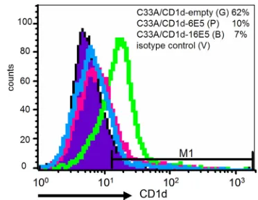
[image:4.585.136.449.533.668.2]C33A/CD1d cells (Fig. 4A). In contrast, the 48-kDa, mature glycosylated form of the CD1d heavy chain (HC) that was detected in naive C33A/CD1d and C33A/CD1d-empty cells was completely abrogated in C33A/CD1d-6E5 and barely de-tectable in the C33A/CD1d-16E5 cells (Fig. 4B, lanes 1, 4, 2, and 3, respectively). The presence of HPV6 and HPV16 E5 drastically inhibited the maturation of CD1d HCs. Flow cy-tometry was used to analyze the effect of HPV E5 on cell surface expression of CD1d in the C33A/CD1d cells harboring E5-expressing or empty vector (Fig. 5). CD1d was expressed by
the majority of C33A/CD1d-empty cells but absent in⬎70% of C33A/CD1d-6E5 or -16E5 cells (Fig. 5).
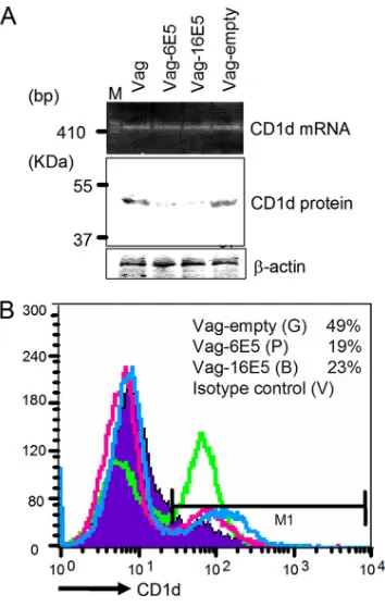
To confirm the effect of E5 on endogenous CD1d, we used a vaginal epithelial cell line immortalized via HPV16 E6/E7 transduction of primary cells collected from normal human vaginal epithelium and subsequently well characterized as pos-sessing histological and immunological characteristics identical to those of primary epithelial cells (18). We have previously reported the endogenous expression of functional CD1d molecules on the surface of these cells (25). Since vaginal epithelial cells are well-known targets of genital HPV, these cells were considered to be useful as anin vitromodel forin vivo HPV infections. FLAG-tagged HPV6 or HPV16 E5 genes were transduced into these vaginal cells by using ret-rovirus vectors (Vag-6E5 and -16E5). We then examined the expression of CD1d at various levels in the presence or absence of HPV E5 (Fig. 6). RT-PCR and Western blotting revealed that CD1d transcription was unaffected by the presence of E5, but the 48-kDa CD1d HC product clearly decreased in Vag-6E5 and -16E5 cells compared to naive and Vag-empty cells (Fig. 6A). Flow cytometry confirmed the decreased cell surface expression of CD1d in 6E5-ex-pressing vaginal epithelial cells (Fig. 6B).
E5-expressing epithelial cells retain CD1d in the ER. To
[image:5.585.45.285.64.205.2]demonstrate the intracellular localization of CD1d heavy chains in C33A/CD1d cells harboring HPV-6E5 and -16E5, immunofluorescence confocal microscopy was performed with an CD1d MAb (NOR3.2) combined with either an anti-FLAG MAb that detects anti-FLAG-E5 proteins, an ER-specific marker (ER tracker) or DAPI (Fig. 7). In C33A/CD1d-empty control cells, dual labeling for CD1d and the nucleus (DAPI) verified that CD1d could be detected in a diffuse pattern throughout the intracellular space, with increased accumula-tion near the cell surface but not in the perinuclear area (Fig. 7, upper image). In contrast, decreased amounts of CD1d could be detected in C33A/CD1d-6E5 and -16E5 cells and CD1d proteins were localized to perinuclear areas near the ER. CD1d and ER signals merged in perinuclear areas (pink
[image:5.585.326.512.67.211.2]FIG. 3. HPV E5 detection in HPV E5-transformed C33A/CD1d cells. (A) Transcription of HPV E5. cDNA was produced via RT of 1 g of total RNA from each cell line and amplified by PCR with primer pairs specific for HPV16 E5 and HPV6 E5. PCR products were sep-arated over an agarose gel containing ethidium bromide. Lanes 5 and 6 display PCR products derived from C33A/CD1d-6E5 and -16E5 cDNA, respectively, while lanes 2 and 3 show PCR products derived from corresponding expression plasmid DNA. Lanes 4 and 7 represent negative control plasmid and cell lines lacking E5, respectively. (B) Translation of HPV E5. Fifty-microgram aliquots of protein lysates from each cell line were analyzed by Western immunoblotting with antibodies against the FLAG tag (1:500 dilution) and-actin (loading control).
[image:5.585.58.267.486.650.2]FIG. 4. CD1d heavy-chain transcription and translation in C33A/ CD1d, C33A/CD1d-empty, C33A/CD1d-6E5, and C33A/CD1d-16E5 cells. (A) Transcription of CD1d HC. The mRNA levels of CD1d were analyzed by quantitative RT-PCR using SYBR green methodology. CD1d mRNA levels were normalized to-actin. (B) Fifty-microgram aliquots of protein lysates from each cell line were analyzed by West-ern immunoblotting with a peroxidase-labeled anti-CD1d NOR3.2 MAb (1:200 dilution) and a-actin loading control.
FIG. 5. Cell surface expression of CD1d in C33A/CD1d-empty (green line), C33A/CD1d-6E5 (pink line), and -16E5 (blue line) cells. All cells were stained with an anti-CD1d primary MAb (NOR3.2; 1:100 dilution) and a PE-conjugated goat anti-mouse immunoglobulin secondary antibody (1:20 dilution). Background staining of the cells with an isotype-matched control antibody is also shown (filled region). Cells were suspended in 1% paraformalde-hyde and analyzed using a FACSCalibur flow cytometry system.
on November 8, 2019 by guest
http://jvi.asm.org/
signals), suggesting that the majority of CD1d is within the ER (Fig. 7, images on the left). Dual labeling for CD1d and FLAG-E5 verified the colocalization of CD1d and E5 within the ER (orange to yellow signals), while nonmerged FLAG-E5 signals were present in the perinuclear area (pure green), sug-gesting the presence of E5 in the GA in the absence of CD1d (Fig. 7, images on the right). The results of immunofluores-cence microscopy support our biochemical and flow cytometry data showing that mature CD1d protein levels decrease and CD1d fails to traffic to the cell surface in HPV E5-expressing cells.
HPV E5 interacts with calnexin in the ER. Previous
bio-chemical studies have reported that HPV16 E5 interacts with calnexin and that these interactions interfere with the modifi-cation of HLA class I heavy chains that typically occurs in the ER (21). The role of calnexin and/or calreticulin in the forma-tion of the second disulfide bond of CD1d HCs in the ER is well described (24). We therefore hypothesized that E5
inter-acts with calnexin in the ER and may impair calnexin-mediated CD1d folding. This, in turn, could interrupt appropriate traf-ficking of CD1d to the surface of HPV-infected cells. To ad-dress the hypothesis, we examined the interaction of E5 with calnexin using immunoprecipitation. Total cell lysates obtained from C33A/CD1d-empty, -6E5, and -16 E5 cells were incu-bated with anti-FLAG MAb conjugated beads. FLAG-E5-bound proteins were immunoprecipitated and analyzed by im-munoblotting with an anti-calnexin MAb. A band with an apparent molecular mass of 90 kDa and corresponding to calnexin was detected in C33A/CD1d-6E5 and -16 E5 cells, but not C33A/CD1d-empty cells, biochemically demonstrating in-teraction between E5 with calnexin (Fig. 8A).
[image:6.585.72.249.66.345.2]To visually demonstrate the colocalization of CD1d and calnexin, C33A/CD1d-empty, -6E5, and -16E5 cells were du-ally stained with anti-CD1d NOR3.2 and anti-calnexin MAbs and examined by using confocal microscopy. Again, NOR3.2-reactive CD1d was detected throughout the intracellular space in C33A/CD1d-empty cells. In contrast, the majority of CD1d molecules in C33A/CD1d-6E5 or -16E5 cells localized to the perinuclear area (Fig. 8B, images on left). Calnexin detection was rendered as green signals. These mostly localized to pe-rinuclear areas in E5-expressing cells and correspond to the location of ER (Fig. 8B, center images). Although some merge images (yellow signals) could be detected in each cell line, the merge patterns differed between C33A/CD1d-empty and E5-expressing cells (Fig. 8B, images on the right). In C33A/CD1d-empty cells, the calnexin and CD1d signals were mostly distinct and but those that did colocalize appeared to follow the syn-thetic pathway for type I proteins. In contrast, CD1d in the E5-expressing cells completely colocalized with calnexin, con-firming our biochemical data demonstrating physiologic inter-action between calnexin and CD1d in the C33A/CD1d-6E5 and -16E5 cells.
FIG. 6. CD1d downregulation in alternate genital keratinocytes in the presence of 6E5 and 16E5. (A) HPV6 and HPV16 E5 genes were transduced into vaginal epithelial cells established from normal human vaginal epithelium (17) and named Vag-6E5 and Vag-16E5, respec-tively. PCR products derived from cDNA generated by reverse tran-scription using 1g of total RNA from each of the vaginal cell lines were separated over an ethidium bromide-containing agarose gel. Fifty-microgram aliquots of protein lysates from each vaginal cell line were analyzed by Western immunoblotting with a peroxidase labeled anti-CD1d NOR3.2 MAb (1:200 dilution) and a-actin loading con-trol. (B) Vag-empty (green line), Vag-6E5 (pink line), and Vag-16E5 (blue line) were stained with an anti-CD1d primary MAb (NOR3.2; 1:100 dilution) and a PE-conjugated goat anti-mouse immunoglobulin secondary antibody (1:20 dilution). Background staining of the cells using an isotype-matched control antibody is also shown (filled region). Cells were suspended in 1% paraformaldehyde and analyzed by using a FACSCalibur flow cytometry system.
FIG. 7. CD1d trafficking in the presence or absence of E5. C33A/ CD1d-empty, C33A/CD1d-6E5, or C33A/CD1d-16E5 cells were seeded onto coverslips. All of the cells were immunostained with an anti-CD1d MAb (NOR3.2, red). C33A/CD1d-empty were also posed to DAPI (blue), and C33A/CD1d-6E5 or -16E5 cells were ex-posed to ER tracker (blue) and an anti-FLAG MAb (green). Cells were then visualized by using fluorescence confocal microscopy. Or-ange to yellow signals represent colocalization of CD1d and E5 within the ER.
on November 8, 2019 by guest
http://jvi.asm.org/
[image:6.585.328.512.70.245.2]CD1d was rescued by treatment of proteasome inhibitor.
We have previously demonstrated that surface expression of CD1d in human genital epithelial cells is downregulated byC.
trachomatisinfection and that downregulation involves
chla-mydial protein-mediated proteasomal pathways (26). We hy-pothesized that HPV infection could utilize posttranslational cellular proteasomal degradation to inhibit cell surface expres-sion of CD1d HC. To address the role of the cellular protea-some in E5-associated CD1d degradation, C33A/CD1d-empty, -6E5, or -16E5 cells were exposed to the proteasome inhibitor, MG132, and CD1d HC levels in cell lysates compared to those in unexposed cells (Fig. 9). Using the NOR3.2 MAb for im-munoblotting, the reduced or abrogated expression of the 48-kDa mature CD1d HC in E5-expressing cells could be rescued by the presence of MG132 (Fig. 9A). To visually replicate this effect, immunofluorescence microscopy was performed with the NOR3.2 MAb and DAPI in MG132 exposed and unex-posed E5-expressing and control cells (Fig. 9B). In C33A/ CD1d-empty cells, NOR3.2-reactive CD1d was detected throughout the intracellular space (Fig. 9B, upper left image). In contrast, NOR3.2-reactive CD1d was barely detected or undetectable in the majority of unexposed C33A/CD1d-6E5 or -16E5 cells (Fig. 9B, upper, right two images). In the presence of proteasomal inhibition with MG132, E5-expressing cells again show CD1d signals throughout the intracellular space (Fig. 9B, lower panels). HPV E5-expressing cells completely recover their expression mature CD1d molecules upon inhibi-tion of cellular proteasomal degradainhibi-tion.
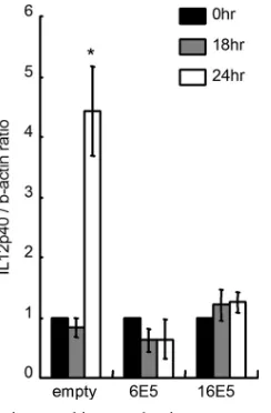
HPV E5 abrogates CD1d-mediated cytokine production in
the epithelial cells. Surface CD1d interacts specifically with
iNKT cells bearing an iTCR. The interaction not only activates NKT cells but also induces phosphorylation of CD1d, intracel-lular signaling, and the release of cytokines from the CD1d-bearing cell. We have previously demonstrated that human reproductive tract epithelial cells expressing CD1d on their cell surfaces have the capacity to produce cytokines, especially IL-12, after CD1d ligation (25). IL-12 is a central mediator in both innate and adaptive immunity and is crucial in the pre-vention of many infectious diseases and tumors (40). IL-12 induces IFN-␥-producing NK, NKT, T helper, and cytotoxic T
[image:7.585.135.449.68.205.2]cells. Since our investigations had demonstrated a decrease in cell surface expression of CD1d in the presence of HPV and specifically of HPV E5, we next examined whether CD1d-mediated IL-12 production was abrogated in E5-expressing epithelial cells (Fig. 10). An anti-CD1d 51.1 MAb can be used for CD1d cross-linking and represents an in vitro model for
FIG. 8. CD1d and calnexin have direct interactions and colocalize in the perinuclear area in the presence of HPV E5. (A) Protein lysates from C33A/CD1d-empty, C33A/CD1d-6E5, and C33A/CD1d-16E5 cells were immunoprecipitated with an anti-FLAG MAb. Immunoprecipitants were then separated by SDS-PAGE and immunoblotted with an anti-calnexin antibody. (B) C33A/CD1d-empty, C33A/CD1d-6E5, or C33A/CD1d-16E5 cells were seeded onto coverslips. All cells were exposed to an anti-CD1d MAb (NOR3.2, red) and to an anti-calnexin MAb (green) labeled with Zenon Alexa Fluor 488 using a mouse IgG labeling kit. Cells were then visualized by using fluorescence confocal microscopy. Yellow images represent colocalization of CD1d and calnexin.
FIG. 9. Proteasome inhibition rescues CD1d from E5-mediated degradation. (A) C33A/CD1d-empty, C33A/CD1d-6E5, or C33A/ CD1d-16E5 cells were cultured for up to 24 h in the presence or absence of the cytosolic proteasome inhibitors MG132 (10M) in DMSO. Fifty micrograms of protein lysates from each cell line were analyzed by Western immunoblotting with a peroxidase-labeled anti-CD1d MAb (NOR3.2; 1:200 dilution) and a-actin loading control. (B) C33A/CD1d-empty, C33A/CD1d-6E5, or C33A/CD1d-16E5 cells were seeded onto coverslips and cultured for up to 24 h in the presence (lower) or absence (upper) of MG132 (10M) in DMSO. All cover-slips were fixed in 4% paraformaldehyde, permeabilized with 0.2% Triton-X, blocked with 6% BSA, and incubated for 1 h at room tem-perature with an anti-CD1d NOR3.2 MAb (red) directly conjugated with Zenon Alexa Fluor 555 using a mouse IgG1 labeling kit. Cells were then counterstained with a DAPI (blue) nucleic acid stain.
on November 8, 2019 by guest
http://jvi.asm.org/
[image:7.585.333.506.380.588.2]CD1d ligation (14, 43). C33A/CD1d-empty, -6E5, or -16E5 cells were first exposed to an anti-CD1d 51.1 MAb and then to a secondary anti-mouse IgG cross-linker and then examined for IL-12 production (Fig. 10). IL-12 p40 transcription in-creased 24 h after cross-linking in the C33A/CD1d-empty cells. This effect was abrogated completely in E5-expressing cells. Decreased cell surface expression of CD1d in E5-expressing cells inhibits the ability of antibodies to cross-link CD1d and thereby halts the downstream signaling that drives IL-12 pro-duction.
DISCUSSION
In this study, we attempted to elucidate a mechanism to explain our finding that CD1d was expressed at lower levels in tissues infected with high-risk and low-risk HPV subtypes (16 and 6, respectively). The CD1d protein levels were lower, but the mRNA levels were unaffected in HPV E5-expressing cells, indicating that CD1d is downregulated at a posttranscriptional level in the presence of HPV E5. Modification of CD1d was interrupted at the level of the ER by interactions between HPV E5 and calnexin. Improper folding and/or ubiquitination of CD1d HC in the presence of HPV then targets CD1d to cellular proteasomal degradation. Others (21) have previously demonstrated that interaction of E5 with calnexin appears to interfere with calnexin-assisted folding of HLA class I mole-cules. Like the well-described quality control system assuring proper HLA class I HC production and maturation, delayed exit of improperly folded CD1d HC from the ER in the pres-ence of HPV E5 appears to result in movement of CD1d HC
In planning for these investigations, we chose to use two cell lines. One was a cervical cancer cell line that is unique in being HPV-negative C33A. The other was an endogenous CD1d-bearing keratinocyte cell line established from normal human vaginal epithelial cells. The HPV-negative C33A cell was par-ticularly useful for this study because it allowed us to control for the influence on CD1d of proteins other than E5 that could have been potentially present in an HPV-positive cervical cell line. Via transfection, CD1d could be stably expressed in a cervical cell, and specific HPV proteins could added in isola-tion to assess their effect on CD1d. Our previous and current immunohistochemical data demonstrated that cells in the basal and parabasal cell layers of a variety of squamous genital epithelia react strongly with anti-CD1d MAbs, in patterns that replicate those seen in normal human skin (9, 25). The distri-bution of CD1d-bearing epithelial cells within the basal and parabasal cell layers may be required for effective interactions between CD1d and the iNKT cells that reside within submu-cosal tissues. These interactions may occur primarily through CD1d expressed on the basilar membrane. The immortalized vaginal epithelial cell lines used in the present study have been characterized by Fichorova et al. as being similar to epithelial cells present in basal or parabasal cell layersin vivo(17, 18). We have also seen similar patterns of CD1d expression in nondiseased genital tract tissues (25). The data derived from vaginal epithelial cells in the present study allowed us to mimic
in vivoinfection of normal human keratinocytes by HPV and to
confirm that the retrovirus vectors used to transduce E5 genes into our cell models did not affect the endogenous CD1d pro-moter.
[image:8.585.103.225.67.253.2]CD1d transcription was barely detectable in both C33A cells and HPV-positive cervical cancer cells (HeLa, Caski, and clin-ical samples). Immunohistochemclin-ical data verified that immune reactivity for CD1d was completely abrogated in all cervical cancer lesions. Lack of CD1d expression in cancer-derived cells is unlikely to be associated with HPV E5 protein expres-sion since the E5 gene is deleted when the HPV genome integrates into the host genome. Rather, CD1d may be genomically inactivated during carcinogenesis. Two of eigh-teen cases with CIN2 and CIN3 showed immune reactivity with the anti-CD1d MAb, although all lesions were positive for high-risk HPV. CD1d expression is known to be induced by inflammatory cytokines such as IFN-␥(9, 25). In some cases, an enhancement in CD1d expression secondary to the immu-nological microenvironment in the cervix in vivomay super-cede E5-mediated downregulation. Alternatively, E5-mediated CD1d downregulation in CIN3 lesions may be lessened be-cause most cells may have already integrated the HPV genome
FIG. 10. Autocrine cytokine production upon CD1d cross-linking in C33A/CD1d-empty, C33A/CD1d-6E5, and C33A/CD1d-16E5 cells. An anti-CD1d 51.1 MAb was added at a dosage of 10g/ml to cultured epithelial cell monolayers, followed by incubation for 1 h. After being washed with PBS, 10g of a goat mouse immunoglobulin anti-body/ml was added as a cross-linker for 30 min. The cells were incu-bated in serum-free growth medium without any antibiotics for 0 to 24 h. cDNA was produced via reverse transcription of 1g of total RNA extracted and amplified by PCR with primer pairs for IL-12 p40 and-actin. IL-12 p40 mRNA levels were normalized to-actin. Mean values with standard deviations are presented. Asterisks indicate the comparisons (before versus after cross-linking) with statistical signifi-cance (P⬍0.05;n⫽4).
on November 8, 2019 by guest
http://jvi.asm.org/
and little E5 remains within the lesion. Statistical analysis, however, reveals a trend toward decreased CD1d expression with progressing CIN.
Previous investigations on HPV-associated immune evasion strategies have highlighted interference with adaptive immune responses against HPV through disruption of HLA molecules (19, 30). Here we focused on CD1d, which serves not only as a sentinel molecule in innate immune response but as a bridge between innate and adaptive immunity. Reports of CD1d ex-pression in epithelial cells lagged behind its detection and functional studies in classic immune cells such as dendritic cells, macrophages, and B cells. In epithelial cells, CD1d en-counters a wide array of pathogens and helps to orchestrate innate and adaptive immune responses to these immunologic challenges via interactions with CD1d-restricted iNKT. The interaction of CD1d with CD1d-restricted iNKT cell is lipid antigen dependent; however, this lipid antigen can be derived from invading microbes or from host cellular lipids. In re-sponse to some microbes, the rapid effects of CD1d-restricted NKT cells do not require recognition of microbial specific antigens (6, 34, 35). Since HPV has no envelope and therefore no HPV-specific lipid antigens, CD1d may present self lipid antigen for activation of iNKT cells in response to HPV-in-fected epithelial cells. Recognition of CD1d by iNKT cells can cause rapid release of both IL-4 and IFN-␥from the NKT cell (6). This would be predicted to activate CD1d-restricted iNKT cells and rapidly induce an adaptive immune response to in-vading microbes. Our previous investigations have also dem-onstrated that human reproductive tract epithelial cells that express CD1d on their cell surfaces are able to produce cyto-kines, including IL-12, in response to CD1d ligation (25). IL-12 is a central mediator in both innate and adaptive immunity and is crucial in the prevention of infectious diseases and tumors (40). IL-12 induces IFN-␥-producing NK, NKT, T helper, and cytotoxic T cells and thereby bridges innate and adaptive im-mune responses. Yue et al. have demonstrated that cross-linking of CD1d rapidly induces phosphorylation of IB. This, in turn, promotes NF-B activation and IL-12 production in monocytes and immature dendritic cells (43). As shown here, the induction of IL-12 production in response to CD1d cross-linking is completely abrogated in HPV E5-expressing epithe-lial cells but never to levels below those produced at baseline. The inhibition of CD1d-mediated cytokine production may be a mechanism by which HPV-infected cells evade (at least tem-porarily) the bridging of innate and adaptive immune re-sponses that would otherwise occur upon interaction between cell surface CD1d and iNKT cells.
HPV E5 has been reported to play a role in HPV immuno-evasion through the downregulation of cell surface HLA class I molecules. Several investigators have demonstrated that the papillomavirus E5 product inhibits the acidification of or-ganelles, including the GA and endosomes (28, 32, 38). Ashrafi et al. have reported that the inhibition of GA acidification mediated by bovine papillomavirus E5 is associated with re-tention of MHC class I molecules in the GA (4, 41) and that HPV16 E5 retains HLA-A and -B, but not HLA-C and -E, within the GA. These authors hypothesize that the selectivity of HLA class I subtype downregulation may suggest that mech-anisms other than GA acidification may be involved (2, 3). Gruener et al. demonstrated that interactions between HPV16
E5 and calnexin interfere with modification of HLA class I HCs and results in heavy-chain retention in the ER (21). Since the synthetic pathways for CD1d and HLA class I HCs are identical, we hypothesized that inhibition of calnexin folding capabilities by HPV E5 was a likely mechanism for decreased cell surface expression of CD1d in HPV-infected cells. Using confocal microscopy, we supported this hypothesis over the acidification mechanism by demonstrating that CD1d HC and calnexin colocalize in the ER rather than the GA. Interest-ingly, the CD1d HC that was rescued by MG132 treatment in E5-expressing cells was a 48-kDa mature form that was present in a diffuse pattern throughout the intracellular space (Fig. 8 and 9). It appears that CD1d synthesis and trafficking may be fairly robust in the presence of HPV E5 if proteasomal deg-radation is inhibited. This suggests that HPV E5 does not interfere with the synthesis of CD1d HC but rather delays its exit from the ER and alters its maturation so that CD1d HCs are targeted to proteasomal degradation. Interactions between HPV E5 and calnexin do not appear to interrupt all of the functions of calnexin, but just enough to co-opt the cellular cytosolic proteolytic pathway and effectively degrade CD1d and temporarily inhibits CD1d-mediated innate and adaptive immune pathways early in HPV infection.
CD1d expression and CD1d activation of neighboring iNKT cells may play an important role in the generation of innate and adaptive immune responses to microbial infection of the ectocervix. Our previous and current immunohistochemical data have shown that CD1d immunoreactivity and distribution patterns in ectocervix are similar to those in the penile urethra and vagina, where epithelial cells exhibit CD1d-mediated Th1-type cytokine production (25). It was likely that CD1d-bearing ectocervical epithelial cells were also capable of CD1d-medi-ated Th1-type cytokine production, and we have here shown that CD1d cross-linking on C33A/CD1d cells promotes the synthesis of IL-12. We therefore suggest a mechanism whereby CD1d downregulation in the presence of low- and high-risk HPV subtypes allows the infecting virus to evade host immune surveillance and establish persistent infection at the primary transmission site. The magnitude of HPV E5 expression and resultant CD1d downregulation may vary among CIN lesions, as shown in our clinical data. If so, variations in CD1d immu-noreactivity in biopsy specimens of CIN lesions may be a pre-dictive marker for the fate of early CIN. This topic is currently under investigation.
ACKNOWLEDGMENTS
This study was supported by a grant-in-aid from the Ministry of Health, Labor, and Welfare of Japan for the Third-Term Comprehen-sive 10-Year Strategy for Cancer Control; by a cancer research grant from the Ministry of Education, Culture, Sports, Science, and Tech-nology of Japan; by a grant from Kanzawa Medical Research Foun-dation; and by a grant from the Okinawa New Industry Creation Project.
We are grateful to R. Blumberg (Harvard Medical School, Boston, MA), K. Oda (University of Tokyo, Tokyo, Japan), and D. J. Anderson (Boston University, Boston, MA) for kindly providing the CD1d-ex-pressing retroviral plasmid pSR␣-neo, the retrovirus expression sys-tem, and the vaginal epithelial cell line, respectively.
REFERENCES
1.Araibi, E. H., B. Marchetti, G. H. Ashrafi, and M. S. Campo.2004. Down-regulation of major histocompatibility complex class I in bovine papillomas. J. Gen. Virol.85:2809–2814.
on November 8, 2019 by guest
http://jvi.asm.org/
7.Bilenki, L., S. Wang, J. Yang, Y. Fan, A. G. Joyee, and X. Yang.2005. NK T-cell activation promotesChlamydia trachomatisinfection in vivo. J. Im-munol.175:3197–3206.
8.Blumberg, R. S., C. Terhorst, P. Bleicher, F. V. McDermott, C. H. Allan, S. B. Landau, J. S. Trier, and S. P. Balk.1991. Expression of a nonpolymorphic MHC class I-like molecule, CD1D, by human intestinal epithelial cells. J. Immunol.147:2518–2524.
9.Bonish, B., D. Jullien, Y. Dutronc, B. B. Huang, R. Modlin, F. M. Spada, S. A. Porcelli, and B. J. Nickoloff.2000. Overexpression of CD1d by kerati-nocytes in psoriasis and CD1d-dependent IFN-␥production by NK-T cells. J. Immunol.165:4076–4085.
10.Brigl, M., and M. B. Brenner.2004. CD1: antigen presentation and T-cell function. Annu. Rev.22:817–890.
11.Brigl, M., L. Bry, S. C. Kent, J. E. Gumperz, and M. B. Brenner.2003. Mechanism of CD1d-restricted natural killer T-cell activation during micro-bial infection. Nat. Immunol.4:1230–1237.
12.Canchis, P. W., A. K. Bhan, S. B. Landau, L. Yang, S. P. Balk, and R. S. Blumberg.1993. Tissue distribution of the non-polymorphic major histocom-patibility complex class I-like molecule, CD1d. Immunology80:561–565. 13.Cho, S., K. S. Knox, L. M. Kohli, J. J. He, M. A. Exley, S. B. Wilson, and
R. R. Brutkiewicz.2005. Impaired cell surface expression of human CD1d by the formation of an HIV-1 Nef/CD1d complex. Virology337:242–252. 14.Colgan, S. P., R. M. Hershberg, G. T. Furuta, and R. S. Blumberg.1999.
Ligation of intestinal epithelial CD1d induces bioactive IL-10: critical role of the cytoplasmic tail in autocrine signaling. Proc. Natl. Acad. Sci. U. S. A. 96:13938–13943.
15.Conrad, M., V. J. Bubb, and R. Schlegel.1993. The human papillomavirus type 6 and 16 E5 proteins are membrane-associated proteins which associate with the 16-kilodalton pore-forming protein. J. Virol.67:6170–6178. 16.Fehrmann, F., and L. A. Laimins.2003. Human papillomaviruses: targeting
differentiating epithelial cells for malignant transformation. Oncogene22: 5201–5207.
17.Fichorova, R. N., A. O. Cronin, E. Lien, D. J. Anderson, and R. R. Ingalls. 2002. Response toNeisseria gonorrhoeaeby cervicovaginal epithelial cells occurs in the absence of Toll-like receptor 4-mediated signaling. J. Immunol. 168:2424–2432.
18.Fichorova, R. N., and D. J. Anderson.1999. Differential expression of im-munobiological mediators by immortalized human cervical and vaginal epi-thelial cells. Biol. Reprod.60:508–514.
19.Frazer, I. H.2009. Interaction of human papillomaviruses with the host immune system: a well evolved relationship. Virology384:410–414. 20.Gravitt, P. E., C. L. Peyton, T. Q. Alessi, C. M. Wheeler, F. Coutlee, A.
Hildesheim, M. H. Schiffman, D. R. Scott, and R. J. Apple.2000. Improved amplification of genital human papillomaviruses. J. Clin. Microbiol.38:357– 361.
21.Gruener, M., I. G. Bravo, F. Momburg, A. Alonso, and P. Tomakidi.2007. The E5 protein of the human papillomavirus type 16 downregulates HLA-I surface expression in calnexin-expressing but not in calnexin-deficient cells. Virol. J.4:116.
22.Gumperz, J. E., C. Roy, A. Makowska, D. Lum, M. Sugita, T. Podrebarac, Y. Koezuka, S. A. Porcelli, S. Cardell, M. B. Brenner, and S. M. Behar.2000. Murine CD1d-restricted T-cell recognition of cellular lipids. Immunity12: 211–221.
23.Ho, G. Y. F., R. Bierman, L. Beardsley, C. J. Chang, and R. D. Burk.1998. Natural history of cervicovaginal papillomavirus infection in young women. N. Engl. J. Med.338:423–428.

28.Marchetti, B., G. H. Ashrafi, E. Tsirimonaki, P. M. O’Brien, and M. S. Campo.2002. The bovine papillomavirus oncoprotein E5 retains MHC class I molecules in the Golgi apparatus and prevents their transport to the cell surface. Oncogene21:7808–7816.
29.Mattner, J., K. L. Debord, N. Ismail, R. D. Goff, C. Cantu III, D. Zhou, P. Saint-Mezard, V. Wang, Y. Gao, N. Yin, K. Hoebe, O. Schneewind, D. Walker, B. Beutler, L. Teyton, P. B. Savage, and A. Bendelac.2005. Exoge-nous and endogeExoge-nous glycolipid antigens activate NKT cells during microbial infections. Nature434:525–529.
30.O’Brien, P. M., and M. Saveria Campo.2002. Evasion of host immunity directed by papillomavirus-encoded proteins. Virus Res.88:103–117. 31.Sanchez, D. J., J. E. Gumperz, and D. Ganem.2005. Regulation of CD1d
expression and function by a herpesvirus infection. J. Clin. Invest.115:1369– 1378.
32.Schapiro, F., J. Sparkowski, A. Adduci, F. Suprynowicz, R. Schlegel, and S. Grinstein.2000. Golgi alkalinization by the papillomavirus E5 oncoprotein. J. Cell Biol.148:305–315.
33.Shiraki, Y., Y. Ishibashi, M. Hiruma, A. Nishikawa, and S. Ikeda.2006. Cytokine secretion profiles of human keratinocytes duringTrichophyton ton-suransandArthroderma benhamiaeinfections. J. Med. Microbiol.55:1175– 1185.
34.Skold, M., X. Xiong, P. A. Illarionov, G. S. Besra, and S. M. Behar.2005. Interplay of cytokines and microbial signals in regulation of CD1d expression and NKT cell activation. J. Immunol.175:3584–3593.
35.Skold, M., and S. M. Behar.2003. Role of CD1d-restricted NKT cells in microbial immunity. Infect. Immun.71:5447–5455.
36.Stanic, A. K., J. S. Bezbradica, J. J. Park, L. Van Kaer, M. R. Boothby, and S. Joyce.2004. Cutting edge: the ontogeny and function of Va14Ja18 natural T lymphocytes require signal processing by protein kinase Cand NF-B. J. Immunol.172:4667–4671.
37.Stoler, M. H., C. R. Rhodes, A. Whitbeck, S. M. Wolinsky, L. T. Chow, and T. R. Broker.1992. Human papillomavirus type 16 and 18 gene expression in cervical neoplasias. Hum. Pathol.23:117–128.
38.Straight, S. W., B. Herman, and D. J. McCance.1995. The E5 oncoprotein of human papillomavirus type 16 inhibits the acidification of endosomes in human keratinocytes. J. Virol.69:3185–3192.
39.Taniguchi, M., and T. Nakayama.2000. Recognition and function of V[al-pha]14 NKT cells. Semin. Immunol.12:543–550.
40.Trinchieri, G.2003. Interleukin-12 and the regulation of innate resistance and adaptive immunity. Nat. Rev. Immunol.3:133–146.
41.Tsirimonaki, E., R. Ullah, B. Marchetti, G. H. Ashrafi, L. McGarry, B. Ozanne, and M. S. Campo.2006. Similarities and differences between the E5 oncoproteins of bovine papillomaviruses type 1 and type 4: cytoskeleton, motility and invasiveness in E5-transformed bovine and mouse cells. Virus Res.115:158–168.
42.Yuan, W., A. Dasgupta, and P. Cresswell.2006. Herpes simplex virus evades natural killer T-cell recognition by suppressing CD1d recycling. Nat. Immu-nol.7:835–842.
43.Yue, S. C., A. Shaulov, R. Wang, S. P. Balk, and M. A. Exley.2005. CD1d ligation on human monocytes directly signals rapid NF-B activation and production of bioactive IL-12. Proc. Natl. Acad. Sci. U. S. A.102:11811– 11816.
44.zur Hausen, H.2002. Papillomaviruses and cancer: from basic studies to clinical application. Nat. Rev. Cancer2:342–350.