JOURNAL OFVIROLOGY, Nov. 2008, p. 10756–10767 Vol. 82, No. 21 0022-538X/08/$08.00⫹0 doi:10.1128/JVI.00802-08
Copyright © 2008, American Society for Microbiology. All Rights Reserved.
Noroviruses Distinguish between Type 1 and Type 2 Histo-Blood
Group Antigens for Binding
䌤
Haruko Shirato,
1* Satoko Ogawa,
1Hiromi Ito,
2Takashi Sato,
2Akihiko Kameyama,
2Hisashi Narimatsu,
2Zheng Xiaofan,
3Tatsuo Miyamura,
1Takaji Wakita,
1Koji Ishii,
1and Naokazu Takeda
1Department of Virology II, National Institute of Infectious Diseases, 4-7-1 Gakuen, Musashi-Murayama, Tokyo 208-0011, Japan1;
Research Center for Medical Glycoscience, National Institute of Advanced Industrial Science and Technology, 1-1-1 Umezono,
Tsukuba, Ibaraki 305-8568, Japan2; and Blood Centre of Zhejiang Province, 345 Wulin Road, Hangzhou,
Zhejiang Province 310006, China3
Received 15 April 2008/Accepted 6 August 2008
Norovirus (NoV) is a causative agent of acute gastroenteritis. NoV binds to histo-blood group antigens (HBGAs), namely, ABH antigens and Lewis (Le) antigens, in which type 1 and type 2 carbohydrate core structures constitute antigenically distinct variants. Norwalk virus, the prototype strain of norovirus, binds to the gastroduodenal junction, and this binding is correlated with the presence of H type 1 antigen but not with that of H type 2 antigen (S. Marionneau, N. Ruvoen, B. Le Moullac-Vaidye, M. Clement, A. Cailleau-Thomas, G. Ruiz-Palacois, P. Huang, X. Jiang, and J. Le Pendu, Gastroenterology 122:1967–1977, 2002). It has been unknown whether NoV distinguishes between the type 1 and type 2 chains of A and B antigens. In this study, we synthesized A type 1, A type 2, B type 1, and B type 2 pentasaccharides in vitro and examined the function of the core structures in the binding between NoV virus-like particles (VLPs) and HBGAs. The attachment of five genogroup I (GI) VLPs from 5 genotypes and 11 GII VLPs from 8 genotypes, GI/1, GI/2, GI/3, GI/4, GI/8, GII/1, GII/3, GII/4, GII/5, GII/6, GII/7, GII/12, and GII/14, to ABH and Le HBGAs was analyzed by enzyme-linked immunosorbent assay-based binding assays and Biacore analyses. GI/1, GI/2, GI/3, GI/4, GI/8, and GII/4 VLPs were more efficiently bound to A type 2 than A type 1, and GI/8 and GII/4 VLPs were more efficiently bound to B type 2 than B type 1, indicating that NoV VLPs distinguish between type 1 and type 2 carbohydrates. The dissociation of GII/4 VLPs from B type 1 was slower than that from B type 2 in the Biacore experiments; moreover, the binding to B type 1 was stronger than that to B type 2 in the ELISA experiments. These results indicated that the type 1 carbohydrates bind more tightly to NoV VLPs than the type 2 carbohydrates. This property may afford NoV tissue specificity. GII/4 is known to be a global epidemic genotype and binds to more HBGAs than other genotypes. This characteristic may be linked with the worldwide transmission of GII/4 strains. GI/2, GI/3, GI/4, GI/8, GII/4, and GII/7 VLPs bound to Lea
expressed by nonsecretors, suggesting that NoV can infect individuals regardless of secretor phenotype. Overall, our results indicated that HBGAs are important factors in determining tissue specificity and the risk of transmission.
Norovirus(NoV), a member of the familyCaliciviridae(1), is
a major cause of water- and food-borne acute nonbacterial gastroenteritis and comprises many morphologically similar but antigenically diverse groups of viruses (8, 11, 22). Recent progress in genetic studies has enabled us to divide human NoV into at least three genogroups, genogroup I (GI), GII, and GIV, which contain at least 15, 18, and 1 genotype, re-spectively (20).
Histo-blood group antigens (HBGAs) are carbohydrates that contain structurally related saccharide moieties. H antigen (Fuc␣1-2Gal), i.e., O-type antigen, is generated by a fucose transfer to a galactose residue with an␣1-2 linkage. A antigen [GalNAc␣1-3(Fuc␣1-2)Gal] and B antigen [Gal␣1-3(Fuc␣ 1-2)Gal] of ABH HBGAs are generated by a transfer of GalNAc and Gal, respectively, to an H structure irrespective of the carbohydrate core structure. The core structures are classified into four major structures, type 1 (Gal1-3GlcNAc), type 2
(Gal1-4GlcNAc), type 3 (Gal1-3GalNAc␣), and type 4 (Gal1-3GalNAc). The fucose transfer of ABH antigens in erythrocytes is catalyzed by FUT1, a member of the fucosyl-transferase family, whereas it is catalyzed by a different fuco-syltransferase, FUT2, in saliva and mucosal secretions (31). Individuals who have nullFUT2alleles cannot synthesize ABH antigens in secretions and are called nonsecretors, although ABH antigens can be expressed in erythrocytes via FUT1 (21).
FUT2alleles of Caucasian nonsecretors are completely inacti-vated by nonsense mutations, whereas those of Asian nonse-cretors are incompletely inactivated by missense mutations (23, 41). Thus, Asian nonsecretors are incomplete nonsecretors and produce a small amount of ABH HBGAs in secretions. FUT2 is essential for the fucose transfer to the type 1 structure (Gal1-3GlcNAc) required to generate the H type 1 structure (Fuc␣1-2Gal1-3GlcNAc), a precursor structure for Lewis b (Leb) [Fuc␣1-2Gal1-3(Fuc␣1-4)GlcNAc], in secretions. The
FUT3 enzyme is required for the fucose transfer to type 1 or H type 1 to generate Lewis a (Lea) [Gal1-3(Fuc␣1-4)GlcNAc]
or Leb, respectively.
Virus-like particles (VLPs) derived from the prototype strain of NoV, Norwalk virus (NV/68), bind to HBGAs in saliva from secretor individuals. They preferentially bind to H
* Corresponding author. Mailing address: Department of Virology II, National Institute of Infectious Diseases, Gakuen 4-7-1, Musashi-murayama, Tokyo 208-0011, Japan. Phone: (81)-42-561-0771. Fax: (81)-42-561-4729. E-mail: harukos@nih.go.jp.
䌤Published ahead of print on 13 August 2008.
10756
on November 8, 2019 by guest
http://jvi.asm.org/
type 1 and Lebsynthetic carbohydrates (12, 14, 15, 24, 26).
Although NV/68 VLPs bind to type A antigens in saliva and synthetic type A carbohydrates, they bind to neither type B synthetic carbohydrates nor the majority of type B antigens in saliva (12, 14, 15, 24). From previous volunteer challenge stud-ies, there is strong evidence that this carbohydrate binding is essential for NV/68 infection (16, 24). Nonsecretors were not infected after the challenge with NV/68. Furthermore, type O secretors are more likely to be infected with NV/68; conversely, type B secretors are less likely to be infected with NV/68. However, other NoV VLPs display different ABH and Lewis carbohydrate-binding profiles (12–15): indeed, epidemiologi-cal studies have shown that some NoV strains could infect individuals with other ABH or secretor phenotypes (34).
Type 1 core structures are widely expressed in endodermally derived tissues, such as lining epithelia and glandular epithelia (32). On the other hand, type 2 core structures are found mainly in ecto- or mesodermally derived tissues, including skin and erythrocytes (7, 10, 32). In the human gastroduodenal junction, type 1 structures are found exclusively at the level of the surface epithelia whereas type 2 structures are preferen-tially found at the glandular level (27). Immunohistochemical analysis showed that the binding of recombinant NV/68 (rNV/ 68) to the gastroduodenal junction correlated with the pres-ence of H type 1 antigen but not with that of H type 2 antigen (26). In a human GII/4 infection experiment, pigs that ex-pressed either A or H antigen on the intestinal mucosa had significantly higher rates of diarrhea and seroconversion in response to the strain, and fecal shedding of virus was also significantly higher (5). Therefore, in pigs and in humans, the expression of HBGAs may lead to increased susceptibility to NoV infection. The rNV/68 VLP has been reported to bind synthetic H carbohydrates in the following order of strength: H type 1 trisaccharides, H type 2 trisaccharides, and H disaccha-rides (17). Meanwhile Harrington et al. reported that this strain had higher binding activity for H type 1 trisaccharides than for H type 3 trisaccharides (12). Although these reports suggest that rNV/68 VLP may recognize the linkages and com-ponents in the core structures of the HBGAs, this has not yet
been proven. Moreover, it has not been known whether NoV VLPs distinguish between the type 1 and type 2 chains of A and B antigens.
To investigate the binding properties of NoV strains for HBGAs and to determine whether NoV distinguishes between type 1 and type 2 chains of HBGAs, the attachment of 5 GI VLPs from 5 genotypes and 11 GII VLPs from 8 genotypes to ABH and LeaLebHBGAs was analyzed by enzyme-linked
im-munosorbent assay (ELISA)-based binding assays and Biacore analysis.
MATERIALS AND METHODS
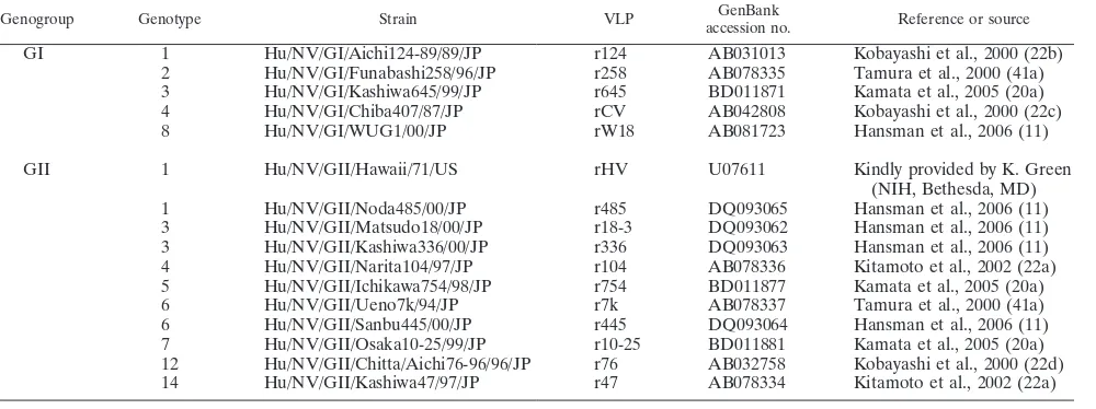
Recombinant VLPs.The recombinant VLPs used in this study are shown in Table 1 (11, 20a, 22a, 22b, 22c, 22d, 41a). VLPs were prepared by infecting subconfluent Tn5 insect cells with the recombinant baculoviruses as described previously (11). Briefly, the culture medium was harvested at 6 days after infec-tion, centrifuged at 1,000⫻gfor 10 min to remove the cell debris, and further centrifuged at 10,000⫻gfor 30 min to remove the baculoviruses. The VLPs in the supernatant were concentrated by centrifugation at 100,000⫻gfor 2 h at 4°C in an SW28 rotor (Beckman Instruments, Inc., Palo Alto, CA). The pellet was resuspended in a solution containing CsCl (1.9 g/4.5 ml) and centrifuged at 120,000⫻gfor 20 h at 10°C in an SW50.1 rotor (Beckman). Peak fractions containing the VLPs were pooled, diluted with phosphate-buffered saline (PBS) (pH 7.5), and centrifuged at 200,000⫻gfor 2 h at 4°C in an SW50.1 rotor. The purified VLPs were examined by electron microscopy, and it was confirmed that the preparations form particles similar to native NoV and do not contain VP2 aggregates, as described previously (11). The VLPs were also examined by sodium dodecyl sulfate-polyacrylamide gel electrophoresis. The protein concen-trations were determined using the Bio-Rad protein assay kit (Bio-Rad Labora-tories, Hercules, CA) with bovine serum albumin as the protein standard. Tn5 cells, an insect cell line fromTrichoplusia ni(Invitrogen, San Diego, CA), were grown at 27°C with Ex-CELL 400 medium (JRH Biosciences, Lenexa, KS). The phylogenetic analysis of the 16 NoV strains, which classified them into 13 clus-ters, was described previously (11).
Saliva samples and blood group antigens.Saliva samples were collected from 29 healthy Japanese donors: specifically, 13 (45%) males (average age, 41 years old; range, 29 to 59 years old) and 16 (55%) females (average age, 36 years old; range, 23 to 62 years old). The saliva samples were boiled for 10 min immediately after collection and centrifuged for 5 min at 13,000⫻g. The clear supernatant was collected and stored at⫺30C° until use.
[image:2.585.43.543.82.269.2]The samples were assayed for the presence of H, A, and B antigens by hemagglutination inhibition. Either 100l of anti-H lecithin (Gamma Biologi-cals, Inc., Houston, TX), 50l of anti-A antibody (Gamma Biologicals, Inc.), or anti-B antibody (Gamma Biologicals, Inc.) was mixed with an equal volume of TABLE 1. Recombinant VLPs used in this study
Genogroup Genotype Strain VLP GenBank
accession no. Reference or source
GI 1 Hu/NV/GI/Aichi124-89/89/JP r124 AB031013 Kobayashi et al., 2000 (22b)
2 Hu/NV/GI/Funabashi258/96/JP r258 AB078335 Tamura et al., 2000 (41a) 3 Hu/NV/GI/Kashiwa645/99/JP r645 BD011871 Kamata et al., 2005 (20a)
4 Hu/NV/GI/Chiba407/87/JP rCV AB042808 Kobayashi et al., 2000 (22c)
8 Hu/NV/GI/WUG1/00/JP rW18 AB081723 Hansman et al., 2006 (11)
GII 1 Hu/NV/GII/Hawaii/71/US rHV U07611 Kindly provided by K. Green
(NIH, Bethesda, MD)
1 Hu/NV/GII/Noda485/00/JP r485 DQ093065 Hansman et al., 2006 (11)
3 Hu/NV/GII/Matsudo18/00/JP r18-3 DQ093062 Hansman et al., 2006 (11) 3 Hu/NV/GII/Kashiwa336/00/JP r336 DQ093063 Hansman et al., 2006 (11) 4 Hu/NV/GII/Narita104/97/JP r104 AB078336 Kitamoto et al., 2002 (22a) 5 Hu/NV/GII/Ichikawa754/98/JP r754 BD011877 Kamata et al., 2005 (20a)
6 Hu/NV/GII/Ueno7k/94/JP r7k AB078337 Tamura et al., 2000 (41a)
6 Hu/NV/GII/Sanbu445/00/JP r445 DQ093064 Hansman et al., 2006 (11)
7 Hu/NV/GII/Osaka10-25/99/JP r10-25 BD011881 Kamata et al., 2005 (20a) 12 Hu/NV/GII/Chitta/Aichi76-96/96/JP r76 AB032758 Kobayashi et al., 2000 (22d) 14 Hu/NV/GII/Kashiwa47/97/JP r47 AB078334 Kitamoto et al., 2002 (22a)
on November 8, 2019 by guest
http://jvi.asm.org/
10758
on November 8, 2019 by guest
http://jvi.asm.org/
each undiluted saliva sample and incubated for 10 or 20 min at 26°C. Then, 50
l of a 3 to 4% suspension of indicator O (Gamma Biologicals, Inc.), A1 (Gamma Biologicals, Inc.), or B (Gamma Biologicals, Inc.) was added. The mixture was left at 26°C for 5 min and centrifuged at 125⫻gfor 1 min. The amount of HBGAs was measured in a semiquantitative manner using serially diluted saliva (40). Serial twofold dilutions of the samples were prepared (1- to 256-fold dilution) and assayed by hemagglutination inhibition assay as described above. Informed consent was obtained from all donors in 2003 before their participation according to ethical code 28 of the National Institute of Infectious Diseases, Japan.
Enzymatic preparation of monovalent carbohydrate-biotin reagents.For the preparation of the GlcNAc1-3Gal1–biotin derivative, a reaction mixture con-taining 25 mM HEPES buffer (pH 7.0), 10 mM MnCl2, biotin-labeled galactose,
an appropriate concentration of UDP-GlcNAc, and purified1,3-N -acetylglu-cosaminyltransferase 2 (3GnT2) (38) was used. Biotin-labeled galactose, UDP-GlcNAc, and3GnT2 were the acceptor, donor substrate, and enzyme, respec-tively. The substrates and enzyme were added to a reaction mixture containing 25 mM HEPES buffer and 10 mM MnCl2and incubated at 37°C for 36 h. The
enzyme was removed with an Ultrafree-MC column (Millipore, Bedford, MA), and the product was purified using reversed-phase high-performance liquid chro-matography. For the preparation of the galactosylated derivatives Gal 1-3GlcNAc1-3Gal1–biotin and Gal1-4GlcNAc1-3Gal1–biotin, a reaction mixture containing GlcNAc1-3Gal1–biotin and UDP-Gal was used. 1,3-Galactosyltransferase-5 (18) and1,4-galactosyltransferase-1 (28) were added to the solution to synthesize type 1 and type 2 structures, respectively. After incu-bation at 37°C for 20 h, the removal of the enzyme and the purification of the products were performed as described above. For the preparation of the fuco-sylated derivatives Fuc␣1-2Gal1-3GlcNAc1-3Gal1–biotin and Fuc␣ 1-2Gal1-4GlcNAc1-3Gal1–biotin (H types 1 and 2, respectively, in Fig. 1A), a reaction mixture containing GDP-Fuc, FUT2, and the galactosylated derivatives as the acceptor substrate was used. After incubation at 37°C for 24 h, the prod-ucts were purified as described above. For the preparation of the derivatives with the A-antigen structure, GalNAc␣1-3(Fuc␣1-2)Gal1-3GlcNAc1-3Gal1– biotin and GalNAc␣1-3(Fuc␣1-2)Gal1-4GlcNAc1-3Gal1–biotin (A types 1 and 2, respectively, in Fig. 1A), a reaction mixture containing UDP-GalNAc, the A enzyme, and the fucosylated derivatives as the acceptor substrate was used. After incubation at 37°C for 15 h, the product was purified. For the preparation of derivatives with B-antigen structure, Gal␣1-3(Fuc␣1-2)Gal1-3GlcNAc 1-3Gal1–biotin and Gal␣1-3(Fuc␣1-2)Gal1-4GlcNAc1-3Gal1–biotin (B types 1 and 2, respectively, in Fig. 1A), a reaction mixture containing UDP-Gal, the B enzyme, and the fucosylated derivatives as the acceptor substrate was used. After incubation at 37°C for 15 h, the products were purified. The monovalent carbohydrate-biotin reagents were identified by matrix-assisted laser desorption ionization–time-of-flight mass spectrometry (Reflex IV; Bruker-Daltonik GmbH) as previously described (19).
ELISA-based binding assay.Two ELISA-based assays were used to detect and quantify NoV VLP attachment to HBGAs. In the first assay, a 96-well microplate (Thermo Labsystems, Franklin, MA) was coated with 100l of serially twofold-diluted saliva with coating buffer (50 mM carbonate-bicarbonate buffer, pH 9.6) (Sigma, St. Louis, MO) and incubated overnight at 37°C in a wet atmosphere. The wells were washed three times with 300l of PBS containing 0.05% Tween 20 (PBS-T) and then blocked with 200l of PBS containing 5% skim milk (SM/PBS) for 1 h at room temperature. After the well was washed three times with PBS-T, the VLPs (1g/ml) in 100l of 1% SM/PBS-T were added and incubated for 1 h at 37°C. The coating buffer was used for the negative control. The plates were washed six times with PBS-T, and 100l of the rabbit anti-recombinant NoV VLP antiserum (1:2,000) in 1% SM/PBS-T was added and incubated for 1 h at 37°C. After the well was washed six times with PBS-T, 100
l of horseradish peroxidase-conjugated anti-rabbit immunoglobulin G (Zymed Laboratories Inc., San Francisco, CA) in 1% SM/PBS-T was added and incu-bated for 1 h at 37°C. The plates were washed six times with PBS-T, 100l of O-phenylenediamine (Sigma) was added as a substrate, and incubation pro-ceeded at room temperature. After 30 min, 50l of 4 N H2SO4was added to stop
the reaction and the optical density at 492 nm was measured. A
convalescent-phase serum from a patient infected with the GI/2 258 strain and r258 were used for the internal standard.
In the second assay, multivalent carbohydrate-biotin reagents conjugated to polyacrylamide (CHO-PAA-biotin; Glycotech, Rockville, MD) (Fig. 1B) were rehydrated to 1 mg/ml with 0.3 M sodium phosphate buffer and diluted to 2.5
g/ml with Tris-buffered saline. The carbohydrates (100l per well) were added to streptavidin-precoated plates (Thermo Electron Corporation, Vantaa, Fin-land) and incubated for 2 h at 37°C. The plates were then blocked with 300l of 5% SM/PBS overnight at 4°C. VLPs (1g/ml) in a 100-l volume of 5% SM/PBS were added and incubated for 4 h at 37°C. The plates were washed six times with PBS-T, and 100l of the rabbit anti-recombinant NoV VLP antiserum (1:2,000) in 5% SM/PBS was added and incubated for 2 h at 37°C. After the well was washed six times with PBS-T, 100l of horseradish peroxidase-conjugated anti-rabbit immunoglobulin G in 5% SM/PBS was added and incubated for 1 h at 37°C. The plates were washed six times with PBS-T, and binding was detected usingO-phenylenediamine. To measure dose-dependent binding of VLPs to the monovalent carbohydrate-biotin reagents (Fig. 1B), the reagents were rehy-drated with 0.3 M sodium phosphate buffer and diluted to 1 pmol/l with Tris-buffered saline. Serial twofold dilutions of the regents were prepared (1.0 to 0.016 pmol/l) and used to coat streptavidin-precoated plates. The binding of the VLPs was detected by polyclonal rabbit anti-VLPs.
SPR assay.The interaction between the VLPs and monovalent carbohydrate-biotin reagents (Fig. 1A) was examined by surface plasmon resonance (SPR) assay at 25°C with a Biacore 2000 instrument. The running buffer used in this assay was 20 mM Tris-HCl, pH 7.4, 150 mM NaCl, 2 mM MgCl2, and 2 mM
CaCl2containing 0.005% p20. A research-grade streptavidin-coated sensor chip
(Biacore AB, Uppsala, Sweden) was pretreated with three injections of 1 M NaCl–50 mM NaOH at the flow rate of 5l/min/injection. The monovalent carbohydrate-biotin reagents (Fig. 1A) were rehydrated to 1 mg/ml with 0.3 M sodium phosphate buffer and diluted to 10-fmol/l with the same buffer. The biotinylated oligosaccharides were captured on the chip. A 10-fmol/l solution of each biotinylated oligosaccharide was then injected at 5l/min until an amount corresponding to 80 resonance units (RU) for H type 1 and H type 2 or 100 RU for A type 1, A type 2, B type 1, and B type 2 was captured on each independent surface of the sensor chip. A signal of 100 RU corresponds approximately to a surface concentration change of 0.1 ng/mm2
. The carbohydrate-free surface of a sensor chip was used as a negative control. The VLPs (100g/ml) in the running buffer were injected at a flow rate of 20l/min for 120 s over all surfaces of the sensor chip to monitor the associations between VLPs and oligosaccharides. After injection, the VLPs were replaced with the running buffer to monitor their dissociations.
RESULTS
Saliva phenotypes.HBGAs and secretor status are known to be associated with susceptibility to NoV infection, and these characteristics are represented in saliva (9). To determine the phenotypes of saliva samples, a hemagglutination inhibition assay was performed as described in Materials and Methods. According to the presence or absence of H, A, and B antigens, the 29 saliva samples were divided into 5 groups (Table 2), in which 1 specimen, no. 26, had an unexpected phenotype, H negative and A positive (Table 2). Therefore, this phenotype was further examined by an ELISA-based binding assay using r124, a VLP derived from a GI/1 strain which has 98% amino acid identity with the prototype Norwalk virus (NV/68) in the P2 domain. Two amino acid differences, at residues 370 and 376, are known not to be related to HBGA binding (3, 4, 42). The abilities of recombinant NV/68 to bind to H and A anti-gens have been well documented (12, 14–16, 24, 26). GII/1
FIG. 1. Diagram of carbohydrate structures used in this study. Monovalent carbohydrate-biotin reagents (A) were synthesized and used in the experiments shown in Fig. 5 and 6, and multivalent carbohydrate-biotin reagents (B) were used in the experiments shown in Fig. 4. Glc, glucose; Fuc, fucose; Gal, galactose; GlcNAc,N-acetylglucosamine; Lac, lactose; GalNAc,N-acetylgalactosamine; R1, biotin; R2, polyacrylamide with biotin.
on November 8, 2019 by guest
http://jvi.asm.org/
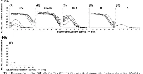
rHV was used as a negative control for the binding assay (12). As depicted in Fig. 2, the binding of r124 occurred in a dose-dependent manner and high optical density values were ob-served up to 1:6,400 saliva dilutions (Fig. 2A, B, and D). The binding of r124 to the saliva containing B antigens was low, probably due to the inefficient binding ability of the terminal galactose of the type B epitope, as described previously (Fig. 2C) (12, 14, 15, 24). Although the saliva included A antigens and they were recognized by NV/68, the binding of r124 to the saliva from donor no. 26 was weak (Fig. 2E). In addition, four saliva samples containing H and A antigens had weak binding ability (Fig. 2A, no. 25 and 27 to 29). None of the saliva bound to rHV, as described previously (Fig. 2F) (12, 15).
Identification of incomplete nonsecretors.To further char-acterize these low binding abilities, the amounts of HBGAs in saliva samples were measured by semiquantitative hemagglu-tination inhibition assay. The phenotypes according to the amounts of HBGAs are summarized in Table 3. The saliva was grouped into five categories, characterized by A, AB, B, and O
secretor status and nonsecretor status. These results were con-sistent with the results shown in Table 2. The phenotype of secretors was characterized by high secretion of H, A, or B antigens in saliva, as observed for donors 1 to 24. Each group of secretors was characterized by antigen content: H and A antigens for phenotype A; H, A, and B antigens for the AB phenotype; H and B antigens for phenotype B; and H antigen for phenotype O. The phenotype of the nonsecretors was char-acterized by low secretion of ABH antigens in saliva as ob-served for donors 25 to 29. These nonsecretors differ from those observed in Europe, because Japanese nonsecretors se-crete a small amount of either H, A, or B antigen or both (23, 41). Therefore, they are described as incomplete nonsecretors. Measurement of the HBGAs enabled us to clarify the phenotypic difference between donors 1 to 5 and donors 25 and 27 to 29.
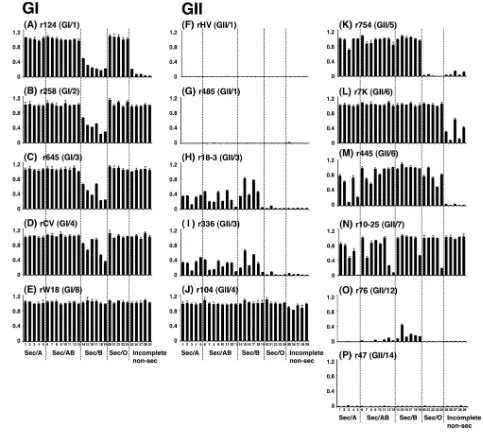
Binding of GI NoV VLPs to HBGAs.The binding between GI VLPs and saliva was analyzed by using an ELISA-based binding assay. The saliva dilution of 1:1,600 was used because the strength of the binding to r124 was observed clearly at this dilution (Fig. 2). GI/1 r124 bound more efficiently to saliva from type O, A, and AB secretors than to that from type B secretors and incomplete nonsecretors (Fig. 3A), indicating that GI/1 r124 recognizes the H and A antigens. These results were consistent with the observation shown in Fig. 2. Three GI VLPs, GI/2 r258, GI/3 r645, and GI/4 rCV, had the same binding profiles and high binding activities for type O, A, and AB secretors and incomplete nonsecretors (Fig. 3B, C, and D). These results indicated that these three GI strains recognize the Leaantigen as well as the H and A antigens, because the
[image:5.585.44.284.89.171.2]saliva of the nonsecretors is characterized by high secretion of
TABLE 2. Detection of soluble ABH antigens in saliva by hemagglutination inhibition assay
Donor no. Grouping
Presence of HBGA
H A B
1–5, 25, 27–29 H/A ⫹ ⫹ ⫺
6–13 H/A/B ⫹ ⫹ ⫹
14–19 H/B ⫹ ⫺ ⫹
20–24 H ⫹ ⫺ ⫺
26 A ⫺ ⫹ ⫺
FIG. 2. Dose-dependent binding of GI/1 r124 (A to E) or GII/1 rHV (F) to saliva. Serially twofold-diluted saliva samples, at 50- to 102,400-fold dilution, were used to coat the microplates. Convalescent-phase serum from a patient infected with the GI/2 258 strain and r258 were used for the internal standard. Coating buffer was used for the blank. The binding of the VLPs was detected by using polyclonal rabbit anti-VLPs as described in Materials and Methods.
10760 SHIRATO ET AL. J. VIROL.
on November 8, 2019 by guest
http://jvi.asm.org/
[image:5.585.41.542.428.687.2]Leaantigen in saliva (40). GI/8 rW18 bound efficiently to all
saliva samples regardless of secretor phenotype or blood type, indicating that GI/8 rW18 recognized the H, A, B, and Lea
antigens (Fig. 3E). To further examine which HBGAs are involved in GI NoV attachment, a carbohydrate-VLP binding assay was performed with seven multivalent carbohydrate-biotin reagents: H type 1, H type 2, H type 3, tri-A, tri-B, Lea,
and Leb. Basically, no discrepancy was found between the
carbohydrate- and saliva-VLP binding assays, although the sensitivity of the former was higher than that of the latter (Fig. 4A to E; Table 4). Moreover, the following observations were obtained from the carbohydrate-VLP binding assay: (i) the results provide direct evidence that GI/2, GI/3, GI/4, and GI/8 VLPs recognized the Leacarbohydrate (Fig. 4B, C, D, and E);
(ii) GI/1 and GI/2 VLPs bound to H type 3 (Fig. 4A and B); and (iii) GI/1, GI/4, and GI/8 VLPs recognized the Leb
carbo-hydrate (Fig. 4D and E). We concluded that the binding prop-erties of GI NoV VLPs for HBGAs were variable. However, four of five genotypes, GI/2, GI/3, GI/4, and GI/8, had high abilities to bind to the Leaantigen. This was a unique
charac-teristic of GI which was not seen with GII, as mentioned below. Binding of GII NoV VLPs to HBGAs. Similarly, a binding assay was performed with 11 GII VLPs. Two GII/1 VLPs (rHV and r485) and one GII/14 VLP (r47) did not bind to any of the saliva samples (Fig. 3F, G, and P), indicating that these three
GII strains did not recognize the HBGAs in saliva. Two GII/3 VLPs (r18-3 and r336) and one GII/5 VLP (r754) bound to saliva samples from type A, AB, and B secretors, whereas they bound neither to type O secretors nor to incomplete nonse-cretors (Fig. 3H, I, and K). The results indicate that these strains recognized the A and B antigens. GII/4 r104 and GII/7 r10-25 bound to all saliva samples (Fig. 3J and N), indicating that these two GII strains recognized the H, A, B, and Lea
antigens. Two GII/6 VLPs, r7K and r445, had high binding ability for all blood-type secretors (Fig. 3L and M), indicating that these GII/6 strains recognize the H, A, or B antigen. GII/12 r76 bound to saliva from type AB and B secretors, although the binding levels were extremely low (Fig. 3O), in-dicating that r76 attached weakly to the B antigen. The saliva-VLP binding assay clearly showed that the strains of the same genetic clusters, rHV and r485 in GII/1, r18-3 and 336 in GII/3, and r7K and r445 in GII/6, have the same carbohydrate-bind-ing patterns. To address which HBGAs are involved in the GII NoV attachment, a carbohydrate-VLP binding assay was per-formed as described above. The sensitivity was higher in the saliva- than the carbohydrate-VLP binding assay, and no dis-crepancy was found between the two binding assays, as was also the case with the GI assays (Fig. 4F to P and Table 4). Five other observations from the carbohydrate-VLP binding assay were made: (i) GII/3, GII/4, GII/6, and GII/7 VLPs bound to H type 3 (Fig. 4J, L, M, and N); (ii) GII/7 VLPs recognized the Lea carbohydrate (Fig. 4N); (iii) r104 hardly recognized the
Lea carbohydrate (Fig. 4J), although the strain bound to
in-complete nonsecretors strongly (Fig. 3J); (iv) r7K had binding activities for the Leacarbohydrate (Fig. 4L) (it had been hard
to predict the r7K binding to the Lea antigen based on the
saliva-VLP binding assay due to low binding activity for incom-plete nonsecretors [Fig. 3L]); and (v) GII/4, GII/6, and GII/7 VLPs recognized the Lebcarbohydrate (Fig. 4J, L, and N). We
concluded that the binding properties of GII NoV VLPs for HBGAs were variable. However, there was no strain which strongly bound to the Leaantigen, although the strains of GII/6
[image:6.585.43.286.79.425.2]and GII/7 had weak binding abilities for this antigen. Both␣1,2-fucosyl residue and carbohydrate core structures are needed for binding to HBGAs.To investigate the effect of the terminal saccharide residue(s), the binding activities between VLPs and synthetic type 1, 2, and 3 carbohydrates and synthetic H disaccharides that do not include the core structures were exam-ined. As depicted in Fig. 4, none of the VLPs bound to synthetic type 1, 2, and 3 disaccharides, although GI/1, GI/2, GI/3, GII/3, GII/4, GII/6, and GII/7 VLPs had binding activities for H type 1, 2, or 3 (Fig. 4A to C, H to J, and L to N). These results suggested that the terminal␣1,2-fucosyl residue on those H trisaccharides is one of the determinants responsible for the NoV binding. More-over, NoV VLPs recognized the components in the core struc-tures, because (i) seven VLP genotypes, including GI/2, GI/3, GII/3, GII/6, and GII/7 VLPs, unequally bound H type 1, 2, and 3 trisaccharides (Fig. 4B, C, H, I, and L to N); (ii) the binding abilities of those VLPs for H disaccharides were undetectable (Fig. 4B, C, H, I, and L to N); and (iii) the binding abilities of GI/1 r124 and GII/4 r104 for H disaccharides were lower than those for the H type 1, 2, and 3 trisaccharides (Fig. 4A and J). Next, we addressed whether the VLPs bound to synthetic A and B disac-charides that lack the␣1,2-fucosyl residue. As shown in Fig. 4A to E, J, K, L, and N, those strains did not bind to the synthetic A and
TABLE 3. Semiquantitation of soluble ABH
Secretion status of donor HBGA secretion phenotype Donor no.
Titer of HBGA
H A B
Secretor A 1 32 128 0
2 32 64 0
3 64 64 0
4 128 ⬎256 0
5 32 128 0
AB 6 4 128 8
7 16 128 8
8 8 ⬎256 1
9 4 128 8
10 16 ⬎256 32
11 8 ⬎256 8
12 64 128 64
13 64 128 ⬎256
B 14 16 0 128
15 8 0 ⬎256
16 16 0 ⬎256
17 8 0 ⬎256
18 16 0 128
19 16 0 ⬎256
O 20 ⬎256 0 0
21 ⬎256 0 0
22 128 0 0
23 ⬎256 0 0
24 ⬎256 0 0
Incomplete 25 16 16 0
nonsecretor 26 0 1 0
27 4 8 0
28 4 1 0
29 4 1 0
on November 8, 2019 by guest
http://jvi.asm.org/
B disaccharides, with the exception of GI/2 r258. Therefore, be-sides the r258 strain, the␣1,2-fucosyl residue is one of the deter-minants responsible for NoV binding, not only to H antigens but also to A and B antigens. We concluded that both the␣1,2-fucosyl residue and core structures are needed for the binding of VLPs to the HBGAs.
NoV VLPs distinguish between type 1 and type 2 carbohy-drates.Six monovalent carbohydrate-biotin reagents, H type 1, H type 2, A type 1, A type 2, B type 1, and B type 2 (Fig. 1A), were used in SPR Biacore experiments to determine whether NoV VLPs distinguish between type 1 and type 2 chains. These oligosaccharides were prepared using human recombinant gly-cosyltransferases. Unlike the organic synthesis of oligosaccha-rides, the reaction using glycosyltransferases is quite clear and efficient, and the monovalent carbohydrate-biotin reagents
[image:7.585.52.535.68.501.2]were identified by matrix-assisted laser desorption ionization-time-of-flight mass spectrometry (Reflex IV; Bruker-Daltonik GmbH) (data not shown). Six strains, including GI/1, GI/2, GI/3, GI/4, GI/8, and GII/4 strains, bound to the monovalent carbohydrate-biotin reagents (Fig. 5), whereas 10 strains, in-cluding GII/1, GII/3, GII/4, GII/5, GII/6, GII/7, GII/12, and GII/14 strains, did not (data not shown). Basically, no discrep-ancy was found between the results of the ELISA-based bind-ing assay with multivalent biotinylated reagents and those of the SPR Biacore experiments with monovalent biotinylated reagents, although the sensitivity of the former was higher than that of the latter (Fig. 4 and 5). GI/1, GI/2, GI/3, and GI/4 VLPs bound to A, but not to B, carbohydrates in the Biacore experiments, whereas GI/8 and GII/4 VLPs bound to both A and B carbohydrates. These results were consistent with the
FIG. 3. Binding between VLPs and saliva samples. The saliva samples were tested at a dilution of 1:1,600. The binding of the VLPs was detected by polyclonal rabbit anti-VLPs as described in Materials and Methods. The experiments were performed in triplicate and reproduced at least twice. Each data point represents the mean value (with error bar).
10762 SHIRATO ET AL. J. VIROL.
on November 8, 2019 by guest
http://jvi.asm.org/
observation shown with the ELISA-based binding assay. More-over, the following three observations were obtained from the Biacore experiments: (i) GI/3 and GII/4 VLPs were more ef-ficiently bound to H type 2 than to H type 1 tetrasaccharides (Fig. 5C and F); (ii) five GI VLPs and r104 were more effi-ciently bound to A type 2 than to A type 1 pentasaccharides (Fig. 5G to L); and (iii) GI/8 and GII/4 VLPs were more efficiently bound to B type 2 than to B type 1 pentasaccharides (Fig. 5Q and R). These results indicate that NoV VLPs are able to distinguish between type 1 and type 2 carbohydrates. Moreover, type 1 carbohydrates are likely to bind more tightly to NoV VLPs than do the type 2 carbohydrates, because the dissociation of GII/4 r104 was slower in B type 1 than B type 2 (Fig. 5R). To further characterize this strong binding ability of type 1 carbohydrates, the binding between r104 and the six
monovalent carbohydrate-biotin reagents was examined using an ELISA-based binding assay. The bindings of GII/4 VLPs were stronger in B type 1 pentasaccharides than in B type 2 pentasaccharides (Fig. 6). The Biacore assay allowed us to visualize each binding step in the 120-s reaction time, whereas ELISA allowed us to visualize only the last step in a total reaction time of about 7.5 h. Therefore, these results indicated that NoV VLPs bind more tightly to type 1 carbohydrates than to type 2 carbohydrates.
DISCUSSION
To date, the interaction between carbohydrates and 16 NoV VLPs, including 5 GI and 11 GII VLPs, has been described, with the conclusion that the carbohydrate-binding properties
FIG. 4. Binding between VLPs and synthetic histo-blood group carbohydrates. The multivalent carbohydrate-biotin reagents conjugated to polyacrylamide were tested at a concentration of 2.5g/ml. H type 1 trisaccharides and r124 were used for the internal standard. Tris-buffered saline was used for the blank. The binding of the VLPs was detected by polyclonal rabbit anti-VLPs as described in Materials and Methods. The experiments were performed in duplicate and reproduced at least twice. Each data point represents the mean value (with error bar).
on November 8, 2019 by guest
http://jvi.asm.org/
of NoV VLPs vary (12–15, 39). Our experiments with 16 VLPs from 5 GI genotypes and 8 GII genotypes have confirmed and extended these findings: not only the outmost sugar residues but also the type 1 and 2 core structures are important for VLP recognition.
In this study, we investigated the function of the core struc-tures in the binding between NoV and H/A/B antigens. The GI/1, GI/2, GI/3, GI/4, GI/8, GII/3, GII/4, GII/6, and GII/7 VLPs were able to distinguish between type 1 and type 2 carbohydrates (Fig. 4 and 5). Moreover, the type 1 carbohy-drates bound more tightly to NoV VLPs than did the type 2 carbohydrates, as indicated in the following results: (i) the dissociation of GII/4 VLPs from B type 1 pentasaccharides was slower than that from B type 2 pentasaccharides in the Biacore experiments (Fig. 5), and (ii) the binding of GII/4 VLPs to B type 1 pentasaccharides was stronger than that to B type 2 pentasaccharides in the ELISA experiments (Fig. 6). Avian and equine influenza viruses are known to preferentially bind to the terminal sialic acid␣2-3Gal (SA␣2-3Gal) linkage, while human influenza viruses preferentially bind to the SA␣2-6Gal linkage (6, 35, 36), affording a major impact on the host spec-ificity in the infection of influenza viruses. A similar relation-ship may exist between NoV carbohydrate recognition and its tissue specificity, because the binding of rNV/68 to the gas-troduodenal junction has been reported to correlate with the presence of the H type 1 antigen but not that of the H type 2 antigen (26).
The importance of the terminal␣1,2-fucosyl residue in the binding between NoV and H/A/B antigens has been analyzed in detail with the GI/1 NV/68 and GII/4 VA387 strains (3, 12, 26). In this study, we confirmed these findings with GI/1 and GII/4 strains. Moreover, we revealed that other genotypes which bind to H, A, and/or B antigen also require the␣ 1,2-fucosyl residue for the binding. Although the GI/1, GI/2, GI/3, GII/3, GII/4, GII/6, and GII/7 VLPs bound to H type 1, type 2,
and/or type 3 carbohydrates (Fig. 4A to C, H to J, and L to N), none of these VLPs bound to type 1, type 2, and type 3 car-bohydrates (Fig. 4A to C, H to J, and L to N), suggesting that the terminal ␣1,2-fucosyl residue on those H trisaccharides may be the determinant responsible for the binding between NoV and the H antigen. Moreover, as shown in Fig. 4A to E, J to L, and N, GI/1, GI/2, GI/3, GI/4, GI/8, GII/4, GII/5, GII/6, and GII/7 VLPs, which bound to A and/or B trisaccharides, did not bind to A and B disaccharides, with the exception of GI/2 VLPs. Therefore, besides the GI/2 strain, the␣1,2-fucosyl res-idue is the determinant for NoV binding not only to H antigens but also to A and B antigens. We had predicted that NoV would require additional terminal sugars, such as␣1,2-fucose, for HBGA recognition. Unexpectedly, however, we found that GI/2 r258 recognized synthetic A disaccharide, which does not include fucose (Fig. 4B). Therefore, both core structures and additional terminal sugars may contribute to the virus-carbo-hydrate interaction.
[image:9.585.46.541.82.293.2]In a previous work, the strain specificities of NoV-HBGA binding were reported (15). Those authors concluded that NoV-HBGA binding patterns could be classified into two groups, an A/B-binding group and a Lewis-binding group, and that there was no correlation between the binding patterns and the genogroup. Our results were consistent with their results when GI/1, GI/2, GII/1, GII/4, and GII/5 VLPs were used. However, GI/3 and GII/3 VLPs gave different results. This may be due to the difference in the amino acid residues at the putative carbohydrate-binding sites. The amino acid residues 267N, 291R, 292G, 293D, 300N, 322D, 327D, 328W, 329H, 331N, 333T, 334Q, 335F, 339S, 341T, 364I, 368N, 373L, 374S, 375W, 377S, and 430A (NV/68 numbering) on the P2 domain were predicted to be important for HBGA binding (3, 4, 42). There were no differences in residues between our GI/1, GI/2, GII/1, GII/4, and GII/5 strains and their corresponding strains, whereas some different residues were found in the GI/3 and
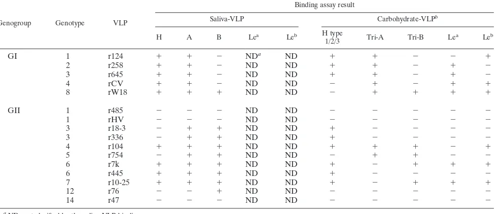
TABLE 4. HBGA recognition predicted by carbohydrate- and saliva-VLP binding assay
Genogroup Genotype VLP
Binding assay result
Saliva-VLP Carbohydrate-VLPb
H A B Lea
Leb H type
1/2/3 Tri-A Tri-B Le
a
Leb
GI 1 r124 ⫹ ⫹ ⫺ NDa ND ⫹ ⫹ ⫺ ⫺ ⫹
2 r258 ⫹ ⫹ ⫺ ND ND ⫹ ⫹ ⫺ ⫹ ⫺
3 r645 ⫹ ⫹ ⫺ ND ND ⫹ ⫹ ⫺ ⫹ ⫺
4 rCV ⫹ ⫹ ⫺ ND ND ⫺ ⫹ ⫺ ⫹ ⫹
8 rW18 ⫹ ⫹ ⫹ ND ND ⫺ ⫹ ⫹ ⫹ ⫹
GII 1 r485 ⫺ ⫺ ⫺ ND ND ⫺ ⫺ ⫺ ⫺ ⫺
1 rHV ⫺ ⫺ ⫺ ND ND ⫺ ⫺ ⫺ ⫺ ⫺
3 r18-3 ⫺ ⫹ ⫹ ND ND ⫹ ⫺ ⫺ ⫺ ⫺
3 r336 ⫺ ⫹ ⫹ ND ND ⫹ ⫺ ⫺ ⫺ ⫺
4 r104 ⫹ ⫹ ⫹ ND ND ⫹ ⫹ ⫹ ⫺ ⫹
5 r754 ⫺ ⫹ ⫹ ND ND ⫺ ⫹ ⫹ ⫺ ⫺
6 r7k ⫹ ⫹ ⫹ ND ND ⫹ ⫺ ⫹ ⫹ ⫹
6 r445 ⫹ ⫹ ⫹ ND ND ⫹ ⫺ ⫺ ⫺ ⫺
7 r10-25 ⫹ ⫹ ⫹ ND ND ⫹ ⫺ ⫹ ⫹ ⫹
12 r76 ⫺ ⫺ ⫹ ND ND ⫺ ⫺ ⫺ ⫺ ⫺
14 r47 ⫺ ⫺ ⫺ ND ND ⫺ ⫺ ⫺ ⫺ ⫺
aND, not clarified by the saliva-VLP binding assay.
bRelative optical densities (optical density at 492 nm/internal standard) greater than 0.178 were considered positive in the carbohydrate-VLP binding assay.
10764 SHIRATO ET AL. J. VIROL.
on November 8, 2019 by guest
http://jvi.asm.org/
GII/3 strains. The finding that a single amino acid change in the P domain resulted in a change in the pattern of HBGA binding (42) could explain the inconsistency of the results.
Differences in the reactivities between saliva samples and synthetic carbohydrates may be due to structural differences between the synthetic products and authentic antigens, which are thought to be present on mucin or mucin-like molecules (15, 39). In our experiment, the sensitivity of the saliva-VLP binding assay was better than that of the carbohydrate-VLP binding assay. On the other hand, the carbohydrate-VLP bind-ing assay demonstrated H type 3, Lea, and Lebantigen
recog-nition by NoV, which could not be detected with the saliva-VLP binding assay. This is why we have performed the binding assay with two ELISA methods.
[image:10.585.47.542.68.451.2]In Biacore assays, we used 11 GII strains; however, only one strain (GII/4 genotype) revealed binding (Fig. 5). The remaining 10 GII strains did not bind to either A or B pentasaccharides (Fig.
[image:10.585.53.273.538.660.2]FIG. 6. Dose-dependent binding of GII/4 r104 to the monovalent biotin reagents. Serially twofold-diluted carbohydrate-biotin reagents, at 1.0 to 0.016 pmol/l, were used to coat streptavidin-precoated plates. Tris-buffered saline was used for the blank. The binding of the VLPs was detected by polyclonal rabbit anti-VLPs as described in Materials and Methods. The optical densities at 492 nm are plotted against the dilutions.
FIG. 5. Interaction between NoV and synthetic histo-blood group carbohydrates. The monovalent carbohydrate-biotin reagents were captured on a streptavidin-coated sensor chip as described in Materials and Methods. Sensorgrams show the binding of the VLPs to immobilized carbohydrates, H type 1 and type 2 (A to F), A type 1 and type 2 (G to L), or B type 1 and type 2 (M to R). At 180 s, 401 of the VLP was injected at a flow rate of 20l/min and was replaced by the running buffer at 300 s. The binding curves of 180 to 300 s showed the association, whereas those of 300 to 500 s showed the dissociation. The binding curves between VLPs and two different carbohydrates were compared by overlaying the sensorgrams obtained on each surface. The experiments were reproduced at least twice. Theyaxis indicates the resonance signal as shown in resonance units (RU).
on November 8, 2019 by guest
http://jvi.asm.org/
5). GII/4 VLPs may recognize complicated carbohydrate struc-tures as the authentic antigens and pentasaccharides, as shown in Fig. 3 and 5, whereas the remaining 10 GII viruses may only recognize simple structures, as shown in Fig. 4.
In conclusion, it is obvious that HBGAs are important fac-tors in determining tissue specificity, although it is still unclear whether the HBGAs function as the primary receptor or en-hance NoV infectivity and/or attachment to a common cellular receptor. GII/4 is known to be a global epidemic genotype (25, 29, 30) and to bind more HBGAs than other strains (15) (confirmed in this study). This characteristic may be linked with the worldwide transmission of GII/4 strains. Lewis HBGAs are also carbohydrate antigens expressed in the epi-thelial cells of gastrointestinal tracts (2, 33, 37). Interestingly, some strains of GI and GII bound to Leaexpressed by
nonse-cretors. This means that NoV can infect individuals regardless of secretor phenotype. We are going to synthesize various Lewis-type glycans to further characterize NoV-Lewis HBGA binding abilities. In this study, the linkages and carbohydrate core structures appeared to be important in NoV-carbohydrate interaction. Since NoV forms many antigenically diverse groups, identification of the common NoV binding epitopes on host cells, if any, should be useful for the development of possible antiviral agents.
ACKNOWLEDGMENTS
We thank Katsuo Natori, Tsutomu Kageyama (National Institute of Infectious Diseases, Tokyo, Japan), and Kunio Kamata (Denka-Seiken Co., Niigata, Japan) for their valuable assistance.
This work was supported in part by a grant for Research on Food Safety from the Ministry of Health, Labor and Welfare of Japan and by the R&D Project of the Industrial Science and Technology Frontier Program, supported by the New Energy and Industrial Technology Development Organization.
REFERENCES
1.Atmar, R. L., and M. K. Estes.2001. Diagnosis of noncultivatable gastroen-teritis viruses, the human caliciviruses. Clin. Microbiol. Rev.14:15–37. 2.Bjork, S., M. E. Breimer, G. C. Hansson, K. A. Karlsson, and H. Leffler.
1987. Structures of blood group glycosphingolipids of human small intestine. A relation between the expression of fucolipids of epithelial cells and the ABO, Le and Se phenotype of the donor. J. Biol. Chem.262:6758–6765. 3.Cao, S., Z. Lou, M. Tan, Y. Chen, Y. Liu, Z. Zhang, X. C. Zhang, X. Jiang,
X. Li, and Z. Rao.2007. Structural basis for the recognition of blood group trisaccharides by norovirus. J. Virol.81:5949–5957.
4.Chakravarty, S., A. M. Hutson, M. K. Estes, and B. V. Prasad.2005. Evo-lutionary trace residues in noroviruses: importance in receptor binding, an-tigenicity, virion assembly, and strain diversity. J. Virol.79:554–568. 5.Cheetham, S., M. Souza, R. McGregor, T. Meulia, Q. Wang, and L. J. Saif.
2007. Binding patterns of human norovirus-like particles to buccal and in-testinal tissues of gnotobiotic pigs in relation to A/H histo-blood group antigen expression. J. Virol.81:3535–3544.
6.Connor, R. J., Y. Kawaoka, R. G. Webster, and J. C. Paulson.1994. Receptor specificity in human, avian, and equine H2 and H3 influenza virus isolates. Virology205:17–23.
7.Dabelsteen, E., P. Vedtofte, S. I. Hakomori, and W. W. Young.1982. Car-bohydrate chains specific for blood group antigens in differentiation of hu-man oral epithelium. J. Investig. Dermatol.79:3–7.
8.Estes, M. K., J. M. Ball, S. E. Crawford, C. O’Neal, A. A. Opekun, D. Y. Graham, and M. E. Conner.1997. Virus-like particle vaccines for mucosal immunization. Adv. Exp. Med. Biol.412:387–395.
9.Grubb, R.1948. Correlation between Lewis blood group and secretor char-acter in man. Nature162:933.
10.Hakomori, S.1981. Blood group ABH and Ii antigens of human erythro-cytes: chemistry, polymorphism, and their developmental change. Semin. Hematol.18:39–62.
11.Hansman, G. S., K. Natori, H. Shirato-Horikoshi, S. Ogawa, T. Oka, K. Katayama, T. Tanaka, T. Miyoshi, K. Sakae, S. Kobayashi, M. Shinohara, K. Uchida, N. Sakurai, K. Shinozaki, M. Okada, Y. Seto, K. Kamata, N. Nagata, K. Tanaka, T. Miyamura, and N. Takeda.2006. Genetic and anti-genic diversity among noroviruses. J. Gen. Virol.87:909–919.
12.Harrington, P. R., L. Lindesmith, B. Yount, C. L. Moe, and R. S. Baric.2002. Binding of Norwalk virus-like particles to ABH histo-blood group antigens is blocked by antisera from infected human volunteers or experimentally vac-cinated mice. J. Virol.76:12335–12343.
13.Harrington, P. R., J. Vinje, C. L. Moe, and R. S. Baric.2004. Norovirus capture with histo-blood group antigens reveals novel virus-ligand interac-tions. J. Virol.78:3035–3045.
14.Huang, P., T. Farkas, S. Marionneau, W. Zhong, N. Ruvoen-Clouet, A. L. Morrow, M. Altaye, L. K. Pickering, D. S. Newburg, J. LePendu, and X. Jiang.2003. Noroviruses bind to human ABO, Lewis, and secretor histo-blood group antigens: identification of 4 distinct strain-specific patterns. J. Infect. Dis.188:19–31.
15.Huang, P., T. Farkas, W. Zhong, M. Tan, S. Thornton, A. L. Morrow, and X. Jiang.2005. Norovirus and histo-blood group antigens: demonstration of a wide spectrum of strain specificities and classification of two major binding groups among multiple binding patterns. J. Virol.79:6714–6722. 16.Hutson, A. M., R. L. Atmar, D. Y. Graham, and M. K. Estes.2002. Norwalk
virus infection and disease is associated with ABO histo-blood group type. J. Infect. Dis.185:1335–1337.
17.Hutson, A. M., R. L. Atmar, D. M. Marcus, and M. K. Estes.2003. Norwalk virus-like particle hemagglutination by binding to H histo-blood group an-tigens. J. Virol.77:405–415.
18.Isshiki, S., A. Togayachi, T. Kudo, S. Nishihara, M. Watanabe, T. Kubota, M. Kitajima, N. Shiraishi, K. Sasaki, T. Andoh, and H. Narimatsu.1999. Cloning, expression, and characterization of a novel UDP-galactose:beta-N-acetylglucosamine beta1,3-galactosyltransferase (beta3Gal-T5) responsible for synthesis of type 1 chain in colorectal and pancreatic epithelia and tumor cells derived therefrom. J. Biol. Chem.274:12499–12507.
19.Ito, H., A. Kameyama, T. Sato, M. Sukegawa, H. K. Ishida, and H. Narimatsu. 2007. Strategy for the fine characterization of glycosyltransferase specificity using isotopomer assembly. Nat. Methods4:577–582.
20.Kageyama, T., M. Shinohara, K. Uchida, S. Fukushi, F. B. Hoshino, S. Kojima, R. Takai, T. Oka, N. Takeda, and K. Katayama.2004. Coexistence of multiple genotypes, including newly identified genotypes, in outbreaks of gastroenteritis due to Norovirus in Japan. J. Clin. Microbiol.42:2988–2995. 20a.Kamata, K., K. Shinozaki, M. Okada, Y. Seto, S. Kobayashi, K. Sakae, M. Oseto, K. Natori, H. Shirato-Horikoshi, K. Katayama, T. Tanaka, N. Takeda, and K. Taniguchi.2005. Expression and antigenicity of virus-like particles of norovirus and their application for detection of noroviruses in stool samples. J. Med. Virol.76:129–136.
21.Kaneko, M., S. Nishihara, N. Shinya, T. Kudo, H. Iwasaki, T. Seno, Y. Okubo, and H. Narimatsu.1997. Wide variety of point mutations in the H gene of Bombay and para-Bombay individuals that inactivate H enzyme. Blood90:839–849.
22.Kapikian, A. Z.1996. Overview of viral gastroenteritis. Arch. Virol. Suppl. 12:7–19.
22a.Kitamoto, N., T. Tanaka, K. Natori, N. Takeda, S. Nakata, X. Jiang, and M. K. Estes.2002. Cross-reactivity among several recombinant calicivirus virus-like particles (VLPs) with monoclonal antibodies obtained from mice immunized orally with one type of VLP. J. Clin. Microbiol.40:2459–2465. 22b.Kobayashi, S., K. Sakae, Y. Suzuki, K. Shinozaki, M. Okada, H. Ishiko, K.
Kamata, K. Suzuki, K. Natori, T. Miyamura, and N. Takeda.2000. Molec-ular cloning, expression, and antigenicity of Seto virus belonging to geno-group I Norwalk-like viruses. J. Clin. Microbiol.38:3492–3494.
22c.Kobayashi, S., K. Sakae, K. Natori, N. Takeda, T. Miyamura, and Y. Suzuki. 2000. Serotype-specific antigen ELISA for detection of Chiba virus in stools. J. Med. Virol.62:233–238.
22d.Kobayashi, S., K. Sakae, Y. Suzuki, H. Ishiko, K. Kamata, K. Suzuki, K. Natori, T. Miyamura, and N. Takeda.2000. Expression of recombinant capsid proteins of chitta virus, a genogroup II Norwalk virus, and develop-ment of an ELISA to detect the viral antigen. Microbiol. Immunol.44:687– 693.
23.Kudo, T., H. Iwasaki, S. Nishihara, N. Shinya, T. Ando, I. Narimatsu, and H. Narimatsu.1996. Molecular genetic analysis of the human Lewis histo-blood group system. II. Secretor gene inactivation by a novel single missense mutation A385T in Japanese nonsecretor individuals. J. Biol. Chem.271: 9830–9837.
24.Lindesmith, L., C. Moe, S. Marionneau, N. Ruvoen, X. Jiang, L. Lindblad, P. Stewart, J. LePendu, and R. Baric.2003. Human susceptibility and resis-tance to Norwalk virus infection. Nat. Med.9:548–553.
25.Lopman, B., H. Vennema, E. Kohli, P. Pothier, A. Sanchez, A. Negredo, J. Buesa, E. Schreier, M. Reacher, D. Brown, J. Gray, M. Iturriza, C. Galli-more, B. Bottiger, K. O. Hedlund, M. Torven, C. H. von Bonsdorff, L. Maunula, M. Poljsak-Prijatelj, J. Zimsek, G. Reuter, G. Szucs, B. Melegh, L. Svennson, Y. van Duijnhoven, and M. Koopmans.2004. Increase in viral gastroenteritis outbreaks in Europe and epidemic spread of new norovirus variant. Lancet363:682–688.
26.Marionneau, S., N. Ruvoen, B. Le Moullac-Vaidye, M. Clement, A. Cailleau-Thomas, G. Ruiz-Palacois, P. Huang, X. Jiang, and J. Le Pendu.2002. Norwalk virus binds to histo-blood group antigens present on gastroduode-nal epithelial cells of secretor individuals. Gastroenterology122:1967–1977. 27.Mollicone, R., J. Bara, J. Le Pendu, and R. Oriol.1985. Immunohistologic
10766 SHIRATO ET AL. J. VIROL.
on November 8, 2019 by guest
http://jvi.asm.org/
pattern of type 1 (Lea, Leb) and type 2 (X, Y, H) blood group-related antigens in the human pyloric and duodenal mucosae. Lab. Investig.53:219– 227.
28.Narimatsu, H., S. Sinha, K. Brew, H. Okayama, and P. K. Qasba.1986. Cloning and sequencing of cDNA of bovine N-acetylglucosamine (beta 1-4)galactosyltransferase. Proc. Natl. Acad. Sci. USA83:4720–4724. 29.Noel, J. S., R. L. Fankhauser, T. Ando, S. S. Monroe, and R. I. Glass.1999.
Identification of a distinct common strain of “Norwalk-like viruses” having a global distribution. J. Infect. Dis.179:1334–1344.
30.Okada, M., T. Ogawa, I. Kaiho, and K. Shinozaki.2005. Genetic analysis of noroviruses in Chiba Prefecture, Japan, between 1999 and 2004. J. Clin. Microbiol.43:4391–4401.
31.Oriol, R.1990. Genetic control of the fucosylation of ABH precursor chains. Evidence for new epistatic interactions in different cells and tissues. J. Im-munogenet.17:235–245.
32.Oriol, R., J. Le Pendu, and R. Mollicone.1986. Genetics of ABO, H, Lewis, X and related antigens. Vox Sang.51:161–171.
33.Orntoft, T. F., E. H. Holmes, P. Johnson, S. Hakomori, and H. Clausen. 1991. Differential tissue expression of the Lewis blood group antigens: en-zymatic, immunohistologic, and immunochemical evidence for Lewis a and b antigen expression in Le(a-b-) individuals. Blood77:1389–1396.
34.Rockx, B. H., H. Vennema, C. J. Hoebe, E. Duizer, and M. P. Koopmans. 2005. Association of histo-blood group antigens and susceptibility to noro-virus infections. J. Infect. Dis.191:749–754.
35.Rogers, G. N., and J. C. Paulson.1983. Receptor determinants of human and animal influenza virus isolates: differences in receptor specificity of the H3 hemagglutinin based on species of origin. Virology127:361–373. 36.Rogers, G. N., T. J. Pritchett, J. L. Lane, and J. C. Paulson.1983. Differential
sensitivity of human, avian, and equine influenza A viruses to a glycoprotein inhibitor of infection: selection of receptor specific variants. Virology131: 394–408.
37.Sakamoto, J., K. Furukawa, C. Cordon-Cardo, B. W. Yin, W. J. Rettig, H. F. Oettgen, L. J. Old, and K. O. Lloyd.1986. Expression of Lewisa, Lewisb, X,
and Y blood group antigens in human colonic tumors and normal tissue and in human tumor-derived cell lines. Cancer Res.46:1553–1561.
38.Shiraishi, N., A. Natsume, A. Togayachi, T. Endo, T. Akashima, Y. Yamada, N. Imai, S. Nakagawa, S. Koizumi, S. Sekine, H. Narimatsu, and K. Sasaki. 2001. Identification and characterization of three novel beta 1,3-N-acetyl-glucosaminyltransferases structurally related to the beta 1,3-galactosyltrans-ferase family. J. Biol. Chem.276:3498–3507.
39.Shirato-Horikoshi, H., S. Ogawa, T. Wakita, N. Takeda, and G. S. Hansman. 2007. Binding activity of norovirus and sapovirus to histo-blood group anti-gens. Arch. Virol.152:457–461.
40.Sidmann, F. K. (ed.).1981. Appendix 2: saliva testing for ABH and Lewis, p. 122–123.InTechnical Manual of the American Association of Blood Banks, 8th ed. J. B. Lippincott, Philadelphia, PA.
41.Soejima, M., and Y. Koda.2005. Molecular mechanisms of Lewis antigen expression. Leg. Med. (Tokyo)7:266–269.
41a.Tamura, M., K. Natori, M. Kobayashi, T. Miyamura, and N. Takeda.2000. Interaction of recombinant Norwalk virus particles with the 105-kilodalton cellular binding protein, a candidate receptor molecule for virus attachment. J. Virol.74:11589–11597.
42.Tan, M., P. Huang, J. Meller, W. Zhong, T. Farkas, and X. Jiang.2003. Mutations within the P2 domain of norovirus capsid affect binding to human histo-blood group antigens: evidence for a binding pocket. J. Virol. 77: 12562–12571.