Cochrane
Database of Systematic Reviews
Clinically-indicated replacement versus routine replacement
of peripheral venous catheters (Review)
T A B L E O F C O N T E N T S
1 HEADER . . . .
1 ABSTRACT . . . .
2 PLAIN LANGUAGE SUMMARY . . . .
3 SUMMARY OF FINDINGS FOR THE MAIN COMPARISON . . . .
5 BACKGROUND . . . .
6 OBJECTIVES . . . .
6 METHODS . . . .
[image:2.595.73.527.84.599.2]9 RESULTS . . . .
Figure 1. . . 10
Figure 2. . . 12
Figure 3. . . 13
Figure 4. . . 15
Figure 5. . . 15
Figure 6. . . 16
Figure 7. . . 16
Figure 8. . . 17
Figure 9. . . 17
Figure 10. . . 18
Figure 11. . . 18
Figure 12. . . 18
19 DISCUSSION . . . . 20 AUTHORS’ CONCLUSIONS . . . . 21 ACKNOWLEDGEMENTS . . . . 21 REFERENCES . . . . 23 CHARACTERISTICS OF STUDIES . . . . 34 DATA AND ANALYSES . . . . Analysis 1.1. Comparison 1 Clinically-indicated versus routine change, Outcome 1 Catheter-related blood stream infection. . . 34
Analysis 1.2. Comparison 1 Clinically-indicated versus routine change, Outcome 2 Phlebitis. . . 35
Analysis 1.3. Comparison 1 Clinically-indicated versus routine change, Outcome 3 Phlebitis per device days. . . . 36
Analysis 1.4. Comparison 1 Clinically-indicated versus routine change, Outcome 4 All-cause blood stream infection. 36 Analysis 1.5. Comparison 1 Clinically-indicated versus routine change, Outcome 5 Cost. . . 37
Analysis 1.6. Comparison 1 Clinically-indicated versus routine change, Outcome 6 Infiltration. . . 38
Analysis 1.7. Comparison 1 Clinically-indicated versus routine change, Outcome 7 Catheter blockage. . . 39
Analysis 1.8. Comparison 1 Clinically-indicated versus routine change, Outcome 8 Local infection. . . 40
Analysis 1.9. Comparison 1 Clinically-indicated versus routine change, Outcome 9 Mortality. . . 40 40 APPENDICES . . . .
41 WHAT’S NEW . . . .
42 CONTRIBUTIONS OF AUTHORS . . . .
42 DECLARATIONS OF INTEREST . . . .
42 SOURCES OF SUPPORT . . . .
43 DIFFERENCES BETWEEN PROTOCOL AND REVIEW . . . .
[Intervention Review]
Clinically-indicated replacement versus routine replacement
of peripheral venous catheters
Joan Webster1,2,3, Sonya Osborne4, Claire M Rickard2,5, Karen New6
1Centre for Clinical Nursing, Royal Brisbane and Women’s Hospital, Brisbane, Australia.2NHMRC Centre of Research Excellence in Nursing, Centre for Health Practice Innovation, Menzies Health Institute Queensland, Griffith University, Brisbane, Australia.3School of Nursing and Midwifery, University of Queensland, Brisbane, Australia.4School of Nursing, Queensland University of Technology, Kelvin Grove (Brisbane), Australia.5Royal Brisbane and Women’s Hospital, Brisbane, Australia.6The University of Queensland, School of Nursing, Midwifery and Social Work, Brisbane, Australia
Contact address: Joan Webster, Centre for Clinical Nursing, Royal Brisbane and Women’s Hospital, Level 2, Building 34, Butterfield Street, Brisbane, Queensland, 4029, [email protected],[email protected].
Editorial group:Cochrane Vascular Group.
Publication status and date:New search for studies and content updated (no change to conclusions), published in Issue 8, 2015. Citation:Webster J, Osborne S, Rickard CM, New K. Clinically-indicated replacement versus routine replacement of peripheral venous catheters.Cochrane Database of Systematic Reviews2015, Issue 8. Art. No.: CD007798. DOI: 10.1002/14651858.CD007798.pub4. Copyright © 2015 The Cochrane Collaboration. Published by John Wiley & Sons, Ltd.
A B S T R A C T
Background
US Centers for Disease Control guidelines recommend replacement of peripheral intravenous (IV) catheters no more frequently than every 72 to 96 hours. Routine replacement is thought to reduce the risk of phlebitis and bloodstream infection. Catheter insertion is an unpleasant experience for patients and replacement may be unnecessary if the catheter remains functional and there are no signs of inflammation. Costs associated with routine replacement may be considerable. This is an update of a review first published in 2010. Objectives
To assess the effects of removing peripheral IV catheters when clinically indicated compared with removing and re-siting the catheter routinely.
was no significant between group difference in the CRBSI rate (clinically-indicated 1/2365; routine change 2/2441). The risk ratio (RR) was 0.61 (95% CI 0.08 to 4.68; P = 0.64). No difference in phlebitis rates was found whether catheters were changed according to clinical indications or routinely (clinically-indicated 186/2365; 3-day change 166/2441; RR 1.14, 95% CI 0.93 to 1.39). This result was unaffected by whether infusion through the catheter was continuous or intermittent. We also analysed the data by number of device days and again no differences between groups were observed (RR 1.03, 95% CI 0.84 to 1.27; P = 0.75). One trial assessed all-cause bloodstream infection. There was no difference in this outcome between the two groups (clinically-indicated 4/1593 (0.02%); routine change 9/1690 (0.05%); P = 0.21). Cannulation costs were lower by approximately AUD 7.00 in the clinically-indicated group (mean difference (MD) -6.96, 95% CI -9.05 to -4.86; P≤0.00001).
Authors’ conclusions
The review found no evidence to support changing catheters every 72 to 96 hours. Consequently, healthcare organisations may consider changing to a policy whereby catheters are changed only if clinically indicated. This would provide significant cost savings and would spare patients the unnecessary pain of routine re-sites in the absence of clinical indications. To minimise peripheral catheter-related complications, the insertion site should be inspected at each shift change and the catheter removed if signs of inflammation, infiltration, or blockage are present.
P L A I N L A N G U A G E S U M M A R Y
Replacing a peripheral venous catheter when clinically indicated versus routine replacement
Background
Most hospital patients receive fluids or medications via an intravenous catheter at some time during their hospital stay. An intravenous catheter (also called an IV drip or intravenous cannula) is a short, hollow tube placed in the vein to allow administration of medications, fluids or nutrients directly into the bloodstream. These catheters are often replaced every three to four days to try to prevent irritation of the vein or infection of the blood. However, the procedure may cause discomfort to patients and is quite costly.
Study characteristics and key results
This review included all of the randomised controlled trials (current up to March 2015), which have compared routine catheter changes with changing the catheter only if there were signs of inflammation or infection. We measured catheter-related blood stream infection, phlebitis and other problems associated with peripheral catheters, such as local infection and catheter blockage. There was no difference between the groups on any of these measures. However, we did find that it costs less, on average, when catheters were replaced when there was a clinical indication to do so, compared with routine changes.
Quality of the evidence
S U M M A R Y O F F I N D I N G S F O R T H E M A I N C O M P A R I S O N [Explanation]
Clinically- indicated versus routine changes for peripheral venous catheter- related complications
Patient or population:patients with peripheral venous catheter-related com plications Settings:Hospitals and com m unity settings
Intervention:clinically-indicated versus routine changes
Outcomes Illustrative comparative risks* (95% CI) Relative effect (95% CI)
No of Participants (studies)
Quality of the evidence (GRADE)
Comments
Assumed risk Corresponding risk
Control
Clin-ically indicated versus routine changes
Catheter- related bloodstream infection Positive blood culture f rom a peripheral vein; clinical signs of
in-Study population RR 0.61 (0.08 to 4.68)
4806 (5 studies)
⊕⊕⊕
moderate1,2,3,4
68 per 1000 78 per 1000 (63 to 95)
All- cause bloodstream infection
Study population RR 0.47 (0.15 to 1.53)
3283 (1 study)
⊕⊕⊕⊕
high1,3
5 per 1000 3 per 1000 (1 to 8)
M oderate
5 per 1000 2 per 1000 (1 to 8)
Cost
Estm ated. Based on m aterials and staf f costs5,6
The m ean cost in the in-tervention groups was AUD $6.96 lower (9.05 to 4.86 lower)
4244 (3 studies)
⊕⊕⊕⊕
high
* The basis f or theassumed risk(e.g. the m edian control group risk across studies) is provided in f ootnotes. Thecorresponding risk(and its 95% conf idence interval) is based on the assum ed risk in the com parison group and therelative effectof the intervention (and its 95% CI).
CI:Conf idence interval;RR:Risk ratio;
GRADE Working Group grades of evidence
High quality:Further research is very unlikely to change our conf idence in the estim ate of ef f ect.
M oderate quality:Further research is likely to have an im portant im pact on our conf idence in the estim ate of ef f ect and m ay change the estim ate. Low quality:Further research is very likely to have an im portant im pact on our conf idence in the estim ate of ef f ect and is likely to change the estim ate. Very low quality:We are very uncertain about the estim ate.
1Although patients and those recording outcom es were aware of group allocation, it seem s unlikely that this knowledge would
have af f ected results. None of those recording outcom es were investigators and the diagnosis was based on verif iable
B A C K G R O U N D
Among hospitalised patients, vascular access is the most common invasive procedure with 80% of hospital admissions involving an average of two vascular access devices per patient (Hadaway 2012). Peripheral intravenous access is associated with a phlebitis rate of between 1.5% (Malyon 2014) and 60% (Gupta 2007) and a peripheral intravenous catheter-related bacteraemia (CRBSI) rate of approximately 0.1% (Maki 2006). Current guidelines recom-mend that “there is no need to replace peripheral catheters more frequently than every 72 to 96 hours to reduce risk of infection and phlebitis in adults” (O’Grady 2011) but most hospitals interpret this to mean ’change peripheral catheters every 72-96 hours’. The 2011 recommendation carries a category rating of 1B (strongly recommended for implementation and supported by some exper-imental, clinical or epidemiological studies). In support of the rating, the guideline cites two observational studies (Lai 1998; Tager 1983) and one RCT. The first observational study followed 3094 patients through their period of IV peripheral catheterisation and found that the phlebitis rate was 3.2% among those whose catheters remaining in situ for > seven days, compared with a rate of 4.1% and 3.9% for those whose dwell times were three and four days respectively (Tager 1983). The second observational study compared intravenous catheters left in place for 72 hours or 96 hours and found equivalent phlebitis rates (Lai 1998). The one RCT that was cited was designed to compare two types of catheter material, not dwell times (Maki 1991). The guideline also exempts children or patients with poor veins from the recommendation. In recent years, there have been improvements in catheter design and composition and more recent studies, including an earlier version of this review (Webster 2010), indicate that the recommendation may need to be revised. On the other hand, based on level 1 evi-dence, the most recent Infusion Nursing Standards of Practice and the epic3 National Evidence Based Guidelines recommend that short peripheral catheters should be replaced when clinically indi-cated, unless the patient is receiving parenteral nutrition peripher-ally (Infusion Nurses Society 2011;Loveday 2014). The projected 5-year savings from implementing clinically indicated peripheral intravenous catheter removal policies is US$300 million and 1
symptoms. The scales are limited because not all symptoms may be present, or they may not always be present in the clusters described in the scales. Consequently, many investigators define PVT based on two or more of pain, tenderness, warmth, erythema, swelling, and a palpable cord (Maki 1991; Monreal 1999), even though it may be difficult to distinguish between pain and tenderness. More recently, a new definition for phlebitis has been proposed, one based on a more objective assessment of the insertion site (Rickard 2012). Although the precise pathogenesis of thrombus formation remains unclear, it is thought to be related to inflam-mation of the vein wall. Studies have been unable to demonstrate a high correlation between phlebitis and catheter infection and Maki has suggested that phlebitis may primarily be a physical re-sponse (Maki 1991). This was supported by Catney and colleagues when investigating the aetiology of phlebitis; they found that drug irritation, size of catheter, and the person inserting the catheter were all predictors (Catney 2001). Utrasonographic imaging has demonstrated thrombus formation in two thirds of catheterised veins studied and it has been suggested that catheter design may be implicated (Everitt 1997). Thus, possible causes of phlebitis are mechanical irritation from the catheter and the properties of the infusate or intravenously administered medications.
Description of the intervention
The intervention under consideration is replacing an intravenous peripheral catheter only if there are clinical indications to do so. Clinical indications include blockage, pain, redness, infiltration, swelling, leakage, and phlebitis.
How the intervention might work
periph-than at 72 hours (2.5%) (Catney 2001). Similarly, in a prospec-tive investigation of 305 peripheral catheters there were 10 cases of infusion phlebitis amongst patients who had their catheter in situ for less than 72 hours whereas none were reported in patients where the dwell time was longer (White 2001). In the same study, there were three cases of post-infusion phlebitis; these all occurred amongst patients whose peripheral vein infusion catheter had been in place for less than 72 hours. Even among a high risk popula-tion of oncology and infectious diseases patients, phlebitis rates were no different when length of cannulation was dichotomised to three days or less and more than three days (Cornely 2002).
Why it is important to do this review
These observational studies create uncertainty around the US Cen-ters for Disease Control (CDC) guidelines relating to peripheral intravenous catheter management. This uncertainty has led some hospitals to adopt the practice of re-siting only where there is evi-dence of inflammation or infiltration (personal communication). Included in the new CDC recommendations is a statement related to clinically-indicated (Cl I) replacement in adults, advising that this was an “unresolved issue” and referencing the previous version of this review (Webster 2010), which showed ’no difference’ be-tween the two approaches to re-siting. Making the guidelines even more difficult to rationalise is the recommendation for peripheral catheter replacement in children, which states “replace peripheral catheters in children only when clinically indicated” (O’Grady 2011). References supporting the 2011 recommendation were un-related to dwell times (Band 1980;Maki 1973) and may indicate a mistake in the CDC’s reference list (p61) (O’Grady 2011). In-sertion of a peripheral intravenous catheter can be a painful and traumatic process and, if unnecessary, adds not only to a patient’s discomfort but also has significant cost implications for the in-stitution. There is a clear need to provide direction for clinicians through systematically reviewing existing studies.
O B J E C T I V E S
To assess the effects of removing peripheral intravenous (IV) catheters when clinically indicated compared with removing and re-siting the catheters routinely.
M E T H O D S
All randomised controlled trials (RCTs) comparing routine re-moval of peripheral IV catheters with rere-moval only when clinically indicated were considered. Cross-over trials were not eligible for inclusion.
Types of participants
Any patient requiring a peripheral IV catheter to be in situ for at least three days for the administration of intermittent or continu-ous therapy (this may include patients in hospitals, nursing homes, or in community settings). Participants receiving parenteral fluids were excluded.
Types of interventions
Any duration of time before routine replacement versus clinically-indicated replacement will be included. Catheters made from any type of material (for example metal, plastic); non-coated or coated with any type of product (for example antibiotic, anticoagulant); or covered by any type of dressing (for example gauze, clear occlusive) were eligible.
Types of outcome measures
Primary outcomes
• Catheter-related blood stream infection (CRBSI) (defined as a positive blood culture from a peripheral vein; clinical signs of infection; no other apparent source for the bloodstream infection except the intravenous catheter; and colonised intravenous catheter tip culture with the same organism as identified in the blood)
• Thrombophlebitis (using any definition identified by the trial author)
• All-cause bloodstream infection (defined as a any positive blood culture drawn from a peripheral vein while an intravenous catheter is in situ or for 48 hours after removal)
• Cost (in terms of materials and labour associated with IV catheter-related insertion)
Secondary outcomes
• Infiltration (defined as permeation of IV fluid into the interstitial compartment, causing swelling of the tissue around the site of the catheter)
Search methods for identification of studies
There was no restriction on language. If foreign language studies had been found, we intended to seek initial translation of abstracts for the application of the inclusion and exclusion criteria. Where necessary, the methods, results, and discussion sections would have been translated for inclusion in the review.
Electronic searches
For this update the Cochrane Vascular Trials Search Co-ordinator (TSC) searched the Cochrane Vascular Specialised Register (last searched March 2015) and the Cochrane Register of Studies (CRS) (http://www.metaxis.com/CRSWeb/Index.asp) (2015, Issue 2). SeeAppendix 1for details of the search strategy used to search the CRS. The Cochrane Vascular Specialised Register is maintained by the TSC and is constructed from weekly electronic searches of MEDLINE, EMBASE, CINAHL, AMED, and through hand-searching relevant journals. The full list of the databases, journals, and conference proceedings which have been searched, as well as the search strategies used, are described in theSpecialised Register section of the Cochrane Vascular module in theCochrane Library (www.cochranelibrary.com).
Searching other resources
We contacted researchers and manufacturers in order to obtain any unpublished data. Reference lists of potentially useful articles were also searched.
We also searched the following clinical trials registries;. • ClinicalTrials.gov (http://clinicaltrials.gov/) (10 April 2015) using the terms peripheral and catheter and routine
• World Health Organization International Clinical Trials Registry Platform (ICTRP) (http://apps.who.int/trialsearch/) (10 April 2015) using the terms peripheral and catheter
Data extraction and management
Following Cochrane Vascular recommendations, two review au-thors independently extracted data to a pre-tested data extraction form. Disagreements were resolved by discussion and, where nec-essary, by a third review author. We contacted authors of published and unpublished trials for additional information.
We extracted the following main sets of data from each included study:
• lead author, date;
• study participant inclusion criteria; • country where the research was conducted; • participants’ gender and age;
• study design, randomisation processes, allocation concealment;
• intervention descriptions;
• intervention setting (hospital, home, residential aged care facilities);
• numbers of participants in each trial arm, withdrawals and dropouts;
• outcome measures, time(s) at which outcomes were assessed.
The first review author entered the data into RevMan, with another review author checking the data entry accuracy.
Assessment of risk of bias in included studies
Summary of findings tables
To assess the overall body of evidence, we developed a ’Summary of findings’ table for the four primary outcomes (catheter-related bloodstream infection; phlebitis; all-cause bloodstream infection; and cost) using GRADEprofiler. The quality of the body of evi-dence was assessed against five principle domains: 1) limitations in design and implementation; 2) indirectness of evidence or gener-alisability of findings; 3) inconsistency of results, for example un-explained heterogeneity and inconsistent findings; 4) imprecision of results where confidence intervals were wide; and 5) other po-tential biases, for example publication bias or high manufacturer involvement (Sch nemann 2011).
Unit of analysis issues
It is inadequate merely to compare longer and shorter dwell time intravenous devices (IVDs) on crude incidence of complications; this does not take into account the cumulative daily risk inherent with IVD use. There is clearly a ‘per day risk’ that is present, and grows with each day of IVD treatment, regardless of how many IVDs are used over the period of therapy. This cannot be extrapolated to mean that restricting (removing) individual IVDs will reduce overall risk. That is, an IVD in situ for seven days has seven days of exposure to risk compared with an IVD in use for only three days, but if the patient requires therapy for seven days in total then using multiple catheters over the period may not reduce risk but merely divide the same risk between multiple catheters. Appropriate time comparisons need to be made using statistics such as Kaplan-Meier analysis, logistic regression, or Cox proportional models. It is vital that the patient is used as the unit of measurement (denominator for comparison), not the IVD. If a patient requires therapy for example for five days, the patient may have one catheter used for the entire time or alternately multiple IVDs used over the five days. If the multiple catheters are viewed independently they may appear to have lower risk per catheter but the total risk for the patient over the five days may be the same. We dealt with this by only including studies where data were available per patient rather than per catheter. Where data were not originally analysed in this format we contacted the investigators (for exampleVan Donk 2009) to get these data. For comparison, we have also included an analysis of phlebitis per catheter days where this information was available.
Cross-over trials were not eligible. There were no cluster ran-domised trials.
Dealing with missing data
we analysed the available information. If standard deviations were missing, we planned to impute them from other studies or, where possible, compute them from standard errors using the formula SD = SE X√N where these were available (Higgins 2008).
Assessment of heterogeneity
We explored clinical heterogeneity by examining potentially influ-ential factors, for example intervention dwell time, care setting, or patient characteristics. We assessed statistical heterogeneity using the I2statistic (Higgins 2008). This examines the percentage of total variation across studies due to heterogeneity rather than to chance. Values of I2between 50% and 90% may represent sub-stantial heterogeneity and values over 75% indicate a high level of heterogeneity. We carried out statistical pooling on groups of studies which were considered to be sufficiently similar. Where heterogeneity was absent or low (I2= 0% to 25%) we used a fixed-effect model; if there was evidence of heterogeneity (I2> 25%) we used a random-effects model. If heterogeneity was high (I2> 65%) we did not pool the data (Higgins 2003).
Assessment of reporting biases
Reporting bias was assessed using guidelines in theCochrane Hand-book for Systematic Reviews of Interventions(Higgins 2011a). Where sufficient study data were available for individual outcomes, fun-nel plots were developed and inspected for evidence of publication bias.
Data synthesis
Where appropriate, results of comparable trials were pooled using a fixed-effect model and the pooled estimate together with its 95% CI were reported. We conducted a narrative review of eligible studies where statistical synthesis of data from more than one study was not possible or considered not appropriate.
Subgroup analysis and investigation of heterogeneity We planned to analyse potential sources of heterogeneity using the following subgroup analyses.
1. Type of randomisation (truly randomised versus not reported).
2. Concealment of allocation (adequate versus not reported). 3. Blinding (patients and clinicians blinded versus open-label). 4. Statement of withdrawals and losses to follow up in each group (stated versus not stated).
2. Size of studies (< 100 patients versus at least 100 patients). 3. Duration of follow up.
4. Unpublished studies.
R E S U L T S
Description of studies
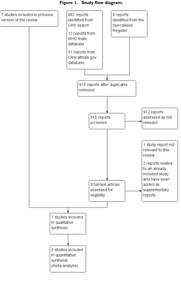
For this update, there were three additional citations which were considered potentially relevant following screening of the search results. Two of these were publications related to an already in-cluded study (Rickard 2012) and have been added as supplemen-tary papers. The third was not relevant to this review. No addi-tional trials were found in our search of trials registries.
Included studies
Because three of the authors of this review were also investigators in trials under consideration, we allocated the assessment of those trials to review authors who were not investigators for those par-ticular studies.
Seven RCTs (Barker 2004;Nishanth 2009;Rickard 2010;Rickard 2012;Van Donk 2009;Webster 2007;Webster 2008) met the inclusion criteria (seetable:Characteristics of included studiesfor details).
The seven trials involved a total of 4895 participants, with in-dividual trial sizes ranging between 42 and 3283. One trial was carried out in England (Barker 2004), one in India (Nishanth 2009), the remaining five trials were Australian (Rickard 2010; Rickard 2012;Van Donk 2009;Webster 2007;Webster 2008). Five of the trials were conducted in single-centre, acute inpatient settings (Barker 2004; Nishanth 2009; Rickard 2010; Webster 2007;Webster 2008), one was a multi-centre trial in three Aus-tralian hospitals (Rickard 2012), and one was undertaken in a community setting (Van Donk 2009).
In six trials (Barker 2004;Nishanth 2009;Rickard 2010;Rickard 2012; Webster 2007; Webster 2008) patients were included if they were receiving either continuous infusions or intermittent infusions for medication therapy, whereas the catheters in the Van Donk 2009trial were used for intermittent medication ther-apy only. In five trials (Rickard 2010;Rickard 2012;Van Donk 2009; Webster 2007; Webster 2008) the comparison was be-tween routine care (planned three-day changes) and clinically-in-dicated changes.Barker 2004andNishanth 2009compared
48-hour changes with removal for clinical indications such as pain, catheter dislodgement, or phlebitis.
Five of the trials (Barker 2004; Rickard 2010; Rickard 2012; Webster 2007;Webster 2008) used a standard definition of two or more of the following: pain, warmth, erythema, swelling, or a palpable cord.Barker 2004andNishanth 2009further classi-fied phlebitis as either mild, moderate, or severe depending on the area of erythema (Barker 2004) or on the number of symptoms (Nishanth 2009).Van Donk 2009included the same symptoms as other trials but scored them as either one or two depending on the severity. A score of two or more was classified as phlebitis, con-sequently a patient may have had only one symptom, for example pain, to receive a positive diagnosis.
Power calculations were reported by Nishanth 2009; Rickard 2010;Rickard 2012;Webster 2007;Webster 2008; andVan Donk 2009but not byBarker 2004. All of the studies had institutional ethical approval.
Excluded studies
The tableCharacteristics of excluded studies contains the rea-sons for excluding nine trials. In summary, two were very small studies involving the administration of peripheral parenteral nu-trition. Neither trial compared straightforward routine replace-ment with clinically-indicated removal (Kerin 1991;May 1996). One trial (Panadero 2002) compared one group that used the same catheter both intraoperatively and postoperatively with a group using two catheters, one during surgery and one postoper-atively. TheHaddad 2006trial compared 72-hour changes with 96-hour changes, and theCobb 1992;Eyer 1990;Nakae 2010; andRijnders 2004trials involved central venous catheters. The other excluded study was not an RCT (Arnold 1977).
Risk of bias in included studies
Allocation
Generation of random allocation sequence
All of the investigators reported that they used a computer-based sequence generator (Barker 2004;Nishanth 2009;Rickard 2010; Rickard 2012;Van Donk 2009;Webster 2007;Webster 2008).
Allocation concealment
Sealed envelopes were used for allocation concealment byBarker 2004;Nishanth 2009; andVan Donk 2009; the remaining four trials used a central telephone or computer-based service (Rickard 2010;Rickard 2012;Webster 2007;Webster 2008).
Blinding
It was not possible to blind either the participants or the healthcare providers in any of the trials.
Outcome assessment
The chief investigator assessed outcomes in theBarker 2004and theNishanth 2009trial. In theVan Donk 2009;Webster 2007; andWebster 2008trials, assessment was made by nurses caring for the patient or by a dedicated IV service nurse. None of the nurses were blinded to the group allocation but nor were any of them associated with the trial. In theRickard 2010andRickard 2012trials, outcome assessment was undertaken by a dedicated research nurse who was also aware of the allocation.
Incomplete outcome data
A flow chart was not provided byBarker 2004, so the numbers screened and eligible were unclear, nor were any dropouts reported. There was also an imbalance in the number of participants re-ported by group in this trial, which may indicate either a failure in the randomisation process in such a small trial or incomplete reporting. The number of protocol violations by group was not reported. There was complete reporting in the other six trials, all of which provided a flow of participants through each stage and used intention-to-treat analysis (Nishanth 2009;Rickard 2010; Rickard 2012;Van Donk 2009;Webster 2007;Webster 2008). In theWebster 2007;Webster 2008; andVan Donk 2009trials,
approximately one third of the participants had protocol viola-tions and in theRickard 2012trial, protocol violations occurred in 16% of the participants. Primarily these were in the routine replacement groups, where catheters were not replaced within the specified time period, reflecting day to day clinical practice.
Selective reporting
Study protocols were available for five trials (Rickard 2010; Rickard 2012; Van Donk 2009;Webster 2007;Webster 2008) and reporting followed pre-planned analyses.Barker 2004 and Nishanth 2009reported on the expected primary outcomes.
Other potential sources of bias
In theBarker 2004trial there were two definitions of phlebitis, one of which stated that two symptoms were necessary; yet it ap-pears that erythema alone was diagnosed as phlebitis, with sever-ity based on the area of inflammation. The extreme results in the Nishanth 2009trial, where 100% of participants in the clinically-indicated group developed phlebitis compared with 9% in the two-day change group, suggests that chance or other unknown bias affected results in this small trial.
Effects of interventions
See:Summary of findings for the main comparison Clinically-indicated versus routine changes for peripheral venous catheter-related complications
Routine changes versus clinically-indicated changes
Figure 4. Forest plot of comparison: 1 Clinically-indicated versus routine change, outcome: 1.1 Catheter-related bloodstream infection.
Phlebitis (Analysis 1.2 and Analysis 1.3)
All of the included studies reported incidence of phlebitis (4895 patients). When results of all trials were combined, heterogene-ity was 65%. Consequently, we conducted a sensitivheterogene-ity analy-sis and removed the two trials with less than 100 participants, both of which used a two-day replacement schedule (Barker 2004; Nishanth 2009). Removing the two trials eliminated the
hetero-geneity (I2= 0). Data from the remaining studies (4806 partici-pants) were combined (Rickard 2010;Rickard 2012;Van Donk 2009;Webster 2007;Webster 2008). There was no difference in this outcome whether catheters were changed according to clini-cal indications or routinely (cliniclini-cally-indicated 186/2365; 3-day change 166/2441; RR 1.14, 95% CI 0.93 to 1.39; P = 0.20). This result was unaffected by whether the infusion was continuous or intermittent (Figure 5).
[image:17.595.102.516.111.215.2] [image:17.595.100.518.368.564.2]group.
Figure 6. Forest plot of comparison: 1 Clinically-indicated versus routine change, outcome: 1.3 Phlebitis per device days.
All-cause bloodstream infection (Analysis 1.4)
One trial assessed this outcome (Rickard 2012). There was no difference in the all-cause bloodstream infection rate between the two groups (clinically-indicated: 4/1593 (0.02%); routine change 9/1690 (0.05%); P = 0.21) (Figure 7).
Figure 7. Forest plot of comparison: 1 Clinically-indicated versus routine change, outcome: 1.4 All-cause bloodstream infection.
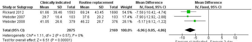
Cost (Analysis 1.5)
[image:18.595.102.517.145.245.2] [image:18.595.104.512.445.493.2]Figure 8. Forest plot of comparison: 1 Clinically-indicated versus routine change, outcome: 1.5 Cost.
Infiltration (Analysis 1.6)
A total of four trials assessed infiltration in 4606 participants (Rickard 2010;Rickard 2012;Webster 2007;Webster 2008). In-filtration of fluid into surrounding tissues was reported less often in the routine change group (452/2346; 19.3%) compared with the clinically-indicated group (518/2260; 22.9%). The RR was 1.17 (95% Ci 1.05 to 1.31; P = 0.004) (Figure 9).
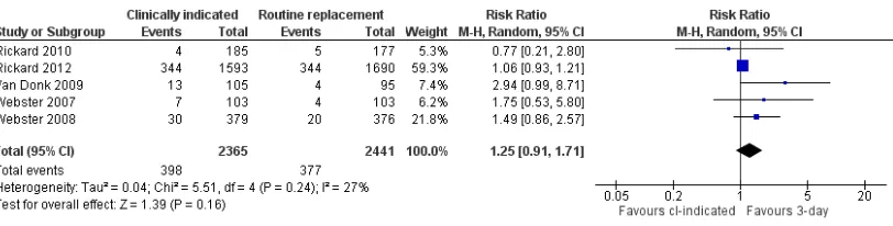
[image:19.595.100.516.103.173.2] [image:19.595.101.518.331.428.2]Figure 10. Forest plot of comparison: 1 Clinically-indicated versus routine change, outcome: 1.7 Catheter blockage.
Local infection (Analysis 1.8)
Among the four trials measuring local infection (Rickard 2010; Rickard 2012;Webster 2007;Webster 2008) no differences were found between groups (clinically-indicated 2/2260 (0.09%); rou-tine replacement 0/2346 (0.0%); RR 4.96, 95% CI 0.24 to 102.98; P = 0.30) (Figure 11).
Figure 11. Forest plot of comparison: 1 Clinically-indicated versus routine change, outcome: 1.8 Local infection.
Mortality (Analysis 1.9)
Four deaths occurred in each group in the one trial (Rickard 2012) that assessed this outcome (RR 1.06, 95% CI 0.27 to 4.23; P = 0.93) (Figure 12).
[image:20.595.107.513.112.215.2] [image:20.595.105.513.368.460.2] [image:20.595.97.516.607.656.2]The pre-planned outcomes ’number of catheter re-sites per pa-tient’, ’pain’ and ’satisfaction’ were not reported by the studies in-cluded in the review.
Subgroup and sensitivity analysis
We planned to conduct subgroup analyses on 1) Type of randomi-sation (truly randomised versus not reported); 2) Concealment of allocation (adequate versus not reported) and; 3) Statement of withdrawals and losses to follow up in each group (stated versus not stated). However, there were too few studies in these subgroups to make any meaningful comparisons. Similarly, blinding was not possible in any of the studies. Nor did we conduct any of our pre-planned sensitivity analysis (except size of studies for the outcome ’phlebitis’) for similar reasons.
D I S C U S S I O N
Summary of main results
This systematic review analysed catheter-related bloodstream in-fection, phlebitis, other reasons for catheter failure, and cost with the intention of comparing routine catheter changes (at between two and four days) with replacing the catheter only if clinical signs were apparent.
The primary outcomes of this review suggest that patients are not adversely affected if the catheter is changed based on clinical indi-cations rather than routinely, as recommended by the US Centers of Disease Control (O’Grady 2011). The rate of catheter-related bloodstream infection was similar in both groups, between 0.0% and 0.3%, and comparable to that previously reported in prospec-tive studies (Maki 2006). A marginal but non-significant increase in the phlebitis rate in the clinically-indicated group was apparent when data were analysed by patient but became less perceptible when data were analysed per 1000 device days, which is a more clinically useful measure. In addition, most cases of phlebitis are
Cost was significantly less, around AUD 7, in the clinically-in-dicated group. This result was based on three studies and results were consistent and intuitively logical (fewer catheters, less clin-ician time and equipment). Although, this is a seemingly small amount, it corresponds to approximately 11% of catheter-related expenditure, which may represent a considerable saving to organ-isations with high use (Figure 8).
Overall completeness and applicability of evidence
Limitations in study design and implementation Risk of bias was assessed according to six components: sequence generation, allocation concealment, blinding, selective outcome reporting, incomplete follow up, and other potential biases. All of the studies avoided selection bias and ensured allocation con-cealment. The methodological quality of most of the RCTs was high with one exception. It was not possible to blind the primary outcome in any of the trials. Blinding was not possible because it was necessary to identify the catheter as either ’routine change’ or ’clinically indicated’, to prevent inadvertent routine replacement of catheters in the intervention group. It is unclear if this had any bearing on outcomes but the review authors argue that it is unlikely (Figure 2;Figure 3). In theBarker 2004andNishanth 2009trials, the investigator was directly involved in diagnosing phlebitis; in all of the other studies either medical staff, ward nurses, IV therapy staff, or research nurses evaluated the outcomes. As one author noted, it is routine practice to record reasons for removal of an intravenous catheter in the medical record, and it is unlikely that such entries would be falsified based on group allocation (Webster 2008).
Indirectness of evidence
All of the trials compared routine changes with clinically-indicated changes. However, five trials used a three to four-day change sched-ule and two trials changed catheters every two days. Consequently, three to four-day results may provide indirect evidence for two-day changes, conversely two-day changes provide indirect evidence for a three to four-day change schedule. Additionally, only one study (Nishanth 2009) included patients who were from a developing country and who were “usually asthenic, many underhydrated/ dehydrated on admission” (personal correspondence), so the evi-dence may be regarded as indirect for these types of patients.
Unexplained heterogeneity or inconsistency of results When we combined results of studies that investigated the effect of different catheter replacement schedules on phlebitis, the het-erogeneity was high. This was probably due to the different sched-ules for the routine catheter changes or population differences, or both. Small sample sizes may also have contributed to the extreme results, which caused the heterogeneity. We tested these assump-tions by performing a sensitivity analysis, removing two of the seven studies. Results of the five trials are presented in the review text and the Summary of findings table (Summary of findings for the main comparison).
uncertainty around the effect size. Further research is therefore very likely to have an important impact on the confidence in the estimate of effect for these outcomes.
Publication bias
We feel confident that our comprehensive electronic searches iden-tified all existing, published, randomised controlled trials address-ing the review question.
Potential biases in the review process
Although the authors were investigators in one or more of the in-cluded trials, clearly described procedures were followed to prevent potential biases in the review process. A careful literature search was conducted and the methods we used are transparent and re-producible. None of the authors has any conflict of interests.
Agreements and disagreements with other studies or reviews
Our results concur with several prospective observational studies, which found no additional risk in extending IVD dwell times (Bregenzer 1998;Catney 2001;Homer 1998;White 2001). We believe the reason for this is the similarity in the mean dwell times between the intervention and control arms. Each of the included studies were pragmatic trials and, in real life, many catheters are not changed within the prescribed time frames. For example, in three-day protocols the 72-hour period may occur in the middle of the night; or a decision may be made to leave an existing catheter in place if the patient is due for discharge the following day or if they are thought to have poor veins. Conversely, the catheter may need to be removed early in any clinically-indicated group if the patient’s catheter becomes blocked or infiltration or phlebitis occurs, or the patient is discharged within a couple of days of catheter insertion.
Our results also support the CDC guidelines for peripheral catheter replacement in children, which state “replace peripheral catheters in children only when clinically indicated” (O’Grady 2011). Similarly, in a guideline for timing peripheral intravenous replacement (Ho 2011) findings from the original version of this review were replicated (Webster 2010).
indicated. The consistency in these results, which include a very large multi-site study, indicate that healthcare organisations should adopt a clinically-indicated replacement policy. This would pro-vide significant cost savings and would also be welcomed by pa-tients, who would be spared the unnecessary pain of routine re-sites in the absence of clinical indications. Busy clinical staff would also reduce time spent on this intervention. To minimise periph-eral catheter-related complications, the insertion site should be in-spected at each shift change and the catheter removed if signs of inflammation, infiltration, or blockage are present.
Implications for research
Any future trial should use standard definitions for phlebitis and be sufficiently large to show true differences. Based on results from the meta-analysis in this review, at least 2500 participants would be required in each arm of any future trial to show a lowering of
phlebitis rates from 8% to 6% (α= 0.05 and 80% power). Neither
pain nor satisfaction were measured in any of the reviewed studies and would be a useful addition to any future trial. Although costs were estimated in some of the included trials, a careful economic analysis of routine versus clinically-indicated replacement would be helpful for healthcare administrators. There was also some ev-idence from this review that different results may occur when the population is drawn from a developing country. Consequently, trials conducted in a wider variety of healthcare systems would add to the external validity of the review’s results.
A C K N O W L E D G E M E N T S
We are grateful to Marlene Stewart, Cochrane Vascular Managing Editor, for her support and speedy responses, and to the editors Mr Paul Tisi and Dr Jackie Price for their useful comments.
R E F E R E N C E S
References to studies included in this review
Barker 2004 {published and unpublished data}
Barker P, Anderson ADG, Macfie J. Randomised clinical trial of elective re-siting of intravenous cannulae.Annals of the Royal College of Surgeons of England2004;86(4):281–3.
Nishanth 2009 {published data only}
Nishanth S, Sivaram G, Kalayarasan R, Kate V, Ananthakrishnan N. Does elective re-siting of intravenous cannulae decrease peripheral thrombophlebitis? A randomized controlled study. The International Medical Journal of India2009;22(2):60–2.
Rickard 2010 {published and unpublished data}
Rickard CM, McCann D, Munnings J, McGrail M. Routine resite of peripheral intravenous devices every 3 days did not reduce complications compared with clinically indicated
Van Donk 2009 {published and unpublished data} Van Donk P, Rickard CM, McGrail MR, Doolan G. Routine replacement versus clinical monitoring of peripheral intravenous catheters in a regional hospital in the home program: A randomized controlled trial.Infection Control and Hospital Epidemiology2009;30(9):915–7.
Webster 2007 {published and unpublished data} Webster J, Lloyd S, Hopkins T, Osborne S, Yaxley M. Developing a research base for intravenous peripheral cannula re-sites (DRIP trial). A randomised controlled trial of hospital in-patients. International Journal of Nursing Studies2007;44(5):664–71.
Webster 2008 {published and unpublished data}
Eyer 1990 {published data only}
Eyer S, Brummitt C, Crossley K, Siegel R, Cerra F. Catheter-related sepsis: prospective, randomized study of three methods of long-term catheter maintenance.Critical Care Medicine1990;18(10):1073–9.
Haddad 2006 {published data only}
Haddad FG, Waked CH, Zein EF. Peripheral venous catheter inflammation. A randomized prospective trial.Le Journal Médical Libanais2006;54:139–45.
Kerin 1991 {published data only}
Kerin MJ, Pickford IR, Jaeger H, Couse NF, Mitchell CJ, Macfie J. A prospective and randomised study comparing the incidence of infusion phlebitis during continuous and cyclic peripheral parenteral nutrition. Clinical Nutrition 1991;10(6):315–9.
May 1996 {published data only}
May J, Murchan P, MacFie J, Sedman P, Donat P, Palmer D, et al. Prospective study of the aetiology of infusion phlebitis and line failure during peripheral parenteral nutrition. British Journal of Surgery1996;83(8):1091–4.
Nakae 2010 {published data only}
Nakae H, Igarashi T, Tajimi K. Catheter-related infections via temporary vascular access catheters: a randomized prospective study.Artificial Organs2010;34(3):E72–6.
Panadero 2002 {published data only}
Panadero A, Iohom G, Taj J, Mackay N, Shorten G. A dedicated intravenous cannula for postoperative use. Effect on incidence and severity of phlebitis.Anaesthesia2002;57
(9):921–5.
Rijnders 2004 {published data only}
Rijnders BJ, Peetermans WE, Verwaest C, Wilmer A, Van Wijngaerden E. Watchful waiting versus immediate catheter removal in ICU patients with suspected catheter-related infection: a randomized trial.Intensive Care Medicine2004;
30(6):1073–80.
Additional references
Band 1980
Band JD, Maki DG. Steel needles used for intravenous therapy. Morbidity in patients with hematologic malignancy. Archives of Internal Medicine1980;140(1): 31–4.
Bregenzer 1998
Bregenzer T, Conen D, Sakmann P, Widmer AF. Is routine replacement of peripheral intravenous catheters necessary?. Archives of Internal Medicine1998;158:51–6.
Catney 2001
Catney MR, Hillis S, Wakefield B, Simpson L, Domino L,
and duration of cannulation.Infection Control and Hospital Epidemiology2002;23:249–53.
Everitt 1997
Everitt NJ, Krupowicz DW, Evans JA, McMahon MJ. Ultrasonographic investigation of the pathogenesis of infusion thrombophlebitis. British Journal of Surgery1997;
84:642–5.
Gupta 2007
Gupta A Mehta Y, Juneja R, Trehan N. The effect of cannula material on the incidence of peripheral venous thrombophlebitis.Anaesthesia2007;62:1139–42.
Hadaway 2012
Hadaway. Short peripheral intravenous catheters and infections.Journal of Infusion Nursing2012;35:230–40.
Higgins 2003
Higgins JPT, Thompson SG, Deeks JJ, Altman DG. Measuring inconsistencies in meta-analysis.BMJ2003;327
(7414):557–60.
Higgins 2008
Higgins JPT, Deeks JJ. Selecting studies and collecting data. In: Higgins JPT, Green S editor(s).Cochrane Handbook for Systematic Reviews of Interventions. Wiley-Blackwell, 2008.
Higgins 2011a
Higgins JPT, Altman DG, and Sterne JAC on behalf of the Cochrane Statistical Methods Group and the Cochrane Bias Methods Group. Chapter 8: Assessing risk of bias in included studies. In: Higgins JPT, Green S, editor(s). Cochrane Handbook for Systematic Reviews of Interventions Version 5.1.0 [updated March 2011]. The Cochrane Collaboration, 2011. Available from www.cochrane-handbook.org.
Ho 2011
Ho KHM, Cheung DSK. Guidelines on timing in replacing peripheral intravenous catheters.Journal of Clinical Nursing 2011;21(11-12):1499–506.
Homer 1998
Homer LD, Holmes KR. Risks associated with 72- and 96-hour peripheral intravenous catheter dwell times.Journal of Intravenous Nursing1998;21:301–5.
Infusion Nurses Society 2011
Infusion Nurses Society. Infusion Nursing Standards of Practice.Journal of Infusion Nursing2011;34(1S):S57.
Lai 1998
Lai KK. Safety of prolonging peripheral cannula and i.v. tubing use from 72 hours to 96 hours. American Journal of Infection Control1998;26:66–70.
Loveday 2014
prevent cephalothin-induced phlebitis.American Journal of Hospital Pharmacy1977;34:29–34.
Maki 1973
Maki DG, Goldman DA, Rhame FS. Infection control in intravenous therapy.Annals of Internal Medicine1973;79
(6):867–87.
Maki 1991
Maki DG, Ringer M. Risk factors for infusion-related phlebitis with small peripheral venous catheters. A randomized controlled trial. Annals of Internal Medicine 1991;114:845–54.
Maki 2006
Maki DG, Kluger DM, Crnich CJ. The risk of bloodstream infection in adults with different intravascular devices: a systematic review of 200 published prospective studies. Mayo Clinic Proceedings2006;81(9):1159–71.
Maki 2008
Maki DG. Improving the safety of peripheral intravenous catheters.BMJ2008;337(7662):122–3.
Malyon 2014
Malyon L, Ullman AJ, Phillips N, Young J, Kleidon T, Murfield J, et al. Peripheral intravenous catheter duration and failure in paediatric acute care: a prospective cohort study.Emergency Medicine Australasia2014;26:602–8.
Monreal 1999
Monreal M, Quilez F, Rey-Joly C, Vega J, Torres T, Valero P, et al. Infusion phlebitis in patients with acute pneumonia: a prospective study.Chest1999;115:1576–80.
O’Grady 2011
O’Grady NP, Alexander M, Burns LA, Dellinger EP, Garland J, Heard SO, et al. 2011 Guidelines for the prevention of intravascular catheter-related infections. http: //www.cdc.gov/hicpac/bsi/bsi-guidelines-2011.html.
Ray-Barruel 2014
Ray-Barruel G, Polit DF, Murfield JE, Rickard CM. Infusion phlebitis assessment measures: a systematic review. Journal of Evaluation in Clinical Practice2014;20:191–202.
Tager 1983
Tager IB, Ginsberg MB, Ellis SE, Walsh NE, Dupont I, Simchen E, et al. The Rhode Island Nosocomial Infection Consortium. An epidemiologic study of the risks associated with peripheral intravenous catheters.American Journal of Epidemiology1983;118(6):839–51.
Tuffaha 2014
Tuffaha HW, Rickard CM, Webster J, Marsh N, Gordon L, Wallis M, et al. Cost-effectiveness analysis of clinically indicated versus routine replacement of peripheral intravenous catheters.Applied Economics and Health Policy 2014;12:51–8.
Tuffaha 2014a
Tuffaha HW, Rickard CM, Inwood S, Gordon L, Scuffham P. The epic3 recommendation that clinically indicated replacement of peripheral venous catheters is safe and cost-saving: How much would the NHS save?. Journal of Hospital Infection2014;87(3):183–4.
Uslusoy 2008
Uslusoy E, Mete S. Predisposing factors to phlebitis in patients with peripheral intravenous catheters: a descriptive study.Journal of the American Academy of Nurse Practitioners 2008;20:172–80.
White 2001
White SA. Peripheral intravenous therapy-related phlebitis rates in an adult population.Journal of Intravenous Nursing 2001;24:19–24.
References to other published versions of this review
Webster 2009
Webster J, Osborne S, Hall J, Rickard C. Clinically indicated replacement versus routine replacement of peripheral venous catheters.Cochrane Database of Systematic Reviews 2009, Issue 2. DOI: 10.1002/14651858.CD007798
Webster 2010
C H A R A C T E R I S T I C S O F S T U D I E S
Characteristics of included studies [ordered by study ID]
Barker 2004
Methods Study design:Single-centre RCT.
Method of randomisation:Computer generated. Concealment of allocation:Sealed envelopes.
Participants Country:England.
Number: 47 patients in general medical or surgical wards. Clinically indicated: 43 catheters were inserted in 26 patients. Routine replacement: 41 catheters were inserted in 21 patients
Age:Clinically indicated 60.5 yrs (15.5); routine replacement 62.7 yrs (18.2) Sex (M/F):Clinically indicated 15/11; routine replacement 14/7.
Inclusion criteria:Hospital inpatients receiving crystalloids and drugs. Exclusion criteria:Not stated.
Interventions Clinically indicated:Catheters were removed if the site became painful, the catheter dislodged or there were signs of PVT
Routine replacement: Catheters were replaced every 48 hours.
Outcomes Primary:Incidence of PVT defined as “the development of two or more of the following: pain, erythema, swelling, excessive warmth or a palpable venous cord”
Notes PVT was defined as “the development of two or more of the following: pain, erythema, swelling, excessive warmth or a palpable venous cord”. However, in the discussion, the author stated that “even a small area of erythema was recorded as phlebitis” (i.e., only one sign)
It is unclear what proportion of patients were on continuous infusion Catheters were inserted “at the instruction of the principal investigator”
“All patients were reviewed daily by the principal investigator, and examined for signs of PVT at the current and all previous infusion sites”
Risk of bias
Bias Authors’ judgement Support for judgement
Random sequence generation (selection bias)
Low risk Comment:Computer generated (personal
communication with author).
Barker 2004 (Continued)
Incomplete outcome data (attrition bias) All outcomes
High risk Comment:In this small sample, there were five fewer patients in the routine replace-ment group. No explanation was provided for the unequal sample size. No dropouts or loss to follow up were reported Selective reporting (reporting bias) Low risk Comment:Phlebitis was the only outcome
planned.
Other bias High risk Comment:The chief investigator allocated
patients and was responsible for outcome evaluation
No sample size calculation.
Nishanth 2009
Methods Study design:Single-centre RCT.
Method of randomisation:Not stated
Concealment of allocation:Sequentially numbered sealed envelopes.
Participants Country:India.
Number:42 patients in surgical wards. Clinically indicated: 21. Routine replacement: 21
Age:Clinically indicated 40.2 yrs (15.0); routine replacement 42.9 yrs (15.0) Sex (M/F):Clinically indicated 17/4; routine replacement 16/5.
Inclusion criteria:Hospital inpatients admitted for major abdominal surgery Exclusion criteria:Receiving total parenteral nutrition, duration of therapy expected to be < three days, if a cannula was already in situ, terminally ill patients
Interventions Clinically indicated:Catheters were removed if the site became painful, the catheter dislodged or there were signs of PVT
Routine replacement: Catheters were replaced every 48 hours.
Nishanth 2009 (Continued)
(SNOSE).”
Comment:Presumably the authors meant ’in’ an opaque serially numbered sealed en-velope - based on subsequent information Blinding (performance bias and detection
bias) All outcomes
High risk Evidence for participants: Quote
“un-blinded study”.
Evidence for personnel:As above. Evidence for outcomes:As above. Incomplete outcome data (attrition bias)
All outcomes
Low risk Comment:Data for all patients were avail-able.
Selective reporting (reporting bias) Low risk Comment:Stated outcomes were reported but original protocol not sighted
Other bias Unclear risk Extreme results: In this small trial, 100%
of participants in the clinically indicated group developed phlebitis compared with 9% in the 2-day change group, which sug-gests that chance or other unknown bias af-fected results
Rickard 2010
Methods Study design:Single-centre RCT.
Method of randomisation:Computer generated. Concealment of allocation:Telephone service.
Participants Country:Australia.
Number:362 patients requiring IV therapy in general medical or surgical wards. Clin-ically indicated: 280 catheters were inserted in 185 patients. Routine replacement: 323 catheters were inserted in 177 patients
Age:Clinically indicated 62.7 yrs (15.5); routine replacement 65.1 yrs (17.3) Sex (M/F):Clinically indicated 82/103; routine replacement 81/91.
Inclusion criteria:Patients in over 18 years, expected to have a peripheral intravenous device (IVD), requiring IV therapy for at least 4 days
Exclusion criteria:Patients who were immunosuppressed, had an existing bloodstream infection or those in whom an IVD had been in place for > 48 hours
Interventions Clinically indicated: Catheters were removed if there were signs of phlebitis, local infection, bacteraemia, infiltration or blockage
Rickard 2010 (Continued)
Notes Approximately 75% of patients were receiving a continuous infusion
Risk of bias
Bias Authors’ judgement Support for judgement
Random sequence generation (selection bias)
Low risk Comment:Computer generated.
Allocation concealment (selection bias) Low risk Quote “assignment was concealed until randomisation by use of a telephone ser-vice”
Blinding (performance bias and detection bias)
All outcomes
High risk Comment: Neither study personnel nor
participants were blinded.
Incomplete outcome data (attrition bias) All outcomes
Low risk Comment:Results from all enrolled
pa-tients were reported.
Selective reporting (reporting bias) Low risk Comment:The protocol was available. All nominated outcomes were reported
Other bias Unclear risk Comment:Significantly more patients in
the routine change group received IV an-tibiotics (73.1% versus 62.9%)
Rickard 2012
Methods Study design:Multi-centre RCT.
Method of randomisation:Computer generated, stratified by site.
Rickard 2012 (Continued)
Interventions Clinically indicated: Catheters were removed if there were signs of phlebitis, local infection, bacteraemia, infiltration or blockage
Routine replacement:Catheters were replaced every 72 - 96 hours.
Outcomes Primary:Phlebitis during catheterisation or within 48 hrs of removal (defined as two or more of the following: pain, erythema, swelling, purulent discharge, palpable venous cord)
Secondary:Catheter-related bloodstream infection, all-cause bloodstream infection, lo-cal venous infection, colonisation of the catheter tip, infusion failure, number of catheters per patient, overall duration of intravenous therapy, cost, mortality
Notes
Risk of bias
Bias Authors’ judgement Support for judgement
Random sequence generation (selection bias)
Low risk Quote: “Random allocations were
com-puter-generated”.
Allocation concealment (selection bias) Low risk Quote: “Random allocations were com-puter-generated on a hand-held device, at the point of each patient’s entry, and thus were concealed to patients, clinical staff and research staff until this time”
Blinding (performance bias and detection bias)
All outcomes
High risk Evidence for participants: Quote
“Pa-tients and clinical staff could not be blinded”.
Evidence for personnel: Quote“Research nurses were similarly not masked”. Evidence for outcomes: Quote“... lab-oratory staff were masked for rating of all microbiological end-points, and a masked, independent medical rater diag-nosed catheter-related infections and all bloodstream infections”
Incomplete outcome data (attrition bias) All outcomes
Low risk ITT analysis reported.
Van Donk 2009
Methods Study design:RCT.
Method of randomisation:Computer generated. Concealment of allocation:Sealed envelopes.
Participants Country:Australia.
Number:200. Clinically indicated: 105 patients. Routine replacement: 95 patients Age:Clinically indicated 62.8 yrs (18.2); routine replacement 54.5 yrs (19.0) Sex (M/F):Not stated.
Inclusion criteria:Adult patients who could be treated at home for an acute illness and had a 20, 22, or 24 gauge catheter inserted in an upper extremity
Exclusion criteria:Not stated.
Interventions Clinically indicated: Catheters were removed if there were signs of phlebitis, local infection, bacteraemia, infiltration or blockage
Routine replacement:Catheters were replaced every 72 - 96 hours.
Outcomes Primary:Phlebitis per patient and per 1000 device days (phlebitis was defined as a total score of 2 or more points from the following factors: pain (on a 10-point scale, 1 = 1 point, and 2 or more = 2 points; redness (less than 1cm = 1 point, and 1 or more cm = 2 points); swelling (as for redness); and discharge (haemoserous ooze under dressing = 1 point, and haemoserous ooze requiring dressing change or purulence = 2 points) Also reported on:Suspected IVD-related bacteraemia and occlusion/blockage. Notes
Risk of bias
Bias Authors’ judgement Support for judgement
Random sequence generation (selection bias)
Low risk Comment:Computer generated allocation
(personal communication with author) Allocation concealment (selection bias) Low risk Quote: “Randomization was concealed
Webster 2007
Methods Study design:Single-centre RCT.
Method of randomisation:Computer generated.
Concealment of allocation:Allocation concealed until telephone contact made with an independent person
Participants Country:Australia.
Number:206. Clinically indicated: 103 patients. Routine replacement: 103 patients Age:Clinically indicated 60.2 yrs (16.2); routine replacement 63.1 yrs (17.3) Sex (M/F):Clinically indicated 53/50; routine replacement 54/49.
Inclusion criteria:At least 18 yrs of age, expected to have a peripheral intravenous device (IVD) in situ, requiring IV therapy for at least 4 days, catheter inserted by a member of the IV team
Exclusion criteria:Immunosuppressed patients and those with an existing bloodstream infection
Interventions Clinically indicated:Catheters removed if there were signs of phlebitis, local infection, bacteraemia, infiltration or blockage
Routine replacement:Catheters replaced every 3 days.
Outcomes Primary:Composite measure of any reason for an unplanned catheter removal Secondary:Cost (For intermittent infusion: 20 minutes nursing/medical time, a can-nula, a 3 way tap, a basic dressing pack, gloves, a syringe, transparent adhesive dressing, skin disinfection and local anaesthetic per insertion. For patients receiving a continuous infusion: all the above costs plus the additional cost of replacing all associated lines, solutions and additives which are discarded when an IV catheter is changed (based on an intravenous administration set, 1 litre sodium chloride 0.09%)
Notes
Risk of bias
Bias Authors’ judgement Support for judgement
Random sequence generation (selection bias)
Low risk Quote:“randomization was by computer
generated random number list, stratified by oncology status”
Allocation concealment (selection bias) Low risk Quote:“Allocation was made by phoning a person who was independent of the re-cruitment process”
Blinding (performance bias and detection bias)
All outcomes
High risk Evidence for participants: Comment:
Webster 2007 (Continued)
phlebitis”
“Staff in the microbiological laboratory were blind to group assignment of catheters submitted for testing”
Incomplete outcome data (attrition bias) All outcomes
Low risk Comment:All recruited patients were
ac-counted for in the results.
Selective reporting (reporting bias) Low risk Comment: Protocol was available. All planned outcomes were reported.
Other bias Low risk No other known risks of bias.
Webster 2008
Methods Study design:Single-centre RCT.
Method of randomisation:Computer generated. Concealment of allocation:Telephone randomisation.
Participants Country:Australia.
Number:755. Clinically indicated: 379 patients. Routine replacement: 376 patients Age:Clinically indicated 60.1 yrs (17.1); routine replacement 58.8 yrs (18.8) Sex (M/F):Clinically indicated 248/131; routine replacement 233/143.
Inclusion criteria:At least 18 yrs of age, expected to have a IVD in situ, requiring IV therapy for at least 4 days
Exclusion criteria:Immunosuppressed patients and those with an existing bloodstream infection
Interventions Clinically indicated:Catheter removed if there were signs of phlebitis, local infection, bacteraemia, infiltration or blockage
Routine replacement:Catheter replaced every 3 days.
Webster 2008 (Continued)
Random sequence generation (selection bias)
Low risk Quote: “Block randomisation was by a
computer generated random number list” Allocation concealment (selection bias) Low risk Quote:“.... telephoned a contact who was
independent of the recruitment process for allocation consignment”
Blinding (performance bias and detection bias)
All outcomes
High risk Neither study personnel nor participants
were blinded.
Incomplete outcome data (attrition bias) All outcomes
Low risk All recruited patients were accounted for in the results.
Selective reporting (reporting bias) Low risk Protocol was available. All planned out-comes were reported.
Other bias Low risk No other known risks of bias.
IV: intravenous
IVD: peripheral intravenous device
PVT: peripheral vein infusion thrombophlebitis RCT: randomised controlled trial
Characteristics of excluded studies [ordered by study ID]
Study Reason for exclusion
Arnold 1977 Not a randomised controlled trial Cobb 1992 Involved central, not peripheral lines
Eyer 1990 Involved pulmonary artery or arterial catheters, not peripheral catheters Haddad 2006 End point was lymphangitis
(Continued)
D A T A A N D A N A L Y S E S
Comparison 1. Clinically-indicated versus routine change
Outcome or subgroup title studiesNo. of participantsNo. of Statistical method Effect size
1 Catheter-related blood stream infection
5 4806 Risk Ratio (M-H, Fixed, 95% CI) 0.61 [0.08, 4.68]
2 Phlebitis 5 4806 Risk Ratio (M-H, Fixed, 95% CI) 1.14 [0.93, 1.39]
2.1 Continuous infusion 4 4606 Risk Ratio (M-H, Fixed, 95% CI) 1.11 [0.89, 1.39] 2.2 Intermittent infusion 1 200 Risk Ratio (M-H, Fixed, 95% CI) 1.29 [0.85, 1.96] 3 Phlebitis per device days 5 26191 Risk Ratio (M-H, Fixed, 95% CI) 1.03 [0.84, 1.27] 4 All-cause blood stream infection 1 Risk Ratio (M-H, Fixed, 95% CI) Totals not selected
5 Cost 3 4244 Mean Difference (IV, Fixed, 95% CI) -6.96 [-9.05, -4.86]
6 Infiltration 4 4606 Risk Ratio (M-H, Fixed, 95% CI) 1.17 [1.05, 1.31]
7 Catheter blockage 5 4806 Risk Ratio (M-H, Random, 95% CI) 1.25 [0.91, 1.71]
8 Local infection 4 4606 Risk Ratio (M-H, Fixed, 95% CI) 4.96 [0.24, 102.98]
9 Mortality 1 Risk Ratio (M-H, Fixed, 95% CI) Totals not selected
Analysis 1.1. Comparison 1 Clinically-indicated versus routine change, Outcome 1 Catheter-related blood stream infection.
Review: Clinically-indicated replacement versus routine replacement of peripheral venous catheters
Comparison: 1 Clinically-indicated versus routine change
Outcome: 1 Catheter-related blood stream infection
Study or subgroup Clinically indicated Routine replacement Risk Ratio Weight Risk Ratio
n/N n/N M-H,Fixed,95% CI M-H,Fixed,95% CI
Rickard 2010 0/185 0/177 Not estimable
Rickard 2012 0/1593 1/1690 59.2 % 0.35 [ 0.01, 8.67 ]
Van Donk 2009 0/105 0/95 Not estimable
Webster 2007 0/103 0/103 Not estimable
Webster 2008 1/379 1/376 40.8 % 0.99 [ 0.06, 15.80 ]
Total (95% CI) 2365 2441 100.0 % 0.61 [ 0.08, 4.68 ]