Continued imaging of the transport of a
single neutral atom
Y. Miroshnychenko, D. Schrader, S. Kuhr, W. Alt, I. Dotsenko, M. Khudaverdyan, A. Rauschenbeutel,
D. Meschede
Institut f¨ur Angewandte Physik, Universit¨at Bonn, Wegelerstr. 8, 53115 Bonn, Germany, [email protected]
http://www.iap.uni-bonn.de/ag meschede/
Abstract: We have continuously imaged the controlled motion of a single atom as well as of a small number of distinguishable atoms with observation times exceeding one minute. The Cesium atoms are confined to potential wells of a standing wave optical dipole trap which allows to transport them over macroscopic distances. The atoms are imaged by an intensified CCD camera, and spatial resolution near the diffraction limit is obtained.
© 2003 Optical Society of America
OCIS codes: (020.7010) Trapping , (140.7010) Trapping
References and links
1. W. Neuhauser, M. Hohenstatt, P. E. Toschek, H. Dehmelt, “Localized vizible Ba+mono-ion oscillator,” Phys. Rev. A 22, 1137 (1980).
2. S. Kuhr, W. Alt, D. Schrader, M. M ¨uller, V. Gomer, D. Meschede, “Deterministic Delivery of a Single Atom,” Science 293, 278 (2001).
3. D. Schrader, S. Kuhr, W. Alt, M. M ¨uller, V. Gomer, D. Meschede, “An optical conveyor belt for single atoms,” Appl. Phys. B 73, 819 (2001).
4. S. Kuhr, W. Alt, D. Schrader, I. Dotsenko, Y. Miroshnychenko, W. Rosenfeld, M. Khudaverdyan, V. Gomer, A. Rauschenbeutel, D. Meschede, “Coherence properties and quantum state transportation in an optical conveyor belt,” Phys. Rev. Lett. 91, 213002 (2003).
5. H. C. N¨agerl, D. Leibfried, F. Schmid-Kaler, J. Eschner, R. Blatt, “Coherent excitation of normal modes in a string of Ca+ions,” Optics Express 3, 89 (1998).http://www.opticsexpress.org/abstract.cfm?URI=OPEX-3-2-89
6. N. Schlosser, G. Reymond, I. Protsenko, P. Grangier, “Sub-poissonian loading of single atoms in a microscopic dipole trap,” Nature 411, 1024 (2001).
7. D. Haubrich, H. Schadwinkel, F. Strauch, B. Ueberholz, R. Wynands, D. Meschede, “Observation of individual neutral atoms in magnetic and magneto-optical traps,” Europhys. Lett. 34, 663 (1996).
8. W. Alt, “An objective lens for efficient fluoresence detection of single atoms,” Optik 113, 3 (2002). 9. H. J. Metcalf, P. van der Straten, “Laser cooling and trapping,” (Springer Verlag, 1999).
10. D. Frese, B. Ueberholz, S. Kuhr, W. Alt, D. Schrader, V. Gomer, D. Meschede, “Single Atoms in an Optical Dipole Trap: Towards a Deterministic Source of Cold Atoms,” Phys. Rev. Lett. 85, 3777 (2000).
1. Introduction
to realize a coherent interaction between two or more particles in order to implement quantum logic operations.
In this context, we demonstrated that a standing wave optical dipole trap allows to control the external degrees of freedom of individual neutral atoms and to transport them over millimeter scale distances [2, 3]. Furthermore, we showed that the electronic states of the atoms in our trap exhibit long coherence times, making them a good candidate for storing and processing quantum information [4]. However, for an efficient and flexible state preparation and detection as well for a controlled interaction the absolute position of the atoms in the trap has to be known and controlled.
This can readily be achieved in ion trapping experiments where the position of the ions is determined by the fixed trapping potential including the Coulomb repulsion of the ions. Fur-thermore, due to their strong spatial confinement, trapped ions can resonantly scatter light with-out influencing their motional state. As an impressive example of this observation method, the molecule-like vibrations of a chain of trapped ions have been recorded in [5] using a strobo-scopic method. Imaging of single neutral atoms in a stationary dipole trap potential has also been achieved [6].
In this letter, we present a continued observation of the controlled transport of individual neutral atoms in a standing wave dipole trap with an observation time of the order of one minute. Our technique should enable us to control the absolute position of the atoms with sub-micrometer precision and to address the atoms individually. Our results therefore represent a major step towards the preparation and manipulation of a string of trapped neutral atoms for use as a quantum register.
2. Experimental setup
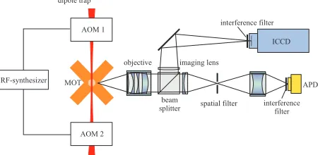
[image:2.612.195.420.503.613.2]A specially designed six-beam magneto-optical trap (MOT) [7] serves as a source of cold neu-tral cesium atoms. A high magnetic field gradient of 340 G/cm localizes the atoms to a region of dMOT=12µm in diameter. The diameter of the MOT beams is about 1 mm and the saturation parameter of each beam is s=0.5. The high magnetic field gradient reduces the loading rate of the MOT, which allows us to work at the level of single atoms. The fluorescence light from the MOT is collected and collimated by a home made diffraction limited objective (NA=0.29) [8] and is evenly split by a beam splitter (Fig. 1). The transmitted light is spatially and spec-trally filtered before it is focused onto an avalanche photodiode (APD). The light reflected by the beam splitter is only spectrally filtered and imaged onto the photocathode of an intensified CCD camera (ICCD).
The APD (SPCM-200, EG&G) has a quantum efficiency of 50% for the fluorescence light at the wavelength of 852 nm. For typical MOT parameters this yields a count rate of 35000/s for each trapped atom at a stray light background of 25000/s. This allows us to determine the exact number of up to 20 atoms trapped in the MOT at any moment in less than 10 ms, limited by shot noise.
The photocathode of the camera intensifier (Gen III HQ, Roper Scientific) is connected to an intensifying microchannel plate. For each photoelectron the microchannel plate together with the fluorescence screen of the intensifier produces a photon burst which is recorded by the CCD camera (PI-MAX:1K,HQ,RB, Princeton Instruments). On average this photon burst results in 350 counts on the 1024×1024 pixel CCD chip. The counts are concentrated in a 3×3 pixel area with 50% in the central pixel. The quantum efficiency of the intensified camera is approximately 10% at 852 nm. Since our imaging optics has a magnification of 13.99(±0.06), one CCD pixel of 13×13µm2corresponds to 0.929(±0.004)µm at the position of the atom. The expected full width at half maximum (FWHM) of the point spread function of our imaging optics is wPSF=1.4µm (all further widths in this letter are FWHM).
We use a red-detuned standing wave dipole trap (DT) in order to trap and move the atoms. The DT consists of two counter-propagating Gaussian beams (λ=1064 nm) in linlin polariza-tion configurapolariza-tion (Fig. 1). Each beam is focused to a waist of 2w0=32µm at the position of the MOT and has 1.3 W of optical power. The resulting interference pattern yields a chain of traps,
λ/2 apart, each with a potential depth of U/kB=2.9 mK and a radial FWHM of b=19µm. The atoms are transferred from the MOT into the DT with an efficiency close to 100%. Acousto-optic modulators (AOMs) control the frequencies of both beams of the DT. They are driven by a digital dual-frequency synthesizer (DFD100, APE) with two phase synchronized RF outputs. This allows us to phase-continuously detune the frequencies of each of the beams, causing the standing wave pattern to move in a controlled way along the dipole trap axis. The direction of the transport depends on the sign of the relative detuning between the two beams. Using this “optical conveyor belt” the atoms can be transported over macroscopic distances of up to 1 cm with submicrometer precision on a millisecond timescale [2, 3].
3. Experimental results
3.1. Imaging of an atom in the MOT
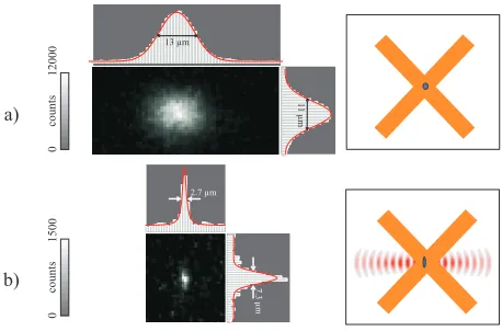
Under continuous illumination the fluorescence light of a single atom in the MOT generates nMOT = 6400 photoelectrons/s on the photocathode. Figure 2(a) shows an image of a single atom trapped in the MOT with an exposure time of 1 s. The size and the position of the MOT are determined by binning the pixels of the picture in the vertical and horizontal directions and by fitting the resulting histogram with a Gaussian. Here, the MOT has a size of 11µm in the vertical and of 13µm in the horizontal direction.
A trajectory of an atom in the MOT corresponds to a trace of sequentially recorded single photon events. In order to reconstruct the atomic trajectory with a spatial resolution at the diffraction limit of our optics, the mean spacing between consecutive photons should not exceed the diffraction limited spot size wPSF. This results in the upper limit of the atomic velocity of vdmax=wPSF·nMOT=9 mm/s. This number is much smaller than the Doppler velocity of
Fig. 2. a) CCD image of one atom stored in the MOT (exposure time 1 s). The counts are binned in the vertical and horizontal directions and fitted with Gaussians to determine the size and the absolute position of the trap. b) One atom in a potential well of the DT illuminated by an optical molasses (Exposure time 0.5 s).
3.2. Imaging of atoms in the dipole trap
An exactly known number of atoms is prepared in the MOT and transferred into the conser-vative potential of the DT. For continuous observation, we illuminate the atoms in the DT by a 3-D optical molasses. We use the MOT beams for this purpose, however, we red-detune them by 13.5Γfrom the light-shifted transition of the trapped atom and reduce the saturation parameter to 0.004 for each beam (Γ = 2π× 5.2 MHz is the linewidth of the excited state). Figure 2(b) shows an image of one atom trapped in a potential well of the DT recorded with an exposure time of 0.5 s. The observed fluorescence spot corresponds to about 70 detected photons (nDT=140 photoelectrons/s). The trapping region has a size of arad=7.3µm in the vertical direction, which is a few times smaller than the radial width b=19µm of the trap. This shows that the atom is trapped close to the minimum of the DT potential and remains cold during the illumination. From the standing wave geometry we know that the axial width of the trapping region is smaller thanλ/2=532 nm. However, the observed width of the fluo-rescence spot is aax=2.7µm. Therefore, two atoms are optically resolved if they are separated by more than aax, which corresponds to 6 or more potential wells. The deviation of aaxfrom the expected diffraction limited spot size of wPSF=1.4µm is mainly due to the fact that one photon detected by the camera produces a spot on the CCD chip with a width that corresponds to wDet=2.50(±0.03)µm at the position of the atoms. The width of the observed fluorescence spot aax, in combination with the current count rate in the dipole trap nDT, results in a lower limit of the precision of the detection of the axial atomic position of aax/√nDT=230 nm/
√ Hz. The radial distribution aradof the imaged atom depends on its temperature T . Using a Fokker-Planck equation model [9] we find that T = (U/2kB)(a/b)2. Here, the width a is extracted from the radial extent of the fluorescence spot corrected for the point spread function of the camera, a= (a2rad−a2ax)1/2. This results in a temperature of 188(±40)µK, which is of the same order as the Cs Doppler temperature of 125µK.
3.3. Controlled motion of trapped atoms
[image:4.612.192.421.76.227.2]switched on and the second picture is taken, again with 1 s exposure time. We then displace the atom by 2µm within 2 ms and the next picture with 1 s exposure time is recorded. The sequence of displacement and imaging is repeated and yields a series of pictures of the same atom.
The movie corresponding to the screenshot in Fig. 3 shows a transport of a single atom over a distance of 60µm within about one minute. The transport ends with the loss of the atom from the DT.
time = 54.0 sec
10mm
Fig. 3. (89 KB) Controlled transport of one neutral atom over a distance of 60µm during some tens of seconds.
In the second movie, see Fig. 4, we show the synchronous transport of three spatially re-solved atoms. The reversal of the transport direction was initiated by manually changing the sign of the relative detuning between the dipole trap laser beams. The stochastic nature of the loss mechanism results in random departure times of the atoms. The mean observation time before atom loss is of the order of 30 s. This time is close to the limit expected for loss due to background gas collisions [9, 10].
10mm time = 15.6 sec
Fig. 4. (316 KB) Transport of three neutral atoms.
4. Summary
We have spatially resolved and continuously observed individual neutral atoms stored in a standing wave dipole trap. Our optical conveyor belt transports them over macroscopic dis-tances with submicrometer precision. The combination of these two techniques allows us to continuously observe the controlled motion of the same atoms trapped in the dipole trap during several tens of seconds.
In future experiments we now aim at the control of the absolute position of the atoms in our dipole trap. This could be achieved by measuring the position of the atom using a CCD image and by then actively transporting the atom to a predetermined position. Such an absolute position control is essential for deterministically and reproducibly coupling one or more atoms to the mode of an optical high finesse resonator in order to realize elementary quantum logic operations in the framework of optical cavity quantum electrodynamics.
Acknowledgments
[image:5.612.163.451.160.205.2] [image:5.612.166.454.332.380.2]