INTERACTIONS BETWEEN NERVE AND MUSCLE
by
J. F. Y. Hoh
Thesis submitted for the Degree of Doctor of Philosophy in the Australian National University
i
Some of the investigations described in this thesis have been done in collaboration with Dr. R. Close, and these have all been published or accepted
for publication and are listed in the following page. All other investigations described in this thesis are my own original work.
As a result of investigations in which I have collaborated during the tenure of my Scholarship at the Australian National University, the following papers have appeared or have been accepted for
publication:-Hoh; J.F.Y. & Close, R. (1967). Effects of nerve cross- union on twitch and slow-graded muscle fibres in the toad. Aust. J. exp, Biol, med. Sei. 45, P51. (Abstract)
Close, R. & Hoh, J.F.Y. (1967) . Force-velocity properties of kitten muscles. J. Physiol. 192, 815-822.
Close, R. & Hoh, J.F.Y. (1968a) . Influence of temperature on isometric contractions of rat skeletal muscles. Nature,217, 1179-1180.
Close, R. & Hoh, J.F.Y. (1968b). The after-effects of repetitive stimulation on isometric twitch con traction of rat fast skeletal muscle. J. Physiol. 197, 461-477.
iii
7
Close, R. & Hoh, J.F.Y. (1968d). Post-tetanic poten tiation of twitch contractions of cross-innervated
iv ACKNOWLEDGEMENTS
I wish to thank Dr. R. Close and Professor P.0. Bishop, my supervisors, for their assistance, advice and encouragement during the course of this research.
I am indebted also to Dr. E.M. Landau for the com puter program used for statistical analyses, and to
Mr. LcM. Davies, Mr. A. Chapman, Mrs. E. Murray, Miss I. Sheaffe, Mr. J.S. Coombs, Mr. B. Maher, Mr. L. Carpenter, Mr. R. Jackson and other members of the Department of Physiology, A . N . U . , Mr. R. Westen and the staff of the Photographic Unit, John Curtin School of Medical Research,
for technical assistance.
V
TABLE OF CONTENTS
Page
SECTION I GENERAL INTRODUCTION: INTERACTIONS
BETWEEN NERVE AND MUSCLE 1
1. Effects of the nervous system on muscle 2
(a) Muscle differentiation 2
(b) Trophic influences of the
moto-neurone 2
2. Effects of muscle on the nervous
system 10
(a) During development 10
(b) Nerve regeneration 11
3. Specificity of nerve-muscle connexions 13
(a) Specificity of synaptic connexions 13
(b) Affinities between nerves and
muscles 17
Scope of investigations reported in this
thesis 20
SECTION II EFFECTS OF NERVE CROSS-UNION ON FAST-TWITCH AND SLOW-GRADED MUSCLE FIBRES IN
THE TOAD 22
SECTION III THE EFFECTS OF REPETITIVE STIMULATION AND TEMPERATURE ON ISOMETRIC CONTRACTIONS OF NORMAL AND CROSS-INNERVATED RAT FAST
AND SLOW MUSCLES 25
Introduction 25
Methods 29
Results 35
S E C T I O N IV SELECTIVE R E I N N E R V A T I O N OF F A S T - T W I T C H AND SLOW-GRADED MUSCLE FIBRES IN THE TOAD
vi
Pa qe
Introduction Methods
Results D i s c u s s i o n
50 52 56 62 S E C T I O N V THE P R O BLEM OF S E L E C T I V I T Y IN THE
R E I N N E R V A T I O N OF FAST AND SLOW RAT MUSCLES
Introduction Methods
Results D i s c u s s i o n
67 68 70 75 S E C TION VI G E N E R A L D I S C U S S I O N
Neural r e gulation of the speed of muscle contraction
S p e c i f i c i t y of synaptic connexions
82 84
vii
Fig.
TABLE OF FIGURES
1. Records of contractions and time courses
of post-tetanic potentiation or depression in normal rat muscles
2. Records of contractions and time
courses of post-tetanic potentiation or depression in self-innervated rat muscles
3. Records of contractions and time courses
of post-tetanic potentiation or depression in cross-innervated rat muscles
4. Averaged time courses for post-tetanic
potentiation or depression
5. Effect of temperature on peak tensions
of pre-train and post-train twitches
6. Relation between maximum isometric
tetanic tension and temperature
7. Relation between contraction time and
temperature
8. Relation between the ratio of twitch
tensions and the reciprocal of the contraction time
9. Records of contractions of toad muscles
10. Threshold curves for twitch and tetanic
contractions of toad muscles
11. Records of contractions of toad muscles
showing axon reflexes
Between Pages 34-35 34-35 34-35 37-38 37-38
41- 42
42- 43
43- 44
55-56
55-56
viii
Between Pages
Fig. 12. Records of contractions of motor
units in toad muscles 59-60
13. Records of contractions of rat muscles
innervated by two nerves 69-70
14. Degree of reinnervation of rat muscles
by their own nerves 70-71
15. Records of contractions of rat muscle
innervated by two nerves 72-73
16. Time courses of changes in contraction
times and twitch:tetanus ratios of rat
muscles 73-74
TABLES
Table I Summary of properties of normal,
self-innervated and cross-self-innervated rat
muscles at 35°C 37-38
II Contraction and half relaxation times
of normal, self-innervated and
cross-innervated rat muscles at 25°C 42-43
III Summary of properties of normal and
operated toad muscles and their nerves 56-57
IV Summary of properties of motor units
1
SECTION I
GENERAL INTRODUCTION ; INTERACTIONS BETWEEN NERVE AND MUSCLE
The functional relationships between the skeletal muscle and its nerve supply has been extensively studied with electrophysiological techniques so that there exists
now a fairly clear understanding of the phenomena as sociated with the transmission of electrical impulses to and from skeletal muscle. There are other relationships between muscle and nerve which are as yet poorly under stood, such as their mutual interaction during morpho genesis, the long-term "trophic" relations and the
specificity of their connexions. These phenomena have been studied in a wide range of vertebrates, mainly during development, following denervation and during nerve re generation. A brief survey of this field is presented here from the following points of
2
1. Effects of the nervous system on muscle.
(a) Muscle differentiation.
In the last century, it was thought that the nervous
system played a decisive role in the initial differenti
ation of muscle from undifferentiated blastema (Zelena,
1962). This view was disproved by experimental studies
on embryos of amphibians (Harrison, 1907; Hooker, 1911;
Hamburger, 1939a; Piatt, 1942, 1952) and birds (Hoadly,
1925a, 1925b; Hunt, 1932; Hamburger, 1939a; Eastlick,
1943; Eastlick and Wortham, 1947) in which innervation
of premuscle tissue was avoided by operative procedures.
Muscle fibres with myofibrils developed in the absence cf
innervation even though uninnervated muscle tissue was
atrophic and might finally degenerate. No comparable
experiments have been done on mammals. However, Zelena
(1962) has been able to denervate developing muscles at
the myotube stage and has shown that mature extrafusal
muscle fibres can develop in the absence of reinnervation.
Tissue culture experiments (Shimada, Fischman & Moscona,
1967) have also demonstrated that muscle fibres can
develop from myoblasts in the absence of innervation.
3
the extrafusal muscle fibres is directly dependent on the influence of the nerve (Zelena & Szentagothai, 1957). Denervation of developing rat muscles at the myotube stage when neuromuscular junctions are just being formed lead
to rapid disappearance of cholinesterase activity which characterizes the subneural apparatus»
Extrafusal muscle fibres of a wide range of adult vertebrates can be differentiated into fast and slow types having distinct contractile properties with or without distinct morphological features and patterns of
innervation (for literature on fish, see Barets, 1961; anu r a n s : Peachey, 1961; birds: Ginsborg, 1960b; mammals: Close & Hoh, 1967). At the present time, it is not known whether these muscle fibre types differentiated spontane
ously or as a result of some influence originating from the nerve, though it has been suggested that both mecha nisms play a part in the differentiation of cat fast and slow muscles (Builer, Eccles & Eccles, 1960a).
4
If muscles are denervated at this stage, no muscle
spindles are formed. If denervation of muscles is
carried out at birth, when the nuclear bag of muscle
spindles has just been formed, muscle spindles do not
differentiate further and disintegrate within a few days
§
(Zelena, 1957) . Similarly, the differentiation of
tendon organs is completely arrested after this post
natal denervation (Zelena and Hnik, 1963).
(b) Trophic influences of the motoneurone.
It has long been known from clinical observations
that the nervous system is necessary for the maintenance
of normal structure and function of muscles. For example,
if the nerve supply to a muscle is cut, it gradually
undergoes atrophic changes which are reversed following
reinnervation. This long-term influence of the nervous
system on muscle is referred to as "trophic influence",
and it is mediated by motor nerve fibres (Tower, 1935)
while sensory and sympathetic nerve fibres play* no
significant role (Tower, 1931a, 1931b).
Histological, biochemical and physiological changes
in muscle following denervation and their reversal during
5
exists now an extensive literature on this subject (for
f
reviews, see Tower, 1939; Gutmann & Hnik, 1962; Gutmann,
1964). Most of the histological and biochemical changes
in denervated muscle possibly do not differ significantly
from those occurring in muscle atrophy induced by disuse
or tenotomy (Tower, 1937; Zak, 1962), but a few changes
in membrane properties, such as fibrillation (Hnik &
Skorpil, 1962) , lengthening of electrical time constants
(Desmedt, 1950a, 1950b) and increase and spreading of
acetylcholine sensitivity (Kuffler, 1943; Miledi, 1960;
Thesleff, I960, 1961) in some mammalian and amphibian
muscles;are specific changes of muscle fibres to de
nervation .
The weight of present evidence suggests that the
trophic influence of the motoneurone which prevents
changes that occur following denervation does not depend
on impulse transmission. The onset of post-denervation
changes in muscle depend on the length of the distal
stump of the severed nerve; these changes commence
earlier in muscles with shorter distal stumps (Luco &
Eyzaguirre, 1955; Emmelin & Malm, 1965). When
6
of the spinal cord, there is atrophy from disuse, but
fibrillation and spreading of acetylcholine sensitivity
do not occur (Tower, 1937; Johns & Thesleff, 1961).
Interruption of nerve conduction by a local anaesthetic
does not lead to the rapid biochemical changes in muscle
w /
which follows denervation (Gutmann & Zak, 1961). Pro
longed curarization does not produce changes in contractile
properties which occur following denervation of the same
duration (Lanari & Lopez Amalfara, 1966). On the other
hand, neuromuscular block produced by botulinum toxin
lead to changes which are indistinguishable from dener
vation (Thesleff, I960, 1961; Josefsson & Thesleff, 1961;
I I /
Jirmanova, Sobotkova, Thesleff & Zelena, 1964). Since
botulinum toxin blocks even the spontaneous quanta1
release of acetylcholine which is not. blocked by other
measures of preventing neuromuscular transmission
(Thesleff, 1960, 1961; Josefsson & Thesleff, 1961),
it has been suggested that acetylcholine released spon
taneously at the neuromuscular junction may normally
prevent fibrillation and the increase and spreading of
acetylcholine sensitivity. Recent experiments on develop
7
observed when botulinum toxin, curare and hemicholmium were used to block the neuromuscular junction have been interpret/ed as supporting the view that acetylcholine released spontaneously at the neuromuscular junction may
function as the transmitter of the motor nerves' trophic influence (Drachman, 1964, 1967, 1968). These results did not rule out the possibility that disuse of muscles during development without neuromuscular block could lead to the observed degree of atrophy even though it was shown that tenotomy resulted in only a mild degree of atrophy.
A new approach to the study of trophic influences of the motoneurone on skeletal muscle was introduced by Buller, Eccles and Eccles (1960b) who cross-united nerves to fast and slow muscles in cats and found that the contraction t.ime£ of cross-innervated fast muscles was increased and that of cross-innervated slow muscles was reduced. As one of the principal differences in the
dynamic properties between fast and slow mammalian muscles is in the force:velocity properties (Close, 1964, 1965a; Close & Hoh, 1967) it would be of interest to know if
8
Further work on cross-innervated cat muscles in which
force:velocity properties were not directly measured
suggested that these properties were changed only in the
originally fast muscles (Buller & Lewis, 1965b). In
contrast, cross-innervation of fast and slow muscles in
the rat leads to the reversal of force:velocity proper
ties in both fast and slow muscles (Close, 1965b).
Two possible mechanisms whereby motoneurones
mediate the influence on the speed of contraction of
the muscle fibres they innervate have been suggested.
These are the pattern of nerve impulses passing down
the motor nerve and a hypothetical trophic substance
secreted by the motoneurone and passing to the muscle
fibres (Buller, Eccles & Eccles, 1960b). Several attempts
have been made to elucidate the role of the pattern of
nerve impulses. These experiments have either produced
small changes in contraction times (Eccles, Eccles &
Kozak, 1962; Vrbova, 1966) or large changes in con
traction time accompanied by proportional changes in the
twitch:tetanus ratio (Salmons & Vrbova, 1967). However,
there is as yet no evidence that these changes are the
9
could occur with an increase in the duration of the active state of the twitch with no change in the force: velocity-properties (Hill & Macpherson, 1954).
Nerve cross-union has been used increasingly to investigate other possible neural influences on various properties of fast and slow mammalian muscles which may account for the differences between them. It has been shown that some biochemical differences between these muscles, such as intracellular glycogen and potassium levels (Drahota & Gutmann, 1963), electrophoretic pattern of soluble proteins (Guth & Watson, 1967), soluble enzymes
(Prewitt & Salafsky, 1967; Guth, Watson & Brown, 1968), the principal pathway of energy metabolism (Romanul & Meulen, 1966, 1967; Dubowitz & Newman, 1967; Dubowitz,
10
2. Effect of muscle on the nervous system.
(a) During development.
It has been recognized for some time that the
non-nervous periphery exerts a profound influence on the
development of nervous elements innervating it. Thus,
when the peripheral field of innervation is reduced, there
is a reduction in the number of motoneurones innervating
that field. This has been shown in urodeles (Stulz, 1942),
anurans (May, 1930; Beaudoin, 1955; Flanigan, 1960;
Hughes, 1962; Prestige, 1967b), birds (Hamburger, 1934;
Bueker, 1943; Barron, 1948; Mottet, 1952; Dunnebacke,
1953) and mammals (Curtis & Helmholz, 1911; Barron and
Barcroft, 1938) . Conversely, the number of motoneurones
are increased in animals with supernumerary limbs or
digits (Hamburger, 1939b; Tsang, 1939; Baumann &
Landauer, 1943; Bueker, 1945) . In many different verte
brates spinal ganglion cells have been found to be simi
larly affected by a decrease or increase in the peripheral
field of innervation (Detwiler, 1920, 1924; May, 1930;
Hamburger, 1934, 1939b; Barron, 1945; Hall &
Sehneider-han, 1945; Prestige, 1967a). Both muscle and skin exert
11
The nature of this peripheral influence on the
differentiation of nerve cells is not known. Since the
number of cells in a given region of the nervous system of
at any stage of development is the result^cellular pro
liferation, migration, maintenance and degeneration
(Hamburger & Levi-Montalcini, 1950), an adequate analysis
of this mechanism must take these factors into account.
Recent attempts in this direction (Hughes & Tschumi, 1958;
Hughes, 1962, 1964; Prestige, 1967a, 1967b) suggest that
ganglion cells and motoneurones undergo a labile stage
in their development during which they depend for their
further differentiation and maintenance on essential
factors carried centripetally in their axons. Failure
to make adequate contact with the periphery at this stage
leads to degeneration of the neuron. This concept pro
vides a plausible explanation of cell deaths previously
observed during normal neurogensis (Romanes, 1946;
Hamburger & Levi-Montalcini, 1949; Glücksman, 1951;
Hughes, 1961, 1962; Prestige, 1965).
(b) Nerve regeneration.
Studies on the regeneration of peripheral nerves
12
the diameter of nerve fibres. W h e n peripheral nerve fibres are crushed and per m i t t e d to reinnervate the original end-organs, a normal fibre size spectrum is r e gained (Gutmann & Sanders, 1943). If, however, r e g e nerating nerve fibres are p r e v ented from entering an e n d - o r g a n (Weiss, Edds & Cavanaugh, 1945; Cavanaugh,
1951) or are directed into end-organs w i t h w h i c h they do not make functional connexions (Simpson & Young, 1945; Sanders & Young, 1946; Aitken Sharman & Young,
1947) both the regenerated and the proximal segments of sensory and motor nerve fibres suffer marked reduction in size. Muscle nerve fibres regenerating into dener- vated muscles mature more rapidly than those regenerat
ing into normally innervated muscles (Aitken, 1949).
Separation of the nerve from its muscle periphery at an early age results in complete cessation of nerve growth
(Evans & Vizoso, 1 9 5 1 ). Chronic disconnexion of an axon from its periphery leads to a permanent reduction in the size of its cell body, nucleus and nucleolus (Cavanaugh,
13
3. Specificity of nerve-muscle connexions. (a) Specificity of synaptic connexions.
Highly specific connexions between muscle and nerve are established during development. Not only are differ ent components in a muscle nerve connected to their
appropriate end-organs, but also individual muscles are connected to appropriate centres in the nervous system to enable innervated organs to function in a coordinated manner. The current concepts underlying the establish ment of these specific connexions between nerves and appropriate end-organs are matters of long-standing con troversy.
i
Ramon y Cajal (1928, 1960) introduced the concept of "neurotropism" to account for highly specific contacts between axons and end-organs during development and nerve regeneration. He postulated that an end-organ could
specifically attract, or at least selectively contact its appropriate nerve fibre, and speculated that the forces responsible were probably physico-chemical in nature.
Attempts to demonstrate neurotropism in tissue
14
1941). These experiments pointed to the importance of
the orientation of the structural matrix of the growth
medium in determining the orientation of growing nerve
fibres. Weiss postulated that peripheral tissues have
no influence on the direction of nerve growth, but that
mechanical factors in the path of growing axons are
decisive in this respect. Nerve regeneration experi
ments in mammals (Weiss & Taylor, 1944; Weiss & Hoag,
1946) show that peripheral tissues do not influence the
direction of growth of regenerating nerve fibres.
On the other hand, there is now overwhelming
evidence in favour of selective forces operating during
the re-establishment of synaptic contacts between nerves
and end-organs. The development of the current concepts
of selective synaptic contacts was closely intertwined
with the related physiological controversy centred around
the phenomenon of "homologous response".
When a supernumerary limb is grafted in a larval
amphibian and is innervated by nerves derived from the
nearby normal limb, individual muscles in the transplanted
limb move with the same timing as muscles of the same
15
to as "homologous response" (Weiss, 1936). To explain it, Weiss proposed the theory of "myotypic specification" or "nerve modulation" which may be summarized as follows: (i) each individual muscle has some constitutive specifi city by which it is distinguished from all other muscles
(except homologous ones), (ii) each muscle is reinnervated in a random, non-specific way, following which it imparts its specificity to the motoneurones and thereby determines the properties of the motoneurones and in some way enables them to become selectively sensitive to impulse patterns intended for that muscle. According to this view, the pattern of central and peripheral synaptic connexions has no relevance to function; nevertheless, motoneurones
connected to a given muscle are able to respond selective ly to appropriate impulses by a process analogous to
"resonance" which cannot be accounted for in terms of current neurophysiological concepts (Weiss, 1952).
A more plausible alternative explanation for homo logous response is that the pattern of central and peri pheral synaptic connexions are decisive in bringing about coordinated function. During the innervation of the
16
are re-established either by specific changes in the
pattern of synapses on the motoneurone induced by changes
in peripheral connexions (Sperry, 1941, 1951a, 1951b,
1955, 1958, 1965), or by selective reinnervation of
muscles (Sperry & Arora, 1965; Mark, 1965; Mark,
Campenhausen & Lischinsky, 1966). It is assumed that
lasting functional synaptic connexions are established
only between cells with matching chemical affinities.
The significance of these concepts is that they have
general application not only in connexion with homologous
response, but also in the formation of specific synaptic
contacts during neurogenesis and regeneration of the
nervous system.
The early experimental evidence for selectivity in
the establishment of synaptic contacts was derived chief
ly from behavioural studies in lower vertebrates which
showed that many fibre systems in the central nervous
system were capable of regeneration with recovery of
function (for review, see Sperry, 1950a). More recently,
demonstrations of selective synaptic contacts during
regeneration of the nervous system using
17
Jacobson & Szekely, 1963; Guth & Bernstein, 1961;
Westerman, 1965) and anatomical (Attardi & Sperry, 1960,
1963; Arora & Sperry, 1962; Arora, 1963) methods have
become available. Muscle reinnervation experiments have
also provided evidence for selective synaptic contacts
(see below).
The results of these studies provide strong evidence
that there are selective forces in the formation of
synaptic contacts and it would seem impossible to inter
pret selective phenomena purely in terms of mechanical
factors as proposed by Weiss (1955) . The nature of these
selective forces is an extremely important problem in
neurobiology, but presumably owing to the complexity of
interconnexions in the nervous system, these forces are
at present very poorly understood. The neuromuscular
junction offers a simple and accessible system for the
analysis of these selective forces.
(b) Affinities between nerves and muscles.
Early interest in the problem of affinities between
nerves and muscles stem from studies in the comparative
anatomy of muscles and nerves of tetrapods. The pervading
18
(1888) and Cunningham (1882, 1890) to postulate their
theories of nerve-muscle specificity, according to which
there exists an inherent, specific ontogenetic and phylo
genetic relationship between individual striated muscles
and nerves. Results of studies in experimental embryolo
gy in which attempts were made to alter the normal re
lationship between muscles and nerves (reviewed by Straus,
1946) are against the existence of a rigid specificity
between individual muscles and nerves.
Recent studies on the transplantation of the spinal
cord in the chick embryo (Szekely & Szentagothai, 1962?
Straznicky, 1963, 1967) reveal complex patterns of
nerve-muscle affinity. These experiments show that while the
brachial and the lumbar segments of the spinal cord are
interchangeable as far as the establishment of neuro
muscular end-plates and maintenance of muscles are con
cerned, the thoracic cord segment cannot innervate wing
muscles, and brachial and lumbar cord segments cannot
innervate thoracic musculature.
Muscle reinnervation experiments in general show
that nerve fibres can reinnervate foreign muscles in the
19
junctions are re-established following nerve cross-union
(for review of early literature see Sperry, 1945; Buller,
Eccles & Eccles, 1960b; Close, 1965b; Buller & Lewis,
1965b). Forelimb nerves can reinnervate hindlimb muscles
and hindlimb nerves can reinnervate forelimb muscles in
the rat (Barron, 1934). In fish, pelvic nerves can inner
vate pectoral muscles (Sperry & Deupree, 1956), but
pectoral muscles receiving the foreign nerve showed
atrophy while those reinnervated by pectoral nerves are
normal.
Attempts to demonstrate stronger affinities between
muscles and their original nerves have produced conflict
ing results in mammals. Eisberg (1917) reported that when
a rabbit muscle was given its own and a foreign nerve, it
was selectively reinnervated by its original nerve. No
selective reinnervation was observed in the rat by Weiss
and Hoag (1946) and Bernstein and Guth (1961). However,
selective reinnervation was reported in fish extraocular
(Sperry & Arora, 1965) and pectoral fin (Mark, 1965)
muscles. In the chick, Feng, Wu and Yang (1965) showed
that nerve fibres which normally innervated fast and slow
20
However, both fish and chick muscles, which show selective
reinnervation, could also be reinnervated by foreign
§
nerves (Sperry & Deupree, 1956; Mark, 1965; Feng, Wu
& Yang, 1965; Hnik, Jirmanova, Vyklicky & Zelena, 1967)
when innervation by their original nerves is prevented.
In summary, it may be said that a rigid
nerve-muscle specificity as originally conceived by Fürbringer
and Cunningham is untenable since nerve fibres generally
show the ability of innervating muscles other than those
they normally innervate. Nevertheless, limb muscles show
greater affinity for limb nerves than for nerves to the
trunk, and some nerve fibres show greater affinity for its
original muscles than for another. The pattern of affini
ties between nerves and muscles appear to be rather complex
and require further elucidation.
Scope of investigations reported in this thesis
This thesis examines some aspects of the inter
actions between nerves and muscles during nerve regener
ation in toads and rats. Section II deals principally
with the question whether nerve cross-union in a lower
21
of muscle contxactions as described previously for mammals. Section III describes the differences in the responses of fast and slow rat muscles to repetitive stimulation and to changes in temperature, and examines the question whether these differences are under neural control by studying the effects of nerve cross-union on these properties. Sections IV and V deal with the
question of selective reinnervation on the fast-twitch and slow-graded muscle fibres of the toad (Section IV) and of the fast and slow muscle fibres of the rat
22
SECTION II
EFFECTS OF NERVE CROSS-UNION ON FAST-TWITCH
AND SLOW-GRADED MUSCLE FIBRES IN THE TOAD
The demonstration of neural control of the speed of
contraction of mammalian skeletal muscles (Buller, Eccles
& Eccles, 1960b; Buller & Lewis, 1965b; Close, 1965b) has
raised the question whether similar influences are exerted
through motoneurones innervating muscle fibres with
different speeds of contraction in other vertebrates.
In anurans there are fast-twitch and slow-graded
muscle fibres which differ in structure, innervation and
function. Fast-twitch muscle fibres show Fibrillen
struktur in cross-sectional appearance (Kruger, 1952;
Gray, 1958) and are focally innervated by low threshold
nerve fibres of large diameter (Tasaki & Mizutani, 1944;
Kuffler & Vaughan Williams, 1953a; Gray, 1957) with
nerve endings of the en plague type (Gray, 1957) . These
muscle fibres respond to direct or indirect stimulation
with a propagated action potential followed by an
23
membrane is subjected to persistent depolarization, there is a transitory contracture response (Kuffler & Vaughan Williams, 1953a; Hodgkin & Horowicz, 1960; Miledi &
Orkand, 1966).
Anuran slow-graded muscle fibres show Felderstruktur in cross-sectional appearance (Kruger, 1952; Gray, 1958) and show many ultrastructural differences from fast-twitch muscle fibres (Peachy & Huxley, 1962; Page, 1965). These muscle fibres are innervated by high threshold nerve
fibres of small diameter (Tasaki & Mizutani, 1944; Kuffler & Vaughan Williams, 1953a; Gray, 1957) with nerve endings of the en grappe type which occur diffusely along the
muscle fibres so that each muscle fibre is innervated by several nerve fibres (Gray, 1957; Hess, 1960). Slow-
graded muscle fibres respond to stimulation of their nerves with a locally spreading small-nerve junctional potential, but are unable to respond with a propagated action po
tential to either indirect or direct stimulation (Kuffler & Vaughan Williams, 1953a; Burke & Ginsborg, 1956). These
24
Mizutani, 1944; Kuffler & V a u g h a n Williams, 1 9 5 3 b ) .
D e p o l a r i z i n g agents acting on these fibres cause a
p e r s i s t e n t contracture tension (Kuffler & V a u g h a n
/
Williams, 1953b; N a s l e d o v Zachar & Zacharova, 1966;
M i l e d i & Orkand, 1966).
In the p r e sent work, an attempt has b e e n made to
determine w h e t h e r functional neuromuscular connexions
d e v e l o p following cross-union of the nerves to
fast-twitch muscle fibres and slow-graded muscle fibres of
toad skeletal muscles, and w h e t h e r the characteristic
a l l - o r - n o t h i n g twitch and graded contractions of these
two kinds of muscle fibres are altered b y nerve c r o s s
union. The results of these investigations have b een
p u b l i s h e d and are p r e s ented as a paper (Close & Hoh,
25
SECTION III
THE EFFECTS OF REPETITIVE STIMULATION AND
TEMPERATURE ON ISOMETRIC CONTRACTIONS OF
NORMAL AND CROSS-INNERVATED RAT FAST AND
SLOW MUSCLES
INTRODUCTION
Recent experiments have shown that some physio
logical and biochemical properties of mammalian muscles
are under neural control. Cross-union of nerves to fast
and slow muscles have been shown to result in a reversal
of a number of properties characteristic of mammalian
fast and slow muscles, such as the speed of contraction
(Buller, Eccles & Eccles, 1960b; Close, 1965b; Buller
& Lewis, 1965b), the electrophoretic pattern of soluble
proteins (Guth & Watson, 1967), and enzyme profiles
(Romanul & Meulen, 1966, 1967; Dubowitz, 1967).
A brief period of repetitive stimulation causes
transitory post-tetanic potentiation (PTP) of the
isometric twitch contractions of fast skeletal muscles
there is usually a post-tetanic depression (PTD) of
slow muscles (Brown & Euler, 1938; Euler & Swank, 1940;
Bernhard, Euler & Skoglund, 1941; Bowman, Goldberg &
Raper, 1962; Standaert, 1964; Buller & Lewis, 1965a;
Desmedt & Hainnaut, 1968; Close & Hoh, 1968b). The
possibility arises that this difference between fast
and slow muscles is also under neural control. This
possibility was explored by examining the effects of
repetitive stimulation on cross-innervated fast and
slow muscles in the rat and preliminary results have
been reported elsewhere (Close & Hoh, 1968&).
Before the experiments on cross-innervated muscles
were performed, it was deemed desirable to investigate
in detail the effects of repetitive stimulation on
isometric contractions of normal rat muscles in order to
provide some information which may be useful in planning
the experiments and interpreting the findings on
cross-innervated muscles. For this purpose, the fast extensor
digitorum longus (EDL) muscle of juvenile rats was studied
in vitro. The results of these investigations are sub
mitted in the form of a publication (Close & Hoh, 1968b).
26
27
aspects of potentiation of the isometric twitch of rat
EDL following repetitive stimulation at 35°C. With
repetitive trains of up to about 200 stimuli, there was
an increase in the post-train peak tension with little
or no change in time course of the twitch. When the
number of stimuli exceeded that required to give maximum
PTP, there occurred a prolongation in both the contraction
and relaxation phases of the post-train twitch. It was
suggested that PTP with little or no change in the post
train twitch time course resulted from an increase in
the degree of activation of muscle fibres, and that
changes following prolonged stimulation resulted from
an increase in the duration of the active state.
During the course of this work it was found that
the peak twitch tension of EDL increased approximately
two-fold with a fall in temperature from 35 to 20°C,
whereas the peak twitch tension of the slow soleus (SOL)
muscle fell slightly over the same change in temperature.
A similar difference between the temperature dependence
of isometric twitch tensions of cat fast and slow muscles
has recently been reported by Buller, Ranatunga and
28
differences in the temperature dependence of twitch
contractions of rat EDL and SOL muscles, and the differ
ences in the effects of repetitive stimulation on these
muscles were examined using _in vitro preparations. The
results of these investigations are also submitted in
the form of a publication (Close & Hoh, 1968a). It was
shown that, with a fall in temperature, PTP in EDL
decreased as the peak twitch tension increased until
there was little or no PTP at 20°C, at which temperature
the peak twitch tension was about the same as the peak
tension of the maximally potentiated twitch at other
temperatures between 20 and 35°C. It was suggested that
a fall in temperature increased the degree of activation
of muscle fibres in EDL in a manner similar to that
postulated to follow repetitive stimulation (Close & Hoh,
1968b), whereas neither a fall in temperature nor repe
titive stimulation seems to alter the degree of activation
of SOL muscle fibres.
As a result of the investigations outlined above,
observations on the effects of repetitive stimulation on
isometric twitch contractions of cross-innervated EDL and
29
observations were also made on normal and self-innervated EDL and SOL muscles.
METHODS
Operations. Twelve female rats of the Wistar strain were used. In 10 of these animals, the EDL and SOL muscles in one hindlimb were cross-innervated when the animals were 3 weeks old. The contralateral limbs of these animals were either left intact (4 animals) or the SOL and EDL nerves were sectioned and resutured in turn (self-union: 6 animals). The operations were per formed under aseptic conditions and the anaesthetic was 40-50 mg sodium pentobarbital/kg body weight injected
intraperitoneally). No operation was performed on the other two animals which were 18-19 weeks old at the time of the experiment.
Dissections. The experiments were carried out 140 to 346 days after operations. The average body weight of the animals at the time of the experiment was
30
followed by 20-30% of the initial dose every 1-1^ hr. The trachea was cannulated in all animals and atropine sulphate 0.1 mg/kg was given intraperitoneally to suppress excessive secretions in the respiratory tract. The EDL and SOL muscles in both legs were dissected free except for their attachments through the proximal tendons. The main blood supplies to these muscles were kept intact. The tibial and peroneal branches of the sciatic nerve were separated and transected near the sciatic notch. All branches of these nerves were cut except those to EDL and SOL muscles.
Equipment. The nerves and muscles in situ were placed in a Perspex bath in the way described previously
(Close, 1967a). The muscles were bathed in about 120 ml of Ringer's solution (NaCl 137 mM, KCl 5 mM, CaCl2 2 mM,
MgCl2 1 mM, NaE^PO^ ImM, NaHCO^ 2 g/1., glucose 2 g/1.) .
This solution was aerated with a mixture of 95% O^ and 5% CO^• The solution in the bath was replaced at the rate of about 2-3 ml/min.
Isometric contractions of the muscles were recorded with the proximal tendon clamped securely to a rigid frame
31
(Statham Gl-8-350 or Gl-80-350), the total compliance -5
of the recording system was no more than 3.3 x 10 cm/g. The strain gauge was used in conjunction with a carrier amplifier (Tektronics, Q ) , the output of which was ampli
fied and displayed on a dual beam oscilloscope (Tektronics, 565) with plug-in amplifiers (Tektronics 3A3 and 72). One beam of the oscilloscope was used for displaying responses to single stimuli while the other beam, on a different time base and with a different gain, was used for dis playing responses to repetitive stimulation. The outputs of the amplifiers were also displayed on a slave oscillo scope and the traces were photographed with a Grass C4 camera. Time marks were triggered from a time mark generator (Tektronics 180A) and were displayed simul taneously with the tension records on either beam with the plug-in amplifiers in the chopped mode.
A Grass S4 stimulator with isolation unit was used for indirect stimulation of the muscles via the nerves in the Ringer's solution. The stimulus was a square pulse of 20 ^usec duration and supramaximal in intensity, usually 15 V, through platinum wire electrodes. In all the
32
beginning of each sweep.
Procedure for determining PTP or PTD. The optimal
length for twitch contractions was determined for every
muscle at 35°C and all subsequent measurements were made
at this length. In a few preparations the optimal length
at 20°C was also determined and in every case the value
was identical to that determined at 35°C. For each muscle
a series of measurements of the effect of repetitive
stimulation were made at 35°C. In some muscles, measure
ments were made also at 30, 25 and 20°C in a random
sequence and finally again at 35°C. Changes in tempera
ture were brought about by replacing the muscle bath
with Ringer^s solution at the desired temperature which
was then maintained by a temperature regulating circuit.
Following each change of temperature a period of equili
bration lasting at least 10 minutes was allowed before
measurements at that temperature began. The muscles
remained in good condition at the end of the experiment
as indicated by the ratio of the final peak twitch tension
at 35°C to the initial peak twitch tension at 35°C, the
mean for all the muscles studied was 1.015. At each
33
after-effects of repetitive indirect stimulation was to
record a series of 2 or 3 pre-train responses to single
stimuli at intervals of 20 sec followed by responses to
a train of repetitive stimuli which were timed to end
20 sec after the last pre-train stimulus, and the res
ponses to single post-train stimuli beginning 10 seconds
after the end of the train and thereafter every 20
seconds for 5 minutes. The number of stimuli in the
train was always 200 and the frequency of stimulation
was 200 c/s at 35°C, 154 c/s at 30°C, 100 c/s at 25°C
and 80 c/s at 20°C. This number of stimuli was chosen
because it produced nearly maximal PTP in normal EDL
muscles _in vitro with little or no change in the time
course of the post-train twitch (Close & Hoh, 1968b).
The frequency used for stimulation at 35°C is close to
the optimal frequency for isometric contractions of
both normal EDL and SOL muscles (Close, 1964). The
lower frequencies used at lower temperatures were based
on the assumption that optimal frequency for isometric
contractions of these muscles is halved for a 10°C drop
in temperature.
34
of the muscle at which the peak twitch tension, in excess
of initial tension, is maximal at 35°C.
Maximum isometric twitch tension (Pt) at any temper
ature is the peak twitch tension in excess of initial
tension at LQ .
Maximum isometric tetanic tension (PQ) is the maxi
mum tension in excess of initial tension at LQ during
repetitive stimulation using 200 c/s at 35°C, 154 c/s at
30°C, 100 c/s at 25°C and 80 c/s at 20°C.
Contraction time (Tc) at any temperature is the
time from onset of contraction to the peak of the iso
metric twitch at L0 .
Half-relaxation time (T^R) at any temperature is
the time for decay of tension from the peak of the iso
metric twitch to one half of the peak tension at LQ .
Cross-sectional area of muscle in square centi
metres was estimated by dividing the weight of the muscle
(M) in grams by the average fibre length (L) at LQ in
centimetres.
The degree of potentiation or depression at any
temperature is the ratio of the peak tension of a post
N-EDL N-SOL
Minutes Minutes
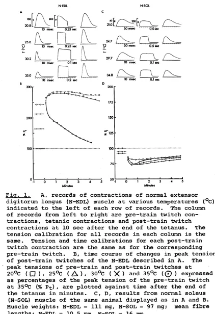
Fig. 1, A, records of contractions of normal extensor digitorum longus (N-EDL) muscle at various temperatures (°C) indicated to the left of each row of records. The column of records from left to right are pre-train twitch con tractions, tetanic contractions and post-train twitch contractions at 10 sec after the end of the tetanus. The tension calibration for all records in each column is the same. Tension and time calibrations for each post-train twitch contraction are the same as for the corresponding pre-train twitch. B, time course of changes in peak tension of post-train twitches of the N-EDL described in A. The peak tensions of pre-train and post-train twitches at
20°C ( □ ) , 25°C (
A
) / 30°C (X
) and 35°C(0
) expressed as percentages of the peak tension of the pre-train twitch at 35°C (% Pt) / are plotted against time after the end of the tetanus in minutes. C, D, results from normal soleus (N-SOL) muscle of the same animal displayed as in A and B. Muscle weights: N-EDL = 111 mg, N-SOL = 97 mg; mean fibre [image:44.536.69.491.35.651.2]S-EDL S-SOL
A
50
20.0
10 msec 0 .2 5 sec
10 msec Ö.V sec
3 4 .6
/V
10 msecr
0.1 sec 10 msec 6.1 sec
200
M inutes M inutes
Ficr. 2. Records of contractions and time course of
changes in peak tension of post-train twitches at various temperatures for self-innervated extensor
digitorum longus (S-EDL) muscle (A, B) and self-innervated
soleus (S-SOL) muscle (C, D) displayed as in Fig. 1. The
experiment was done 346 days after operation. Muscle
X-EDL X-SOL
A C
50 msec 0 .5 sec
U 10 msec Ö i5 s e c
10 msec ö!t sec
10 msec 0.1 sec
5 0 msec
25.4 L j
5Ö msec sec
29.9 J
0.1 sec msec
10 msec Ö.l sec
B D
□ - 0--0- 0—0— 0
Minutes Minutes
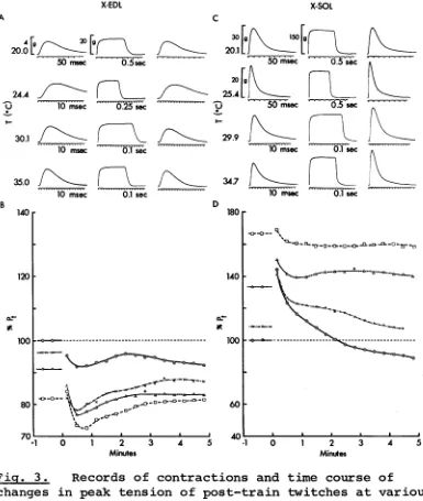
Fig. 3. Records of contractions and time course of
changes in peak tension of post-train twitches at various temperatures for, cross-innervated extensor digitorum
longus (X-EDL) muscle (A, B) and cross-innervated soleus (X-SOL) muscle (C, D ) , displayed as in Fig. 1 except that the tension calibration for X-SOL twitch records at 20.1°C differ from that at other temperatures. These muscles and the normally innervated muscles described in Fig. 1 are from the same animal examined 338 days after the operation. Muscle weights: X-EDL = 69 mg (this muscle was reinnervated by nerve fibres from soleus and peroneal
[image:46.536.83.468.53.508.2]35
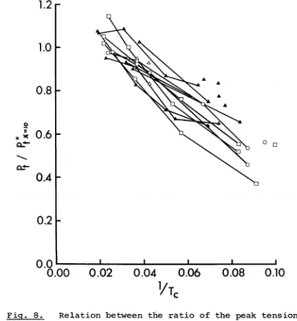
twitch (Pt) , i.e. P t*/Pt .
RESULTS
Representative records of isometric responses of normal (N-EDL, N-SOL) , self-innervated (S-EDL, S-SOL) and cross-innervated (X-EDL, X-SOL) EDL and SOL muscles at various temperatures are shown in A and C of Figs. 1-3. For each muscle, each row of records are, from left to right, the control twitch recorded about 10 sec before the tetanus, the tetanic contraction in response to 200 stimuli, and the post-tetanic twitch recorded 10 sec after the end of the tetanic train. The records shown for each muscle are part of a series described in the graphs below them (i.e. in B and D of Figs. 1-3) in which the peak twitch tensions before and after
repetitive stimulation at 20°C ( Q ), 25°C ( ) , 30°C ( X )
and 35°C (
O
)/ expressed as percentages of the pre-train Pt at 35°C, are plotted against time after the end of repetitive stimulation.36
the same as those of corresponding normal muscles (Fig. 1) # the speed of contraction of X-EDL (Fig. 3A) is decreased
I if*»
and that of X-SOL (Fig. 3C) is decreased. These effects of nerve cross-union on the speed of muscle contraction in mammals have already been described in detail (Buller, Eccles & Eccles, 1960b; Close, 1965b; Buller & Lewis,
1965b).
At 35°C, the post-train records of muscles innervated by the EDL nerve (i.e. N-EDL, S-EDL and X-SOL shown in
Figs. 1A, 2A & 3 C , respectively) show PTP with little or no change in the post-train contraction time. The degree of potentiation for N-EDL and S-EDL is between 2.1-2.5, and this is comparable to the maximal value of 1.9 re ported for EDL muscles from juvenile rats stimulated in vitro (Close & Hoh, 1968b), but the value for X-SOL is much less, being only 1.45. At lower temperatures, the pre-train P fc of these muscles rise, and at 20°C the pre train Pt is approximately the same as the peak tension for the post-train twitch at 10 sec after the end of the
37
tensions of these muscles are raised to about the same
level by repetitive stimulation using 200 stimuli at
temperatures between 20 and 35°C or by lowering the
temperature from 35 to 20°C, and that PTP is maximal
at 35°C and disappears at 20°C. These features, which
are essentially the same as those in EDL muscles in
vitro (Close & Hoh, 1968a), are clearly shown in the
graphs below the records for each muscle (i.e. Figs.
IB, 2B & 3D for N-EDL, S-EDL & X-SOL, respectively).
These graphs also show that the time course of decay of
PTP at 35°C for N-EDL and S-EDL is about the same while
that for X-SOL is more rapid and is followed by a tran
sient phase of depression which probably results from
superimposed PTD. At lower temperatures, PTP decays
more slowly in all muscles.
The peak twitch tensions of muscles innervated by
SOL nerve (i.e. N-SOL, S-SOL and X-EDL, shown in C & D of
Figs. 1 & 2 and A & B of Fig. 3) are depressed by a fall
in temperature as well as by repetitive stimulation. In
N-SOL and S-SOL, PTD is more pronounced at 35°C and the
Pt*x=10 is relatively temperature independent. At 35°C,
A B
1.7 r
r
s
I I I I I___________ I
0 1 2 3 4 5
Minutes
I___________ I
Minutes
1.5
r
Fier. 4. Averaged time course of PTP or PTD at 35°C following 200 stimuli at 200c/s in 4 N-EDL (A),
5 N-SOL (B) , 3 S-EDL (C) , 5 S-SOL (D) , 7 X-EDL (E)
and 9 X-SOL (F) muscles. Ordinates: ratio of peak
tension of post-train twitch (Pt*) to peak tension
of pre-train twitch (Pt) , i.e. Pt*/Pt ; abscissae,
A B
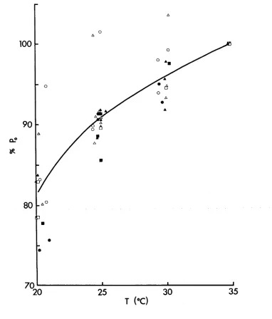
Fig, 5. Relation between peak tension of pre-train and post-train twitches and temperature for N-EDL (A),
N-SOL (B) , S-EDL (C) , S-SOL (D) , X-EDL (E) and X-SOL (F) . The peak tension of pre-train ( O ) and the first post train ( Zik. ) twitches for each muscle at different
temperatures are expressed as percentages of the peak
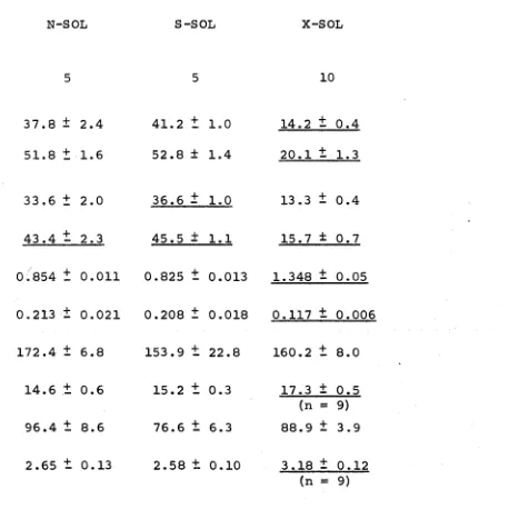
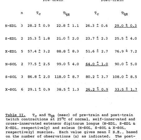
TABLE I
N o . of muscles Pre-train
T c (msec) T^r (msec) Post-train
T c (msec)
T^r (msec)
(Pt*x=10)/Pt
P t/po
p0 (g)
L (mm)
M (mg)
P0L/M (kg/cm2 )
N-EDL
4
11.3 + 0.5 8.8
t
0.312.1 ± 1.0
9.3
t
0.32.143 ± 0.187
0.155
t
0.010283.8
t
9.311.0
t
0.2109.3
t
9.32.90
t
0.17S-EDL
3
11.2 ± 0.3 9.0
t
0.011.5
±
0.89.3 ± 0.3
1.958 ± 0.146
0.147
t
0.002237.5 ± 5.4
12.8
t
0.6104.0
t
6.92.91 ± 0.10
X-EDL
8
27.9
±
1.2 40.9 - 2.726.3
±
0.935.1 ± 2.2
1.021 t 0.034
0.309
t
0.02851.6 ± 6.8
12.2 ± 0.6 (n = 6) 54.0 - 4.5
(n = 7) 1.35
±
0.16(n = 6)
Table I . Summary of properties for normal, self-innervated and cross-innervated extensor digitorum longus (N-EDL,
TABLE I (continued)
N-SOL
5
37.8 ± 2.4 51.8 + 1.6
33.6
±
2.043.4
t
2.30.854 t 0.011
0.213
±
0.021172.4 ± 6.8
14.6 ± 0.6
96.4
t
8.62.65 ± 0.13
S-SOL
5
41.2
t
1.0 52.8 ± 1.436.6 ± 1.0
45.5 ± 1.1
0.825
t
0.0130.208 ± 0.018
153.9
t
22.815.2
t
0.376.6 ± 6.3
2.58 ± 0.10
X-SOL
10
14.2 ± 0.4 20.1 ± 1.3
13.3 ± 0.4
15.7 ± 0.7
1.348 ± 0.05
0.117 ± 0.006
160.2
t
8.017.3 ± 0,5 (n = 9) 88.9 ± 3.9
3.18
±
0.12 (n = 9)10 sec after the end of repetition stimulation at 200c/s for 1 sec. The mean values for post-train T c and T^R which are significantly different from corresponding pre-train values, and those for all other properties of self-innervated and cross-innervated muscles which are significantly different from corresponding mean values for normal muscles, as indicated by the t-test
[image:55.536.59.517.62.529.2]38
muscles are minimal at about 30 sec after the end of
repetitive stimulation and return to pre-train values
over a period of about 10 minutes. The time course of
recovery from PTD at lower temperatures is very similar
to that at 35°C.
The results obtained from other normal,
self-innervated and cross-self-innervated EDL and SOL muscles are
very similar to those described above. A summary of the
contractile properties of these groups of muscles is shown
in Table I. Fig. 4 shows the averaged time courses of
PTP or PTD of these groups of muscles at 35°C and Fig. 5
shows the relations between temperature and pre-train and
post-train peak twitch tensions of these groups of muscles.
The t-test shows that self-innervation of EDL and
SOL muscles produced no significant changes in all the
properties listed in Table I except that the mean PQ of
S-EDL is less (P < 0.0125), and the mean fibre length of
S-EDL is longer (P < 0.05), than that for N-EDL. Fig. 4
shows the similarity between the averaged time course of
PTP for N-EDL (A) and S-EDL (C) , and of PTD for N-SOL (B)
and S-SOL (D). The curves for the decay of PTP in N-EDL
39
massively stimulated juvenile EDL muscles _in vitro (Close
& Hob, 1968b), and this may be due to PTD or neuromuscular
depression superimposed on PTP in these muscles. Fig. 5
shows that the relations between temperature and the pre
train and post-train peak twitch tensions for N-EDL (A)
and S-EDL (C) , and for N-SOL (B) and S-SOL (D) , are the
same.
PTP in N-EDL and S-EDL is accompanied by small
increases in Tc and Ti^r , but t-tests show that these are
not significant (P>0.20) . PTD in all N-SOL muscles is
accompanied by a decrease in Tc and Tj^r , and t-tests
indicate that the post-train Tc of S-SOL (P< 0.01) and
post-train T^R of N-SOL (P< 0.025) and S-SOL (P<0.05)
are significantly reduced. These changes are probably
due to a decrease in the duration of the active state as
suggested by Bowman, Goldberg and Raper (1962) who re
ported a decrease in Tc associated with PTD in cat SOL
muscle.
All the 8 X-EDL muscles studied were innervated
accidentally by nerve fibres from the peroneal nerve in
addition to the intended innervation by SOL nerve fibres.
40
nerve were excited during these experiments, and this
P L
accounts for the low mean values for -°— and P^. The
M °
PQ is low also because the diameter of X-EDL muscle fibres
innervated by SOL nerve fibres is about half of that jor
muscle fibres innervated by peroneal nerve fibres (Close,
personal communication). The smaller size of X-EDL muscle
fibres innervated by the SOL nerve would partly account
for significant differences in the mean muscle weight
between X-EDL and N-EDL (P < 0.0005) and between X-EDL
and S-EDL (P < 0.0005) .
The mean pre-train Tc and Ti^r of X-EDL are signi
ficantly greater than those for N-EDL (Tc , P < 0.0005;
T^r , P < 0.0005) and S-EDL (Tc , P < 0.0005; T^R , P<0.0005),
but these values are also significantly lower than those
for N-SOL (Tc , P < 0.0025; T^R , P < 0.01) and S-SOL (Tc ,
P < 0.0005; T^r , P < 0.01). Four X-EDL muscles whose pre
train Tc range from 29 to 31 msec show PTD at 35°C, while
4 others with pre-train Tc ranging from 23.5 to 26 msec
show a small degree of PTP at 35°C. The (Pt*x=10)/Pt for
all these muscles range from 0.91 to 1.21. The Pt of
those X-EDL muscles which show PTD are depressed by a