CYCLOPHYLLIDEAN CESTODES
by
Jennifer M. Shield
A thesis submitted for the degree of Doctor of Philosophy in the
Australian ational University
The work reported in this thesis, except where specifically mentioned, was performed by me.
presence of a nervous system. Most features of animals are shared with other forms of life - for example, metabolism, growth, and inheritance. But the behavior of animals endowed with a nervous system sets them apart by its complexity. The organization and function of the machinery of behavior are surely the highest achievements in the natural world.
Fronti~piece: Dipylidium ~niE~ scolex.
The whole mount is stained by the 5-bromoindoxyl acetate technique for esterases and much of the nervous system is evident.
V
ACK OWLEDGEMENTS
I should like to express my sincere appreciation to my supervisor, Professor J,D, Smyth of the Australian National University for his guidance, inspiration and assistance during this study.
I am also grateful for the assistance given by
members of staff and fellow students of the Department of Zoology in the Australian ational University. In
particular, I would like to thank Dr W.L. Nicholas who read and commented on the manuscript, Dr M.J. Howell who discussed much of the work with me and read and commented ~n the manuscript, Dr V.A.P. Harris for help and advice
in the physiology experiments, Mr D.D. Heath and Mr R. Pengilley who assisted in the supply of material, Mr I. Fox who processed the photographs, Mr A.B. Howkins who gave technical assistance in the early months of this study, and Mrs V. Blackburn who translated a number of German and Russian papers and typed part of the manuscript.
My thanks are also due to Professor G. Burnstock of the Department of Zoology in the University of
Melbourne for the use of the facilities in his department for the study of the fluorescent histochemistry of bio-genic monoamines, I am also very grateful to Mr J.R. Mclean and Mrs J.B. Hurley of the Department of Zoology in the University of Melbourne for teaching me the
fluorescence technique and for their comments on the manuscript of chapter
5,
I would like to thank Dr C. Harrigan of the Veterinary School, and the Department of Pharmacology in the University of Melbourne who assisted considerably in obtaining material while I was working at the University of Melbourne.TABLE OF CONTE TS
Acknowledgements
Chapter 1. GENERAL I TRODUCTION The problem
Materials
The structure of the cestode nervous system - a review. Introduction
The morphology of the cestode nervous system
I. Historical
II. The nervous system in cestode groups
1. Order Lecanicephalidea 2 • Order Diphyllidea
J.
Order Spathebothriidea 4 . Order Pseudophyllidea5.
Order Tetraphyllidea 6 . Order Trypanorhyncha7 .
Order Cyclophyllidea III. Basic variation in the~orphology of the nervous system
IV. Sense Organs V. eurocords
[image:7.613.43.599.27.704.2]Chapter 2. A CO TRIBUTIO TO THE MORPHOLOGY OF THE NERVOUS SYSTEM IN SIX
CYCLOPHYLLIDEAN CESTODES
Introduction
Materials and Methods Results
I. The central nervous system 1. Arrangement
2. Histology
vii Page
32
32
32
34
34
34
36
II. The peripheral nervous system
37
1. The inner plexus
37
2 . The peripheral plexus 383. erve endings 39
4.
Innervation of the muscles40
5,
Innervation of the reproductive system6.
Innervation of therostellar gland
7.
Innervation of the excretory system8.
Esterase-positive cellsin the tail of T.
Hydatigena cycticercus
41
43
43
43
Discussion
44
I. T chniques
44
II. Discussion of nervous structures
47
1. Arrangement
47
2. Histology 52
Chapter
J.
5 . Nerve endings
6
.
Innervation of the muscles7 . Innervation of the reproductive organs
8. Innervation of the rostellar gland
RESPONSES OF DIPYLIDIUM CANI UM PROGLOTTIDS TO CHOLINERGIC DRUGS
Introduction
Materials and Methods Results
Discussion
Chapter
4.
A HISTOCHEMICAL STUDY OF CHOLIN-ESTERASES I FOUR CYCLOPHYLLIDEAN CESTODESIntroduction
Materials and Methods
Results
I. Whole mounts:
5-bromoindoxyl acetate technique
ix
Page
Chapter
5.
MONOAMINES IN THE NERVOUS SYSTEM OF97
DIPYLIDIUM CANINUM, HYMENOLEPIS ANA
AND TAENIA HYDATIGENA Introduction
Materials and Methods
Results
I . II.
~ l i d i ~ caninum Hymenolepis ~
III. Taenia hydatigena
Discussion
Chapter
6.
HISTOCHEMISTRY AND ATTACHME T OF THE SCOLEXIntroduction
Materials and Methods Results
I . Anatomy of the scolex of
Dipylidium caninum 1. Tegument
2 . Musculature
J.
Rostellum4.
Nervous systemII. Histochemistry of the scolex of Dipylidium caninum
1. Tegument
2. Musculature
J.
Rostellum4.
Nervous systemPage
III. Attachment in ~ l i d i ~ caninum 116
and Taenia ~datigena
Discussion 117
1. Tegument
2. Rostellum
J.
ervous System117
119
123
Chapter 7. GENERAL DISCUSSION 124
124
SUMMARY
APPENDIX 1
APPENDIX 2
REFERENCES
1. Evidence for neurohumoral
transmission in cestodes
2 • Possible functions of the two
types of nerves
127
3. Evidence for neurosecretion in 127
cestodes
4. Role of the nervous system in l28
movement
5.
The nervous system inoncospheres
6
.
Comparison of the nervoussystem with that of other
animals
129
130
lJl
133
CHAPI'ER 1
GENERAL INTRODUCTION
THE PROBLEM
The importance of the nervous system, in controlling
physiological and developmental processes by the mediation
of neurosecretory substances,has become widely recognised in recent years,and neurosecretory cells have been
described in most major animal groups. In the
Platyhelminthes, there is some evidence that neuro-secretory cells occur in turbellarians (Ude, 1964) and trematodes (Ude, 1962; Gresson & Threadgold, 1964), and this may indicate that a neuroendocrine system exists in this group.
It has been established (Smyth, 1964) that the rostellar gland cells of 35-day adult Echinococcus
granulosus are secretory. Smyth speculated (p.523) that
1the secretion could be hormonal in nature and its
release may be related to the regulation and maturation of the strobila'. This stimulated the idea that the rostellar gland i n ~" granulosus might be part of a
neurosecretory mechanism. Since this study was commenced,
neurosecretory cells have been described in the rostellum of the cestode Hymenolepis diminuta (Davey & Breckenridge, 1967) 0
Thus, the present study was originally directed towards elucidating the morphological features of the nervous system of~· granulosus, with special reference to any nerve cells which might be neurosecretory. With this aim in view, the paraldehyde fuchsin and
haematoxylin techniques, which typically stain
neuro-se retory cells (Gabe, 1966) were applied, but no cells
stained satisfactorily. Moreover, less striking and less
selective stains were not satisfactory owing to the
difficulty in distinguishing the ganglion cells in the
small scolex of this species. Several other techniques
for nervous tissue were also applied, but none of these
stained the nervous system satisfactorily.
I t was early realised that E. granulosus, in view of
its small size and its refractoriness to selective
histologi al staining, was not very suitable as an experimental organism for this particular problem and
that a broader and more basic approach to the problem of
the physiology of the nervous system was required.
Accordingly Dipylidium caninum, a species common in
local dogs, was chosen as the main experimental organism,
but some other cestode species, including E. granulosus ,
were also examined as they became available.
Using a histochemical approach supplemented by other
techniques, i t has been possible to describe the
morphology of the nervous system in some detail, and to broa h some of the problems in understanding the
physiology of the cestode nervous system.
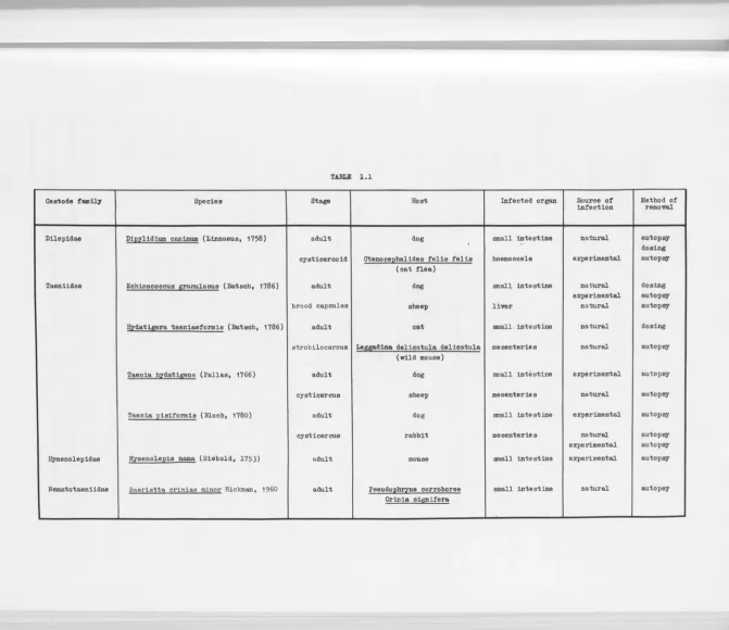
MATERIALS
The estode species used, together with their hosts
and method of olle tion, are listed in Table 1.1. The following details supplement this l i s t .
Most of the adult Dipylidium caninurn and Echinococcus
from properties near Canberra and Goulburn with
t
t o t grain of arecoline hydrobromide (1Hydarex11 ). AdultHydatigera taeniaeformis was obtained by dosing an adult cat with l / J2 grain of arecoline hydrobromide.
Eggs of fleas (Ctenocephalides felis felis) were collected from naturally infested cats and cultured in the medium and under the environmental conditions
recommended by Bruce (1948). The flea larvae were fed small pieces of partially dried gravid proglottids of D. caninum and this procedure resulted in a high
incidence of infection. Adult fleas which were infected as larvae were fed for 1 hour daily by holding the gauze-covered end of their glass container in contact with the shaved belly of a kitten (Fig.1.1). However, difficulty was encountered in keeping them alive until the
cysticercoids were infective, and only one apparently normal cysticercoid was recovered.
Echinococcus granulosus brood capsules and Taenia
QYdatigena cysticerci were obtained from infected livers and mesenteries respectively of sheep slaughtered at the Goulburn abattoir. The larval material used for
experimental infections of dogs with adult~. granulosus and T. hydatigena originated from the same source.
A !!y:datigera taeniaeformis strobilocercus was
obtained from a naturally infected wild mouse collected by C.S.I.R.O. Division of Wildlife at Port Essingdon, Coburg Peninsula, orthern Territory of Australia, and supplied through Miss J.L. Hunter, Department of Zoology, A.N.U.
1
Parke, Davis & Co. , Sydney.
Taenia pisiformis cysticerci from naturally infected
rabbits were fed to dogs in order to obtain adult worms. Oncospheres o f ! • pisiformis were cultured in vitro by Mr D.D. Heath, Department of Zoology, A.N.U., for JO days,
and small undifferentiated cysticerci were obtained. Hymenolepis ~ adults were obtained from infected mice. The original infected mice were derived from an
infected stock maintained at the Department of Parasitology,
University of Queensland.
Baerietta criniae minor adults were obtained from
frogs , juvenile Pseudophryne corroboree and adult Crinia
signifera, collected by Mr R. Pengilley, Department of
Zoology, A.N.U., from Goree Flats, Brindabella Ranges,
near Canberra.
Details of the techniques used in various aspects
TABLE l . l
Ceetode fam~ I Species Stage Hoot Infected organ Source of Method of infection removal.
DUepidaa I D1J2zlidium caninum (Linnaeus, 1758) adult dog small intestine natural autopsy
- dosin8
cysticercoid Ctenoce]2balides felis felis baemocoele experimental autopsy
(cat nea)
Taaniidse I Echinococcus granulosus (Batscb, 1786)
I
adult dog small intestine natural dosingexperimental autopsy
I
I
brood capsules sheep liver naturalautopsy
I
!!izdstij!!ra taeniaaformis (Batsch, 1786) adult
cat
I
small intestineI
natural
I
dosing
strobilocercus Leggadina delicatula delicatula meeenteries natural autopsy
(wild mouse)
Taania bzdstigena (Pallas, 1766)
I
cysticercus adult sheep dog mesenteries small inti, stine experimental natural autopsy autopsyTaania pisiformis (Bloch, 1780)
I
cysticercus adult rabbit dog me senterie small intestine s experimental natural autopsy autopsyexperimental autopsy
ll,ymenolepidaa l Hymenole]2iS nana (Siebold, 1753) adult mouse small intestine experimental autopsy
Nematotaaniidae I Ba~rietta criniae minor Hickman, 1960 adult Peeud0]2~8 corroboree small intestine natural autopsy
Crinia siJl!!_ifera
[image:18.722.39.711.22.602.2]THE STRUCTURE OF THE CESTODE ERVOUS SYSTEM
-A REVIEW
Introduction
What is a nervous system? How do we recognise whether a given animal tissue is, in fact, nervous?
Bullock & Horridge
(1965)
define the nervous system as 1an organized constellation of cells (neurons)specialized for the repeated conduction of an excited state from receptor sites or from other neurons to
effectors or to other neurons'. This definition provides anatomical and physiological criteria to be satisfied by any true nervous system. Thus, presumptive nerve cells must be shown to conne t receptor sites with effectors or to onne t with each other, and physiological evidence of specialisation for repeated conduction of excitation
should support anatomical evidence.
Bullo k & Horridge
(1965)
also point out that there a e two pract· al problems involved in dealing with the nervous system of any one animal: firstly, how torecognise a nerve cell, and secondly, how to prove whether the nervous sytem is, in fact, involved in a parti ular response to a stimulus. The most basic signs of a nerve cell, even when the axon is reduced in
length~ are electronmicroscopic or oscillographic evidence of real functional contact with other cells, themselves clearly neurons, and a reasonably brief action potential and refractory period permitting repeated
In the Cestoda, the nerve cells and nervous systems
have been described on anatomical grounds only - very
l i t t l e physiological evidence on nervous function has
been gathered so far.
Interest in the cestode nervous system has been
stimulated by the following facts:
6
(i) The cestodes belong to the phylum Platyhelminthes,
the most primitive group of animals at the level of
organ-grade construction. The nervous system of these
animals is more highly organised than that of the
Coelenterata1 which, in general, has only an uncentralised
network of connected nerve cells. The first centralised
nervous systems , including distinct brains, are found in
the Platyhelminthes.
(ii) The cestodes are a group of animals extremely
adapted to the parasitic habit . They have no gut, no
morphologically obvious specialised sense organs, and
have no free-living stages apart from embryonic forms
which play no active part in entering the host. A
knowledge of the nervous system is important in
understanding how the parasite interacts with its
environment (a) in activation and establishment in the
host in which organs sensitive to physico-chemical
factors would be important 1 (b) in co- ordination of
movement (probably governed by tactile and
rheo-responses of sense organs) which would be important
in attachment, and , in apolytic forms, in movement
of gravid proglottids in and probably out of the faeces
of the host9 and (c) in, perhaps, an endocrine control
Some of the preceding remarks apply as well to the
Trematoda, which is also a highly specialised parasitic
group, but which has free-living stages provided with
sense organs to deal with their brief sojourn in the
outside world.
Although there have been numerous descriptions of
the gross morphology of the nervous system of cestodes,
very l i t t l e of the microanatomy and physiology is known.
There is considerable difficulty involved in making
suitable histological preparations in which the nervous
tissue is differentiated sufficiently from the parenchyma
for correct identification. This fact has been noted by
many authors, and may explain why there are so many
discrepancies in detail between descriptions of the
nervous system of a particular species by different
authors, for example, according to Dollfus (1942),
Pintner (1880) described four nerves proceeding
anteriorly from the lateral ganglia of Eutetrarhynchus
ruficollis; iemie (1885) , on the other hand, found
eight anterior nerves in the same species. It would seem
unlikely that this dis repancy is due to intraspecific
variation.
M an ·ngful physiological experimentation on the
nervous system of cestodes is difficult, mainly because
they remain viable for only a short time on removal from
the host. Modern methods of in vitro culture have
considerably improved on this state of affairs for
The morphology of the cestode nervous system. I. Historical.
Until recently, the nervous system of cestodes had been described using morphological criteria only.
Because of tbe difficulty in staining cestode nervous
tissue, usually only the main outline of the nervous system was given, with some histological detail.
The first description of the nervous system of a cestode appeared in 1836, when Mueller described a small
flat swelling in the scolex of Tetrarhynchus attenuatus (an insufficiently described species of the order
Trypanorhyncha). This was followed by the work of
Blanchard (1847) who described the nervous system of
some taeniids, bothriocephalids and cysticerci_. He
found in the middle of the scolex a transverse commissure
with a ganglionic swelling at both ends. Each ganglion
gave rise to a band of nerve fibres which ran posteriorly
and anteriorly.
Little was added to this knowledge until the 1880s,
when Golgi impregnation techniques, devised for
vertebrate nervous tissue, were applied to cestodes.
Niemiec (1885) reviewed the literature on the cestode
nervous system, and resolved many of the controversies
which had arisen since 1836. He gave detailed
descriptions of the pattern of the nerve cords and commissures of four cyclophyllidean species, thus providing a basis for further work.
The first microanatomical details of the cestode
nervous system were described from silver impregnation
and methylene blue preparations. Blochmann (1895)
described a subtegumental nerve plexus, sensory cells
and free nerve endings. In 1911 he considered the last
mentioned to be parenchymal cells. Zernecke (1895) also
described sensory cells and an outer plexus of nerve fibres
just beneath the epithelial cells, and, in addition,
ganglion cells and an inner plexus at the same level as
the nerve cords and ring commissures . Despite the
difficulty in distinguishing nervous elements from other
tissues e .g. myoblasts , these descriptions have since
been found to be remarkably accurate, and very l i t t l e has
been added to this information.
The nervous system of the Platyhelminthes in general,
consists of a 'brain' a complex of ganglia and large
commissures near the anterior end - from which a number
of longitudinal nerve cords proceed posteriorly. These
longitudinal nerve cords lie between the outer, muscular
layer, and the internal medullary tissue, and are joined
by a number of transverse commissures on the circumference
of the medullary tissue . This type of nervous system has
been described by Hyman (1951) as the 'barrel-type'
longitudinal system. As well as this, they have a fine
peripheral network of nerves, the 'diffuse' system of
Hyman (1951), which may be homologous with the nerve net
of the Coelenterata.
Within this outline, there are variations between and
within the Platyhelminth groups. Species of Turbellaria
differ considerably from one another in the number of
longitudinal nerve cords and commissures, and in the
complexity of the brain. The shape of the brain and the
number of longitudinal nerve cords (six posterior and
The numerically small groups - the Aspidogastrea and the Cestodaria - both have different plans. The Cestoda, although fairly constant in the structure of the 'brain', have a variable number of longitudinal nerve cords
(Fig.1.2).
II. The nervous system in cestode groups.
10
In the following pages, much of our present knowledge of the nervous system of cestodes is summarised.
Individual species whose nervous systems have been
described are considered together with other members of
the order to which they belong. The system of classification used is that given by Yamaguti (1959), unless the species has been described since Yamaguti 1s treatise was compiled.
1. Order Lecanicephalidea.
So far, knowledge of the nervous system in this order is confined to that of Tylocephalum dierama Shipley &
Hornell, 1906 (Lecanicephalidae), whose nervous system was described by Subramaniam (1941 a). The brain is
described as a cap-shaped plate, from which approximately 19 pairs of longitudinal nerve cords arise and continue posteriorly. In mature segments this number varies from J2 to 42. These nerve cords are interconnected
irregularly by fine fibres, and, at the posterior end of each proglottid, there is a 'plate' commissure, which consists of a meshwork of fibrils. From the main nerves, fibres supply the ovary, testes, vitelline glands, and unicellular sense organs on the inner edge of the
2. Order Diphyllidea.
Again, knowledge of the nervous system in this order
is confined to one species, Ditrachybothrium macrocephalum
Rees,
1959,
This species resembles closely thediphyllidean genus Echinobothrium, but i t does not fit
precisely into the order Diphyllidea, since i t is similar
to the Trypanorhyncha and Pseudophyllidea in the
distribution of the yolk glands. The nervous system of
this species was described by Rees
(1959).
Two lateralganglia are present immediately posterior to the apical
organ, and are linked by a transverse commissure. From
each lateral ganglion arises one pair of anterior nerves
-one dorsal and -one ventral, each of which divides into an
anterior nerve and a posterior nerve passing backwards
to the region of the scolex behind the brain. Two
longitudinal nerve cords arise, one in each ganglion,
and continue posteriorly. In the scolex, 16 pairs of
bothridial nerves originate at intervals in the
longitudinal nerve cords.
J.
Order Spathebothriidea.Cooper
(1918)
briefly described the nervous systemof Bothriomonus intermedius Cooper,
1917
(Diplocotylidae).There are two lateral longitudinal nerve cords, each of
which gives off a large branch to the lateral wall of the
bothria. Anterior to the bothria, the trunks begin to
approach each other and each gives off small branches to
the other, thus forming an irregular transverse
commissure , The tip of the scolex is supplied with a
4.
Order Pseudophyllidea.The nervous system has been described, frequently not in great detail, for a large number of species in the order Pseudophyllidea.
Amphicotylidae.
12
Cooper (1918) briefly described the nervous system for three species of the Amphicotylidae: Eubothrium crassum (Bloch, 1779), Eubothrium rugosum (Batsch, 1786) and Marsipometra hastata (Linton, 1897). In Eubothrium crassum and~· rugosum, Cooper described two lateral longitudinal nerve cords situated dorsally to the cirrus and the vagina. I n [ , crassum, the two lateral
longitudinal nerve cords enlarge in the scolex, and are united by a small transverse commissure in both E. crassum and E. rugosum. In~. hastata, there are similarly
two lateral longitudinal nerve cords which give off a
number of branches to the walls of the bothria and enlarge anteriorly to form two ganglia. Each ganglion divides into two large short branches which are joined to the
corresponding branches on the opposite side by commissures. An apical branch proceeds anteriorly from each ganglionic branch.
Triaenophoridae.
The nervous system of Fistulicola plicatus (Rudolphi, 1819) was described briefly by Cooper (1918) and that of the plerocercoid of an unidentified species of
plerocercoids of Triaenophorus sp. which, in the scolex, form two lateral ganglionic swellings joined by a median commissure. From each lateral ganglion arises an anterior nerve which probably supplies the hooks.
Diphyllobothriidae.
The nervous systems of a number of species of Diphyllobothriidae have been described: Ligula intestinalis (Linnaeus,
1758)
by Cooper(1918),
Schistocephalus solidus' (Muller,
1776)
by Cohn(1898),
an unidentified species of Schistocephalus andplerocercoids of an unidentified species of Diphyllobo-thrium by Lacey
(1955).
The arrangement of nerves inL. intestinalis is very similar to that in Schistocephalus. Each main longitudinal nerve cord has six accessory
longitudinal nerves associated with i t , arranged in three groups of two (i.e. two lateral, two dorsal and two
ventral). In~. solidus, Cohn
(1898)
described two ganglia joined by a commissure in the scolex. In the proglottids, the nerve cords provide two series of nerve fibres - one in the transverse plane, supplying thetransverse and longitudinal musculature, and the other in a frontal plane, contributing in places to a ring commissure. Lacey
(1955)
described seven pairs ofaccessory longitudinal nerve cords in Schistocephalus sp. but no accessory nerves were present in plerocercoids of Diphyllobothrium sp. The bothridia in these plerocercoids were supplied with a pair of nerves from each ganglion.
Bothriocephalidae.
Brief descriptions of the nervous system of
- - - - ~ ~
-~~~-(Linton, 1897) and~. manubriformis (Linton, 1889) were given by Cooper (1918), and that of an unidentified species of Bothriocephalus, by Lacey (1955). A more detailed description of the nervous system of~. s~orpii
(Muller, 1776) was given by Rees (1958) who compared her findings with earlier work by Niemiec (1888) and Lonnberg
(1891). Cooper (1918) noted the presence of two lateral longitudinal nerve cords in~· claviceps, B. occidentalis
and B. manubriformis. This author showed for B. manubri-formis only that these enlarge in the scolex to form lateral ganglia whi ch are connected by a transverse
commissure. Lacey (1955) found that, in Bothriocephalus sp., the ganglia are connected by dorsal, ventral and median commissures. In~. scorpii, Rees (1958) described the brain as two bilobed lateral ganglia, connected by a median transverse commissure posteriorly, and dorsal and ventral commissures, anteriorly. A lateral longitudinal nerve cord arises from each ganglion and continues
posteriorly into the strobila. Two anterior nerves from each ganglion divide into one or more nerves supplying the tip of the scolex, and a nerve supplying the region of the scolex posterior to the brain. Eighteen pairs of bothridial nerves arising in each longitudinal nerve cord supply the long bothria.
Ptychobothriidae
Cooper (1918) outlined the nervous system in Clesobothrium crassiceps (Rudolphi, 1819). The two lateral longitudinal nerve cords enlarge in the scolex to form lateral ganglia which are connected by a
transverse commissure. Anterior nerves arise from the ganglia. Rees (1958) described the nervous system
of C. crassiceps in greater detail. The lateral ganglia
are four-lobed structures, connected by dorsal and ventral
commissures anteriorly (forming a hexagonal commissure)
as well as a median commissure posteriorly. An X-commissure
joins diametrically opposite ganglion lobes. Four anterior
nerves arise from each ganglion, each dividing into one
or more nerves supplying the tip of the scolex, and a
nerve supplying that part of the scolex posterior to the
brain. From each of the two lateral longitudinal nerve
cords, six pairs of nerves supply the bothria,
Haplobothriidae.
Cooper (1918) described the nervous system of the
primary scolex (which contains proboscides similar to
those of the Trypanorhyncha), and of the primary
strobila of Haplobothrium globuliforme Cooper, 1914. Two
lateral longitudinal nerve cords are present throughout
the strobila. Each is swollen into a very large
ganglionic mass at some distance posterior to the
proboscis bulbs. The lateral nerve cords are joined
further anteriorly, by a large but very irregular
trans-verse commissure. At the point of junction arise large
nerves which supply the walls of the proboscis sheaths.
5.
Order Tetraphyllidea.Two families within the order Tetraphyllidea contain
species whose nervous systems have been described.
Phyllobothriidae.
The nervous system of Anthobothrium auriculatum
(Rudolphi, 1819) has been described by Rees (194J), of
16
of the proglottids of Scypophyllideum giganteum (van Beneden,
1858),
by Riser(1949)
and of Phyllobothriumsinuosiceps Williams,
1959,
by William~(1959).
The brain is situated near the anterior end of the scolex andconsists of two four-lobed lateral ganglia which are joined by a dorsal and a ventral commissure anteriorly, and a median transverse commissure posteriorly. From each ganglion, a lateral longitudinal nerve cord passes
posteriorly and continues throughout the strobila. Four nerves arise from each ganglion and supply the bothridia. I n ! • auriculatum, each of the four anterior nerves
divides into three branches, the two inner branches
supplying the myzorhynchus, and the outer nerve extending over the face of the bothridial peduncle. The four
ganglia and the commissures o f ! • auriculatum contain large ganglion cells.
E·
sinuosiceps has a pair ofaccessory lateral longitudinal nerve cords supplying each side of the strobila, in addition to the main lateral longitudinal nerve.
Oncobothriidae.
Rees & Williams
(1965)
and Rees(1966)
havedescribed the nervous system of Acanthobothrium coronatum (Rudolphi,
1819),
The brain consists of two bilobedlateral ganglia joined by a transverse commissure posteriorly and a dorsal and a ventral commissure
anteriorly. Eight anterior nerve cords continue forward from the brain and are joined by an anterior ring
commissure. There are five pairs of longitudinal nerve cords throughout the strobila. These are connected by
Each bothridium and its pair of hooks is supplied by six pairs of bothridial nerves; two pairs originate from the lateral ganglion, two pairs from the locus of the first ring commissure, and two pairs from the locus of the second ring commissure.
The nervous system is not set off from the parenchyma by a discrete limiting layer of tissue. Ganglion cells are present only in the median transverse commissure, and nerve cells are present in the main longitudinal nerve cords within the scolex. The so-called 'binding cells' of Tower (1900) are present around the longitudinal nerve cords in the strobila. Probable sensory cells,
which have long process s extending to muscle fibres, occur in the scolex and may be stretch receptors
(Rees, 1966) .
6.
Ord r Trypanorhyncha.Th nervous system in species of the Trypanorhyncha is remarkably constant in general form, possibly because of the pres nee of the proboscides in the scolex. Many of th arlier studies on the nervous sytem of trypano-rhynchs hav been review d by Dollfus (1942), and the information pres nted by th following authors, Pintner
(1880; 1893; 1925; 1930), Lang (1881), iemiec (1885; 1888), Lonnberg (1891), Poyarkoff (1909), and ybelin
(1918), and given her , has been derived from this source. H patoxylidae
Th n rvous syst ms of th adult stag of Hepatoxylon trichiuri and plerocercoid larva of Dibothriorhynchus
18
resp ctiv ly. According to Yamaguti (1959), these species
are synonymous and their correct designation is
Hepatoxylon sguali
(?
Martiniere, 1797) .The lateral ganglia of~· sguali are four-lobed
structures, joined by a dorsal and a ventral commissure
anteriorly, and by a median commissure posteriorly. Two
lateral longitudinal nerve cords arise in the ganglia and
continue throughout the strobila. The anterior region of
the scolex is supplied by two pairs of nerves from each
ganglion, and the bothridia, by nerves from the lateral
ganglia and the lateral longitudinal nerve cords. In
each ganglion two nerves arise, each supplying a proboscis.
Gilquiniidae.
The nervous system of Gilquinia squali (Fabricius,
1794) has been described by Niemiec (1888), Lonnberg
(1891) and Mackenzie (1965), and Aporhynchus norvegicum
(Olsson, 1868), by ybelin (1918) and Rees (1941 b). In
~. squali, the ganglia are bilobed structures, joined by
three commissures as in H. squali . Two anterior nerves arise in ach ganglion, and two pairs of nerves, one
pair dir ct d ant riorly and another post riorly, supply
th bothridia. The probosc·s n rves, one to each
probos is sh ath, arise in the lateral ganglia. Two
lateral longitudinal n rv cords are pres nt throughout
th strobila. Then rvous syst m of Aporhynchus
urn is very s·milar to that of
g.
sguali . The---~---lat ral ganglia are four-lob d structures. From each
ganglion·two pairs of ant rior nerves supply the tip
of th scol x. Six pairs of nerves supply ach bothridium,
lateral longitudinal nerve cord. As proboscides are
absent, there are no proboscis nerves, Lacistorhynchidae.
The nervous system has been described in the
following species: Grillotia erinaceus (van Beneden,
1858) by Johnstone (1911), Q, scolecina (Rudolphi, 1819) by Rees (1950) and Lacistorhynchus tenue proglottids by Riser (1949). The structure of the nervous system is very similar to that in Hepatoxylon ~uali . The lateral ganglia are four-lobed in G. erinaceus and bilobed in
G. scolecina. In G. scol cina, two pairs of anterior
nerves arise from each ganglion, one pair from each
ganglionic lobe, In G. erinaceus, two pairs of
bothridial nerves arise in each ganglion. In G. scolecina, there are three pairs of bothridial nerves, one pair
branching from the anterior nerves, one pair from each
lateral ganglion, and one pair from each lateral longitudinal nerve cord. In G. erinaceus a columnar ganglion containing ganglion cells is present as a
swelling on each lateral longitudinal nerve cord just posterior to the lateral ganglion. The proboscis nerves originat in the columnar ganglia in G. erinaceus, and the lat ral ganglia in Q, scolecina. Interproglottidal
nerve comm·ssures linking th two lateral longitudinal nerve cords are present in L. tenue.
T ntacularndae.
Pintner (1925) d scribed the nervous system of Tentacularia coryphaenae Bose, 1797 (as Stenobothrium
is similar to that of Hepatoxylon sguali. Four anterior
nerves are present, and each main lateral longitudinal
nerve cord gives off two accessory longitudinal nerves which lie very close to the main longitudinal nerve cord.
The proboscis nerves arise in the lateral ganglia.
Eutetrarhynchidae.
The nervous system of Eutetrarhynchus ruficolle
(Eysenhardt, 1829) has been described by Pintner (1880)
and Niemiec (1885). Two pairs of anterior nerves arise
in each ganglion. A single proboscis nerve from each
ganglion divides into two near its origin, each branch
supplying a proboscis sheath. Bothridial nerves also
aris in each lateral ganglion, just posterior to the
junction with the dorsal and ventral commissures .
Gymnorhynchidae.
Lang (1881) described the nervous system in
Gymnorhynchus gigas (Cuvier, 1817) . The nervous system
is very similar to that in Hepatoxylon sguali, except
that eight anterior nerves are present, four arising
from each lateral ganglion.
The nervous system has also been described in a
number of trypanorhynch sp cies which are qpecies
incerta s dis, according to Yamaguti (1959). These
include T trarhynchus papilJifer (by Poyarkoff, 1909),
Tetrarhynchus gracilis (by Lang, 1881), Tetrarhynchus
smaridum (by Pintner, 1893) and Tentacularia macropora
proglottids (by Subramaniam, 1940). Lacey (1955) has
20
d scr·b d th nervous system in an unidentifi d plerocercus
larval stage of a trypanorhynch. These descriptions are
Tentacularia macropora, in which Subramaniam (1940)
claimed that a large number, approximately 60,
longitudinal nerve cords are present in the proglottids,
Judging from copies of his photographs, i t would appear
that the structures described as nerve cords are probably
longitudinal muscle bundles . He described the
innervation of various internal organs including the
ovary by fibrils from a nervous plexus situated between
the vitelline glands and the testicular vesicles.
For the Trypanorhyncha in general, the brain
consists of two lateral ganglia, joined by a do~sal and
a ventral commissure anteriorly, and by a median
commissure posteriorly. Each ganglion consists of two
(a dorsal and a ventral) or four (anterior and
posterior dorsal, and anterior and posterior ventral)
lobes which are fused posteriorly. There may be two or
four anterior nerves arising from each ganglion. Two
lateral longitudinal nerve cords are present throughout
the strobila each arising from the posterior point of
fusion of the dorsal and ventral ganglionic lobes of the
corresponding lateral ganglion. The bothridia are
supplied with anterior, lateral, and/ or posterior nerves
from the lateral ganglia, and/ or lateral nerves from
the longitudinal nerve cords within the scolex. Four
proboscis nerves arise, either two from each ganglion,
or two from each longitudinal nerve cord, close to the
posterior end of the ganglion. These follow the
proboscis sheaths to the base of the bulbs .
The median transverse commissure contains bipolar
and multipolar nerve cells, and neurocordal cells.
There are non rve c lls in th lateral 'ganglia' and the
dorsal and ventral commissures. The lateral 'ganglia' cannot therefore be considered as true ganglia, since
they contain no nerve cells. In Grillotia erinaceus,
the brain is covered by a delicate capsule containing nuclei (Johnstone, 1911). eurocords, which have been
described only in this group of cestodes, will be
considered later.
7.
Order Cyclophyllidea.This is a very large and comparatively diverse order. The main differences in nervous system
morphology found within this group are in the number
of longitudinal nerve cords, and in the arrangement
of nerves in the scolex. The latter is probably
associated with the presence or absence of a rostellum.
Taeniidae.
Descriptions of then rvous system include those
of the following species: Taenia pisiformis (Bloch, 1780), by Niemiec (1885) and Lacey (1955); Hydatigera
taeniaeformis adult by Cohn (1898) and Bart ls (1902),
and strobiloc rcus by Res (1951); Multiceps multiceps
(Lesk, 1780) by Niemiec (1885), and Taeniarhynchus saginatum (Goeze, 1782) by iemiec (1885) and by Cohn
(1898).
Dilepida
Th n rvous system of Dipylidium caninum has been
describ d by i miec (1885).
The p t t rninth n rvous system is essentially the same in sp cies of Ta niidae and Dilepidae. Th re are
ten longitundinal nerve cords throughout the strobila,
where they are linked by ring cornmissures. In the
scolex, the three lateral nerve cords on each side run
together to form the lateral ganglia. The anterior
extensions of the lateral ganglia and the median nerve
cords continue forward and meet the rostellar nerve ring.
The lateral ganglia are joined by a transverse cerebral
commissure, and by one or more polygonal cornmissures which
unite them with the median longitudinal nerve cords. The
latter are also linked by an X-cornmissure. erves from
the rostellar nerve ring supply the rostellum, and nerves
from the lateral ganglia and median longitudinal nerve
cords supply the suckers.
Anoplocephalidae.
Within this family, the number of longitudinal nerve
cords varies from species to species. The nervous system
of three species belonging to the sub-family
Thysano-somatinae have been described: Thysanosoma actinoides
Diesing, 1835 by Lacey (1955); Avitellina lahorea Woodland,
1927 by Subramaniam (1941 b); and
A
·
centripunctata(Rivolta, 1874) by Gough (1911). The nervous systems of
four species of the sub-family Anoplocephalinae have also
been described: Moniezia expansa (Rudolphi, 1805) by
Tower (1896; 1900); ~. benedeni (Moniez, 1879) by Tower
(1896) as~. planissima; Anoplocephala magna (Abildgaard,
1789) by Becker (1922); and
A·
perifoliata (Goeze, 1782)by Cohn (1898).
Thysanosoma actinoides, Anoplocephala magna and
A
·
perfoliata all have ten longitudinal nerve cords, and the
arrangement of ganglia, nerve cords and cornmissures in the
Moniezia expansa and M. benedeni have six
longitudinal nerve cords, the accessory lateral nerve
cords being absent. Within the scolex of~· expansa the 24
nervous system is very similar to that of the Taeniidae, except that the lateral ganglia themselves curve medially
and fuse in the centre of the scolex, instead of being
joined by a cerebral commissure.
Both Avitellina lahorea and A. centripunctata have only two longitudinal nerve cords. In A. centripunctata,
four ganglia, two lateral, one dorsal and one ventral,
have been described in the scolex. Each of these is joined to the adjacent and to the diametrically opposite
ganglion by commissures. A central nerve plate occurs
anterior to this complex, but no connections between
the plate and other elements of the nervous system have
been established.
Davaineidae.
Siddiqi (1961) described the nervous system of
Cotugnia digonopora (Pasquale, 1890). Two longitudinal
nerv cords are present, and, in the scolex, the central nervous system comprises an anterior rostellar nerve ring, a cephalic ganglionic mass dorso-ventrally compressed and
laterally longated, and eight sucker nerves, two to each
suck r.
Tetrabothriidae.
This family has closer affinities to Pseudophyllidea
and Tetraphyllidea (se Wardle & McLeod, 1952) than do
most oth r cyclophyllid an families, and therefore one
patt rn. The nervous system of Tetrabothrius affinis
(Lonnberg, 1891) has been described by Rees (1956).
Six longitudinal nerve cords run throughout the strobila. At the base of the scolex these are linked by a hexagonal
nerve ring. The longitudinal nerves continue forwards,
the lateral nerves becoming the lateral ganglia. These are connected by the median transverse commissure and a l i t t l e further forward by a ring commissure. An
X-commissur is also present. Two anterior nerves on each side supply the apical organ while bothridial nerves
arise from the lateral ganglion and the lateral portion of the hexagonal nerve ring. Spatlich (1909) also described
six longitudinal nerve cords in T. laccocephalus Spatlich,
1909.
Tower (1900) described ganglion cells in the lateral ganglia and n rve cords of Moniezia expansa, and also binding cells covering the lateral ganglia, and the nerve
trunks within mature proglottids. erves occur in the suckers of Avitellina centripunctata and Anoplocephala magna, according to Gough (1911) and Becker (1922) resp ctiv ly.
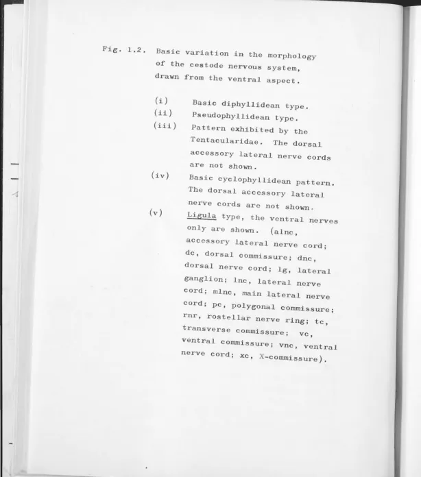
III. Basic variation in the morphology of th nervous system.
t ·s possible to classify the cestode central nervous system into five morphological types based on those
describ d i n the foregoing r view, provided that nerves supplying structures in th scolex such as suckers and
proboscid s ar omitt d from the discussion (s e Fig. 1.2). (i) Th basic patt rn consists of two lateral
~
/
median transverse commissure. From the lateral ganglia
and lateral nerve cords, nerves arise, supplying the
muscular organs and tegument of the scolex and strobila.
The Diphyllidea and Spathebothriidea have this type of
nervous system. In all other cestodes these features are
26
present, together with additional commissures and/ or nerve
cords.
(ii) The second type has linking the lateral ganglia two
commissures, one dorsal and one ventral, in addition to
the median transverse commissure. The dorsal and ventral
commissures together form a 'ring' commissure, which
joins the ganglia in front of the median transverse
cornmissure. The lateral ganglia may consist of two or four
lobes. This condition is found in the Pseudophyllidea
apart from Ligula intestinalis and Schistocephalus solidus,
in the Tetraphyllidea apart from Phyllobothrium sinuosiceps
and Acanthobothrium coronatum, and in the Trypanorhyncha
apart from th Tentacularidae.
(iii) Six longitudinal nerve cords are present in the
third type of nervous system. There may be two accessory
lateral longitudinal nerve cords on each side in addition
to the main lateral longitudinal nerv cords (Fig. 1 .2) ,
or there may be two dorsal and two ventral median
longitudinal nerve cords. The arrangement of the nervous
system in the scolex is similar to the second type
above, th accessory lateral n rve cords fusing with the
main lateral nerve cord on each sid to form the lateral
ganglia. The tetraphyllidean Phyllobothrium sinuos, ceps
and the trypanorhynch family Tentacularidae belong to
(iv) In the fourth type, ten longitudinal nerve cords are present in the strobila, three pairs of lateral nerve
cords, and two dorsal and two ventral median nerve cords.
In the scolex, the arrangement is usually more complex
than the patterns described above. There is an additional
complete or incomplete dorsal and ventral commissure
linking the lateral ganglia with the dorsal and ventral nerve cords, together with an X-commissure linking
diametrically opposite median longitudinal nerve cords via the cerebral (median transverse) commissure, The
median longitudinal nerve cords and the anterior
extensions of the lateral ganglia meet the apical, or rostellar, nerve ring, from which nerves supplying the
apex and rostellar structures arise. This is the basic arrang ment for the Cyclophyllidea. In this group, some
species have a reduction in the number of nerve cords, but th arrangement of nerves in the scolex generally rema·ns the same. Ten longitudinal nerve cords are also
pr sent in the tetraphyllid, Acanthobothrium coronatum
(Rees, 1966) but this species has no apical nerve ring.
(v) The fifth type is a very simple nervous system, and
consists o the basic arrangement, together with
additional long'tudinal nerve cords. Cohn (1898) claimed
that there are 14 longitudinal nerve cords in the
pseudophyllid species Ligula !£:testinalis and
Schistocephalus solidus, while Lacey (1955) described 16 in Schistocephalus sp. Subramaniam (1941 a) described a large number (32 to 42) of longitudinal nerve cords in the lecanicephalid, Tylocephalum dierama. Subramaniam
(1940) also described a large number of longitudinal nerve
Fig. 1.2. Basic variation in the morphology
of the cestode nervous system,
drawn from the ventral aspect.
(i)
(ii)
(iii)
(iv)
(v)
Basic diphyllidean type.
Pseudophyllidean type.
Pattern exhibited by the
Tentacularidae. The dorsal
accessory lateral nerve cords are not shown.
Basic cyclophyllidean pattern.
The dorsal accessory lateral
nerve cords are not shown.
Ligula type, the ventral nerves
only are shown. (alnc,
accessory lateral nerve cord;
de, dorsal commissure; dnc,
dorsal nerve cord; lg, lateral
ganglion; lnc, lateral nerve
cord; mlnc, main lateral nerve
cord; pc, polygonal commissure;
rnr, rostellar nerve ring; tc,
transverse commis~ure; vc,
ventral commissure; vnc, ventral
[image:42.650.0.615.19.715.2](i) (ii)
tc
lg lg
VC
Inc Inc
(iv)
(iii)
cc rnr
lg
lg
XC
pc
mine dnc
mine
olnc vn c
11
olnc
I
(v)I
tc
lg
mine
vnc
II
olncTentacularia macropora. As noted earlier, i t is possible
that in thes two species, Subramaniam may have confused
longitudinal muscle bundl s with nerve cords in his
metallic ·mpregnation preparations.
28
Rees
(1958)
has sugg sted that the nervous system incestodes is intimately connect d with the musculature and
that th re should obviously be a correlation between the
typ of adhes·ve apparatus and the nerves which supply its
const·tu nt parts.
w·thin the Ps udophyllid a, Bothriocephalus scorpii, which has two very long, weakly muscular bothridia, has
18
pairs of bothridial nerves originating at intervalsin th longitudinal nerv cords in the scolex.
Cl sobothrium crassic ps, which has a shorter scolex and
four small r, more muscular bothridia, has six pairs of
bothridial nerv s . Res
(1958)
explained this reduction·n number of bothridial n rves in C. crassiceps as a
- I
cons quenc of th shorter scolex, and smaller, more
muscular bothridia. In th T traphyllidea, this
cone ntration is ven mor marked, as the central zone of th scol x i s greatly reduc d i n size and the
bothridial nerves, which are few in number, arise in
almost the same place, .g. in Anthobothrium auriculatum
(Res,
1943)
and Phyllobothrium dohrnii (Rees,1946)
.
Thos c stodes with an almost spherical scolex and four
highly muscular sucking cups have only on to three
s parat n rv s to ach sucker, e.g. Hydatigera
t formis (R s , 1
51)
and T trabothrius affinis(R
s,1956)
.
Th prin ipl suggest d by R s
(1958)
also appliesIn the Trypanorhyncha, as mentioned earlier, each proboscis sheath is supplied with a nerve arising in or near the
nearest lateral ganglion. The innervation of the proboscides is very similar in all species, since the gross morphology of the proboscides is essentially uniform.
I . Sense organs.
Hyman
(1951)
states that sensory cells are of two types: (i) sensory nerve cells, or neurosensory cells, which are essentially nerve cells with an axon running to the central nervous system, and (ii) non-nervous sensory cells which are modified epithelial cells and must be supplied by fibres from the nervous system.Th sensory cells of cestodes are mostly of the neurosensory type. As mentioned earlier, these were first described by Blochmann
(1895)
and Zernecke(1895).
They consist of a cell body lying deep in the outer, muscular zone of the body, with a process ending below or in the tegument, and an axon to the inner plexus on the periphery of the internal medullary region of theproglottid. Free nerve endings have been frequently described. They have a variety of shapes, and may be a simple process (Morseth,
1966),
or a bulb with(Mors th,
1967
b) or without (Blochmann,1895)
a small process protruding b yond th tegument . There may be a small funnel within th tegument with its base near the ap x of the bulb (Dollfus, 1942). These are possibly long proc sses of sensory cells which could not be traced to the cell body.Eversible sensory pits in the frontal margin of the
bothridia, occur in the Otobothriidae. Each is a compact organ consisting of a pouch with a slit-like opening. Circular and radial muscle fibres are responsible for
the motility of the organ, and i t is supplied with sensory hairs similar to those of the rest of the tegument . Peribothridian sensory grooves are similar
to the eversible pits, apart from their elongate shape. They are found in the margin of the bothridia of
Grillotia scolecina.
A sensory function for these organs is conjectured, purely on morphological grounds and has not been
confirmed by physiological experimentation.
V. Neurocords.
JO
Descriptions of these structures in cestodes were reviewed by Dollfus (1942). eurocords have been
reported only by ·Pintner (1925, 1934), who described them in a number of trypanorhynch species .
The neurocords are giant fibres which closely
Because of their size, structure, and their direct contact with separate parts of the nervous system, they were considered by Pintner (1934) to function in the transmission of motor stimuli,
They are probably analogous to the giant nerve fibres found in many other invertebrate groups (Prosser & Brown, 1961). These giant fibres function in fast conduction of nerve impulses. Thus, in the earthworm, giant fibres mediate quick end-to-end startle contractions, in
crustaceans they control quick flipping of the abdomen, and in cephalopod molluscs, stimulation of a giant fibre elicits maximal contraction of the mantle muscle.
32
CHAPTER 2
A CONTRIBUTION TO THE MORPHOLOGY OF THE NERVOUS SYSTEM IN SIX CYCLOPHYLLIDEAN CESTODES
INTRODUCTION
When some histochemical tests for esterases are used on platyhelminth material, i t has been frequently found that the reaction product of the enzyme hydrolysis is localised in the nervous system (for example, see Lee, Rothman
&
Senturia, 1963; Halton&
Jennings, 1964). Suchtechniques include the 5-bromoindoxyl acetate technique of Holt & Withers (1952) and a number of different methods involving the use of acetylcholine and butyrylcholine and
their corresponding thiol esters and halide salts as
substrates. These techniques are therefore very valuable in augmenting the results from routine histological
procedures which do not stain the cestode nervous system as selectively; moreover, they reveal fine details which are rarely encountered in routinely prepared material.
In the present study, histochemical techniques for esterases have been appli d i n the study of the nervous system of six cyclophyllidean cestodes.
MATERIALS AND METHODS
Taenia pisiformis, Baerietta criniae minor and cysticercoid of D. caninum were studied in some detail . Additional
information on the structure of the cestode nervous system was obtained by observations on~- granulosus brood
capsules,
g.
taeniaeformis strobilocerci, Taenia hydatigena adults and cy~ticerci, and T . pisiformis cysticerci and cultured oncospheres ,The cestode material and the techniques used for studies described in this chapter are summarised in Table 2.1.
Sections of material fixed in 4 per cent formaldehyde at
4°c
were cut in the cryostat, and either the Holt & Withers (1952) technique for non-specific esterases or the Karnovsky & Roots (1964) technique forcholinesterases was employed. Further details are given in chapter
4.
Whole mounts of D. caninum and B. criniae minor were incubated in the media of Holt & Withers and Karnovsky & Roots respectively. Cultured oncospheres of'.I.·
pisiformis and portions of~· granulosus brood capsulewall and
'.I.·
hydatigena cyst wall were also prepared as whole mounts. Sections of adult~· granulosus andg
.
taeniaeformis, after incubation in the medium Karnovsky & Roots, were counterstained in Gower's carmine (seeJohri & Smyth, 1956) in order to show the relationship of other organs to the nervous system,
[image:49.645.44.636.16.680.2]Species
Dipylidium caninum
Echinococcus granulosus
Hydatigera taeniaeformis
Taenia hydatigena
Taenia pisiformis
Baerietta criniae minor
Stage
adult
cysticercoid
adult
daughter cyst adult
strobilocercus
adult
cysticercus
adult
cysticercus
cultured oncospheres
adult
TABLE 2.1
Fixative
4% formaldehyde
Zenker•s fixative
Bouin•s fixative
4% formaldehyde 4% formaldehyde 4% formaldehyde 4% formaldehyde 4% formaldehyde
Zenker•s fixative
4% formaldehyde
4% formaldehyde
Zenker•s fixative
4% formaldehyde
4% formaldehyde
Staining Technique
5-bromoindoxyl acetate
Acetylthiocholine iodide
Maximow
Heidenhain's azan
Paraldehyde fuchsin
Chromalum-haematoxylin Acetylthiocholine iodide Acetylthiocholine iodide
5-bromoindoxyl acetate
Acetylthiocholine iodide Acetylthiocholine iodide
Gabe
Mayer's haemalum & eosin
5-bromoindoxyl acetate Acetylthiocholine iodide
Acetylthiocholine iodide
Gabe
Acetylthiocholine iodide
Acetylthiocholine iodide
Reference
Holt & Withers (1952)
Karnovsky & Roots (1964)
see appendix l
Lillie (1954)
Cameron & Steele (1959) Gomori ( 1939)
Karnovsky & Roots (1964) Karnovsky & Roots (1964) Holt & Withers (1952)
Karnovsky & Roots (1964)
Karnovsky & Roots (1964)
see appendix 1 Culling (1963)
Holt & Withers (1952)
Karnovsky & Roots (1964)
Karnovsky & Roots (1964)
see appendix 1
[image:50.730.6.714.18.619.2]RESULTS
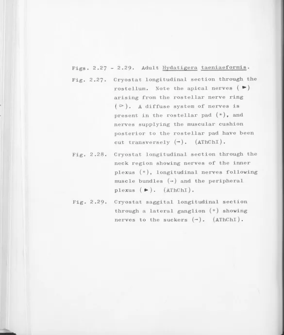
I. The central nervous system: 1. Arrangement .
For the purposes of this description, the central nervous system is considered to comprise the system of nerve cords, ganglia and commissures, and the major nerves supplying structures in the scolex. The
arrangement of these elements in adult Q• caninum, E. granulosus, H. taeniaeformis, T. pisiformis, ~ - criniae minor and cysticercoid of Q• caninum conforms to the basic cyclophyllidean pattern described in chapter 1 .
This is illustrated semi-diagrammatically in Figs. 2.1,
2.2, 2.J, 2.4, 2.5.
There are ten longitudinal nerve cords: two prominent 'main' lateral nerve cords, two pairs of accessory lateral nerve cords, and two ventral and two dorsal median nerve cords (Figs. 2 .6, 2 .8, 2 .10, 2.16,
2.2J, 2.JJ, 2 .J4, 2.40, 2 .46) . In D. caninum adult, the accessory lateral nerve cords are relatively closer to the lateral margin of the proglottids than in the other species studies. Dorsal and ventral nerve cords were not detected in gravid segments of~- criniae minor
(Figs. 2.35, 2.36). The longitudinal nerve cords are linked, in each proglottid, by an interproglottidal nerve ring in D. caninum (Fig, 2.8), H. taeniaeformis and
~- pisiformis,but an organised interproglottidal nerve ring was not observed i n ~- granulosus or B. criniae minor.
In the scolex, the accessory lateral longitudinal
main lateral longitudinal nerve cord and fuse to form the
lateral ganglia (Figs. 2.7, 2 .15, 2.29, 2 .39) . A large
transverse commissure, the cerebral commissure, connects
the lateral ganglia (Figs. 2.9, 2.14, 2 .22, 2.39) . The
lateral ganglia are also linked to the dorsal and ventral
median commissures by a polygonal commissure (Fig. 2 .14).
An X-commissure links diametrically opposite median
commissures via the cerebral commissure in Q, caninum,
35
~. granulosus, g. taeniaeformis (Fig. 2 .22) and
I
·
pisiformis, but was not detected i n ~. criniae minor.
I n g. taeniaeformis, the X-commissure is connected with
the lateral ganglia by small accessory nerves (Fig. 2 .JO) .
Large nerves, two from each lateral ganglion and one from
each of the median longitudinal nerve cords (Figs . 2 .15,
2.29, 2 .JO, 2.50) supply the suckers .
The anterior extensions of the lateral ganglia and
dorsal and ventral median nerve cords meet the rostellar
nerve ring (Figs . 2 .lJ, 2 .21, 2 .38) . In B. criniae minor,
a similar structure is termed the apical nerve ring
(Fig. 2 .32) since no rostellum is present . From the
rostellar nerve ring, apical nerves arise which supply
the rostellar structures - the rostellar pad, the rostellar
gland and the tegument (Figs . 2 .21, 2 .27) . The number of
these apical nerves was difficult to determine. At least
two are present in Q, caninum (Fig. 2 .6) and there are
probably more i n ~· granulosus (Figs. 2 .13) . In H.
taeniaeformis (Figs. 2.21, 2 .27), i t was again difficult
to count the apical nerves. In these preparations,
branches penetrating the rostellar pad number from 18 to
21, while counts of nerves which do not penetrate the