White Rose Research Online URL for this paper:
http://eprints.whiterose.ac.uk/83535/
Version: Published Version
Article:
Bawazer, LA, Ihli, J, Comyn, TP et al. (3 more authors) (2015) Genetic algorithm-guided
discovery of additive combinations that direct quantum dot assembly. Advanced Materials,
27 (2). 223 - 227. ISSN 0935-9648
https://doi.org/10.1002/adma.201403185
[email protected]
https://eprints.whiterose.ac.uk/
Reuse
Unless indicated otherwise, fulltext items are protected by copyright with all rights reserved. The copyright
exception in section 29 of the Copyright, Designs and Patents Act 1988 allows the making of a single copy
solely for the purpose of non-commercial research or private study within the limits of fair dealing. The
publisher or other rights-holder may allow further reproduction and re-use of this version - refer to the White
Rose Research Online record for this item. Where records identify the publisher as the copyright holder,
users can verify any specific terms of use on the publisher’s website.
Takedown
If you consider content in White Rose Research Online to be in breach of UK law, please notify us by
COMMUNICA
TION
Genetic Algorithm-Guided Discovery of Additive
Combinations That Direct Quantum Dot Assembly
Lukmaan A. Bawazer ,* Johannes Ihli , Timothy P. Comyn , Kevin Critchley ,
Christopher J. Empson , and Fiona C. Meldrum*
Dr. L. A. Bawazer, [+] J. Ihli, Dr. C. J. Empson, Prof. F. C. Meldrum
School of Chemistry University of Leeds Leeds , LS1 7HG , UK
E-mail: [email protected]; [email protected] Dr. T. P. Comyn
Institute for Materials Research School of Process
Environmental and Materials Engineering University of Leeds
Leeds , LS1 7HG , UK Dr. K. Critchley
School of Physics and Astronomy University of Leeds
Leeds , LS1 7HG , UK
DOI: 10.1002/adma.201403185
these techniques are limited by the requirement for complex enzymatic reactions. As an alternative strategy that bypasses the need for genetic engineering, combinatorial methods can be employed. These can be used to explore tens to hundreds of reaction conditions, where the most promising or “lead” condi-tions may be selected based on the structures or properties of the resultant material. [ 17,18 ] Lead conditions can then be used to
narrow the reaction landscape in successive screening rounds. Surprisingly, although combinatorial methods are often used in solid-state chemistry to explore, for example, different reagent compositions, [ 18 ] we are not aware of their use in identifying
soluble additives capable of directing mineralization.
In this article we demonstrate how combinatorial methods can be used in conjunction with effi cient screening processes to rapidly identify combinations of small organic molecules that are capable of directing the formation of photolumines-cent quantum dot minerals in aqueous solution and at room temperature. Indeed, a key feature of biomineralization pro-cesses is that control over mineral formation is achieved using many soluble additives that operate in concert. That this feature has seldom been addressed in bioinspired methods is almost certainly due to the vast number of potential variables, which renders a full, systematic exploration intractable. As a solu-tion to this challenge, we here utilize a genetic algorithm as a bioinspired heuristic that mimics natural evolution. Genetic algorithms use selection, recombination, and mutation strate-gies to rapidly identify and optimize the combination of con-ditions (here, soluble additives), which gives rise to materials with target properties. [ 19 ] Using a pipetting robot to prepare
reaction sets and a UV-light table to rapidly assess the reactions for the formation of photoluminescent minerals, we are able to rapidly identify the key additives that promote the formation of quantum dot superstructures from one-pot aqueous reactions.
Our initial library of organic mediators included 23 compo-nents, of which 17 were amino acids and 6 were surfactants. Stock solutions of the amino acids were prepared to initial concentrations of 100 × 10 −3 M , and explored at concentrations
ranging from 0.01 to 50 × 10 −3 M , while surfactants were
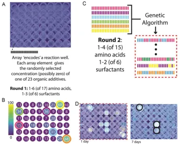
pre-pared to near their solubility limits in water. Surfactants were included as potential structure-directing agents to drive hier-archical assembly in aqueous solution. The overall screening approach used to identify the key additive set is summarized
in Figure 1 . First, library amino acids and surfactants were
ran-domly mixed in 48 wells of a multi-well plate, such that each well contained between 1–6 amino acids and 1–3 surfactants (Figure 1 A). Cadmium chloride and thioacetic acid (as a sulfur source) [ 20 ] were then added to a concentration of 1
× 10 −3 M in
all wells, as precursors for CdS. After 3 d the plate was viewed under UV illumination (Figure 1 A) and with a fl uorimetric Biomineralization, which is the process by which biological
organ-isms form ceramic-based composites, has been widely used as an inspiration for the development of new strategies for materials synthesis. [ 1-3 ] These bioinspired methods are characterized by mild
reaction conditions and promise the ability to generate structures comparable to those found in nature. Central to biogenic control over mineralization is the use of soluble organic additives, where these can even guide the assembly of composite materials [ 4-9 ] with
superior mechanical properties. [ 3,4,10 ] Many bioinspired
minerali-zation strategies therefore utilize either naturally extracted biomac-romolecules or their synthetic analogues, [ 1,2,5,7,9,10 ] and even small
organic species such as amino acids and surfactants can exert con-siderable control over mineralization, sometimes supporting the formation of complex particle assemblies. [ 5,6,8,11-13 ] While
attrac-tive, however, this approach is still hampered by the diffi culty in selecting appropriate organic additives—and in particular combi-nations of additives—to give materials with target structures and properties. As a result, experiments are often performed on a rather ineffi cient “trial-and-error” basis.
Due to the complexity of mineralization processes, we are still not in a position to use theoretical methods to predict the sol-uble additives that will generate materials with specifi c proper-ties. Experimental strategies are therefore required which enable rapid screening of a potentially vast reaction space. As an elegant approach, DNA-based technologies have been used to evolve libraries of nucleic acids, [ 14 ] short polypeptides [ 15 ] and enzymes [ 16 ]
which, when combined with appropriate screening technologies, can be used to identify active biopolymer sequences. However,
[+]Current address: National Institute of Standards and Technology and Stanford University, Stanford, CA 94305, USA
COMMUNICA
TION
plate-reader (Figure 1 B). While no products visibly fl uoresced under UV irradiation, six wells with higher emission intensity were identifi ed using a plate-reader, and were selected as “par-ents.” Each parent's reaction parameters, as described by the concentration of each additive in the well, were then input into a genetic algorithm to create pipetting instructions for a new population of 48 combinatorial reactions (Figure 1 C).
In the second round, additives were dispensed using the new pipetting instructions, and CdS precursors were added as in round 1. Distinct luminescence was now observed by eye under UV illumination in a number of the wells after 1 d of incubation (Figure 1 D, left), demonstrating that the evolution process resulted in increased luminescence. After 7 d, many of the wells lost their distinctive emission (Figure 1 D, right), sug-gesting that either quantum confi nement was lost or the sur-face chemistry of the particles changed (e.g., through ripening or restructuring) as mineralization progressed. However, three reaction conditions maintained their emission properties rela-tive to the rest of the population. Examination of the compo-sitions of these “winning” wells showed that four additives— the amino acids cysteine and aspartic acid, and the surfactants sodium dodecyl sulfate (SDS) and sodium dodecylbenzyl sul-fonate (SDBS)—were conserved in each ( Figure 2 A).
In a fi nal screening round, reactions were prepared such that each well contained each of the four of these conserved
additives (Figure 2 B). The majority of the round 3 wells con-taining surfactant concentrations comparable to those in the round 2 parent wells exhibited intense photoluminescence (Figure 2 B, top three rows). The photoluminescence was dis-tinctly reduced in a control subpopulation in which the sur-factant concentration range was diluted (Figure 2 B, bottom three rows). Just three rounds of screening therefore succeeded in identifying a set of two amino acids and two surfactants that control the formation of fl uorescent CdS products. As the fi nal applied selection pressure was the appearance of distinct fl uo-rescence as visible by eye, additional screening rounds would not necessarily be expected to lead to improved fl uorescence beyond the applied threshold (i.e., qualitative visibility by eye), but could possibly further refi ne the concentrations of the rea-gents required to give this effect.
The morphologies and structures of the products in three of the wells which exhibited the highest photoluminescence in round 3 (Figure 2 B, yellow-highlighted wells) were then charac-terized using transmission electron microscopy (TEM) and elec-tron diffraction. This revealed that CdS nanoparticles ≈2–5 nm
[image:3.595.112.480.74.367.2]COMMUNICA
TION
with CdS (Figure 3 ), while powder X-ray diffraction (PXRD) of materials produced from scaled up (10 mL) reaction conditions demonstrated the formation of wurtzite (hexagonal) CdS. Typical data from particles generated under the conditions of one of the “winning” wells (“Well 1,” 3 × 10 −3 M cysteine,
8 × 10 −3 M aspartate, 0.39
× 10 −3 M SDBS,
0.1 × 10 −3 M SDS, 1.00 × 10 −3 M cadmium
chloride, and 1.00 × 10 −3 M thioacetate) are
shown after 1, 4, and 14 d in Figures S2 and S3 (Supporting Information). For compar-ison, products from two reactions which failed to luminesce in screening round 3 (Figure 2 B, brown-highlighted wells) were also characterized. One of these conditions yielded CdS nanoparticles but no hierarchical assembly (Figure S4A, Supporting Informa-tion), while the other produced only amor-phous materials (Figure S4B, Supporting Information).
The formation of the CdS nanopar-ticle assemblies formed in Well 1 was also investigated over time. Lamellar nanostruc-tures were observed by TEM ( Figure 4 A) after 1 d, where these were amorphous by selected area electron diffraction (SAED), Figure 2. Overview of screening round 3. A) Schematic showing that the arrays encoding the three winning reactions from round 2 (Figure 1 D, black highlights) share in common the four organic additives shown. B) Photo of 48 CdS mineralization reaction wells from round 3 screening after 7 d (excited by UV light). Round 3 reactions only include the additives shown in (A), with concentrations randomly selected within the ranges indicated. Reaction products from wells showing high photoluminescence (highlighted yellow) and no photoluminescence (highlighted brown) were further characterized.
COMMUNICA
TION
and showed very weak and broad diffraction peaks in PXRD (Figure S2, Supporting Information). After 14 d, the composite structures were larger and exhibited more defi ned lamellae consisting of CdS nanocrystals (Figure 4 B; and Figure S3, Supporting Information). UV–vis spectroscopy was also used to study the evolution of these hierarchical composites. Early in the reaction (15 min), an apparent absorption band-edge appeared at ≈365 nm (Figure 4 C), which indicates quantum dot
formation. After 1 d, absorbance increased over all wavelengths, while at 4 d, the absorbance continued to increase with the simultaneous emergence of an additional, red-shifted apparent band-edge at ≈380 nm. UV–vis of the 14 d reaction products
showed an apparent band-edge at ≈410 nm (Figure 4 D), which
corresponds to an individual particle size of ≈4 nm. [ 21 ] The
broad emission peak (centered at ≈460 nm, Figure 4 D, inset)
of the corresponding photoluminescence spectrum, however, suggests some polydispersity in size. By comparison, a con-trol reaction performed under identical conditions but lacking thioacetate exhibited no absorbance at any stage of the reaction (Figure 4 C, “controls”).
The fl uorescence behavior of the CdS superstructures also provides valuable insight into the environment of the parti-cles. The emission maximum of ≈460 nm can be attributed to
excitonic fl uorescence, which arises from the radiative recom-bination of thermally detrapped electrons with valence band holes. [ 22 ] This is typically only observed when hole traps are
blocked in an excess of Cd 2+ ions, due to the formation of S-Cd
bonds. In aqueous solutions, this is achieved in the presence of both excess cadmium and hydroxide ions, but has also been seen on the formation of nanoparticle assemblies. [ 23,24 ] The
latter effect appears to be due to a reduc-tion in the degree of hydrareduc-tion of the CdS nanoparticles in these environments, which reduces the number of surface traps, and thus increases the effi ciency of the excitonic emission.
Considering then the composition of the “winning” reaction solutions, the effi cacy of the four most active additives likely arises from the presence of carboxy, thiol, and sul-fonate moieties, which are known to bind effectively to CdS. [ 26,27 ] At the same time,
how-ever, it is not readily apparent why these four additives were collectively conserved to the exclusion of other structurally similar amino acids (such as glutamate). Indeed, a range of different amino acids have previously been shown to serve as capping ligands for CdS, including Arg, Met, Val, Glu, Asp, Gly, His, Pro, Ser, and Trp. [ 28–30 ] Thus, while the
chem-ical mode by which the four selected additives serve as binding ligands is well established, [ 26 ]
the possibility that they could act in concert to control CdS superstructure formation would have been diffi cult to predict a priori, without the use of genetic algorithm-based optimiza-tion and rapid reacoptimiza-tion screening.
The mechanisms by which these nanopar-ticle assemblies form, and the necessity for the four key additives, are intriguing. Our data show that the surfactant and metal ions initially assemble to form lamellar-type structures (Figure 4 A), where this occurs prior to the evo-lution of highly crystalline CdS. Quantum dot growth then progresses as sulfur is released from thioacetate degradation, where this takes place over several days. [ 20 ] Growth of the
super-structures is then likely to occur by two mechanisms: accretion of the quantum dots onto the existing superstructure templates, and nucleation (and subsequent growth) of CdS quantum dots at the metal sites of the metal-organic scaffolds. This process is consistent with the observed red-shift in the UV–vis absorption (Figure 4 C), where this corresponds to the formation of larger quantum dots. [ 25 ] That the CdS particles grow within a
prestruc-tured composite matrix, in an excess of Cd 2+ ions, is also likely
to give rise to the observed fl uorescence behavior.
[image:5.595.49.359.74.303.2]Further insight into the relative importance of the four addi-tives in generating the fl uorescent quantum dot superstruc-tures was obtained by conducting a series of reactions in which either one or two of the four additives were removed from a “winning” (Figure 3 ) reaction. All of these new reactions exhib-ited either no, or reduced (<40%) fl uorescence (Figure S5, Sup-porting Information), and only the sample with all four addi-tives showed fl uorescence under UV illumination that was visible to the naked eye. This confi rms that all four additives indeed act in concert to generate highly fl uorescent CdS prod-ucts. Interestingly, removal of SDS alone completely eliminated fl uorescence (Figure S5, Supporting Information), but some fl uorescence was observed under conditions featuring the addi-tional removal of one of the other three additives. Thus, none of the additives are essential to fl uorescence, but all contribute Figure 4. Growth of quantum dot superstructures from a “winning” reaction (3 × 10 −3 M cysteine,
8 × 10 −3 M aspartate, 0.39
× 10 −3 M sodium dodecylbenzyl sulfonate, 0.1
× 10 −3 M SDS, 1.00
COMMUNICA
TION
to the fl uorescence properties of the “winning” reactions. One possible concerted mode of action might involve specifi c packing ratios of hydrophobic tails controlling superstructure assembly, while amino acids might cap nascent quantum dots to confi ne them to sizes amenable to accretion with the preex-isting superstructures. The detailed mechanisms operating in this system are clearly worthy of further study.
The work described here therefore demonstrates that a simple aqueous mixing approach, combined with genetic algorithm opti-mization heuristics, permits the rapid identifi cation of small, sol-uble organic molecules that can act in combination to generate fl uorescent CdS quantum dot superstructures in a single-pot reaction. We stress that our method is used to rapidly converge on target properties, and thus circumvents the diffi culties associ-ated with strategies based on predicting—and then exploiting— complex mineralization mechanisms. Variations of this strategy could incorporate diverse starting chemistries and alternate fi t-ness functions (e.g., by selecting materials with narrow emis-sion spectra) to achieve new aqueous routes toward quantum dot materials for applications in labeling, sensing, and photonics. Beyond quantum dots, this general approach can exploit a vast combinatorial reaction space offered by water-soluble minerali-zation-mediating additives, and can be directed to the discovery and optimization of new self-assembly mechanisms and “green” synthetic routes to wide range of advanced materials.
Experimental Section
Prior to each robot dispensing, organic additive stocks were prepared fresh in water (see Supporting Information). Scripts to generate pipetting instructions and perform genetic algorithm optimization were custom written using National Instruments LabVIEW 2011 software (see Supporting Information). A Hamilton MicroLab Star liquid-handling robot was used to prepare combinatorial reactions in 96-well plates (see Supporting Information). After reaction incubation, the multi-well plate was imaged on a UV light table (maximum emission: 305 nm). A Perkin-Elmer EnVision plate reader was used for fl uorimetric measurements, using an excitation fi lter centered at 260 nm (10 nm bandwidth), a dichroic mirror at 315 nm, and an emission fi lter centered at 595 nm (60 nm bandwidth). After reaction incubation, materials were prepared for TEM or XRD characterization by washing precipitates with water and ethanol by pelleting and centrifugation, and drying them in air on TEM grids or silicon substrates for PXRD analysis (see Supporting Information). For UV–vis and fl uorescence spectra shown in Figure 4 D, the washed suspension was measured directly in ethanol; while for the UV–vis time-course study (Figure 4 C), spectra were acquired directly in reaction solution (see Supporting Information).
Supporting Information
Supporting Information is available from the Wiley Online Library or from the author.
Acknowledgements
This work was supported by a UK Engineering and Physical Sciences Leadership Fellowship (FCM and LAB, EP/H005374/1). Electron microscopy was conducted at the Leeds Electron Microscopy and Spectroscopy Centre (LEMAS). The authors thank Rachel Cavill
(Maastricht University) for helpful discussions and Stuart Warriner (University of Leeds) for access to the liquid dispensing robot.
Received: July 16, 2014 Revised: September 12, 2014 Published online:
[1] F. Nudelman , N. A. J. M. Sommerdijk , Angew. Chem Int. Ed. 2012 ,
51 , 6582 .
[2] L. A. Bawazer , MRS Bull. 2013 , 38 , 509 .
[3] J. Aizenberg , J. C. Weaver , M. S. Thanawala , V. C. Sundar , D. E. Morse , P. Fratzl , Science 2005 , 309 , 275 .
[4] J. Seto , Y. Ma , S. A. Davis , F. Meldrum , A. Gourrier , Y.-Y. Kim , U. Schilde , M. Sztucki , M. Burghammer , S. Maltsev , C. Jaeger , H. Coelfen , Proc. Natl. Acad. Sci. USA 2012 , 109 , 3699 .
[5] F. C. Meldrum , H. Coelfen , Chem. Rev. 2008 , 108 , 4332 .
[6] L. A. Estroff , L. Addadi , S. Weiner , A. D. Hamilton , Org. Biomol. Chem. 2004 , 2 , 137 .
[7] A. M. Belcher , X. H. Wu , R. J. Christensen , P. K. Hansma , G. D. Stucky , D. E. Morse , Nature 1996 , 381 , 56 .
[8] M. Li , H. Schnablegger , S. Mann , Nature 1999 , 402 , 393 . [9] R. L. Brutchey , D. E. Morse , Chem. Rev. 2008 , 108 , 4915 .
[10] Y.-Y. Kim , K. Ganesan , P. Yang , A. N. Kulak , S. Borukhin , S. Pechook , L. Ribeiro , R. Kroeger , S. J. Eichhorn , S. P. Armes , B. Pokroy , F. C. Meldrum , Nat. Mater. 2011 , 10 , 890 .
[11] S. Mann , Nat. Mater. 2009 , 8 , 781 .
[12] Y. Yin , A. P. Alivisatos , Nature 2005 , 437 , 664 .
[13] Y. F. Lu , H. Y. Fan , A. Stump , T. L. Ward , T. Rieker , C. J. Brinker ,
Nature 1999 , 398 , 223 .
[14] L. A. Gugliotti , D. L. Feldheim , B. E. Eaton , Science 2004 , 304 , 850 . [15] S. R. Whaley , D. S. English , E. L. Hu , P. F. Barbara , A. M. Belcher ,
Nature 2000 , 405 , 665 .
[16] L. A. Bawazer , M. Izumi , D. Kolodin , J. R. Neilson , B. Schwenzer , D. E. Morse , Proc. Natl. Acad. Sci. USA 2012 , 109 , E1705 .
[17] X. D. Xiang , X. D. Sun , G. Briceno , Y. L. Lou , K. A. Wang , H. Y. Chang , W. G. Wallacefreedman , S. W. Chen , P. G. Schultz ,
Science 1995 , 268 , 1738 .
[18] R. Potyrailo , K. Rajan , K. Stoewe , I. Takeuchi , B. Chisholm , H. Lam ,
ACS Comb. Sci. 2011 , 13 , 579 .
[19] A. K. Sharma , K. H. Son , B. Y. Han , K.-S. Sohn , Adv. Funct. Mater.
2010 , 20 , 1750 .
[20] K. Iwahori , T. Enomoto , H. Furusho , A. Miura , K. Nishio , Y. Mishima , I. Yamashita , Chem. Mater. 2007 , 19 , 3105 .
[21] W. W. Yu , L. Qu , Q. Guo , X. Peng , Chem. Mater. 2003 , 15 , 2854 . [22] U. Resch , A. Eychmiller , M. Haase , H. Weller , Langmuir 1992 , 8 , 2215 . [23] N. A. Kotov , F. C. Meldrum , C. Wu , J. H. Fendler , J. Phys. Chem.
1994 , 98 , 2735 .
[24] L. Spanhel , M. A. Anderson , J. Am. Chem. Soc. 1990 , 112 , 2278 . [25] B. O. Dabbousi , J. Rodriguez-Viejo , F. V. Mikulec , J. R. Heine ,
H. Mattoussi , R. Ober , K. F. Jensen , M. G. Bawendi , J. Phys. Chem. B 1997 , 101 , 9463 .
[26] I. Medintz , H. Uyeda , E. Goldman , H. Mattoussi , Nat. Mater. 2005 ,
4 , 435 .
[27] S. S. Narayanan , S. K. Pal , J. Phys. Chem. B 2006 , 110 , 24403 . [28] W. Qiu , M. Xu , X. Yang , Y. Nan , J. Zhang , H. Chen , J. Alloy Compd.
2011 , 509 , 8413 .
[29] R. F. Domingos , C. Franco , J. P. Pinheiro , Environ. Sci. Pollut. R
2013 , 20 , 4872 .