WORKING GROUP ON ACUTE PURCHASING
The Use of Ultrasound (Viability) Scans
in Early Pregnancy Bleeding
May 1998
GUIDANCE NOTE FOR PURCHASERS 98/06
Series Editor: Nick Payne
Trent Development and Evaluation Committee
The purpose of the Trent Development and Evaluation Committee is to help health authorities
and other purchasers within the Trent Region by commenting on expert reports which
evaluate changes in health service provision. The Committee is comprised of members
appointed on the basis of their individual knowledge and expertise. It is chaired by Professor
Sir David Hull.
The Committee recommends, on the basis of appropriate evidence, priorities for:
the direct development of innovative services on a pilot basis;
service developments to be secured by health authorities.
The statement that follows was produced by the Development and Evaluation Committee at
its meeting on 20 October 1998 at which this Guidance Note for Purchasers (in a draft form)
was considered.
THE
USE OF ULTRASOUND (VIABILITY) SCANS IN EARLY PREGNANCY
BLEED
ING
AUTHORS:
Calvert N W, Singleton C D, Tromans P M. Sheffield: Trent Institute for Health
Services Research, Universities of Leicester, Nottingham and Sheffield 1998. Guidance Note
for Purchasers: 98/06.
EXPERT ADVISORS TO TRENT DEC:
Dr N W Calvert, Research Fellow, Health
Economics, ScHARR; Dr C D Singleton, Consultant in Public Health, North Derbyshire
Health Authority; Mr P M Tromans, Consultant in Obstetrics and Gynaecology, Chesterfield
& North Derbyshire Royal Hospital.
(The recommendations made by the Committee may not necessarily match the personal opinions expressed by the experts)
May 1998
THE USE OF ULTRASOUND (VIABILITY) SCANS IN
EARLY PREGNANCY BLEEDING
N W Calvert
C D Singleton
P M Tromans
Series Editor: Nick Payne
Trent Institute for Health Services Research
Universities of Leicester, Nottingham and Sheffield
GUIDANCE NOTE FOR PURCHASERS 98/06
Published by the Trent Institute for Health Services Research
© 1998 Trent Institute for Health Services Research, Universities of Leicester,
Nottingham and Sheffield.
ISBN: 1 900 733 24 2
Referencing information:
Calvert NW, Singleton CD, Tromans PM.
The Use of Ultrasound (Viability) Scans in
Early Pregnancy Bleeding
Sheffield: Trent Institute for Health Services Research,
Universities of Leicester, Nottingham and Sheffield, 1998. Guidance Note for
Purchasers: 98/06.
Further copies of this document are available (price £10.00) from:-
Alison Ring
Information Resources
Trent Institute for Health Services Research
Regent Court
30 Regent Street
SHEFFIELD S1 4DA
Tel
0114 222 0703
Fax
0114 272 4095
E-mail scharrlib@sheffield.ac.uk
Please make cheques payable to “The University of Sheffield”
AUTHORS
Dr N W Calvert, is a Research Fellow, Health Economics in the School of Health and
Related Research (ScHARR), The University of Sheffield.
Dr C D Singleton is a Consultant in Public Health at North Derbyshire Health Authority.
Mr P M Tromans is a Consultant in Obstetrics and Gynaecology at Chesterfield & North
Derbyshire Royal Hospital.
ACKNOWLEDGEMENTS
The authors wish to thank the following clinicians for their valuable comments during the
drafting of this paper:-
Miss V A Brown, Clinical Director and Consultant Obstetrician & Gynaecologist, The Jessop
Hospital for Women, Sheffield;
Professor I D Cooke, Academic Head, Department of Obstetrics & Gynaecology, The
Jessop Hospital for Women Sheffield;
Mr T C Li, Consultant Obstetrician & Gynaecologist, The Jessop Hospital for Women,
Sheffield;
Mr P Stewart, Consultant Obstetrician & Gynaecologist, Northern General Hospital,
Sheffield.
The authors also wish to thank Suzy Paisley for undertaking the literature search; and
Gill Rooney, Pat Holmes, and Mike Jacobs for their help in preparing, editing, and proof
ABOUT THE TRENT INSTITUTE FOR HEALTH SERVICES RESEARCH
The Trent Institute for Health Services Research is a collaborative venture between the
Universities of Leicester, Nottingham and Sheffield with support from NHS Executive Trent.
The Trent Institute:
undertakes Health Services Research (HSR), adding value to the research through the networks created by the Institute;
provides advice and support to NHS staff on undertaking HSR;
provides a consultancy service to NHS bodies on service problems;
provides training in HSR for career researchers and for health service professionals;
provides educational support to NHS staff in the application of the results of research;
disseminates the results of research to influence the provision of health care.
The Directors of the Institute are: Professor R L Akehurst (Sheffield);
Professor C E D Chilvers (Nottingham); and
Professor M Clarke (Leicester).
Professor Clarke currently undertakes the role of Institute Co-ordinator.
A Core Unit, which provides central administrative and co-ordinating services, is located in
Regent Court within the University of Sheffield in conjunction with the School of Health and
FOREWORD
The Trent Working Group on Acute Purchasing was set up to enable purchasers to share
research knowledge about the effectiveness and cost-effectiveness of acute service
interventions and determine collectively their purchasing policy. The Group is facilitated by
The School of Health and Related Research (ScHARR), part of the Trent Institute for Health
Services Research, the ScHARR Support Team being led by Professor Ron Akehurst and
Dr Nick Payne, Consultant Senior Lecturer in Public Health Medicine.
The process employed operates as follows. A list of topics for consideration by the Group is
recommended by the purchasing authorities in Trent and approved by the Purchasing
Authorities Chief Executives (PACE) and the Trent Development and Evaluation Committee
(DEC). A public health consultant from a purchasing authority leads on each topic assisted
by a support team from ScHARR, which provides help including literature searching, health
economics and modelling. A seminar is led by the public health consultant on the particular
intervention where purchasers and provider clinicians consider research evidence and agree
provisional recommendations on purchasing policy. The guidance emanating from the
seminars is reflected in this series of Guidance Notes which have been reviewed by the
Trent DEC, chaired by Professor Sir David Hull.
In order to share this work on reviewing the effectiveness and cost-effectiveness of clinical
interventions, The Trent Institute’s Working Group on Acute Purchasing has joined a wider
collaboration, InterDEC, with units in other regions. These are: The Wessex Institute for
Health Research and Development, The Scottish Health Purchasing Information Centre
(SHPIC) and The University of Birmingham Department of Public Health and Epidemiology.
Professor R L Akehurst,
LIST OF ABBREVIATIONS
CA125 Carcinoma Associated Antigen Tumour Markers
CNDRH Chesterfield and North Derbyshire Royal Hospital
EPAUs Early Pregnancy Assessment Units
EPF Early Pregnancy Factor
ERPC Evacuation of Retained Products of Conception
HCG Human Chorionic Gonadatrophin
PAPP-A Pregnancy Associated Plasma Protein
PID Pelvic Inflammatory Disease
PSA Prostate Specific Antigen
RCOG Royal College of Obstetricians and Gynaecologists
SDs Standard Deviations
CONTENTS
Page
EXECUTIVE SUMMARY
11.
INTRODUCTION
31.1 Epidemiology 3
1.2
Scale of the Problem in a ‘Typical’ District 5
2.
THE USE OF ULTRASOUND (VIABILITY) SCANS IN EARLY PREGNANCY BLEEDING: SUMMARY OF EVIDENCE OF EFFECTIVENESS6
2.1 Use of Ultrasound in Confirming Viability of Fetus in Early Pregnancy Bleeding
2.2 Management of Women with Threatened Miscarriage
2.3 Use of Vaginal or Abdominal Ultrasound in Diagnosing Ectopic Pregnancy
2.4 Other Diagnostic Tests for Bleeding in Early Pregnancy
2.5 Summary of Evidence
6
10
11
13
15
3.
COST AND BENEFIT IMPLICATIONS
183.1 Introduction
3.2 Model Description
3.3 Results
3.4 Sensitivity Analysis
3.5 Discussion
18
19
24
25
31
4.
OPTIONS FOR PURCHASERS AND PROVIDERS
345.
DISCUSSION AND CONCLUSIONS
38LIST OF TABLES AND FIGURES
Page
Table 1 Sensitivity and Specificity of Transvaginal Ultrasound in the Diagnosis of Ectopic Pregnancy
12
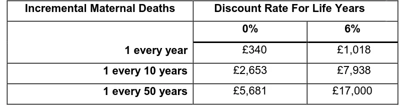
Table 2 Maternal Deaths from Ruptured Ectopic Pregnancies :- A ‘What if’
Analysis of Estimated Cost per Life Year Gained
31
Figure 1a The Scanning For All Policy Decision Tree Model 20
Figure 1b A Protocol-based Access Policy Decision Tree Model 21
Figure 2 Sensitivity Analysis - of a Scan Providing an Unclear Result (p_uncertain) 26
Figure 3 Sensitivity Analysis of an Unclear Blood Test (p_unsure) 27
Figure 4 Sensitivity Analysis of Changes in the Proportion of Non-Viable Pregnancy Patients who are Given a Hospital Intervention (p_hospital)
28
Figure 5 Sensitivity Analysis of the Costs of a Scan (cost_scan1) 28
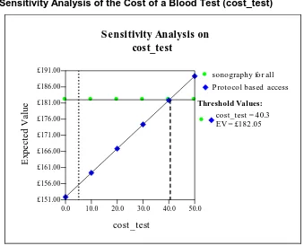
Figure 6 Sensitivity Analysis of the Cost of a Blood Test (cost_test) 29
EXECUTIVE SUMMARY
Introduction - The use of ultrasound scanning in cases of early pregnancy bleeding has increased rapidly as a routine test to assess fetal viability. A result indicating non-viability is
frequently followed by in-patient intervention for evacuation of retained products of
conception (ERPC), although evidence suggests that most non-viable pregnancies would
resolve naturally anyway. In a ‘typical’ district of 500,000 people, some 1,600 women are
estimated to experience early pregnancy bleeding each year. Around two thirds of these will
have a viability scan, of which some one third will require rescans to obtain a clear result.
Question Considered - This guidance note explores the evidence for the effectiveness and costs of investigation of women with bleeding in early spontaneous pregnancy by ultrasound
scanning to assess fetal viability. Although this guidance note accepts that ultrasound is of
proven value for conditions such as ectopic pregnancy, there is concern about the value of
viability scanning in routine spontaneous pregnancy where bleeding occurs at an early
gestational age, (within the first nine weeks). Patients involved in assisted conception or
recurrent abortion are specifically excluded from the analysis.
Evidence Base - There is little documented evidence about appropriateness, or about the safety for the fetus, of scanning at such an early stage. The potential for using biochemical
markers as a substitute for scanning is reported and reference is made to Dutch clinical
guidelines which emphasise the use of non-interventionist primary care and counselling.
Economic Modelling - In the absence of documented economic research evidence, a simple decision tree model for costing three alternative scanning policies is undertaken. The
results indicate that the potential savings for a ‘typical’ district from employing a
protocol-based approach to viability scanning is only about £17,000 per annum (16% of the modelled
cost of the scanning for all policy). Some of these savings would inevitably have to be used
to re-educate patients and staff. Extensive sensitivity analysis shows costs to be relatively
insensitive to changes in the model assumptions. Possible implications for ectopic
management imply relatively good cost-effectiveness ratios for a scanning for all policy,
although whether a protocol-based access policy would affect the identification and
management of ectopic pregnancies is largely unproven.
clearly a need to address the value of such a service and the information made available to
women about the likely risks and benefits of scanning in the early gestation period.
Need for Agreed Protocol - A protocol-based access to viability scanning is likely to be an acceptable option compared to the extremes of allowing free access to scanning at any
gestation or no viability scanning service. However, there is also a clear need for agreement
1.
INTRODUCTION
This guidance note provides a summary of current evidence regarding the investigation of
women with bleeding in early spontaneous pregnancy by ultrasound scanning to assess
fetal viability. The authors consider the benefits and costs of such a service, review
cost-effectiveness and offer options for future provision.
Women who become pregnant through assisted conception techniques, and those with
reproductive problems such as recurrent abortion, previous termination of pregnancy,
previous ectopic pregnancy, a previous episode of pelvic inflammatory disease (PID), or
tubal surgery, are specifically excluded from the discussion in this guidance note.
Blood loss during the first 13 weeks (first trimester) occurs in around 20% of all known
pregnancies. More than 10% of pregnancies ascertained by conventional routine pregnancy
tests end in miscarriage. When tests which are able to indicate pregnancy soon after
conception are used, this percentage is considerably higher. There are several causes of
bleeding in early pregnancy and one of the most important differential diagnoses is that of
ectopic pregnancy.
There has been increasing use of ultrasound scanning to assess fetal viability in
pregnancies where early bleeding occurs since the late 1970s and early 1980s. Mainstream
gynaecological services are increasingly offering ultrasound scans as a routine to women
with bleeding in early pregnancy, in order to assess fetal viability and to ensure the
diagnosis is not that of ectopic pregnancy. In some hospitals, early pregnancy assessment
units (EPAUs) have been set up.
1.1 Epidemiology
Gynaecological complaints are amongst the most common presenting to GPs, and
gynaecological disease accounts for nearly 5% of hospital activity within the NHS. Around
20% of total gynaecological workload is related to early pregnancy problems, which
comprise 50% of emergency gynaecological activity.
Bleeding in early pregnancy is common, occurring in around one-fifth of pregnancies. In
40% - 45% of such pregnancies continuing to term.1 Causes of bleeding in early pregnancy
include:-
threatened miscarriage, missed abortion, blighted ovum;
extrauterine pregnancy;
cervical erosion;
cervical or uterine polyp;
hydatidiform mole.
1.1.1 Miscarriage
Miscarriage is the commonest complication of pregnancy, affecting 12-15% of all pregnant
women.2 The frequency in very early pregnancy is difficult to determine as the pregnancy
may not be recognised. Whittaker et al.3 showed that 8% of human pregnancies are lost at
such an early stage of development that parents are unaware of conception. Estimates
suggest that the occurrence of subclinical pregnancy loss may be as high as 40-60%.4,5
Most (around 60%) of early fetal losses are abnormal karyotypes.
Over the years, there has been a shift towards hospital management and treatment of
miscarriage by uterine curettage. Patients admitted to hospital with miscarriage generally
undergo surgical evacuation of retained products of conception (ERPC), which comprises
75% of all emergency gynaecological operations. Unfortunately, no national statistics on
miscarriage are published except for such hospital activity. It is important to note that
miscarriage is normally a self-regulating process with at least 75% of cases resolving
spontaneously and naturally within a week.
More recently, women with incomplete or missed abortions including blighted ovum are
being managed medically with a combination of anti-progesterone and prostaglandins.
Spontaneous abortion occurs in around 150 per 1,000 pregnancies.
1.1.2 Ectopic Pregnancy
Ectopic pregnancy, the siting of a pregnancy outside the uterine cavity, may result in severe
morbidity and accounts for some 10% of direct maternal deaths,6 mainly from haemorrhage.
It occurred in 1.15% of pregnancies in the UK in 1994-96 and the incidence appears to be
rising in industrialised countries.6,7,8 Factors associated with an increased incidence include
contraceptive devices. Around 50% of cases can be attributed to PID.9 Ectopic pregnancies
can miscarry spontaneously or resorb. They may not present at all or may present as an
acute emergency with pain, bleeding and shock.
Early diagnosis affords the opportunity to use minimal access surgical techniques such as
salpingostomy or medical treatment using methotrexate.10
1.1.3 Other Causes of Bleeding in Early Pregnancy
Cervical ectropion and cervical and uterine polyps may be the cause of bleeding in early
pregnancy. Cervical lesions are usually detectable on clinical examination. The risk of
hydatidiform mole increases with age and it is very rare under the age of 30; diagnosis
requires ultrasound. The frequency differs between countries, but it is very low in all
developed countries.
1.2 Scale of the Problem in a ‘Typical’ District
A district of 500,000 population can expect to have around 5,800 births annually, with
around 7,000 recognised pregnancies, 64 ectopic pregnancies and 920 spontaneous
abortions. Around 1,600 women will experience bleeding during their pregnancy. These
figures do not include those with unrecognised or unsuspected early pregnancy loss.
Between 63% and 73% of women with bleeding in early pregnancy (around 1,000 - 1,160
women in a ‘typical’ district) currently have an ultrasound scan to determine fetal viability.1
2.
THE USE OF ULTRASOUND (VIABILITY) SCANS IN EARLY PREGNANCY
BLEEDING: SUMMARY OF EVIDENCE OF EFFECTIVENESS
There have been several studies which have attempted to define clinical, biological, and
ultrasonic markers which can predict outcome following early pregnancy bleeding. However,
there are no effective treatments which can maintain an intrauterine pregnancy when
adverse factors are recognised. Any investigations or tests performed, therefore, have no
therapeutic consequences in terms of maintaining pregnancy for these patients, although
ERPC may be required.
2.1 Use of Ultrasound in Confirming Viability of Fetus in Early Pregnancy Bleeding
The use of ultrasound in the assessment of fetal viability in patients with early pregnancy
bleeding appears to be generally accepted as good practice in the UK. There are still
questions about the timing and purpose of the test and long-term safety of children who
have been exposed in utero.
Gilling-Smith assessed the use of guidance on routine vaginal examination and ultrasound
in patients with bleeding prior to the 20th week of pregnancy attending an accident and
emergency department in London. Around 70% of patients had first trimester bleeding. A
diagnosis was made on the basis of the clinical examination alone in 47% of cases and
ultrasound was used in 38% of women. The introduction of this guidance on management
reduced the numbers of unnecessary admissions from 28% to 12%, referrals to
gynaecological colleagues from 44% to 22% and the number of re-attendances to the
department from 15% to 4%.12
In 1987, the Dutch College of General Practitioners developed a guideline policy programme
and one of the first evidence-based guidelines to be developed was the management of
(threatened) miscarriage in 1989 (NHG guidelines).13 This is under review at present, but
there are not expected to be any major changes to the guidance.14 The Dutch guidelines
promote the use of expectant management for women with threatened abortion to allow
events to take their natural course, and discourage the use of ultrasound as a routine
investigation. However, they recognise the value of an ultrasound investigation where there
is suspicion of an ectopic pregnancy, where blood loss is heavy, and where it lasts longer
than a week, as these symptoms would justify intervention. Studies have shown that as
many as 10% of women who experience a miscarriage may not contact health services at
were happy to support patients with light spotting in the community, but referred to hospital,
for viability scanning, patients with moderate or heavy bleeding.16 Subsequent work has
shown that almost all women can be managed in the primary care setting, especially if
ultrasound is available.17
Both the above guidelines and the Gilling-Smith study12 confirm that, where clinical
examination demonstrates a dilated cervix, ultrasound is inappropriate (as miscarriage
inevitably occurs). However, in the absence of clinically recognised products of conception
having been passed, an ultrasound scan may exclude retained products of conception and,
therefore, reduce the need to proceed to ERPC either surgically or medically.
EPAUs with direct access from primary care to diagnostic testing facilities reduce the need
for out-of-hours operating, but have not been formally evaluated.18 Of 624 women referred
to an emergency assessment clinic with a provisional diagnosis of threatened miscarriage,
based on a history of amenorrhoea and vaginal bleeding, with or without pain, 25% were not
pregnant and 9.6% had an ectopic pregnancy.19 Although there is some evidence that such
units are efficient, there is little evidence to suggest they provide clinically effective
services.20
Arguments for echographic examinations are usually based on needing to address the
emotional impact of bleeding in early pregnancy for the patient. However, there are no
studies which have explored other options for management. Experience in the Netherlands
suggests that time taken by the primary care physician in counselling and explanation may
be more effective in meeting these needs.13 A recent publication reported on the adherence
to national guidelines by self-selected Dutch midwives.21 This study showed that there was
71% overall compliance with the guidance on not performing an ultrasound scan. For those
cases where a scan was performed, half were related to lack of skills to interpret a physical
examination and half were in response to the clients’ wishes.
2.1.1 Timing of Ultrasound
The gestational sac can be visualised with transvaginal scans at around 4½ to 5 weeks, but
there are no features which are 100% reliable in differentiating viable and non-viable
pregnancy on a single ultrasound examination, when the gestation is less than six weeks.22
Fetal heart activity can be detected from five weeks and six days with good resolution
ultrasound equipment, when the crown rump length is only 3mm. Fetal movements can be
gestation.23,24 The interpretation as to whether an ultrasound scan is correct or not depends
entirely on the gestational age prediction. The fetus grows at approximately 1mm per day
from six weeks gestation, and by seven weeks there should be no difficulty in identifying
fetal cardiac activity, and confirming viability. At a crown rump length of 6mm or more,
absent fetal heart activity definitely indicates non-viable pregnancy (missed abortion).
Following viability demonstrated by cardiac activity using ultrasound at eight weeks’
gestation, between 3.2% and 5.5% of pregnancies were lost subsequently through
miscarriage .2,25
Experience and expertise in gynaecological ultrasound is important to minimise the risk of
misdiagnosis.26 The result of an ultrasound scan depends on the skill of the staff
undertaking the scan and the quality of the machine. This is particularly relevant now that all
junior trainees in obstetrics and gynaecology are expected to be able to scan. Many are
forced to use small machines on the ward at night and weekends, which do not have the
resolution of the larger machines typically used by ultrasonographers or consultant
personnel.
Vaginal ultrasound is superior to abdominal ultrasound for detection of an intrauterine
embryo and its cardiac activity before eight weeks’ gestation. Indeed, any service nowadays
requires a transvaginal scan facility, as it is no longer considered acceptable to rely on
abdominal scanning alone as the diagnostic instrument.27
Local audit has suggested that around 30% of women require two or three scans to
establish a definite diagnosis. The interpretation of ultrasound scanning is clearly dependent
on gestational age.
Other studies have suggested that ultrasound can be misleading in up to 8% of cases
overall.28
Routine ultrasound in early pregnancy (between 10 and 18 weeks) results in earlier
detection of multiple pregnancy and reduced rates of induction for post-term pregnancy, but
it does not improve clinical outcomes,29,30 although it does reduce intervention rates (e.g.
2.1.2 Risk Factors Identified on Ultrasound
Subchorionic bleeding has been recognised as an important prognostic factor. In
pregnancies where there is subchorionic bleeding demonstrated by ultrasound at nine
weeks’ gestation or more, with normal fetal cardiac activity, 80% will progress to term.
Where there is no subchorionic bleeding and normal cardiac activity at this gestation, one
study showed 100% progressing to term.31 The overall miscarriage rate in women with such
signs in the first trimester has been shown to be 9.3%. This rate is 2-3 times higher: with a
large separation compared to moderate or small haematomas, in women aged over 35; and
where vaginal bleeding has occurred at eight weeks gestation or less.32
The use of ultrasound in very early pregnancy with bleeding can create problems. Patients
are difficult to manage if there are uterine contents in the absence of a confirmed non-viable
early pregnancy. Indeed, in early 1998, a woman was granted substantial damages
following scan diagnosed non-viability, which was followed by continuing pregnancy. The
Royal College of Obstetricians and Gynaecologists (RCOG) has produced guidelines
covering ultrasound scanning in early pregnancy.33
The probability of miscarriage following first trimester bleeding ranges from around 6%, if no
adverse factors are present on ultrasound, to 84% if all the following adverse factors are
identified: fetal bradycardia less than 1.2 standard deviations (SDs) from the mean for
gestational age; a discrepancy between the diameter of the gestational sac and the
crown-rump length of more than 0.5SDs below the mean; and a discrepancy between menstrual
and sonographic age of more than one week.34
2.1.3 Safety of Ultrasound
There is a recently compiled Cochrane review of routine early pregnancy ultrasound;35 this
has demonstrated that there appear to be no long-term effects on Norwegian children
exposed to ultrasound early in pregnancy in terms of adverse influence on school
performance or neurobehavioural function. However, fewer of the ultrasound exposed
children than expected were right-handed. Ultrasound was carried out at under 16 weeks’
gestation in only one of the studies reviewed and, even then, not earlier than 10-12 weeks.36
There are no studies which examine the long-term effects of ultrasound on children exposed
There is a view that women are often not informed of the possible risks when offered an
ultrasound scan.
2.2 Management of Women with Threatened Miscarriage
When used to assess fetal viability, ultrasound scans have no management implications for
the care of the patient in that there is no evidence to support the use of curettage if fetal
death is confirmed, although a viable scan can provide some reassurance and planning of
future care. There is a clinical view held by many obstetricians that curettage should be
carried out to evacuate the uterus if fetal death has occurred. This procedure has the
potential to create morbidity by carrying out a surgical intervention in a condition which is
likely to resolve spontaneously within a few days.1 The enthusiasm to evacuate may stem
from fears of hypofibrinogenaemia, which was thought to be a possible consequence of a
slow reabsorbtion of a spontaneous abortion. The psychological consequences of
non-viability and, indeed, of early pregnancy bleeding itself cannot be ignored, however, it must
be remembered that demonstration of viability is not a guarantee of the pregnancy
continuing to term.2,25
The Dutch guidelines promote the use of expectant management for women with threatened
abortion to allow events to take their natural course, and discourage the use of ultrasound,
as miscarriage is not, a priori, an indication for ERPC. In particular, they cite the pressure for
surgical intervention, such as ERPC, if ultrasound results suggest the death of the fetus.
Other studies have suggested that, because no treatment has a documented effect on
maintaining pregnancy, hospitalisation cannot be recommended.37
Curettage has certain risks which include infection, damage to the cervix, uterine
perforation, intra-uterine adhesions, Ashermann syndrome, and risks of necrosis, as well as
the emotional burden for the women concerned. In addition, there is the risk of morbidity
from a general anaesthetic. A recent study has reported that surgical intervention carries a
doubling in the risk of infection compared to expectant management.38 Another study has
shown a 3% infection rate after spontaneous resolution; however, the complication rate for
women undergoing ERPC was 11%.35 Further studies are continuing. ERPC may be
appropriate when there is heavy blood loss, prolonged blood loss (more than one week),
increasing pain or when the emotional burden is such that curettage may be beneficial.13 It
is important to note that the risks associated with ERPC in relation to miscarriage are not as
with the latter have been reduced with the use of routine pre-operative prostaglandins to
soften the cervix. There is now increasing use of medical management of incomplete
abortion, missed abortion, and blighted ovum with progesterone and prostaglandins.
In patients with threatened miscarriage, resolution will occur within four days following
commencement of blood loss in over 2/3 of cases, and over 3/4 will resolve spontaneously
within a week. A prospective randomised trial of expectant management versus ERPC in
miscarriage under 13 weeks showed that both methods of management produced similar
outcomes.35 Spontaneous resolution of the miscarriage occurred in 79% of women within
three days.
The importance of assessing maternal blood group and giving anti-D immunoglobulin in
those who are Rhesus negative must be emphasised.
2.3 Use of Vaginal or Abdominal Ultrasound in Diagnosing Ectopic Pregnancy
Both in the UK and USA, the failure of women with an ectopic pregnancy to seek medical
attention contributes to the numbers of deaths from this condition. Studies have shown that
history and physical examination do not reliably diagnose or rule out ectopic pregnancy. Of
patients with ectopic pregnancy, 9% reported no pain and 36% lacked adnexal tenderness.39
Vaginal or abdominal ultrasound can exclude the possibility of an ectopic pregnancy by
confirming an intrauterine pregnancy on the grounds that a combination of both intra- and
extra-uterine pregnancies is extremely rare (1 per 30,000 pregnancies).
A comparison between vaginal and abdominal ultrasound showed that visualisation of
ectopic pregnancy was 25% with abdominal and 94.7% with vaginal ultrasound. 83% of
women had spotting. Abdominal ultrasound was significantly inferior and vaginal ultrasound
superior to clinical examination.40 In another study, transvaginal ultrasound definitely
identified 82% of ectopic pregnancies on initial examination, with the remainder identified on
repeat examination and Human Chorionic Gonadotrophin (HCG) measurement.41 However,
there can be difficulty in interpreting a pseudogestational sac in 10% to 20% of ectopic
pregnancies at very early gestations.42
Various studies have examined the sensitivity and specificity of ultrasound in the diagnosis
Table 1 Sensitivity and Specificity of Transvaginal Ultrasound in the Diagnosis of Ectopic Pregnancy
Sensitivity Specificity Likelihood ratio# Positive Predictive Value False Positive Rate Study
Diagnosis 69% 90% 99% 88% 69 7.5 16.6% Kaplan39 Durham43 Bonilla44 Transvaginal B mode
98% 86% Chew45
Free pelvic fluid 96.2% 99.4% 160 Sadek46
Tubal mass 81.6% 99.6% 204 Sadek46
Solid adnexal mass
Low High
The combined application of ultrasound and plasma HCG levels seems to be useful in the
diagnosis of ectopic pregnancy when the gestational age is known.47,48 Several studies
have suggested that the risk of ectopic pregnancy is raised by up to four fold with HCG
levels of less than 1,000 iu/l.39,40,49 An intrauterine sac should be visualised with HCG levels
of greater than 1,000iu/l and its absence should raise the suspicion of ectopic pregnancy.50
The diagnostic use of laparoscopy provides a positive diagnosis in more than 90% of cases
and a falsely negative diagnosis in 3-4% of cases, which tend to be early in pregnancy.51
The longer the diagnosis of ectopic gestation is delayed, the more likely it is that rupture will
take place, requiring emergency laparotomy and tubal loss by salpingectomy and increased
risk of maternal death. Furthermore, a ruptured ectopic and collapse is a particularly
traumatic experience for a patient and those with previous ectopics are often anxious about
the management of their early pregnancy.
There is increasing use of laparoscopy in the treatment of ectopic pregnancy. Its use would
be greater if more junior doctors were trained in the use of minimal access surgical
techniques, and out-of-hours theatre staff were familiar with the use of such equipment.
Apart from the obvious advantage to the patient of avoiding large scars, there is less
discomfort, reduced morbidity, a shorter hospital stay and an earlier return to normal
#
activities.52,53 A summary shows that there is fair evidence of benefit, although most studies
are poorly controlled or uncontrolled.54
There is some interest in conservative treatment, including salpingostomy and medical
treatments such as methotrexate, in appropriate patients. The latter has been shown to
have a success rate of 71%-100% with tubal patency rates of 72%-93%.55,56 Spontaneous
resolution of ectopic pregnancy has been observed in at least 15% of patients. Earlier
diagnosis has shown that spontaneous regression of tubal pregnancies is more common
than previously thought.42
The treatment of ectopic pregnancy is the subject of a current Cochrane protocol for a future
review and is not considered further here.10
2.4 Other Diagnostic Tests for Bleeding in Early Pregnancy
A useful summary of the current state of biochemical tests can be found in chapter 10 of
‘Problems in Early Pregnancy: Advances in Diagnosis and Management’.33
i) Human Chorionic Gonadotrophin
HCG estimation should be the primary biochemical diagnostic test used in the differential
diagnosis of abdominal pain in women of reproductive age. Recent recommendations to
establish protocols and algorithms for the management of patients with early pregnancy
bleeding using HCG measurement have been made.33 Free beta-HCG concentrations
have been found to be significantly lower in non-continuing, threatened miscarriage and
tubal pregnancy patients. A cut-off value of 20ng/ml differentiates between viable and
abnormal/non-viable pregnancies with 88.3% sensitivity and 82.6% positive predictive
value.57 In another study the predictive value of HCG in patients with threatened
abortion was 93%, the sensitivity 84%, and the prediction of viability at the given
gestations was 81%.58 However, free beta-HCG testing is currently a research
procedure only and is not available in routine practice.
Generally, the prediction of abortion is more accurate than that of successful outcome.
However, a single normal value of HCG in women with threatened abortion between six
and nine weeks’ gestation, where gestation is accurately known, has been shown in one
study to be associated with a successful outcome in every case.59 HCGs are frequently
for viability. In a viable pregnancy the HCG levels double every 48 hours. A sub-optimal
rise suggests a non-viable intra-uterine pregnancy or ectopic pregnancy.
For HCG measurement, it is important to acknowledge that interpretation is dependent
on gestational age. There is disagreement between professionals about how good
women are at knowing their dates and, thus, how important the need is for ultrasound
scanning to ascertain gestational age.
In one study of women where gestational age was known, a single transvaginal scan
and plasma HCG were used. Using figures for the natural logarithm of HCG value,
Ln(HCG), a result below a pre-defined limit on the distribution of values had a poor
predictive capacity with a sensitivity of 31% and a specificity of 97% for distinguishing
viable from non-viable pregnancy.60
ii) Progesterone
Serum progesterone levels are lower in non-viable pregnancies. Values less than 14.2
ng/ml in asymptomatic pregnancy and 10.5 ng/ml in women with threatened miscarriage
are 100% sensitive in the detection of non-viability.61 A cut-off value of 45 nmol/l as a
single measurement has been found to differentiate between viable and non-viable
pregnancy with a 87.6% sensitivity and 87.5% specificity.62 The addition of serum
progesterone estimations to HCG measurement may increase sensitivity and specificity
in the identification of ectopic pregnancy or failed intrauterine pregnancy, but there is
insufficient evidence at present to recommend routine use.33
Progesterone measurement does not appear to be dependent on gestational age in
early pregnancy.
iii) Other Biochemical Markers
There are several other biochemical markers whose use has been studied in the
management of early pregnancy bleeding, however, none is in current routine clinical
use. These are included for completeness below.
Oestradiol: Levels of oestradiol higher than 30 pg/ml have been shown to correlate in 100% of cases with favourable outcome following bleeding in early pregnancy.22
Carcinoma Associated Antigen Tumour Markers (CA125): Mean serial serum CA125 levels have been found to be higher in women with an unfavourable pregnancy
outcome, as it may help to determine the extent of decidual destruction in threatened
abortion. Levels above 65 U/ml on the first test and 60 U/ml on the second gave a
risk of termination of the pregnancy of 83%; when associated with vaginal bleeding
for three days or more, the risk was 100%.63
Pregnancy Specific Beta 1-Glycoprotein (SP1): Maternal concentrations of pregnancy-specific beta 1-glycoprotein (SP1) have been shown to be reduced in
cases with an unfavourable outcome. The test has been shown to have a sensitivity
of 65%, specificity of 98% and predictive value of 96%.47
Early pregnancy factor (EPF): This has been studied as a potential marker for assessing fetal viability in women with bleeding in early pregnancy. Sensitivity of the
test was 78.9% and specificity 95.6%. The positive predictive value was 93.8% and
the negative predictive value 84.6% and, thus, it could well be a useful prognostic
marker in threatened abortion.64
Pregnancy Associated Plasma Protein A (PAPP-A): This test has been shown to be significantly lower in women with bleeding, but has a positive predictive value of only
18.7% for actual miscarriage and is, therefore, of no clinical value in the assessment
of patients with bleeding in early pregnancy.65,58
Folate:Although folate levels have been shown to be low in a substantial proportion of pregnant women, low folate levels do not appear to be associated with an
increased risk of pregnancy loss or adverse outcome.66
2.5 Summary of Evidence
Bleeding in early pregnancy is a common problem. In over half of these cases the
pregnancy will end in miscarriage. Miscarriage is a natural process and ¾ of cases do not
require medical intervention.
There is a substantial emotional impact on women which should be addressed. However,
there are no studies which have examined the most appropriate and effective way(s) to
counselling from primary care team members is important. There is some disagreement in
the profession about which of the primary and secondary sectors is better placed to provide
such support.
The use of ultrasound to detect fetal viability has gained acceptance as part of routine
medical practice despite a lack of directly applicable studies of good quality. It is important
that the patient and physician understand that there is very little prognostic value in the test.
Although results can allow a care plan to be arranged if fetal viability is shown, there may be
pressure to perform ERPC in non-viability, which will be unnecessary in a high proportion of
women and increases morbidity significantly above that experienced by women undergoing
spontaneous resolution. Medical management of retained products of conception is safer
and uses fewer health service resources than surgical intervention.
There are no studies which have examined women’s views on viability scanning, although
uptake of local services suggests women may find it acceptable and reassuring. There is a
view that women with previous experience of early pregnancy loss and particularly ectopics
are very keen to have early scans.
Ultrasound examinations in early pregnancy should be performed transvaginally.
Evidence suggests that there should be a lower limit on gestation for the use of ultrasound
in threatened miscarriage. Using the test before the eighth or ninth week of pregnancy
requires a high proportion to be repeated in order to determine the diagnosis (although this
proportion is lower with the use of transvaginal compared with abdominal ultrasound). The
few studies which have looked at EPAUs have found a high proportion of women not to be
pregnant. There is, however, some evidence that the use of ultrasound in women who
present to hospital as an emergency can reduce gynaecological referrals and out-of-hours
operations. Misdiagnosis from ultrasound examination is reduced by restricting the use of
the test to those with appropriate experience and expertise, but there will always be a
significant proportion of women who will require a repeat test to establish a diagnosis.
Transvaginal ultrasound scanning has a good sensitivity and specificity for ectopic
pregnancy, although studies reporting high false positive rates are of concern. There is no
published evidence that the use of ultrasound in all women presenting with early pregnancy
There is no published evidence that outcomes for women with ectopic pregnancy are
improved by the use of ultrasound in women presenting with early pregnancy bleeding.
However, there is a clinical view that current best practice should include ultrasound. More
ectopic pregnancies are diagnosed before tubal rupture where there is an EPAU.67
Other diagnostic tests have been studied. Use of HCG measurement has been
demonstrated to be helpful in investigation and management of early pregnancy bleeding.
Progesterone assays may also be of use.
The safety, longer-term effects and outcomes of the use of ultrasound in pregnancies under
10 weeks’ gestation have never been studied. It is important also to offer women balanced
and full information on such matters. Also, the introduction of a Dutch style healthcare
programme of managing early spontaneous abortion, up to nine weeks, in the home for the
majority of women, would require re-education of general practitioners, healthcare workers,
and the general public.
Expectant and medical management of miscarriage, and of ectopic pregnancy, have
3.
COST AND BENEFIT IMPLICATIONS
3.1 Introduction
There are no known published papers looking at the cost-effectiveness of viability scanning
using ultrasound in early pregnancy. Consequently, this section of the report describes a
modelling approach to costing. Three theoretical policy models are presented. In brief,
Option 1 suggests sonography for all women presenting with bleeding in the early stages of
pregnancy irrespective of gestational age. Option 2 is an example of a simplified
protocol-based access policy, which excludes sonography for women presenting with bleeding before
nine weeks’ gestation. Option 3 is a policy of no ultrasound viability scanning for the group
of patients under consideration.
It is worth reiterating here that this guidance note, and hence the costing model discussed
below, is concerned only with women who experience bleeding in early spontaneous
pregnancy. They exclude women who become pregnant through assisted conception
techniques, and those with reproductive problems as listed in the introduction to this
guidance note. They also explicitly exclude ectopic pregnancies in the initial analysis,
although ectopics will be discussed under the sensitivity analysis sub-section of this chapter.
It is important to stress that the following models are not offered as clinical models of care,
but simply as tools with which to estimate likely cost differences between the various policy
options.
A decision tree modelling approach is adopted. This approach makes the assumptions used
in the modelling process transparent. By definition, models are simplifications of reality and
in no way is the model designed to capture all the details of the ‘real world’. In brief, decision
trees describe decision-making processes using activity branches, and decision and chance
nodes. Decision trees can be used to represent decision-making processes for individuals or
cohorts of individuals in a population. Time is represented on the horizontal axis running
from left to right, so that the far right of the tree represents the end-points or outcomes of
the decision making process. The square nodes represent points in time when a decision or
choice is made by decision makers. Round nodes are chance nodes from which branches
emanate representing subsequent events which occur with a given probability. The model
described in this guidance note is a population-based model and, hence, the chance points
usually describe points in time when different sub-groups of the population take different
Activity branches emanating from decision nodes usually imply a financial cost. In our
model, the cost of sonography or the cost of medical or surgical management of miscarriage
are examples of such activity costs. By multiplying the probability of chance events by the
costs of subsequent decision events, the expected cost of each branch of the decision tree
can be calculated and compared.
Decision tree models can be used to model costs alone, outcomes alone, or combinations of
the two, such as cost-effectiveness or cost-benefit analyses. The topic under discussion
makes it difficult to define a specific outcome of interest (see discussion below) and, as
such, the economic analysis in this guidance note is confined to costs only.
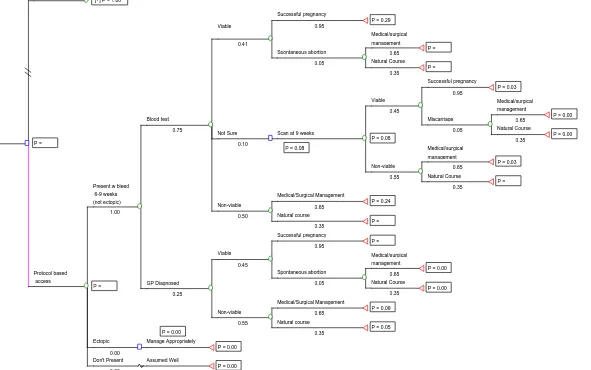
The first two of the three policy options under discussion are represented in Figures 1a and
1b in the form of a decision tree. The third policy option is effectively a sensitivity on the
second and is not, therefore, presented as a separate tree diagram.
3.2 Model Description
The tree models two possible options, the first is where all women presenting between six
and nine weeks’ gestation are given a viability scan using ultrasound. In the second option, a protocol-based policy is assumed in which ultrasound is not given before nine weeks’
gestation. By the very nature of the modelling process, it is necessary to make a number of
simplifying assumptions.
It is assumed that interest is confined to modelling the costs of managing those women who
present with bleeding between six and nine weeks’ gestation. This time period has been
chosen because it is this gestational age for which the value of ultrasound is most in
question. Both ectopic pregnancies and pregnancies with no bleeding between six and nine
weeks are represented in the tree, but assigned a probability of zero occurrence to indicate
Figure 1a
The Scanning For All Policy Decision Tree Model
Successful pregnancy
0.95 P = 0.21 Medical/surgical management
0.65 P = 0.01 Natural Course
0.35 P = 0.00 Miscarriage
0.05 Viable
0.30
Successful pregnancy 0.95 P = 0.07
Medical/surgical management
0.65 P = 0.00 Natural Course
0.35 P = 0.00 Miscarriage
0.05 Viable
0.30
Successful pregnancy 0.95 P = 0.04
Medical/surgical management
0.65 P = 0.00 Natural Course
0.35 P = 0.00 Miscarriage 0.05 Viable 0.45 Medical/surgical management
0.65 P = 0.03 Natural Course
0.35 P = 0.02 Non-viable
0.55 Rescan in 1 week
P = 0.08 Unsure
0.33 P = 0.08
Medical/surgical management
0.65 P = 0.06 Natural Course
0.35 P = 0.03 non-viable
0.37 Rescan in 1 week P = 0.25 Not sure
0.33
P = 0.25
Medical/surgical management
0.65 P = 0.18 Natural Course
0.35 P = 0.10 Non-viable
0.37 Sonography
0.75
successful pregnancy
0.95 P = 0.11 Medical/surgical management
0.65 P = 0.00 Natural Course
0.35 P = 0.00 Spontaneous abortion
0.05 Viable
0.45
Medical/Surgical Management
0.65 P = 0.09 Natural course
0.35 P = 0.05 Non-viable
0.55 GP Diagnosed
0.25 Present w bleeding
6-9 weeks (not ectopic) 1.00 Ectopic 0.00 [+] Don't present 0.00 [+] Sonography for all
P = 1.00
Protocol based access
Figure 1b
A Protocol-based Access Policy Decision Tree Model
Sonography for all
[+] P = 1.00
Successful pregnancy 0.95
P = 0.29
Medical/surgical management 0.65 P = 0.01 Natural Course 0.35 P = 0.01 Spontaneous abortion 0.05 Viable 0.41 Successful pregnancy 0.95
P = 0.03 Medical/surgical management
0.65
P = 0.00 Natural Course
0.35
P = 0.00 Miscarriage 0.05 Viable 0.45 Medical/surgical management 0.65
P = 0.03
Natural Course 0.35 P = 0.01 Non-viable 0.55 Scan at 9 weeks
P = 0.08 Not Sure
0.10
P = 0.08
Medical/Surgical Management 0.65
P = 0.24 Natural course 0.35 P = 0.13 Non-viable 0.50 Blood test 0.75 Successful pregnancy 0.95 P = 0.11 Medical/surgical management 0.65
P = 0.00
Natural Course 0.35
P = 0.00 Spontaneous abortion 0.05 Viable 0.45 Medical/Surgical Management 0.65
P = 0.09
Natural course 0.35
P = 0.05 Non-viable
0.55 GP Diagnosed
0.25 Present w bleed
6-9 weeks (not ectopic)
1.00
Manage Appropriately
P = 0.00 Ectopic
0.00
P = 0.00
Assumed Well
Model 1:- Sonography For All
The top sub-branch of the tree (figure 1a) represents the ‘sonography for all’ policy. Even
with a policy of guaranteed access to ultrasound, Everett and Gilling-Smith indicate that
27% and 17% of referrals with bleeding in the first 20 weeks of pregnancy can be diagnosed
clinically without the need for ultrasound.1,12 On the basis of these results, and given that an
audit at Chesterfield and North Derbyshire Royal Hospital (CNDRH) has indicated that
between 63% and 73% of women presenting with early bleeding are given ultrasound, it has
been assumed that 75% of all women presenting with bleeding between six and nine weeks
are given an ultrasound scan. As such, the model assumes that 25% of presentations can
be managed by GPs, without recourse to scanning.
The model then assumes that the first ultrasound scan indicates either viable pregnancies,
non-viable pregnancy, or gives an unclear result in given proportions. Results from an audit
carried out in Chesterfield,11 indicate that an estimated 1/3 of all ultrasound scans under nine
weeks’ gestation give an unclear result and, therefore, require re-scanning. Another paper17
described a re-scan figure of only 3%, although this was for gestation of up to 20 weeks and
all the re-scans were for pregnancies of less than eight weeks. The model assumes that 1/3
of scans between six and nine weeks’ gestation do not give a clear positive or negative
result.
Everett’s work17
indicates that the ratio of positive to negative scans is more or less 50:50.
To reconcile these results to producing a result of 40-45% of early bleeds leading to a
successful pregnancy,1 our model assumes that 45% of all clear scans give a positive result
(indication of a viable pregnancy) and 55% are negative (indication of a non-viable
pregnancy).
For scans indicating a viable pregnancy, there is then assumed to be a chance event that
the pregnancy goes on to full term or ends in spontaneous abortion. Everett17 has shown
that 95% of positive scans go on to full term. This assumption is used throughout the model.
Using evidence from Everett,1 43% of all women presenting with early bleeding were
admitted to hospital. 86% of these admissions miscarried and required evacuation. That is,
37% of early bleeds required an evacuation in hospital. 42 of the 66 (64%) miscarriages for
which hospitalisation information is known from Everett’s study required ERPC. The model
for evacuation. The other 35% are assumed to resolve naturally. These two assumptions
are used throughout the model.
For the 33% of initial scans assumed to give an indeterminate result, the model allows for a
maximum of two re-scans, so that the second re-scan is forced to indicate a viable or
non-viable result. The sub-tree representing the first re-scan is a clone of the sub-tree
representing the initial scan. The second re-scan sub-tree is similar to those of the first two
scans, but without the branch representing the option of an unclear scan result.
In the sonography for all option, it is assumed that 25% of presenters can be diagnosed by
GPs without recourse to any tests. In line with the assumptions described above, 45% of
these 25% are assumed to be sent home with a viable pregnancy. The other 55% are
assumed to be non-viable. Of the latter, 65% are assumed to require hospital intervention
and 35% to resolve naturally. Of the 45% assumed viable, 95% are assumed to go to full
term, and 5% to abort spontaneously. The latter group is then again subject to the 65:35,
hospital to natural resolution probability assumptions.
In summary, the assumptions used in the modelling of the sonography for all policy imply
that 57% of all women presenting with bleeding between six and nine weeks’ gestation will
have a spontaneous abortion. The other 43% continue to full term. Of the 57% who
miscarry, 65% require a hospital intervention to complete the abortion. The latter implies that
37% of all women presenting with bleeding between six and nine weeks’ gestation will
require a hospital admission for an evacuation or medical intervention. 75% of women
presenting are assumed to require sonography. One third of this 75% are assumed to need
at least 1 re-scan and one ninth of the 75% are assumed to need two re-scans.
Model 2:- Protocol-based Access
The bottom half of the decision tree (figure 1b) represents a protocol-based policy model for
access to sonography for women between six and nine weeks’ gestation. The model assumes that all women presenting with bleeding between six and nine weeks’ gestation
(not suspected ectopics) are given a blood test (e.g. progesterone or serial HCG) to assess
viability. Based upon the indicated sensitivity and specificity of HCG tests, it is assumed that
90% of women presenting can be given a clear positive or negative indication of viability
from the blood test. The assumed ratio of viable to non-viable indications is 45:55 as
explained above. These respective sub-trees then branch out as described under model 1
test are assumed to be asked to ‘watch and wait’ until week nine, at which point they are given an ultrasound scan. Because the scan is taking place beyond the nine weeks’
gestation period, it is assumed that the scan gives a definite positive or negative result. As
such, this scanning sub-tree then takes on the same shape and properties as the second
re-scan sub-tree from the sonography for all policy branch of the decision tree. The proportions
of the population who go to full term, miscarry, and require hospital interventions are the
same as assumed in the sonography for all policy arm of the decision tree.
Model 3:- No Ultrasound
The ‘no ultrasound’ model can be represented in the protocol-based tree by assuming that
the probability of an unclear blood test result is zero. That is, any woman with an unclear
blood test result is asked to watch and wait indefinitely until the GP is able to give a
diagnosis of viable or viable pregnancy. Therefore, all women follow the viable and
non-viable branches in the given ratios.
Input Cost Assumptions
Central case estimates for costing have been made. The costs for an ultrasound scan using
prices quoted by local Trusts range from around £20 to £36. The model uses a central case
estimate of £30 per scan. A paper comparing the costs of treating miscarriage using
surgical and medical management68 implies that the costs of medical management of
patients is about £50 less expensive than surgical management (£347 and £397
respectively). A central assumption estimate of £375 is used in the model. An assumed cost
estimate of £10 is made for the cost of a GP consultation.69 The cost of a blood test (e.g.
progesterone or HCG) is quoted as £5.25 by a local Trust and, as such, the cost of a blood
test has been assumed to be £5.
3.3 Results
Using these cost assumptions, the greatest cost under the sonography for all policy is
estimated at £465 for a woman requiring three scans and who eventually miscarries and
requires a hospital intervention to complete the abortion. The greatest cost under the
protocol-based access is £420 for women who have an unclear blood test and, therefore,
are given a scan at nine week’s gestation, and who then go on to miscarry and require
The lowest cost paths under both policies are for those women whose pregnancies are
diagnosed as viable by their GPs and go on to full term, or those diagnosed as non-viable
by their GP and who abort naturally. The cost assigned to such patients is £10 for the GP
consultation.
The central scenario assumptions described above produce an expected (average) cost per
woman presenting with early pregnancy bleeds of £182 for the sonography for all policy and
£156 for the protocol-based access policy. The expected cost per early bleed for the no
sonography policy is estimated at £153 per patient.
This guidance note has indicated that the number of early bleeds for a district of 500,000
population is 1,600 per annum. Based on miscarriage data presented by Everett,1 it is
assumed that 40% of all bleeds occur in weeks six, seven and eight. Therefore, the
estimated number of women presenting with bleeding between six and nine weeks is 640
per annum. The total costs as modelled for a district are, therefore, £116,512 for the
sonography for all policy, £99,552 for the protocol-based access policy, and £98,112 for the
no sonography policy. The estimated marginal savings of the protocol-based access model
compared with the sonography for all model is, therefore, estimated at £16,960. Having no
sonography at all saves only an estimated additional £1,440 per annum.
3.4 Sensitivity Analysis
i) Proportions and probabilities
The above model contains many assumptions, some of which are based on the results of
research which was not designed to provide the information required for this guidance note.
None of the papers quoted analysed specifically the six to nine weeks’ gestation period. As
such, it is important to assess the sensitivity of the relative costs of each policy model to
changes in input assumptions.
Many of the assumptions are common to the sub-trees representing the two main policies of
interest (sonography for all and a protocol-based access). Changes in these assumptions
will not affect the £26 per patient difference shown in the central case assumptions for the
two policies. Only the magnitude of the relative costs per patient will change. The latter is
true for the probability of hospitalisation, and the probability of a test or scan proving to be
positive or negative. It is also true for the assumed costs of hospitalisation and GP
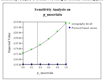
The probability of a scan providing an unclear result is assumed to be 0.33. Figure 2 below
illustrates how the expected costs of the sonography for all policy change as the probability
Figure 2 Sensitivity Analysis of a Scan Providing an Unclear Result (p_uncertain)
S ensiti vi ty Analysis on p_uncertai n
p_uncert ain
E
xp
e
c
te
d
V
al
ue
0.0 0.2 0.4 0.6 0.8 1.0
£2 19.00
£2 11.00
£2 03.00
£1 95.00
£1 87.00
£1 79.00
£1 71.00
£1 63.00
£1 55.00
sonography for all
P rotocol based access
Technically, expected cost is relatively insensitive to a change in the probability of an
uncertain scan in that a change of y per cent in the probability of an uncertain scan result
leads to a less than y per cent change in expected cost. Raising the probability to an
extreme value of 1 increases the expected cost of a sonography for all policy to £217 per
patient. Reducing the probability of an uncertain outcome to zero only reduces the expected
cost to £172 per patient, £16 per patient more than the protocol-based access policy.
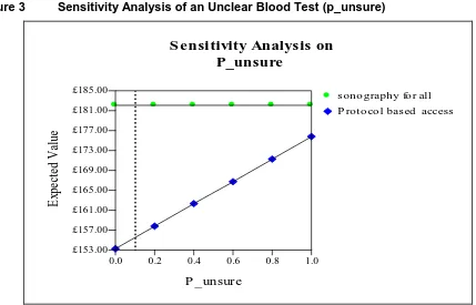
Figure 3 illustrates the results of changing the assumed probability of an unclear blood test
Figure 3 Sensitivity Analysis of an Unclear Blood Test (p_unsure)
S ensitivity Analysis on P_unsure
P _unsure
E
xp
ect
ed
V
al
ue
0.0 0.2 0.4 0.6 0.8 1.0
£185.00
£181.00
£177.00
£173.00
£169.00
£165.00
£161.00
£157.00
£153.00
s ono graphy fo r all
P roto co l bas ed access
Again, the changes in the expected cost of the protocol-based access policy are relatively
insensitive to changes in the probability of an unclear blood test result. Even assuming all
blood tests are unclear only increases the cost per patient to £176.
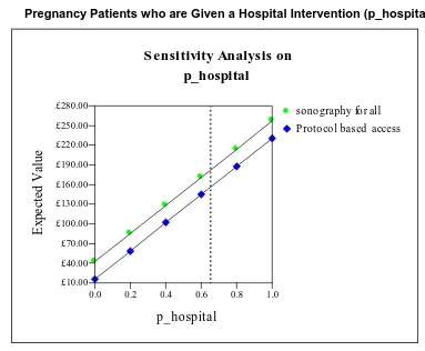
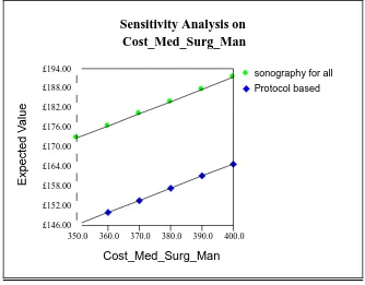
Figure 4 shows the sensitivity of costs to changes in the proportion of non-viable pregnancy
patients who are given a hospital intervention. Because the model assumes that the same
proportion of patients are hospitalised, the lines representing the two policies are parallel. As
such, the difference in expected costs between the two policies would remain as in the
central case. The range of costs per patient are greater than those seen in figures 2 and 3