81
Research
Glutathione S-transferase M1 null genotype: lack of association
with tumour characteristics and survival in advanced breast cancer
Sarab Lizard-Nacol, Bruno Coudert, Pascal Colosetti*, Jean-Marc Riedinger,
Pierre Fargeot and Patrick Brunet-Lecomte
†Centre Georges François Leclerc, Dijon, *University Paris Sud, Orsay, and
†Hôpital du Bocage,
Dijon, France
Abstract
Received: 19 May 1999
Revisions requested: 28 June 1999 Revisions received: 20 July 1999 Accepted: 6 August 1999 Published: 1 September 1999 © Current Science Ltd
Important note about how to cite this article
This article is also available online in the Breast Cancer Research website. To avoid confusion, please ensure that only the online version of the article is cited in any reference, as follows:
Lizard-Nacol S, Coudert B, Colosetti P, et al: Glutathione S-transferase M1 null genotype: lack of association with tumour characteristics and survival in advanced breast cancer [peer-reviewed research]. http://breast-cancer-research.com/vol1no1/01sep99/ research/2
CI = confidence interval; dNTP = deoxynucleotide triphosphate; GST = glutathione S-transferase; OR = odds ratio; PCR = polymerase chain reac-tion; RT = reverse transcription.
Background: Glutathione S-transferase (GST)M1, a member of the µ class GST gene family, has been shown to be polymorphic because of a partial gene deletion. This results in a failure to express the GSTM1gene in 50–60% of individuals. Several studies have demonstrated a possible link with the GSTM1-null genotype and susceptibility to cancer. Further-more, a GSTM1 isoenzyme has been positively associated with protective effect against mutagenic drugs, such as alkylating agents and anthracyclines.
Objectives: To determine whether GSTM1 polymorphisms are associated with tumour characteristics and survival in advanced breast cancer patients, and whether it may constitute a prognostic factor.
Methods: We genotyped 92 patients receiving primary chemotherapy, which included cyclophosphamide, doxo-rubicine and 5-fluorouracil. The relationships between allelism at GSTM1 and clinicopathological parameters including age, menopausal status, tumour size, grade hormone receptors, involved nodes and p53 gene mutations were analysed. Of the patients with GSTM1-positive genotype, tissue samples obtained before and after treatment were available from 28 cases, allowing RNA extraction and GSTM1 expression by reverse transcription polymerase chain reaction. Relationships with clinical response to chemotherapy, and disease-free and
overall survival were also evaluated. The data obtained were analysed using logistic regression to estimate the odds ratio and 95% confidence interval.
Results: Of 92 patients, 57.6% (n= 53) were classified as heritably GSTM1-deficient, and 42.4% (n= 39) were of the GSTM1-positive genotype. There were no statistically significant relationships between GSTM1-null genotype and the clinicopathological parameters analysed. No relationship was observed between GSTM1 RNA expression and objective clinical response to chemotherapy. Objective clinical response to chemotherapy was related only to clinical tumour size (P= 0.0177) and to the absence of intraductal carcinoma (P= 0.0013). GSTM1-null genotype had no effect on disease-free or overall survival. The absence of hormone receptors (P= 0.002), the presence of a mutated p53gene (P= 0.0098) and lack of response to primary chemotherapy (P= 0.0086) were the only factors associated with reduced disease-free or overall survival.
Conclusions: GSTM1-null genotype alone had no effect on tumour characteristics and outcome of patients with advanced breast cancers. The lack of correlation of GSTM1 genotype with clinical tumour features, clinical response to chemotherapy and survival exclude a role for GSTM1 polymorphism as a prognostic factor in advanced breast cancer.
82
Introduction
The human glutathione S-transferases (GSTs) are a multi-gene, isoenzyme family. Cytosolic GST isoenzymes can be classified by their substrate specificities, isoelectric points and amino acid sequence homologies into major classes termed α, µ, π and θ, which are encoded by a superfamily of genes located at different loci [1,2]. There are currently five putative αclass genes encoding subunits GSTA1, GSTA2, GSTA3, GSTA4 and GSTω, whereas the GST π class contains a single gene encoding the GSTP1 protein, and the θ class consists of two genes encoding the GSTT1 and GSTT2 proteins.
The GSTM1gene belongs to the GST µclass gene family, members of which are clustered on chromosome 1p13, and which contains five genes encoding subunits GSTM1, GSTM2, GSTM3, GSTM4 and GSTM5 [3,4]. The pres-ence or abspres-ence of the GSTM1gene constitutes the poly-morphism, and the lack of the GSTM1 gene, which is caused by a gene deletion (the GSTM1-null genotype), affects approximately 50–60% of the population [5,6]. Homozygosity for the GSTM1-null genotype has been found to confer risk for many cancers, including those of the breast [7–17]. The GSTM1-null genotype was posi-tively associated with high DNA adduct levels, suggesting it has a role in carcinogenesis [7]. Smokers with a GSTM1 deficiency had a significantly elevated risk for developing lung, laryngeal and bladder cancer [8,9]. The GSTM1-null genotype has also been associated with higher risk for environmentally related cancer, such as cancers of colon, head and neck, skin, oesophagus and stomach [9–13].
For breast cancer, the GSTM1-null genotype has been found to confer an increased risk in young post-menopausal women [14], whereas other studies did not find any association [18,19]. GST µ deletions have been reported to be associated with higher grade tumours [15], however, and to confer accumulation of epoxides, which are mutagenic [16]. GST isoenzymes catalyze the conjuga-tion of glutathione to several electrophilic compounds, including polyaromatic hydrocarbon, which is lipophilic and stored in adipose tissues, such as those of the breast [20]. Aromatic adducts were found to be higher in women with breast cancer than in healthy control individuals [21]. Most polyaromatic compounds are metabolized to reactive epoxide intermediates by the polymorphic cytochrome p450 (CYP1AI) and detoxified by phase II enzymes, including GST. The variation in conjugation of epoxide substrate intermediates has been observed to segregate with inherited loss of the GSTM1 gene. Therefore, indi-viduals who inherit the homozygous form of the null poly-morphism in the GSTM1 gene will not be capable of conjugating and detoxifying specific substrate epoxide intermediates [16]. In addition, a wide variety of alkylat-ing chemotherapeutic agents used in the treatment of breast cancer have been postulated to act as substrates for
the GSTM1 protein products, thus reducing the effective-ness of these agents as cytotoxins [22].
In an attempt to further characterize the clinical features associated with the GSTM1-null genotype, we examined allelism at the GSTM1 locus in 92 locally advanced breast cancer patients undergoing primary chemotherapy. Allelism at the GSTM1 locus was analysed by polymerase chain reaction (PCR) in paired samples of blood and breast tissue. To determine whether levels of GSTM1 expression in patients with a positive genotype has a pre-dictive or a prognostic value, RNA expression was mea-sured by reverse transcription (RT)-PCR in breast tumour samples obtained before and after treatment. Results were then compared with clinicopathological factors of the patients, including characterization for p53gene mutations [23], clinical response to chemotherapy, and disease-free and overall survival.
Materials and methods
Patients and samplesAfter an initial diagnostic biopsy, including characterization for p53 gene alterations, 92 women who were diagnosed with locally advanced breast carcinoma and who under-went primary chemotherapy were included in this study. The median clinical follow up was 78 months (range 10–120 months). Three of these patients had bilateral lesions, and in these three cases both lesions were exam-ined. No family history for breast cancer was recorded in the 92 women. The patients received chemotherapy treat-ment (four or six courses, each lasting 21 days) with a regimen containing cyclophosphamide, doxorubicine and 5-fluorouracil. The criteria for inclusion were as follows: inflammatory carcinomas, positive nodes and/or large (T3, T4) tumours. Clinical response to primary chemotherapy was categorized according to World Health Organization criteria and was considered as objective response (com-plete or partial response) or no response (stabilization or progression). In all cases neither radiotherapy nor hormone therapy were applied before chemotherapy.
Tumours were characterized before treatment by the clin-ical tumour size (categorized as T2, T3 or T4); the clinclin-ical nodal involvement (categorized as N0, N1 or N2); the his-tologic grade of Scarff, Bloom and Richardson (categorized as SBR1, SBR2 or SBR3); the hormone receptors (catego-rized as HR–or HR+, and considered as positive for
oestro-gen and/or progesterone receptors); the pathological tumour size (categorized as pT0, pT1, pT2 or pT3); and the number of involved axillary nodes (categorized as pN–,
pN1+ and pN3+ for none, one or two, and three or more
involved nodes, respectively).
83 using proteinase, followed by phenol extraction and ethanol
precipitation according to standard procedures [24].
Polymerase chain reaction method
The GSTM1-null genotype was determined by coamplifi-cation with the interferon-β gene, which served as an internal control. Primers for amplification of the GSTM1 gene corresponding to exon 4, intron 5 and exon 5 were 5′ -ctgccctacttgattgatggg-3′ and 5′-ctggattgtagcagatcatgc-3′ (amplified product size, 271 base pairs). Primers for ampli-fication of a part of the interferon-βgene, producing a con-stant 170-base pair band in all samples, were 5′-ggcacaacaggtagtaggcg-3′and 5′-gccacaggagcttctgacac-3′. Because the primers for the GSTM1 locus anneal to sites inside the coding region of the gene, the presence of the gene was determined by the presence of the band, whereas the null-genotype was determined by the lack of the band, using agarose gel electrophoresis (2%).
PCR was performed using 250 ng template DNA in 10 mmol/l Tris-HCl (pH 8.4), 50 mmol/l potassium chloride, 1.5 mmol/l magnesium chloride (Bioprobe Systems, Illkirch, France), 0.2 mmol/l concentrations of each deoxynucleotide triphosphate (dNTP), 500 nmol/l concentrations of each primer and 2.5 units of Taq DNA polymerase (Bioprobe Systems). The reaction (total volume 50µl) was amplified on a Omnigene thermal cycler (Hybaid Ltd, Ashford, Kent, UK). After an initial denaturation at 94°C for 3 min the reaction proceeded for 25 cycles of 50 s at 94°C, 50 s at 55°C and 50 s at 72°C, concluded by a final extension step of 10 min at 72°C. To test for contamination, negative controls (tubes containing the PCR mixture without the DNA tem-plate) were included in every run.
Reverse transcription polymerase chain reaction
Total RNA was obtained by the acid guanidine thio-cyanate–phenol–chloroform extraction method [25]. Of the total RNA 1µg was dissolved in 20µl reverse tran-scriptase buffer (Gibco/BRL, Cergy Pontoise, France) containing 200µmol/l dNTP, 500 ng random hexamer and 200 U avian myeloblastosis virus (AMV) reverse transcrip-tase (Superscript; Gibco/BRL). The reaction was incu-bated at 42°C for 50 min and heated to 70°C for 15 min.
The PCR reaction was carried out in a 50µl volume of PCR buffer (Promega, Madison, Wisconsin, USA) contain-ing 200µmol/l dNTP, 2.5µl complementary DNA tem-plate, 2.5 U Taq DNA polymerase, and 500 nmol/l of both
GSTM1 and β2-microglobulin primers. The reaction was initiated by a heat step at 95°C for 2 min, and carried out for 25 cycles of denaturation at 94°C for 50 s, annealing at 55°C for 50 s and extension at 72°C for 50 s. Blanks for each reaction were included with all samples. Of PCR product 10µl were analysed on a 2% agarose gel with ethidium bromide staining. Band intensities were deter-mined with a gel doc 1000 UV system (Biorad,
Ivry-Sur-Seine, France), and the ratio of GSTM1to β2 -microglobu-lin was calculated.
Determination of p53 mutations
The determination of p53mutations was performed as pre-viously described [23]. Briefly, breast tumour samples were characterized before and after treatment for p53 gene muta-tions by PCR single-strand confirmation polymorphism and/or direct sequencing of exons 5–9 of the p53gene.
Hormonal receptor assay
The oestrogen and progesterone receptor levels were determined in cytosolic tumours using enzyme immunoas-say methods (Abbott Laboratories, Rungis, France). The cutoff level used for oestrogen and progesterone was 20 fmol/mg cytosolic proteins.
Statistical analysis
The Pearson χ2 test was used as a homogeneity test for
proportion. A stepwise logistic regression model with the logoistic regression (LR)-BMDP program (University of California Press, Berkeley, California, USA) [26] was used to assess the contribution of each independent factor to the GSTM1-null genotype and the probability of response to primary chemotherapy. The log-rank test using the Kaplan–Meier method was used to study the relationship between each factor and the probability of disease-free survival (median 50 months) and overall survival (median 78 months). A multivariate analysis using the Cox propor-tional hazards model was performed to assess the contri-bution of each independent factor to the probability of survival. For logistic regression or Cox regression models the enter and remove limits were 0.1 and 0.15, respec-tively. Overall significance of each factor (P value) was given by the likelihood ratio test. Results are expressed as odds ratios (OR) and corresponding 95% confidence inter-val (CI), with the significance of ORs being derived from the ratio of the coefficient divided by its standard error (Wald test). Statistical analysis for RNA GSTM1 expres-sion was performed using the SAS/STAT program (SAS Institute Inc, Cary, North Carolina, USA) by using Student t-test or analysis of variance, Pearson χ2test and
log-rank test. For all tests, optimality of the selected models was verified by all-possible-subsets analyses.
Results
Clinicopathological data
contained significant hormone receptors (HR+), and 39.1%
(n= 36) did not (HR–). Clinical complete response was
found in 19.6% (n= 18) of cases. A high histological grade (SBR3) was found in 34.8% (n= 32) of tumours. An inter-mediate level of pathological tumour size (pT2) was found in 42.4% (n= 39) of samples. Negative lymph nodes were found in 20.7% (n= 19) of the tumours, whereas 23.9% (n= 22) of tumours were associated with > 1 involved nodes and 55.4% (n = 51/92) were associated with > 3 involved nodes. Mutations in p53 were detected in 30% (n= 28) of the cases studied.
Glutathione S-transferase M1 genotype determination The PCR method described above allowed an internal standard controlled classification of GSTM1-deficient (GSTM1-null genotype) individuals. Of the 92 patients, 57.6% (n= 53) were classified as heritably GSTM1 defi-cient, and 42.4% (n= 39) were GSTM1-positive genotype. Paired samples of blood and breast tissue were analysed before treatment with primary chemotherapy from the same individual, and GSTM1 genotype was identical for the two samples (Fig. 1). Among the 39 patients with GSTM1-positive genotype, tissue samples obtained before and after treatment were available from 28 cases, allowing RNA extraction and GSTM1expression using the RT-PCR method. Two of these patients had bilateral lesions, and measurement was determined in the two tumour localizations. Thus, total GSTM1 expression, as measured by the ratio of GSTM1 to β2-microglobulin values, were performed on 30 tumour specimens. GSTM1
RNA signal was detected in all of the tumours analysed before and after treatment. The median GSTM1 expres-sion was 1.38 (range 0.02–23.27) in the untreated tumours, and 1.16 (range 0.01–6.56) in samples obtained after chemotherapy administration.
Glutathione S-transferase M1 and clinicopathological characteristics of the patients
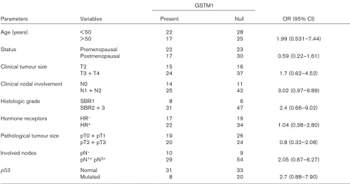
Distribution of GSTM1 genotype and its relation with clini-copathological data of the patients are shown in Table 1. There were no statistically significant associations between GSTM1-null genotype and the parameters analysed: age, menopausal and hormonal status, clinical and pathological tumour size, grade, involved nodes and p53 gene mutations.
GSTM1 expression measured by RT-PCR in 30 samples (corresponding to 28 cases) before and after treatment with primary chemotherapy was also compared with the clinico-pathological characteristics of the patients. None of the parameters tested were related to GSTM1expression deter-mined before or after treatment (data not shown).
Relationship to clinical response to chemotherapy (Table 2) demonstrated that objective response (complete and partial responses) rate of the group with GSTM1-null genotype (75.5%) did not differ from that in those with GSTM1-posi-tive genotype (76.9%). Thus, no significant relation was
found between GSTM1 polymorphism and clinical response to chemotherapy (P= 0.8719). Also, no relation was observed between GSTM1 RNA expression and clinical response to chemotherapy (P= 0.9524 and P= 0.5192 for before and after treatment, respectively). In contrast, clinical tumour size (P= 0.0177) and intraductal carcinoma (P= 0.0013) are strongly associated with clinical response. In multivariate analysis, the clinical tumour size (P= 0.0070, OR = 4.83, 95% CI = 1.45–16.10) and the absence of intraduc-tal carcinoma (P= 0.0002, OR = 14.1, 95% CI = 2.52–78.50) remained the only factors linked to the clinical response.
Impact on survival of the patients
For disease-free survival, no differences were found between individuals with GSTM1-null genotype and those with positive-GSTM1 genotype (P= 0.8094). Accordingly, no impact for RNA GSTM1 expression on disease-free survival (P= 0.8991 and P= 0.9096 for before and after treatment, respectively) was observed (Table 2). In contrast, the absence of hormone receptors (P= 0.0020) and the presence of p53 gene mutations (P= 0.0098) had an impact on disease-free survival. With multivariate analysis, hormone receptor status (P= 0.0002, OR = 3.99, 95% CI = 1.92–8.29) and p53 gene mutations (P= 0.0138, OR = 2.36, 95% CI = 1.22–4.59) remained significantly associated with metastasis recurrence risk.
No impact was also found (P= 0.9729) for GSTM1-null genotype on overall survival (Table 2), or for RT-PCR RNA expression (P= 0.1667 and P= 0.9637 for before and after treatment, respectively). Only the absence of hormone receptors (P= 0.0018), the presence of p53gene mutations (P= 0.0071) and no response to primary chemotherapy (P= 0.0086) were associated with reduced overall survival of the patients. In multivariate analysis, hormone receptor 84
Figure 1
Polymerase chain reaction product of paired lymphocyte (L) and tumour (T) DNA from coamplification of glutathione S-transferase (GST)M1 (271 base pair) and interferon-β(170 base pair) genes. Lane 1 shows a homozygously present GSTM1 allele. Lane 2 shows a homozygously null-GSTM1 allele. M1 is ΦX174-HaeIII digested DNA marker. M2 is ΦX174-HinfI digested DNA marker.
L T L T
271 bp
170 bp 300 bp200 bp
status (P= 0.0003, OR = 5.23, 95% CI = 2.03–13.49) and p53
gene mutations (P= 0.0037, OR = 3.62, 95% CI = 1.53–8.53) were strongly related to the risk for death. Absence of clini-cal response to chemotherapy was less related to the overall survival (P= 0.0530, OR = 2.31, 95% CI = 1.02–5.26).
Discussion
In the present study, the frequency of GSTM1gene defi-ciency among breast carcinoma patients (57%) was similar to that previously reported in this lesion [9,14–19]. Among individuals with GSTM1-positive genotype no deletion was observed in somatic tumour cells, suggesting that the deletion of GSTM1gene was not a characteristic of breast tumour cells.
[image:5.609.56.555.122.385.2]Several series have determined that GSTM1 deletions were involved in the aetiology of breast cancer [9,14,17], whereas other studies did not find any such associations [18,19]. In addition, little is known about clinical charac-teristics that confer GSTM1deletion among breast cancer patients. Only one recently published study [19] reported no relation between clinicopathological parameters and GSTM1 genotype in primary breast tumours. The results obtained in the present study are consistent with these data, because no differences were noted when a variety of known prognostic factors were compared between tumours from patients with and those without GSTM1-null genotype. These factors include age, menopausal status, clinical and pathological tumour size, clinical and 85 Table 1
Association of glutathione S-transferase (GST)M1 genotype and clinicopathological data of breast cancer patients studied
GSTM1
Parameters Variables Present Null OR (95% CI)
Age (years) < 50 22 28
> 50 17 25 1.99 (0.531–7.44)
Status Premenopausal 22 23
Postmenopausal 17 30 0.59 (0.22–1.61)
Clinical tumour size T2 15 16
T3 + T4 24 37 1.7 (0.62–4.52)
Clinical nodal involvement N0 14 11
N1 + N2 25 42 3.02 (0.97–9.89)
Histologic grade SBR1 8 6
SBR2 + 3 31 47 2.4 (0.66–9.02)
Hormone receptors HR– 17 19
HR+ 22 34 1.04 (0.38–2.80)
Pathological tumour size pT0 + pT1 19 26
pT2 + pT3 20 24 0.8 (0.32–2.08)
Involved nodes pN– 10 9
pN1+pN3+ 29 54 2.05 (0.67–6.27)
p53 Normal 31 33
Mutated 8 20 2.7 (0.88–7.90)
CI, confidence interval; OR, odds ratio.
Table 2
Clinical response to primary chemotherapy and breast cancer outcome
GSTM1
Parameters Present Null P
Objective response* 76.9% 75.5% 0.8719†
Disease-free survival (months) 42.00 ± 2.65 54.43 ± 5.67 0.8094‡
Overall survival (months) 74.82 ± 3.95 72.73 ± 4.81 0.9729‡
*Objective response was estimated as complete and partial
responses. †Estimated by Pearson χ2test. ‡Estimated by log rank test,
[image:5.609.54.568.449.538.2]pathological nodal involvement, pathological grade, hormone receptors status and p53gene mutations. These results suggest that clinical tumour features are not associ-ated with GSTM1-null genotype, not only in primary breast cancers, but also in advanced breast cancers.
An improved survival has been observed in patients with GSTM1-null genotype and has been explained by a better response to chemotherapeutic agents related to more effective cell killing, which in turn is related to the absence of a protective effect of the GSTM1 allele [18]. Among the most active chemotherapeutic agents in the treatment of breast cancer are cyclophosphamide and anthracyclines. These compounds are conjugated with thiols through reactions mediated by GST. The πclass of transferase is thought to be the major factor involved in the subsequent prevention of DNA alkylation [22], although there has been some suggestion that the µclass is also involved [27]. In order to determine whether the absence of GSTM1 locus favours the response to chemotherapeutic agents, and whether, on the contrary, GSTM1-positive genotype may confer a resistance to chemotherapy, we analysed GSTM1 polymorphisms in relation to clinical response to chemotherapy. The results demonstrated that the objective response rate of the group with GSTM1-null genotype did not differ from those with GSTM1-positive genotype. Thus, individuals carrying the GSTM1 locus were no more resistant to chemotherapy than those with GSTM1-null genotype.
GSTM1RNA expression levels measured in patients with positive genotype were also not associated with clinical response to chemotherapy. Among clinical parameters analysed, only clinical tumour size and the presence of intraductal carcinoma were found to influence clinical response to primary chemotherapy. The results also revealed that GSTM1-null genotype was not related to survival in advanced breast cancer patients, in contrast to the absence of hormone receptors and to the presence of
p53gene mutations, which are known to be of prognostic value in advanced breast cancer [28–30].
Although the group studied was not very large (n= 92), the clinical characteristics were well known and the results strongly suggest the lack of correlation of GSTM1-null genotype alone with prognostic factors, clinical reponse to chemotherapy and survival. Genotypes of other enzymes (CYP1A1, GSTT1), as well as combinations of genotypes, might be of interest with regard to cancer and carcinogen metabolism.
Taken together, these results show that advanced breast cancers arising in patients with GSTM1-null genotype have no worse clinical tumour characteristics and outcome than those of patients without such deletions. Thus, the lack of correlation of GSTM1-null genotype with clinical tumour features, clinical response to chemotherapy and
survival exclude a role for GSTM1 polymorphism as a prognostic factor in advanced breast cancer.
Acknowledgements
This work was supported by the ‘Comité Départemental de Saône et Loire’, ‘Ligue Bourguignonne Contre le Cancer’ and the ‘Association Régionale pour l’Enseignement et la Recherche Scientifique/Groupe Interrégional de Recherche en Cancérologie’. We thank Mrs L Hahnel and M Arnal for their excellent technical assistance.
References
1. Mannervik B, Alin P, Guthenberg C, et al: Identification of three classes of cytosolic glutathione transferase common to several mammalian species: correlation between structural data and enzy-matic properties.Proc Natl Acad Sci U S A1985, 82:7202–7206. 2. Mannervik B, Danielson UH: Glutathione transferases: structure
and catalytic activity.CRC Crit Biochem1988, 23:283–337. 3. Pearson WR, Vorachek WR, Xu SJ, et al: Identification of class-mu
glutathione transferase genes GSTM1–GSTM5 on human chro-mosome 1p13.Am J Hum Genet1993, 53:220–233.
4. Ross VL, Board PG, Webb GC: Chromosomal mapping of the human mu class glutathione S-transferase to 1p13. Genomics 1993, 18:87–91.
5. Seidegard J, Vorachek WR, Pero RW, Pearson WR: Hereditary dif-ferences in the expression of the human glutathione transferase active on trans-stilbene oxide are due to a gene deletion. Proc
Natl Acad Sci U S A1988, 85:7293–7297.
6. De Jong JL, Chang CM, Whang-Peng J, Knutsen T, Tu CPD: The human liver glutathione S-transferase gene superfamily: expres-sion and chromosome mapping of an Hb subunit cDNA.Nucleic
Acids Res1988, 16:8541–8554.
7. Nazar-Stewart V, Motulsky AG, Eaton DL, et al: The glutathione S-transferase mu polymorphism as a marker for susceptibility to lung carcinoma.Cancer Res1993, 53:2313–2318.
8. Lafuente A, Pujol F, Carretero P, Villa JP, Cuchi A: Human glu-tathione S-transferase mu (GST mu) deficiency as a a marker for susceptibility to bladder and larynx cancer among smokers.
Cancer Lett1993,68:49–54.
9. Zhong S, Wyllie AH, Barnes D, Wolf CR, Spurr NK: Relationship between the GSTM1 genetic polymorphism and susceptibility to bladder, breast and colon cancer. Carcinogenesis 1993, 14: 1821–1824.
10. Trizna Z, Clayman GL, Spitz MR, Briggs KL, Geopfert H: Glutathione S-transferase genotypes as risk factors for head and neck cancer.
Am J Surg1995, 170:499–501.
11. Heagerty AHM, Fitzgerald D, Smith A, et al: Glutathione S-trans-ferase GSTM1 phenotypes and protection against cutaneous tumors.Lancet1994, 343:266–268.
12. Nimura Y, Yokoyama S, Fujimori M, et al: Genotyping of the CYP1A1 and GSTM1 genes in esophageal carcinoma patients with special reference to smoking.Cancer1997, 80:852–857.
13. Harada S, Misawa S, Nakamura T, et al: Detection of GST1 gene deletion by the polymerase chain reaction and its possible corre-lation with stomach cancer in Japanese. Hum Genet 1992, 90:62–64.
14. Ambrosone CB, Freudenheim JL, Graham S, et al: Cytochrome P4501A1 and glutathione S-transferase (M1) genetic polymor-phisms and postmenopausal breast cancer risk.Cancer Res1995, 55:3483–3485.
15. Murray GJ, Weaver RJ, Paterson PJ, et al: Expression of xenobiotic metabolizing enzymes in breast cancer.J Pathol1993, 169:347–353. 16. Wiencke JK, Kelsey KT, Lamela RA, Toscano W: Human glutathione-S-transferase deficiency: a marker of susceptibility to induced chromosome damage.Cancer Res1990, 50:1585–1590. 17. Helzlsower KJ, Selmin O, Huang HY, et al: Association between
glu-tathione S-transferase M1, P1, and T1 genetic polymorphisms and development of breast cancer.J Natl Cancer Inst1998,90:512–518. 18. Kelsey KT, Hankinson SE, Colditz GA, et al: Glutathione S-trans-ferase class µµdeletion polyphorphism and breast cancer: results from prevalent versusincident cases.Cancer Epidemiol Biomarkers Prev1997, 6:511–515.
19. Bailey LR, Roodi N, Verrier CS, et al: Breast cancer and CYP1A1, GSTM1, and GSTT1 polymorphisms: evidence of a lack of associ-ation in Caucasians and African Americans.Cancer Res1998, 58: 65–70.
20. Peresa FP, Estabrook A, Hewer A, et al: Carcinogen-DNA adducts in human breast tissue.Cancer Epidemiol Biomarkers Prev1995, 4: 233–238.
21. Li D, Wang M, Dhingra K, Hittelman WN: Aromatic DNA adducts in adjacent tissues of breast cancer patients: clues to breast cancer etiology.Cancer Res1996, 56:287–293.
22. Fields WR, Li Y, Townsend AJ: Protection by transfected glu-tathione S-transferase isoenzymes against carcinogen-induced alkylation of cellular macromolecules in human MCF-7 cells.
Car-cinogenesis1994, 15:1155–1160.
23. Lizard-Nacol S, Coudert B, Riedinger JM, Fargeot P, Guerrin J: p53 gene alterations are associated with a decreased responsiveness to neoadjuvant chemetherapy in human breast cancer.Int J Oncol 1997, 10:1203–1207.
24. Maniatis T, Fritsch EF, Sambrook J: Molecular Cloning: a Laboratory
Manual.Cold Spring Harbour, New York: Cold Spring Harbour
Labo-ratory, 1982.
25. Chomczynski P, Sacchi N: Single-step method of RNA isolation by acid guanidinium thiocyanate-phenol-chloroform extraction.Anal
Biochem1987, 162:156–159.
26. Engelman L: Stepwise logistic regression. In:BMDP Statistical
Soft-ware Manual.Vol 2. Edited by Dixon WJ, Brown MB, Engelman L,
Jen-nrich RI. University of California Press: Berkeley, 1992:1013–1046. 27. Albin N, Massad L, Toussain C, et al: Main drug-metabolizing
enzyme systems in human breast tumors and peritumoral tissues.
Cancer Res1993, 53:3541–3546.
28. Järvinen TAH, Holli K, Kuukasjärvi T, Isola JJ: Predictive value of topoi-somerase Iiααand other prognostic factors for epiribicin chemother-apy in advanced breast cancer.Br J Cancer1998, 77:2267–2273. 29. Hollstein M, Sidransky D, Vogelstein B, Harris CC: p53 mutations in
human cancers.Science1991, 253:49–53.
30. Sjögren S, Inganäs M, Norberg T, et al: The p53 gene in breast cancer: prognostic value of complementary DNA sequencing versus immunohistochemistry.J Natl Cancer Inst1996, 88:173–182.
Author addresses:Sarab Lizard-Nacol (Laboratory of Molecular Genetics, Centre Georges François Leclerc, Dijon, France), Bruno Coudert, Pierre Fargeot (Oncological Service, Centre Georges François Leclerc, Dijon, France), Pascal Colosetti (INSERM U-442, University Paris Sud, Orsay, France), Jean-Marc Riedinger (Laboratory of Medical Biology, Centre Georges François Leclerc, Dijon, France) and Patrick Brunet-Lecomte (Informatique Médicale, CHU/Hôpital du Bocage, Dijon, France)
Correspondence:Sarab Lizard-Nacol, Laboratory of Molecular Genetics, Centre Georges-François Leclerc, 1 rue du Professeur Marion, 21034 Dijon Cedex, France. Tel: +(33) 3 80 73 75 00; fax: +(33) 3 80 73 77 26; e-mail: Slizard@dijon.fnclcc.fr