Review
Transforming growth factor-
ββ
and breast cancer
Mammary gland development
Mary Helen Barcellos-Hoff and Kenneth BR Ewan
University of California, Berkeley, California, USA
Abstract
Transforming growth factor (TGF)-β1 is a pluripotent cytokine that profoundly inhibits epithelial proliferation, induces apoptosis, and influences morphogenesis by mediating extracellular matrix deposition and remodeling. The physiologic roles of the action of TGF-β in mammary gland, indeed in most tissues, are poorly understood. In order to understand the actions of TGF-β, we need to take into account the complexity of its effects on different cell types and the influence of context on cellular responses. This task is further compounded by multiple mechanisms for regulating TGF-βtranscription, translation, and activity. One of the most significant factors that obscures the action of TGF-βis that it is secreted as a stable latent complex, which consists of the 24-kDa cytokine and the 80-kDa dimer of its prepro region, called latency-associated peptide. Latency imposes a critical restraint on TGF-β activity that is often overlooked. The extracellular process known as activation, in which TGF-βis released from the latent complex, is emphasized in the present discussion of the role of TGF-βin mammary gland development. Definition of the spatial and temporal patterns of latent TGF-βactivation in situis essential for understanding the specific roles that TGF-β plays during mammary gland development, proliferation, and morphogenesis.
Keywords:transforming growth factor (TGF)-β1, activation, latent, mammary, proliferation Received: 13 December 1999
Revisions requested: 10 January 2000 Revisions received: 31 January 2000 Accepted: 1 February 2000 Published: 21 February 2000
Breast Cancer Res2000, 2:92–99
The electronic version of this article can be found online at http://breast-cancer-research.com/content/2/2/092
© Current Science Ltd
ECM = extracellular matrix; LAP = latency-associated peptide; LTGF = latent transforming growth factor; MMTV = mammary mouse tumor virus; TGF = transforming growth factor; WAP = whey acidic protein.
Introduction
This review describes briefly mammary gland develop-ment and discusses the postulated role of TGF-β1 in mammary gland development as evidenced by its effects in mammary epithelial cell culture and animal studies. TGF-β has been strongly implicated in the control of mammary epithelial growth [1] and in breast cancer cells [2,3]. Cell culture studies have also shown that the effects of TGF-βare pleiotropic, context-dependent, con-centration-dependent, and can be indirect. As a conse-quence it is difficult to predict the endogenous action of
Mammary gland development
The seminal work of Daniel and coworkers [4••,5] focused
attention on the role of TGF-βin mammary gland develop-ment soon after its characterization. The morphologic and functional development of the mammary gland is largely postnatal in both humans and mice (for review [6]). Under the effects of hormones of puberty, which begins just after weaning in mice and continues for several weeks (ie 3 weeks to 6–8 weeks of age), the mammary tree is estab-lished within an adipose stroma known as the fat pad. The epithelium, usually a simple epithelium consisting of one or two cell layers, is ensheathed in a fibrous stroma. This period of growth is characterized by a specialized morpho-logic unit called the end-bud, which is a multicellular, mul-tilayered structure whose function is to extend the ductal epithelial tree to the boundaries of the fat pad. The pattern of the resulting tissue can be visualized in a whole-mount preparation and is often used as an end-point for evaluat-ing the role of factors such as TGF-βin guiding morphol-ogy by measuring the extent of branching, the branching pattern by branch-point analysis, and the distribution of end-buds and their character, size, shape, and persis-tence. Once the ductal tree is established, repeated estrus cycles of ovarian hormones elicit further elaboration of the epithelium in some mouse strains, generating small lateral branch points. In a nulliparous animal approximately 10% of the tissue is epithelial.
Under the effects of hormones of pregnancy, a burst of growth and differentiation results in lobuloalveolar differen-tiation of the epithelium into functional secretory units that produce milk that is carried to the teat by large conducting ducts. The resulting fully functional gland is 90% epithelial. Upon weaning, a process of involution destroys the major-ity of secretory epithelium, leaving the ductal tree extant for repeated cycles of growth and differentiation.
The remarkable capacity of the mammary epithelium to undergo development and differentiation provides a research model in which the factors that influence growth, morphologic patterning, proliferation, and differentiation can readily be explored. Furthermore, in mice epithelium can be transplanted to new stroma that is usually cleared of endogenous epithelium, which offers an exceptional research tool for studying the contribution of the stroma to the control of epithelial function and remodeling. Recombi-nations of transgenic and wild-type epithelia and stromas, and the ability to transplant intact fat pads to new hosts, allows determination of whether the source of a particular factor (ie different tissue compartments) affects the matu-ration of the mammary gland.
Transforming growth factor-
ββ
regulation and
expression
There are three mammalian isoforms of TGF-β. All three isoforms bind to the same cell surface receptors and, in cell culture, often appear to elicit similar responses. For the purposes of this review we focus on the well-studied TGF-β1 isoform, its localization in the mammary gland, its effects in cell culture models, and the consequences of manipulating its activity in vivo by various means. The activity of all TGF-βisoforms is restrained by secretion as latent complexes [latent transforming growth factor (LTGF)-β] [7]. (Regarding nomenclature, TGF-βrefers to the active cytokine or its effects, whereas LTGF-β desig-nates the latent form.) LTGF-β consists of the 24-kDa cytokine and a 80-kDa dimer of its prepro region called latency-associated peptide (LAP), which contains the signal sequence for secretion (Fig. 2). To our knowledge, all cells secrete LTGF-β, which underscores the impor-tance of activation events associated with the release of TGF-βfrom LAP.
TGF-β message levels do not usually reflect protein pro-duction, or, more importantly, protein activity. For example, tamoxifen increases both LTGF-β production and activa-tion without any effect on mRNA levels [2]. Differences in mRNA translation are in part due to a 5′-untranslated region stem-loop that binds a regulatory protein, which can result in increased protein production by factors of 10 without changes in mRNA levels [8]. In addition, protein assays are complicated by the fact that tissue extraction protocols activate the endogenous LTGF-β [9]. Because the biologic activity of TGF-β is controlled by its release from the latent complex, elevated expression of latent complex is not likely to have biologic consequences, whereas increased activation, even without changes in synthesis, will profoundly affect physiologic events [10]. The importance of activation is supported by the lack of a phenotype in transgenic mice that overexpress LTGF-βin mammary gland [11•]. A recent study in TGF-β-null
[image:2.612.58.300.94.254.2]het-erozygotes, however, has shown there is a dramatic loss of circulating and tissue TGF-β levels as a result of the
Figure 1
haploid genotype [12•]. This surprising finding indicates
that TGF-βplays a significant role in its own regulation.
A variety of immunostaining studies have been conducted in both murine and human tissues. These are difficult to summarize because of differences in tissue preparation, the antibodies employed, and their uncertain relevance to understanding the functional role of TGF-β in situ. Several studies localized TGF-β to the human breast epithelium and stroma [13,14], but at least one study [15] found it to be associated only with epithelial cells; this is probably due to either the antibody or fixative (see below). TGF-βis also found in the conditioned media of cultured normal human epithelial cells [16–18] and fibroblasts [18,19]. A recent study that immunolocalized TGF-β receptors I, II, and III in normal human breast [20] demonstrated that these are widely expressed in the breast epithelium and stroma.
Robinson et al [21•] used Northern blot analyses, in situ
hybridization, and immunocytochemistry to define TGF-β expression patterns during murine mammary gland
devel-opment and differentiation to establish that all three mam-malian TGF-β isoforms are expressed in the epithelium during all phases of mammary development. TGF-β2was less abundant, however, whereas TGF-β3 was the only isoform found in myoepithelial cells. TGF-β1 transcripts and immunoreactivity decreased during pregnancy, but those of TGF-β2and TGF-β3increased until the onset of lactation [210•]. Intraepithelial immunoreactivity in that
study corresponded to transcript localization, indicating sites of synthesis. Immunolocalization using antibodies to mature TGF-βversus TGF-β1LAP showed an interesting pattern of deposition that was suggested to indicate that TGF-β activity inhibits proliferation and its depletion is necessary for growth [22]. Although activation was implicit in the interpretation of the immunostaining, the model did not provide confirmation of activity, due in large part to the lack of reagents to differentially localize active TGF-βversus LTGF-β.
Studies in our laboratory demonstrate that under certain conditions some antibodies can discriminate between active and latent TGF-β [23]. We determined the speci-ficity of TGF-β antibodies by using a immunodetection method that preserves endogenous LTGF-βin conjunction with engineered control tissues that specifically produce latent versus constituitively active TGF-β[24••]. Our
exten-sive screening of available TGF-β antibodies demon-strated that antibody source, fixation, and tissue preparation influences the ability to discriminate between active and latent forms [25•]; thus, retrospective
interpre-tation of other immunostaining protocols and antibodies combinations is not possible.
One of the most striking features of our immunolocaliza-tion in mammary gland using antibodies that discriminate between TGF-β and LTGF-βis the contrast between the highly restricted localization of TGF-βcompared with the broad distribution of LTGF-β. Our initial studies concerned the effect of ionizing radiation on the mammary gland microenvironment. We observed that the immunoreactivity of activation-specific antibodies LC(1-30) and CC(1-30) is restricted to the normal mammary epithelium [26], whereas LAP immunoreactivity is widely distributed in the epithelium, fibrous stroma, and adipose stroma [24••]. A
remarkably rapid (within 1 h) shift from predominantly LAP to TGF-βimmunoreactivity in all of these tissue compart-ments led us to conclude that ionizing radiation leads to LTGF-βactivation [24••]. Our conclusion is supported by
[image:3.612.56.297.95.263.2]additional studies that demonstrated that radiation induces stromal extracellular matrix (ECM) remodeling (eg collagen III), a known target of TGF-βaction [26], and that administration of TGF-β-neutralizing antibodies blocks ECM remodeling in irradiated animals [27]. These studies indicated that, despite the apparent abundance of LTGF-β in normal, nulliparous mammary gland, active TGF-β is highly localized.
Figure 2
Transforming growth factor-
ββ
responses in
cultured cell models
Much of our understanding of the biology of TGF-βcomes from studies of cultured cells exposed to recombinant or purified active TGF-β. Nonetheless, the specific physio-logic roles of the action of TGF-β in mammary gland are not well understood, which is due in part to the complexity of its effects on different cell types that is compounded by regulation at multiple levels and modified by cellular context [23]. Examples of these effects are summarized below. TGF-βhas been shown to inhibit profoundly prolif-eration of normal rodent and human mammary epithelial cells in cell culture, and may also modify the production of differentiated proteins (for review [28]). Both cell type and context influence TGF-β action. 3T3 cell preadipocytes spontaneously activate their own secreted LTGF-β, whereas mature adipocytes do not, which implicates TGF-β activation as a potent negative regulator of adipocyte differentiation [29]. Fetal versus adult fibro-blasts [30], young versus old smooth muscle cells [31], and normal human mammary epithelial cells grown on ECM versus plastic [32] are differentially sensitive to TGF-β growth inhibition. In addition to reversible growth inhibition, TGF-β can also induce apoptosis. Hormone withdrawal in hormone-dependent tissues such as prostate [33•] and uterus [34•] is a potent inducer of
TGF-β-mediated apoptosis. In mammary gland, TGF-β mRNA increases early in involution and its activity is thought to mediate this apoptotic remodeling process [35]. Notably, mammary secretions during involution contain high levels of active TGF-β and extremely high levels of LTGF-β that regulate the mononuclear phago-cytes involved in apoptotic remodeling of the gland [36].
TGF-β-induced ECM contributes to growth control in cul-tured cells [37] and may be involved in the changes in ECM during mammary gland development [13,22] and the menstrual cycle [38]. The cessation of DNA synthesis and end-bud regression caused by exogenous TGF-β-induced ECM is postulated to involve the concomitant induction of periepithelial ECM around end-buds [39]. This regulation may be offset by feedback mechanisms as evidenced by suppression of TGF-β-induced ECM mRNA levels in murine mammary cells cultured within a basement mem-brane-type ECM [40]. Even cells that lack a proliferative response to TGF-β-induced ECM, such as breast cancer cells, may still respond with specialized ECM production [16], that in turn may modify the tissue microenvironment in such a way as to affect growth. For example, recent mammary cell culture studies [41] also point to a role of ECM in modulating hormone response. The antiestrogen tamoxifen affects stromal production and activity of TGF-β, which has been postulated to contribute to its therapeutic benefit [42]. An interesting model of hormone-mediated remodeling in uterine tissue indicates that stromal activa-tion of TGF-β mediates epithelial production of the
pro-teases that are necessary for degradation of basement membrane [43•].
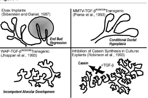
The addition of TGF-βto mammary cell cultures supports a role in regulation of morphogenesis and control of differ-entiation. TGF-β suppressed the ability of mammary explants cultured with lactogenic hormones to secrete casein [44]. It abolished ductal morphogenesis by human mammary epithelial cells cultured within a basement mem-brane-type ECM, whereas antibodies to TGF-βstimulated duct formation [45]. In another study [46], however, cul-tured mammary cell lines exhibited a biphasic response to TGF-β concentration; picomolar concentrations inhibited branching morphogenesis, whereas fentomolar concentra-tions stimulated it. Furthermore, chronic TGF-β exposure can elicit phenotypic transformation under certain condi-tions, resulting in the acquisition of mesenchymal-like transformation of mammary epithelial cells [47], or myofi-broblast characteristics in stromal cells [48]. The role of such phenomena in vivo remains obscure, but may con-tribute to desmoplasia and invasive behavior in neoplasia.
Manipulating transforming growth factor-
ββ
activity in the mammary gland
The consequences of endogenous TGF-β activity in the mammary gland have been evaluated by using exogenous delivery of TGF-β or neutralizing antibodies. The pivotal observations by Daniel and coworkers [4••,5], who
admin-istered exogenous TGF-βvia diffusion from miniature inor-ganic pellets, showed that end-buds undergo reversible regression during puberty, whereas alveolar buds in preg-nancy, which are also actively proliferating, do not. During puberty, end-bud DNA synthesis was profoundly inhibited within 12 h of exposure to TGF-β-containing implants, and by 48 h this resulted in an 80–90% decrease in end-bud number [4••]. In contrast, DNA synthesis in distal ductal
epithelium and fibroblasts was unaffected, indicating an underlying difference in responses and/or access to these cells. End-bud regression was preceded by the deposition of a thick, collagen- and glycosoaminoglycan-rich ECM, which was postulated to force premature differentiation [49]. This selective regression of end-buds was reversible if the implant was removed, even after long-term expo-sures [5], indicating that the gland did not become refrac-tory or lose its multipotent stem-cell population. In contrast, exogenous TGF-β did not inhibit lobuloalveolar growth, morphogenesis or differentiation. This stage specificity belies a simple role for TGF-βas a DNA synthe-sis inhibitor and points to the possible differences in how these two actively proliferating structures are regulated. It may also indicate that endogenous TGF-βactivity is differ-entially regulated during these distinct phases of concomi-tant mammary gland growth and differentiation.
the protein is active. As mentioned above, overexpression of LTGF-β does not produce a mammary phenotype. TGF-βcan be produced in a constitutively active form by expression of a construct in which the cysteines at posi-tions 223 and 225 are mutated to serines, however, pre-venting dimerization of LAP [50]. Two transgenic models have been created with dramatically different phenotypes, depending on the promoter that is used to drive expres-sion of the transgene. Murine mammary tumor virus (MMTV)-long terminal repeat promoter is used to direct transgene expression to the mammary epithelium. When TGF-β223-225is expressed on an MMTV promoter, the gland
is transiently hypoplastic during ductal morphogenesis, but recovers and is able to lactate and support offspring without apparent difficulty [11•]. It has been suggested by Smith
[51] that additional expression of the MMTV-driven trans-gene in the salivary gland may influence this phenotype. If the mutated constitutively active TGF-β is driven by the whey acidic protein (WAP) promoter, however, a milk protein that is highly expressed during pregnancy and lacta-tion, ductal morphogenesis is unaffected, but alveolar devel-opment is greatly compromised [52••].
Subsequent experiments demonstrated that proliferation was uninhibited but that apoptosis was stimulated during both estrous and pregnancy [53••]. Furthermore,
trans-plant experiments indicated that the action of TGF-βwas intrinsic to the epithelial cells and acted in an autocrine manner. For example, wild-type epithelium was unaffected and differentiated normally [53••]. Taking full advantage of
the mammary model, Kordon et al[53••] used serial
trans-plantation of WAP-TGF-β223-225 epithelium to show that
the mammary stem cells were significantly compromised by the transgene expression. Because endogenous WAP protein or reporter expression is restricted to specific luminal epithelial cells and is regulated by the estrus cycle, those investigators postulated that WAP-TGF-β223-225
expression targeted the immediate daughters of mammary epithelial cells, causing inappropriate apoptosis and sub-sequent depletion of this compartment. The severity of the MMTV-TGF-β223-225phenotype may also be influenced by
the timing or cell specificity of expression relative to that of endogenous LTGF-βactivation. Our preliminary data using antibodies that discriminate between active and latent TGF-β supports this concept in that TGF-β activation is suppressed by mid to late pregnancy. Thus, WAP-TGF-β223-225would be driving inappropriate TGF-βactivity
at a point when it is normally inhibited.
The question of how mammary glands of TGF-β-null mice develop has not yet been addressed because the null mice die around the time of mammary gland development [54]. These mice have provided evidence of a previously unrecognized aspect of TGF-β biology, however. Letterio
et al [55] demonstrated that homozygous TGF-β-null off-spring are protected from the gross inflammation that
leads to their early death by maternal transfer of TGF-βvia the placenta and milk. Another surprising aspect of the TGF-β1-null mouse is that the haploid genotype leads to greatly reduced TGF-β production, which is reflected by serum levels that are 11% of the levels in wild-type animals, indicating that TGF-β regulates its own produc-tion and/or stability. This chronic depleproduc-tion of the latent complex pools presumably restricts TGF-β activity, and results in a subtly altered proliferative phenotype in liver and lung. A further consequence is increased tumor devel-opment, indicating that TGF-β is sufficiently depleted to impede its action as a tumor suppressor [12•].
We recently evaluated the mammary gland phenotype of TGF-β-null heterozygote mutant mice (Barcellos-Hoff MH,
et al, unpublished data). Mammary ductal outgrowth during puberty was accelerated, and mammary epithelial proliferation was increased in young TGF-β1haploid geno-type animals. This effect appeared to be stage-specific in that mammary gland whole mounts from adult nulliparous mice were grossly normal in morphology and functionally intact. Despite the apparently normal morphology, we found that the frequency of proliferating cells of the mammary epithelium is significantly elevated in mammary gland, as occurs in other tissues like liver from TGF-β-null mutant heterozygotes [12•].
It is unknown at this time whether systemic, stromal, epithelial or all sources of TGF-βare critical in defining the rate of growth in mammary gland. Gorska et al [56] addressed this question by overexpressing a dominant-negative kinase-deficient TGF-β type II receptor driven by MMTV-long terminal repeat, resulting in primarily epithelial expression, or metallothionein-derived promoter, which is expressed in the stroma. The dominant-negative receptor impedes signaling by all TGF-βisoforms, but circumvents the issues raised by constitutively active TGF-β trans-genes. Blunting the response to TGF-β in the mammary epithelium resulted in alveolar hyperplasia and premature functional differentiation. In contrast, inhibition of the stromal response to TGF-β by this means resulted in increased ductal branching [57]. Thus, both stromal and epithelial responses to TGF-βactivity play a role in defin-ing the maturation of mammary tissue.
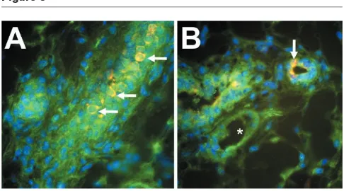
exam-ined mammary glands from a variety of developmental states to determine the physiologic correlates of this pattern. We found that mammary epithelial TGF-β immunoreactivity was heterogeneous and most intense during periods of proliferation and morphogenesis, sug-gesting the presence of a distinct subpopulation. Further characterization of these cells and determination of their ultimate fate in terms of proliferation and differentiation may be informative regarding the cellular mechanisms by which pattern and function are established during mammary gland development.
Conclusion
It is clear from a variety of studies in cell culture that TGF-β can have myriad effects, many of which comple-ment each other, but others that appear paradoxic. In vivo
studies using exposure to exogenous sources or trans-genic expression of constitutively active protein have shown what TGF-βcan do in the mammary gland. These have generally led to the conclusion that TGF-β has a prominent role in regulating pattern formation by the epithelium, perhaps via interactions with the stroma, and is involved in fate decisions by individual cells. In the great majority of animal and cell culture studies, however, activa-tion of the ubiquitous latent TGF-β complex, which exer-cises fundamental control of TGF-β action, has been scarcely addressed, which is due in large part to the lack of appropriate tools and reagents. As evidenced by the WAP-TGF-β223-225 versus the MMTV-TGF-β223-225
trans-genic phenotypes, spatial and temporal patterns of LTGF-β
activation are key elements for to understanding the spe-cific consequences of TGF-β activity during mammary gland development. Likewise, similar studies of the relative activity of the different TGF-β isoforms in the mammary gland may help shed light on the need for this apparent redundancy. Finally, studies that address mechanisms of activation may then contribute to strategies for manipula-tion of TGF-βin situand lead to a better understanding of how its dysregulation contributes to carcinogenesis [58].
Acknowledgements
Work by the authors and cited in the present review was supported by the US Army Medical Research and Materiel Command under DAMD-17-96-1-6716, the California Breast Cancer Research Program and the Office of Health and Environmental Research, Health Effects Research Division, of the US Department of Energy Contract No DE-AC-03-76SF00098.
References
Articles of particular interest have been highlighted as:
• of special interest
•• of outstanding interest
1. Daniel CW, Robinson SD: Regulation of mammary growth and function by TGF-ββ.Mol Reprod Dev1992, 32:145–151.
2. Knabbe C, Lippman ME, Wakefield LM, et al: Evidence that trans-forming growth factor-ββ is a hormonally regulated negative growth factor in human breast cancer cells. Cell 1987, 48: 417–428.
3. Wakefield l, Colletta AA, McCune BK, Sporn MB: Roles for trans-forming growth factors-ββin the genesis, prevention and treatment of breast cancer.In: Genes, Oncogens, and Hormones: Advances in Cellular and Molecular Biology of Breast Cancer. Edited by Dickson RB, Lipman MF. Boston: Kluwer Academic Publishers, 1991:97–136.
4. Silberstein GB, Daniel CW: Reversible inhibition of mammary
•• gland growth by transforming growth factor-ββ.Science1987, 237: 291–293.
This elegant study was the first to show that TGF-β1has a role in mammary gland development. Implantation of slow-release Elvax pellets containing TGF-β1resulted in the inhibition of ductal growth and end-bud regression in
the region nearer the pellet.
5. Daniel CW, Silberstein GB, Van Horn K, Strickland P, Robinson S:
TGF-ββ1-induced inhibition of mouse mammary ductal growth: developmental specificity and characterization. Dev Biol 1989,
135:20–30.
6. Daniel CW, Silberstein GB: Postnatal development of the rodent mammary gland.In: The Mammary Gland: Development, Regulation and Function. Edited by Neville MC, Daniel CW. New York: Plenum Press Publishing Corp, 1987:3–36.
7. Lawrence DA, Pircher R, Jullien P: Conversion of a high molecular weight latent beta-TGF from chicken embryo fibroblasts into a low molecular weight active beta-TGF under acidic conditions.
Biochem Biophys Res Commun1985, 133:1026–1034.
8. Kim S-J, Park K, Koeller D, et al: Post-transcriptional regulation of the human transforming growth factor-ββ1gene.J Biol Chem1992,
267:13702–13707.
9. Lawrence DA: Identification and activation of latent transforming growth factor ββ.Methods Enzymol1991, 198:327–336.
10. Flaumenhaft R, Rifkin DB: The extracellular regulation of growth factor action.Mol Biol Cell1992, 3:1057–1065.
11. Pierce DFJ, Johnson MD, Matsui Y, et al: Inhibition of mammary duct
[image:6.612.56.299.94.229.2]• development but not alveolar outgrowth during pregnancy in transgenic mice expressing active TGF-ββ1.Genes Dev 1993, 7: 2308–2317.
Figure 3
Expression during ductal outgrowth of constitutively active TGF-β1resulting
from the transgene described in [49] led to decreased proliferation in end-buds and consequently slower penetration of the fatpad by ducts during puberty. The MMTV promoter used in this study does not express trans-genes during late pregnancy or lactation.
12. Tang B, Bottinger EP, Jakowlew SB, et al: Transforming growth
• factor-beta1 is a new form of tumor suppressor with true haploid insufficiency.Nature Med1998, 4:802–807.
Heterozygote mice for TGF-β1gene deletion express only 10–30% of the wild-type levels of TGF-β1, depending on the tissue. Treatment of heterozy-gotes with chemical carcinogens results in increased tumorigenesis over that in wild-types.
13. Gomm JJ, Smithe J, Ryall GK, et al: Localization of basic fibroblast growth factor and transforming growth factor ββ1 in the human mammary gland.Cancer Res1991, 51:4685–4692.
14. Lu Y-J, Osin P, Lakhani SR, Palma SD, Gusterson BA, Shipley JM:
Comparative genomic hybridization analysis of lobular carcinoma in situ and a typical lobular hyperplasia and potential roles for gains and losses of genetic material in breast neoplasia.Cancer Res1998, 58:4721–4727.
15. Mizukami Y, Nonomura A, Yamada T, et al: Immunohistochemical demonstration of growth factors, TGF-alpha, TGF-beta, IGF-I and neu oncogene product in benign and malignant human breast tissues.Anticancer Res1990, 10:1115–1126.
16. Stampfer MR, Yaswen P, Alhadeff M, Hosoda J: TGF-ββinduction of extracellular matrix associated proteins in normal and trans-formed human mammary epithelial cells in culture is independent of growth effects.J Cell Physiol1993, 155:210–221.
17. Baillie R, Coombes RC, Smith J: Multiple forms of TGF-beta 1 in breast tissues: a biologically active form of the small latent complex of TGF-beta 1.Eur J Cancer1996, 32A:1566–1573.
18. Dong-Le Bourhis X, Berthois Y, Millot G, et al: Effect of stromal and epithelial cells derived from normal and tumorous breast tissue on the proliferation of human breast cancer cell lines in co-culture.Int J Cancer1997, 71:42–48.
19. Sieuwerts AM, Klijn JG, Henzen-Logmand SC, et al: Urokinase-type-plasminogen-activator (uPA) production by human breast (myo) fibroblasts in vitro: influence of transforming growth factor-beta(1) [TGF factor-beta(1)] compared with factor(s) released by human epithelial-carcinoma cells.Int J Cancer1998, 76:829–835.
20. Chakravarthy D, Green AR, Green VL, Kerin MJ, Speirs V: Expression and secretion of TGF-beta isoforms and expression of TGF-beta-receptors I, II and III in normal and neoplastic human breast.Int J Oncol1999, 15:187–194.
21. Robinson SD, Silberstein GB, Roberts AB, Flanders KC, Daniel CD:
• Regulated expression and growth inhibitory effects of transform-ing growth factor-ββisoforms in mouse mamary gland develop-ment.Development1991, 113:867–878.
The expression patterns of the three TGF-βs were characterized at both the mRNA and protein levelsin situ. TGF-βs were expressed at all stages of development except late pregnancy and lactation. Implantation of slow-release pellets containing TGF-β2or TGF-β3were found to have the same effect as those containing TGF-β1.
22. Silberstein GB, Flanders KC, Roberts AB, Daniel CW: Regulation of mammary morphogenesis: evidence for extracellular matrix-mediated inhibition of ductal budding by transforming growth factor-ββ1.Dev Biol1992, 152:354–362.
23. Barcellos-Hoff MH: Latency and activation in the regulation of TGF-ββ.J Mam Gland Biol Neoplasia1996, 3:353–363.
24. Barcellos-Hoff MH, Derynck R, Tsang ML-S, Weatherbee JA:
Trans-•• forming growth factor-ββactivation in irradiated murine mammary gland.J Clin Invest1994, 93:892–899.
This study was the first to employ differential staining with antibodies to detect active and latent forms of TGF-β1, that were validated using the
approach described in [24•]. Irradiation of the mammary gland resulted in increased levels of the active form of TGF-β1and decreased amounts of the
latent form, which is consistent with activation occurring for up to 3 days.
25. Barcellos-Hoff MH, Ehrhart EJ, Kalia M, et al: Immunohistochemical
• detection of active TGF-ββin situ using engineered tissue.Am J Pathol1995, 147:1228–1237.
See [24••].
26. Barcellos-Hoff MH: Radiation-induced transforming growth factor ββ and subsequent extracellular matrix reorganization in murine mammary gland.Cancer Res1993, 53:3880–3886.
27. Ehrhart EJ, Carroll A, Segarini P, Tsang ML-S, Barcellos-Hoff MH:
Latent transforming growth factor-ββactivation in situ: quantitative and functional evidence following low dose irradiation.FASEB J
1997, 11:991–1002.
28. Pertovaara L, Sistonen L, Bos TJ, et al: Enhanced jun gene expres-sion is an early genomic response to transforming growth factor beta stimulation. Mol Cell Biol1989, 9:1255–1262.
29. Rahimi N, Tremblay E, McAdam L, Roberts A, Elliott B: Autocrine secretion of TGF-beta 1 and TGF-beta 2 by pre-adipocytes and adipocytes: a potent negative regulator of adipocyte differentia-tion and proliferadifferentia-tion of mammary carcinoma cells. In Vitro Cell Dev Biol Anim1998, 34:412–420.
30. Ellis I, Banyard J, Schor SL: Differential response of fetal and adult fibroblasts to cytokines: cell migration and hyaluronan synthesis.
Development1997, 124:1593–1600.
31. McCaffrey TA, Falcone DJ: Evidence for an age-related dysfunction in the antiproliferative response to transforming growth factor-ββin vascular smooth muscle cells.Mol Biol Cell1993, 4:315–322.
32. Takahashi K, Suzuki K, Ono T: Loss of growth inhibitory activity of TGF-ββ toward normal human mammary epithelial cells growth within collagen gel matrix.Biochem Biophys Res Commun1990,
173:1239–1247.
33. Martikainen P, Kyprianou N, Isaacs JT: Effect of transforming growth
• factor-ββ1 on proliferation and death of rat prostatic cells.
Endocrinology1990, 127:2963–2968.
This study and [34•] show that TGF-β1is upregulated and mediates apopto-sis in cultured cells from hormone-dependent tissues when the hormones are withdrawn.
34. Rotello RJ, Lieberman RC, Purchio AF, Gerschenson LE: Coordinated
• regulation of apoptosis and cell proliferation by transforming growth factor ββ1 in cultured uterine epithelial cells. Proc Natl Acad Sci USA1991, 88:3412–3415.
See [33•].
35. Strange R, Li F, Saurer S, Burkhardt A, Friis RR: Apoptotic cell death and tissue remodeling during mouse mammary gland involution.
Development (Camb)1992, 115:49–58.
36. Ayoub IA, Yang TJ: The regulatory role of transforming growth factor-beta in activation of milk mononuclear cells.Am J Reprod Immunol1997, 38:121–128.
37. Newton LK, Yung WKA, Pettigrew LC, Steck PA: Growth regulatory activities of endothelial extracellular matrix mediation by trans-forming growth factor-ββ. Exp Cell Res1990, 190:127–132.
38. Ferguson JE, Schor AM, Howell A, Feruson MWJ: Changes in the extracellular matrix of the normal human breast during the men-strual cycle.Cell Tissue Res1992, 268:167–177.
39. Silberstein GB, Strickland P, Coleman S, Daniel CW: Epithelium-dependent extracellular matrix synthesis in transforming growth factor-ββ1-growth-inhibited mouse mammary gland. J Cell Biol
1990, 110:2209–2219.
41. Xie J, Haslam SZ: Extracellular matrix regulates ovarian hormone-dependent proliferation of mouse mammary epithelial cells.
Endocrinology1997, 138:2466–2473.
42. Butta A, MacLennan K, Flanders KC, et al: Induction of transforming growth factor ββ1 in human breast cancer in vivo following tamox-ifen treatment.Cancer Res1992, 52:4261–4264.
43. Bruner KL, Rodgers WH, Gold LI, et al: Transforming growth factor
• ββmediates the progesterone suppression of an epithelial metal-loproteinase by adjacent stroma in the human endometrium.Proc Natl Acad Sci USA1995, 92:7362–7366.
This in vitrostudy shows that progesterone suppresses matrilysin expres-sion, progesterone induces TGF-β1 expression, TGF-β1 suppresses
matrilysin expression in the absence of progesterone and that pan-specific blocking antibody to TGF-βs blocks the progesterone-mediated suppres-sion of matrilysin expressuppres-sion.
44. Robinson SD, Roberts AB, Daniel CW: TGFββ suppresses casein synthesis in mouse mammary explants and may play a role in controlling milk levels.J Cell Biol1993, 120:245–251.
45. Bergstraesser L, Sherer S, Panos R, Weitzman S: Stimulation and inhibition of human mammary epithelial cell duct morphogenesis in vitro.Proc Assoc Am Phys1996, 108:140–154.
46. Soriano JV, Orci L, Montesano R: TGF-ββ1 induces morphogenesis of branching cords by cloned mammary epithelial cells at subpi-comolar concentrations. Biochem Biophys Res Commun 1996,
220:879–885.
47. Miettinen PJ, Ebner R, Lopez AR, Derynck R: TGF-ββinduced transdif-ferentiation of mammary epithelial cells to mesenchymal cells: involvement of type I receptors.J Cell Biol1994, 127:2021–2036.
48. Desmouliere A, Geinoz A, Gabbiani F, Gabbiani G: Transforming growth factor-ββ1 induces αα-smooth muscle actin expression in granulation tissue myofibroblasts and in quiescent and growing cultured fibroblasts.J Cell Biol1993, 122:103–111.
49. Daniel CW, Robinson S, Silberstein GB: The role of TGF-ββin pat-terning and growth of the mammary ductal tree.J Mam Gland Biol Neoplasia1996, 1:331–341.
50. Brunner AM, Marquardt H, Malacko AR, Lioubin MN, Purchio AF: Site-directed mutagenesis of cysteine residues in the pro region of the transforming growth factor ββ1 precursor.J Biol Chem1989, 264: 13660–13664.
51. Smith GH: TGF-ββand functional differentiation.J Mam Gland Biol Neoplasia1996, 1:343–352.
52. Jhappan C, Geiser AG, Kordon EC, et al: Targeting expression of a
•• transforming growth factor ββ1 transgene to the pregnant mammary gland inhibits alveolar development and lactation.
EMBO J1993, 12:1835–1845.
Expression of constitutively active TGF-β1using this transgene has a dra-matic effect on later alveolar development and lactation. Apoptosis is induced at this stage and also on later alveolar development and lactation. Apoptosis is induced at this stage and also when the transgene is tran-siently expressed in a subset of cells during estrous.
53. Kordon EC, McKnight RA, Jhappan C, et al: Ectopic TGF beta 1
•• expression in the secretory mammary epithelium induces early senescence of the epithelial stem cell population.Dev Biol1995,
168:47–61.
Using transplantation techniques, these authors demonstrate the overex-pression of constitutively active TGF-β1leads to rapid depletion of the ability of the mammary gland to renew itself.
54. Kulkarni AB, Huh CG, Becker D, et al: Transforming growth factor beta 1 null mutation in mice causes excessive inflammatory res-ponse and early death.Proc Natl Acad Sci USA1993, 90:770–774.
55. Letterio JJ, Geiser AG, Kulkarni AB, et al: Maternal rescue of trans-forming growth factor-ββ1 null mice. Science 1994, 264:1936– 1938.
56. Gorska AE, Joseph H, Derynck R, Moses HL, Serra R: Dominant-neg-ative interference of the transforming growth factor-beta type II receptor in mammary gland epithelium results in alveolar hyper-plasia and differentiation in virgin mice.Cell Growth Differ1998, 9: 229–238.
57. Joseph H, Gorska AE, Sohn P, Moses HL, Serra R: Overexpression of a kinase-deficient transforming growth factor-ββtype II receptor in mouse mammary stroma results in increased epithelial branch-ing.Mol Biol Cell1999, 10:1221–1234.
58. Reiss M, Barcellos-Hoff MH: Transforming growth factor-ββ in breast cancer: a working hypothesis.Br Cancer Res Treat1997, 45:81–95.
Authors’ affiliation:Life Sciences Division, Lawrence Berkeley National Laboratory, University of California, Berkeley, California, USA
Correspondence:MH Barcellos-Hoff, Life Sciences Division, Bldg 74-174, 1 Cyclotron Rd, Lawrence Berkeley National Laboratory, Berkeley, CA 94720, USA. Tel: +1 510 486 6371;