R E S E A R C H A R T I C L E
Open Access
Role of ADAM17 in the non-cell autonomous
effects of oncogene-induced senescence
Beatriz Morancho
1†, Águeda Martínez-Barriocanal
1†, Josep Villanueva
1and Joaquín Arribas
1,2,3*Abstract
Introduction:Cellular senescence is a terminal cell proliferation arrest that can be triggered by oncogenes. One of the traits of oncogene-induced senescence (OIS) is the so-called senescence-associated secretory phenotype or senescence secretome. Depending on the context, the non-cell autonomous effects of OIS may vary from tumor suppression to promotion of metastasis. Despite being such a physiological and pathologically relevant effector, the mechanisms of generation of the senescence secretome are largely unknown.
Methods:We analyzed by label-free proteomics the secretome of p95HER2-induced senescent cells and compared the levels of the membrane-anchored proteins with their transcript levels. Then, protein and RNA levels of ADAM17 were evaluated by using Western blot and reverse transcription-polymerase chain reaction, its localization by using biotin labeling and immunofluorescence, and its activity by using alkaline phosphatase-tagged substrates. The p95HER2-expressing cell lines, senescent MCF7 and proliferating MCF10A, were analyzed to study ADAM17 regulation. Finally, we knocked down ADAM17 to determine its contribution to the senescence-associated secretome. The effect of this secretome was evaluated in migration assays in vitro and in nude mice by assessing the metastatic ability of orthotopically co-injected non-senescent cells.
Results:Using breast cancer cells expressing p95HER2, a constitutively active fragment of the proto-oncogene HER2 that induces OIS, we show that the extracellular domains of a variety of membrane-bound proteins form part of the senescence secretome. We determine that these proteins are regulated transcriptionally and, in addition, that their shedding is limited by the protease ADAM17. The activity of the sheddase is constrained, at least in part, by the accumulation of cellular cholesterol. The blockade of ADAM17 abrogates several prometastatic effects of the p95HER2-induced senescence secretome, both in vitro and in vivo.
Conclusions:Considering these findings, we conclude that ectodomain shedding is tightly regulated in oncogene-induced senescent cells by integrating transcription of the shedding substrates with limiting ADAM17 activity. The remaining activity of ADAM17 contributes to the non-cell autonomous protumorigenic effects of p95HER2-induced senescent cells. Because ADAM17 is druggable, these results represent an approximation to the pharmacological regulation of the senescence secretome.
Introduction
Cellular senescence is a terminal cell proliferation arrest characterized by a distinct phenotype. Compared with their proliferating counterparts, senescent cells have enlarged volumes, display a flattened and vacuolated morphology, and express a variety of markers. The most
widely used to identify senescent cells is senescence-associatedβ-galactosidase.
Cellular senescence can be triggered by a variety of stressors, including oncogenes, resulting in what is known as oncogene-induced senescence (OIS) [1]. For example, expression of p95HER2, an oncogenic fragment of the tyrosine kinase receptor HER2, induces OIS in a variety of cell lines [2].
The onset of senescence is characterized by a pro-found change in the secretome (i.e., all factors secreted by a given cell) that results in the so-called senescence-associated secretory phenotype or senescence secretome * Correspondence:jarribas@vhio.net
†Equal contributors
1
Preclinical Research Program, Vall d’Hebron Institute of Oncology (VHIO), Psg. Vall d’Hebron 119-129, Barcelona 08035, Spain
2
Department of Biochemistry and Molecular Biology, Building M, Campus UAB, Bellaterra (Cerdanyola del Valles), , Barcelona 08193, Spain Full list of author information is available at the end of the article
[1]. Depending on the context, the senescence secretome has disparate effects. It may promote [3] or impair [4] immune surveillance against tumor cells in the liver and in the prostate, respectively. In fact, senescent cells may be short-lived or long-lived in vivo, in both immuno-competent [3–5] and immunosuppressed [2, 6] mice. Furthermore, the senescence secretome can suppress [7] or promote [8] tumor growth. These results can be ra-tionalized assuming that the potent tumor suppressive effects of senescence can be reversed, particularly in ad-vanced tumors, by modifying the composition of the senescence secretome and, thus, its effects on target cells.
Because the non-cell autonomous effects of senescent cells can suppress or contribute to tumor progression, the up- or downregulation of the senescence secretome could be a therapeutic strategy to treat cancer and per-haps many other diseases related to cellular senescence [1]. Unfortunately, to date, there are no known strategies to regulate the production of the senescence secretome.
The proteolytic release of the extracellular domain of transmembrane proteins is known as ectodomain shed-ding. This type of limited proteolysis affects a diverse group of functionally unrelated transmembrane proteins, including membrane-anchored growth factors, cytokines, cell adhesion molecules, or transmembrane proteases [9–12]. The proteases that cleave the vast majority of these transmembrane proteins are the metalloprotease disintegrins ADAM17 (also known as tumor necrosis factor-alpha-converting enzyme) or ADAM10 or both (reviewed in [13]).
Some components frequently secreted by senescent cells, such as transmembrane epidermal growth factor (EGF)-like growth factors, are generated through ectodo-main shedding. However, the contribution of ectodoectodo-main shedding to the senescence secretome remains largely un-explored. Although ADAM17 has been recently shown to be active in senescent cells [14], its regulation or func-tional importance during senescence is unknown.
Here, we show that approximately 10 % of the compo-nents of the secretome of p95HER2-induced senescent cells are generated through the shedding of the ectodo-mains of membrane-anchored proteins. The main mech-anism regulating the release of these ectodomains is the transcriptional regulation of the membrane-anchored precursors. Functional analysis shows that ADAM17 plays a major role in these cleavages. However, although ADAM17 protein levels increase during p95HER2-induced OIS, the activity of the metalloprotease does not increase, and this is likely because of the accumula-tion of cholesterol, a negative regulator of ADAM17, in senescent cells. Finally, we show that ADAM17 activity is required for several non-cell autonomous protumori-genic and prometastatic effects of p95HER2-induced
senescent cells. Because the activity of ADAM17 can be pharmacologically downregulated, these results indicate that inhibition of this metalloprotease could be a means to target the undesired non-cell autonomous effects of cellular senescence.
Methods Reagents
Doxycycline (Doxy.), phorbol myristate acetate, biotin, 1-10-phenanthroline, methyl-beta-cyclodextrin (MβCD), in-sulin, EGF, and hydrocortisone were from Sigma-Aldrich (St. Louis, MO, USA). Batimastat (BB94) was from Merck (Schwalbach, Germany).
Antibodies
Rabbit anti-phospho ERK (T202/Y204; #4370), anti-ERK (#9102), anti-phospho Akt (S473; #9271), anti-Akt (#9272), anti-phospho EGF receptor (EGFR) (Y1068; #3777), and anti-EGFR (#4267) were from Cell Signaling Technology (Danvers, MA, USA). Mouse anti-EpCAM (#sc-25308) was from Santa Cruz Biotechnologies (Santa Cruz, CA, USA), and mouse anti-pan actin (#MA5-11869) was from Thermo Scientific (Lafayette, CO, USA). Mouse anti-APP (#MAB348) and rabbit anti-ADAM17 (#AB19027) were from Millipore (Billerica, MA, USA). Rabbit anti-ADAM17 (#ab39162), rabbit ADAM10 (#ab1997), goat anti-Met receptor (#ab10728), and mouse anti-glucose-6-phos-phate (GPI) (#ab66340) antibodies were from Abcam (Cambridge, MA, USA). Rabbit anti-DDR1 (#10730) was from Sino Biological Inc. (Beijing, PR China), rabbit anti-GAPDH (#2275-PC-1) was from Trevigen (Gaithersburg, MD, USA), and mouse anti-HER2 (#MU-134-UC) was from BioGenex (San Ramon, CA, USA). Mouse anti-EphB4 (#AF446) was from R&D Systems (Minneapolis, MN, USA). Fluorochrome-conjugated antibodies were from Molecular Probes (Life Technologies, Grand Island, NY, USA).
Transcriptomic and proteomic analyses
Transcripts and secretome analysis were performed as described in [15] and [2], respectively. GeneChip expres-sion probe array Gene Expresexpres-sion Omnibus reference number is GSE68256. Heatmap hierarchical clustering was performed as in [16].
Plasmids
shRNA vectors targeting ADAM17 or non-silencing control were from Thermo Scientific. p95HER2 in pENTR1Dual (Invitrogen, Paisley, UK) was transferred into pINDUCER20 using LR Clonase II (Invitrogen). For control pINDUCER20 (pINDUCER20-Empty), cloram-phenicol and ccdB genes were removed from pENTR1A Dual (5-SalI and 3’-XhoI) and the minimal MCS was transferred to pINDUCER20. pINDUCER20 was kindly provided by Stephen J Elledge (Howard Hughes Medical Institute, Chevy Chase, MD, USA) [17].
Cell culture and infections
MCF7, MCF7 Tet-Off p95HER2, and MDA-MB-231-Luc were maintained as previously described [2]. MCF10A was maintained in Dulbecco’s modified Eagle’s medium,/ F-12, 10 % fetal bovine serum (FBS), 4 mmol/l L-glutamine (all from Gibco, Carlsbad, CA, USA), 9 μg/ml insulin, 0.5 μg/ml hydrocortisone, and 18 ng/ml EGF. MCF7 Tet-Off p95HER2 AP-Areg, AP-TGF-α, AP-BTC, shControl, and shADAM17 were generated by infecting MCF7 Tet-Off p95HER2 with the lentiviral particles ob-tained transfecting HEK293T cells with the plasmids described above (calcium phosphate method). Viral super-natant was applied to the cells in the presence of 8μg/ml polybrene, and stable clones were selected with 1 μg/ml puromycin. MCF10A was infected with pINDUCER20-p95HER2 by using the same protocol, and selection was performed with 200μg/mL G418. MCF10A infection with lentiviral vectors expressing AP-Areg and AP-TGF-αwas performed with the plasmids and method indicated above.
Protein extraction and immunoblotting
Cells were lysed in 20 mM Tris-HCl pH 7.4, 137 mM NaCl, 2 mM EDTA pH 8.0, 10 % glycerol, 1 % NP-40, pro-tease inhibitor cocktail (Roche, Penzberg, Germany), 1.3 mM sodium orthovanadate, 10 mM 1-10-phenanthroline, and 1μM BB94. Cell lysates were quantified by using the Pierce BCA Protein Assay kit (Pierce, Rockford, IL, USA), and equal amounts of protein were separated on SDS-polyacrylamide gels and transferred onto nitrocellulose membranes. These were blocked with 5 % bovine serum albumin or skim milk in TBS-Tween 0.1 % and then incu-bated with primary antibodies, and bound antibodies were detected with the appropriate peroxidase-conjugated sec-ondary antibodies (Amersham GE Healthcare, Piscataway, NJ, USA) by using the ECL detection system (Millipore). To detect ADAM17, the same amount of protein from cell lysates was concentrated with wheat germ agglutinin (WGA)-agarose beads (Vector Laboratories, Peterbor-ough, UK) for 2 h at 4 °C. Proteins were eluted directly in SDS-polyacrylamide gel electrophoresis loading buffer.
Determination of proteins in the collected conditioned media was performed following the same protocol after concentrating 100× the samples by using centricons
(Amicon Ultracel 3K; Millipore). Densitometric quantifi-cation was performed by using ImageJ software.
Enzyme-linked immunosorbent assay
Cells were plated and the next day were washed twice with 1X phosphate-buffered saline (PBS), and medium was changed to serum-free medium with L-glutamine. Conditioned media were collected 48 h later, spun down at 200×g for 5 min, and transferred to clean tubes. Amphiregulin was determined in accordance with the instructions of the manufacturer (RayBiotech, Norcross, GA, USA).
mRNA expression
RNA was isolated by using an RNeasy Mini kit (Qiagen, Hilden, Germany) and reverse-transcribed by using a High Capacity cDNA Reverse Transcription Kit (Applied Biosystems, Weiterstadt, Germany) in accordance with the instructions of the manufacturers. Quantitative re-verse transcription-polymerase chain reaction was per-formed by using Taqman primers (Applied Biosystems) for ADAM17 (Hs01041915_m1), ADAM10 (Hs00153853_ m1), and GAPDH (Hs03929097_g1).
Biotin labeling
Cells were washed three times with ice-cold 1X PBS pH 8.0 and labeled with 1 mg/ml sulfo-NHS-LC-biotin (Pierce) in 1X PBS pH 8.0 gently shaking for 1 h at 4 °C. Biotin was inactivated by washing with 50 mM Tris pH 7.4, and cells were extensively washed with 1X PBS. Then cells were harvested and lysed, and immunopre-cipitation was performed by using an anti-ADAM17 antibody or a control IgG. Immunoprecipitates were analyzed by Western blotting by using streptavidin-POD conjugate (Roche) or anti-ADAM17 antibody (Abcam).
Confocal microscopy
Cells were seeded on glass coverslips, washed with 1X PBS, fixed with 4 % formaldehyde for 20 min, and perme-abilized with 0.2 % Triton X-100 for 10 min. 1X PBS con-taining 1 % bovine serum albumin, 0.1 % saponin, and 0.02 % NaN3was used for blocking (1 h), primary antibody binding (2 h), and secondary fluorochrome-conjugated antibody binding (40 min in dark). Preparations were mounted by using Vectashield with DAPI (4’,6’ -diamidino-2-phenylindole) (Vector Laboratories). All procedures were performed at room temperature. Images were cap-tured by using an Olympus FV1000 confocal microscope (Olympus Corporation, Tokyo, Japan).
Ectodomain shedding assay
serum or growth factors, and the medium was replaced 1 h later with fresh Opti-MEM with or without the indi-cated stimuli. Cells and supernatants were harvested at the designated time points, and AP activity was measured at an absorbance of 405 nm after incubation with the AP substrate 4-nitrophenyl phosphate (Sigma-Aldrich). At least three wells per condition were performed, and the ratio between the AP activity in the supernatant and the cell lysate plus supernatant was calculated. The fold in-crease in the ratio of AP activity relative to the control is shown.
Proliferation assays
Proliferation was analyzed by cell counting. After trypsi-nization, viable cells determined by trypan blue dye ex-clusion were counted on a Neubauer chamber.
Senescence-associatedβ-galactosidase activity
Cells were plated on coverslips and analyzed by using a senescence β-galactosidase staining kit (Cell Signaling Technology) in accordance with the indications of the manufacturer.
Filipin staining
Cells were washed three times with 1X PBS and fixed with 3 % paraformaldehyde for 1 h at room temperature. Washes were repeated, and cells were incubated with 1.5 mg/ml glycine in 1X PBS for 10 minutes. Staining was performed for 2 h with 0.05 mg/ml filipin complex (Sigma-Aldrich) in 1X PBS/10 % FBS. After the cells were rinsed three times with 1X PBS, they were analyzed by fluorescence microscopy (BX61 Olympus).
Migration assays
Cell migration was determined with 24-well format Boyden chambers containing FluoroBlok PET mem-branes with 8-μm pores (BD Biosciences, Heidelberg, Germany). Conditioned media were obtained after incu-bating cells with serum-free medium with glutamine for 48 h, spun down, and concentrated 100 times by using centricons (Amicon Ultracel 3K). Concentrated condi-tioned media were diluted 10 times in serum-free medium and added to the bottom part of the Boyden chambers. MDA-MB-231 Luc or MCF7 cells were plated in the upper chambers and migration ability was evaluated 24 or 48 h later, respectively, by staining the cells with 1 μM SYTO-9 (Life Technologies) and counting them on the underside of the filters by using fluorescence microscopy.
Mouse model of breast cancer metastasis
MCF7 Tet-Off p95HER2 cells expressing a short hairpin control or targeting ADAM17 were co-injected with MDA-MB-231 Luc cells into the right flanks of 6- to 8-week-old female BALB/c athymic mice (Charles River
Laboratories, Paris, France) in addition to a 17β-estradiol pellet (Innovative Research of America, Sarasota, FL, USA). The expression of p95HER2 was repressed by add-ing doxycycline (50 mg/kg per day) to the drinkadd-ing water until tumors were about 100 mm3. Tumor xenografts were measured with calipers every 3 days, and tumor vol-ume was determined by using the formula: (length × width2) × (pi/6). Tumors were resected when they reached 300 mm3, and metastatic colonization was monitored by in vivo bioluminescence imaging with the IVIS-200 im-aging system (PerkinElmer, Waltham, MA, USA). At the end of the experiment, animals were anesthetized with a 1.5 % isofluorane-air mixture and were sacrificed by cer-vical dislocation. Mice were maintained and treated in ac-cordance with institutional guidelines of Vall d’Hebron University Hospital Care and Use Committee.
Statistics
Data are presented as average ± standard deviation and were analyzed by the two-sided Student t test. Results were considered to be statistically significant at P value of less than 0.05. All statistical analyses were conducted by using the GraphPad Prism 5 Statistical Software (GraphPad Software, Inc., La Jolla, CA, USA).
Results
Shedding of transmembrane molecules during p95HER2-induced senescence
Expression of p95HER2 in different breast cancer cell lines leads to OIS ([2] and Additional file 1: Figure. S1a). Analysis of the media conditioned by control cells or p95HER2-induced senescent cells by quantitative label-free proteomics confirmed the profound change in the secretome composition characteristic of OIS (Additional file 1: Figure. S1b).
chose these proteins because, compared with other trans-membrane proteins, they have long intracellular domains; identification of peptides corresponding to the intracellu-lar domains would indicate that they are released through exosomes or other mechanisms. We concluded that ap-proximately 10 % of the components of the p95HER2-induced senescence secretome are generated through ectodomain shedding.
Regulation of the sheddome of p95HER2-induced senescent cells
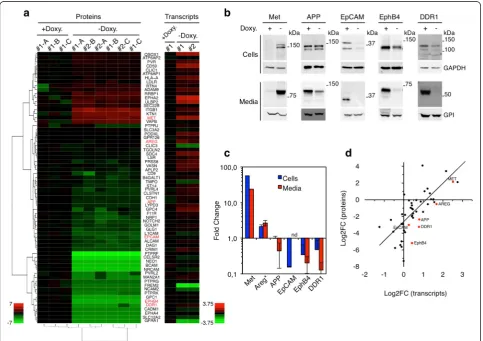
Proteomic analysis of membrane-anchored proteins showed the profound change in the sheddome (i.e., the part of the secretome generated by ectodomain shedding) produced by p95HER2-induced senescent cells (Fig. 1a, Proteins).
Analysis by Western blot of several randomly chosen proteins confirmed the expected full-length species and shed ectodomains in cell lysates and conditioned media, respectively (Fig. 1b and Additional file 6: Table S4). Confirming the proteomic analysis, compared with con-trol proliferating cells, the levels of the soluble extracel-lular domains of the tyrosine kinase receptor Met or the EGF-like growth factor Areg increased during p95HER2-induced senescence. In contrast, the extracellular do-mains of the amyloid precursor protein (APP), the cell adhesion molecule EpCAM, or the tyrosine kinase re-ceptors DDR1 and EphB4 decreased (Fig. 1b, c).
The increased levels of the soluble ectodomains of Met and Areg were concomitant with increased levels of the cell-associated full-length proteins (Fig. 1b, c). Conversely, the decrease in the levels of the soluble ectodomains of
Fig. 1Protein ectodomain shedding during p95HER2-induced senescence.aLeft, MCF7 Tet-Off p95HER2 cells were treated with or without doxycycline as indicated in two independent experiments (#1 and #2). The extracellular media were analyzed by label-free quantitative proteomics in triplicate (A-C) (Additional file 1: Figure. S1). The heatmap shows log2FC of the normalized levels of soluble ectodomains.Right, transcriptomic analysis of the same cells
treated as in left panel (a).bMCF7 Tet-Off p95HER2 cells were treated with or without doxycycline for 7 days. Conditioned media from the last two days and cell lysates were analyzed by Western blotting as indicated.cQuantitative results from at least three independent experiments performed as in (b) were expressed as average ± standard deviation. Note that the levels of Areg were determined by enzyme-linked immunosorbent assay.dlog2FC of the
ectodomain levels and the log2FC of their corresponding transcripts levels, determined as described in (a), are represented. Linear regression line is
shown (R2= 0.528);P<0.001 using the Spearman correlation coefficient.APPamyloid precursor protein,Aregamphiregulin,EpCAMepithelial cell adhesion
[image:5.595.57.541.293.634.2]DDR1, EphB4, and EpCAM paralleled those of their full-length cell-associated counterparts. In the case of APP, the levels of the transmembrane protein did not change; how-ever, the levels of shedding decreased (Fig. 1b, c), indicat-ing that, in senescent cells, this substrate is less accessible to the metalloproteases that cleave its ectodomain or, al-ternatively, that these metalloproteases are inhibited.
The transcriptomic analysis of the factors detected though proteomics showed a direct correlation between the levels of shed ectodomains and those of their corre-sponding mRNAs (Fig. 1a, Transcripts and Fig. 1d). We concluded that, during OIS, ectodomain shedding is reg-ulated largely through the transcriptional control of shedding substrates.
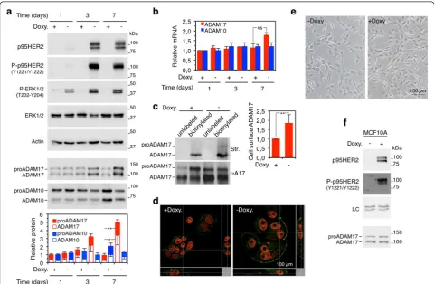
ADAM17 expression in p95HER2-induced senescent cells ADAM17 cleaves Areg [18], APP [19], DDR1 [20], EphB4 [21], Met [22], and EpCAM [23]. Therefore, we analyzed the levels of ADAM17 during the onset of p95HER2-induced senescence.
Expression of p95HER2 during 2 days results in the ir-reversible onset of senescence, and after 5–7 days the cells display the full senescence phenotype [2]. Concomi-tantly, the levels of total ADAM17 increased (Fig. 2a, bottom panels). ADAM17 is synthesized as a zymogen (proADAM17), which contains a pro-domain that in-hibits the metalloprotease active site. During transport through the trans-Golgi network, furin-like pro-protein convertases remove the pro-domain generating mature
[image:6.595.57.538.268.582.2]ADAM17 [24]. The increase in the levels of the metal-loprotease (Fig. 2a, bottom panels) is largely post-transcriptional (Fig. 2b). Analysis of its subcellular distribution by means of biotinylation of intact cells and immunofluorescence showed that ADAM17 is predomin-antly intracellular in proliferating cells and that it accumu-lates at the cell surface in p95HER2-induced senescent cells (Fig. 2c, d). ADAM10 also participates in ectodomain shedding, and some of the substrates of ADAM17, such as APP, are also cleaved by ADAM10 [13]. However, ADAM10 protein was not upregulated during p95HER2-induced senescent cells; in fact, relative to control cells, p95HER2-induced senescent cells showed a slight but re-producible downmodulation of ADAM10 which could not be explained by a reduction in its cognate transcript (Fig. 2a, b).
To determine whether the upregulation of ADAM17 is the result of the activity of p95HER2 independently of the senescence status, we analyzed MCF10A cells. Ra-ther than inducing OIS (Fig. 2e), expression of p95HER2 in this immortalized non-transformed mammary epithe-lium cell line, accelerates proliferation [2]. The levels of ADAM17 remained unchanged in MCF10A expressing p95HER2 (Fig. 2f ), indicating that overexpression of the metalloprotease is likely linked to OIS rather than to the mere expression of the constitutively active fragment of HER2. We concluded that the levels of ADAM17 are up-regulated during p95HER2-induced cellular senescence.
Role of ADAM17 in the shedding of different components of the senescence secretome
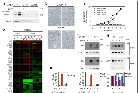
To determine whether ADAM17 plays a role in the shed-ding of transmembrane proteins during OIS, we knocked it down from the MCF7 Tet-Off p95HER2 cells. We used two independent shRNAs targeting ADAM17 to generate two cell lines (Fig. 3a). Although we observed similar re-sults with both cell lines, for simplicity, except in Fig. 3d, we show the results obtained with only one of them. The downmodulation of the protease did not affect the morph-ology or the levels of SAβG in p95HER2-induced senes-cent cells (Fig. 3b). The proliferation of control cells or the inhibition of proliferation induced by p95HER2 expression was also unaffected (Fig. 3c).
In a proteomic analysis of the media conditioned by p95HER2-induced senescent cells expressing control shRNAs or the shRNAs targeting ADAM17, we quanti-fied the soluble ectodomains of 68 % (46 out of 68) of the membrane-anchored proteins shown in Fig. 1b. The levels of 37 % of these ectodomains (17 out of 46) de-creased in the media conditioned by the ADAM17 knockdown cell lines (Fig. 3d and Additional file 7: Table S5), showing that the metalloprotease participates in the cleavage of the corresponding membrane-anchored factors.
To validate these results, we showed that, in p95HER2-induced senescent cells, the knockdown of ADAM17 re-sulted in an approximately 80 % reduction of the shedding of Areg and Met (Fig. 3d, e and f ).
In agreement with the results shown in Fig. 1c, com-pared with that in control proliferating cells, the shedding of APP was reduced in p95HER2-induced senescent cells (Fig. 3g). Confirming the result of the proteomic analysis (Fig. 3d), in ADAM17 knockdown senescent cells, the shedding of APP was further reduced (Fig. 3g).
Collectively, these results show that ADAM17 contrib-utes to protein ectodomain shedding during OIS. We esti-mate that this metalloprotease contributes to the cleavage of approximately one third of the membrane-anchored proteins that undergo ectodomain shedding during OIS.
ADAM17 activity in p95HER2-induced senescent cells Although the results in Fig. 3 clearly show a role of ADAM17 in the generation of the senescence secretome, the inhibition of the shedding of APP during p95HER2-induced senescence (Figs. 1b, c and 3g), despite the in-crease of ADAM17 levels (Fig. 2a), indicates that the activity of the metalloprotease may be partially inhibited in p95HER2-induced senescent cells.
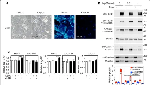
Previous reports have shown that ADAM17 is inhib-ited by high cholesterol levels [25–30]. Senescent cells tend to accumulate cholesterol [31]; in fact, it has been previously shown that HER2 (NeuT)-induced senes-cence results in a marked accumulation of cellular cholesterol [32]. Thus, we reasoned that the high levels of cholesterol in p95HER2-induced senescent cells may lead to the accumulation of partially inactive ADAM17.
As expected, treatment with MβCD, a compound
that selectively extracts membrane cholesterol, re-duced the levels of cholesterol in p95HER2-senescent cells (Fig. 4a).
MβCD did not affect the expression of p95HER2 or its signaling ability (Fig. 4b); however, it completely pre-vented the increase in ADAM17 protein levels that oc-curs during OIS. In contrast, the levels of ADAM10 were largely unaffected by the same treatment (Fig. 4b, bottom panels).
(Fig. 4c). But consistently with an inhibitory effect of chol-esterol, MβCD induced a significant increase in the shed-ding of both AP-tagged growth factors only in p95HER2-induced senescent cells (Fig. 4c). The shedding of betacel-lulin (BTC), a substrate of ADAM10 [18], was also un-affected by the treatment with MβCD, ruling out cholesterol as modulator of the activity of ADAM10 in MCF7 cells (Fig. 4c).
We concluded that the increase in cholesterol content in senescent cells downregulates the activity of ADAM17, but not that of ADAM10, and results in accumulation of partially inactive ADAM17. However, it should be under-scored that the remaining ADAM17 activity is responsible for the shedding of a variety of transmembrane molecules during OIS (Fig. 3).
Role of ADAM17 in non-cell autonomous effects of p95HER2-induced senescence
To analyze the functional importance of ADAM17 activ-ity during OIS, we compared the non-cell autonomous effects of p95HER2-induced senescent cells with those of the same cells knocked down for the metalloprotease on different in vitro and in vivo assays.
Consistently with the production of Areg by senescent cells, incubation of A431 cells, which overexpress the EGFR, with the media conditioned by p95HER2-induced senescent cells increased the levels of phospho-EFGR (Additional file 8: Figure. S3b). Also, as expected, knock-down of the metalloprotease completely inhibited the ef-fect of the senescence secretome on phospho-EGFR levels (Additional file 8: Figure. S3b).
Fig. 3Contribution of ADAM17 to the p95HER2-induced senescence secretome.aMCF7 Tet-Off p95HER2 cells constitutively expressing a control shRNA (NT, non-targeting) or two independent shRNAs targeting ADAM17 (A17#1 and A17#2) were cultured with or without doxycycline for 7 days. Cells were then harvested and lysed, and cell lysates were analyzed by Western blotting with the indicated antibodies.b Senescence-associatedβ-galactosidase was analyzed in the same cells as in (a). Representative bright-field images are shown.cThe same cells as in (a) were plated with or without doxycycline and counted at the indicated time points. The results represent averages of two independent determinations.
dThe conditioned media of the same cells as in (a), treated without doxycycline, were analyzed by label-free quantitative proteomics in triplicate (a-c). The heatmap shows log2FC of the normalized levels of soluble ectodomains.e-gThe same cells as in (a) were treated with or without
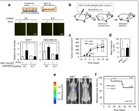
[image:8.595.58.540.90.412.2]Certain senescence secretomes promote cell migration [34]. Accordingly, the secretome of p95HER2-induced senescent cells promoted the migration of different breast cancer cell lines (Fig. 5a). The knockdown of ADAM17 with two independent shRNAs (Fig. 3a) abro-gated the effect of the senescence secretome on cell mi-gration (Fig. 5a and data not shown), indicating that transmembrane factors cleaved by the metalloprotease contribute to the pro-migratory effect of the senescence secretome.
The secretome of p95HER2-induced senescent cells promotes the metastatic growth of non-senescent cells [2]. Because the migratory behavior of cells in vitro (Fig. 5a) can be indicative of their metastatic potential in vivo, we analyzed the effect on ADAM17 knockdown on the non-cell autonomous prometastatic effect of p95HER2-induced senescent cells. To this end, we co-injected MCF7 Tet-Off p95HER2 cells with MDA-MB-231 cells expressing the luciferase reporter (MDA-MB-231 Luc cells) into nude mice and administered doxycycline in the drinking water to initially prevent the expression of
[image:9.595.56.542.91.364.2]prevented the metastatic growth of the reporter cells (Fig. 5e, f ), arguing that factors cleaved by ADAM17 pro-mote the metastatic growth of MDA-MB-231 Luc cells.
The results presented here show the complex regula-tion of ADAM17 during oncogene-induced senescence. While cellular senescence results in the accumulation of ADAM17 partially inhibited by cholesterol, the remaining activity of the metalloprotease is functionally relevant. It preferentially cleaves transmembrane molecules transcrip-tionally upregulated during OIS and its activity is required for some of the non-cell autonomous effects of senescent
cells, including the promotion of cell migration and me-tastasis. These results show that ADAM17 controls the production of a subset of components of the senescence secretome, which are functionally relevant during tumor progression.
Discussion
[image:10.595.62.541.88.468.2]by ADAM17. This report is the first functional analysis of the contribution of ADAM17-mediated ectodomain shed-ding to the non-cell autonomous effects of oncogene-induced senescent cells.
The regulation of ADAM17 during senescence is com-plex. One of the intracellular pathways that activates ADAM17 is the MEK-ERK pathway (reviewed in [35]). Thus, one could assume that ADAM17 is activated in p95HER2-induced senescent cells, where the ERK1,2 pathway is constitutively active [15] (Figs. 2a and 4b). In fact, a recent report shows that ADAM17 is activated in Ras-induced senescent cells [14]. However, our results clearly show that activation of p95HER2 does not result in ADAM17 activation. In fact, p95HER2-induced senes-cent cells accumulate partially inactive ADAM17. This restriction of ADAM17 activity is likely due to the accu-mulation of cholesterol in senescent cells. This result contrasts with that published by Effenberger et al., who showed similar levels of ADAM17 in PC3 proliferating cells and in the same cells after induction of senescence with doxorubicin [14]. The likely explanation(s) for these apparently disparate observation may reside in differ-ences in the cell type (PC3 and MCF7 are derived from prostate and breast cancers, respectively) or in the trigger of senescence (DNA-damage induced by Doxorubicin ver-sus expression of p95HER2) or in both. For instance, ADAM17 levels and activity were differentially regulated in MCF7 and MCF10, which are both of breast origin and express the same oncogene.
Although the exact mechanism of ADAM17 inhibition is not known, it seems to be related to the compartmen-talization of the metalloproteinase in plasma membrane subdomains where shedding substrates are not access-ible [25–30]. In addition to increasing the activity of ADAM17, MβCD decreased the levels of the metallo-protease, particularly those of the processed form (Fig. 4b). A way to interpret this result is by assuming that ADAM17 inhibited by cholesterol has a longer half-life than active ADAM17. Future work will be directed to clarify whether these interpretations of the results ex-plain not only the upregulation of the levels of ADAM17 and the restriction of its activity in p95HER2-induced senescent cells but the differences observed between p95HER2- and Ras-induced senescent cells as well.
The clear correlation between the levels of the tran-scripts encoding shedding substrates and the levels of ectodomains in the senescence secretome (Fig. 1d) indi-cates that the activity of ADAM17 is limiting in senes-cent cells: only substrates whose expression increases, such as Met or Areg, are cleaved. Under these limiting conditions, substrates whose expression does not in-crease during senescence, such as APP, are probably out-competed and, as a result, their shedding decreases during OIS.
Despite this restriction, the remaining activity of ADAM17 clearly contributes to the protumorigenic ef-fects of p95HER2-induced senescent cells. The data in Fig. 5a show that factors whose secretion depends on ADAM17 increase cell motility. This result, along with the fact that the effect of ADAM17 is non-cell autono-mous (Fig. 5c-f ), led us to conclude that the proteolytic activity of ADAM17 acts on factors that, when released, increase the metastatic ability of cancer cells.
Given the lack of modulators of the senescence secre-tome and the fact that the activity of ADAM17 can be upregulated by different compounds or inhibited with small-molecule synthetic inhibitors (reviewed in [36]) or monoclonal antibodies [37], our results open up a possi-bility that part of the effects of the senescence secretome can be modulated by regulating the activity of ADAM17. Thus, the pharmacological modulation of ADAM17 may represent a means to target the non-cell autonomous ef-fects of cellular senescence, which may contribute to dif-ferent diseases, including cancer [1].
Conclusions
p95HER2, an oncogenic fragment of the tyrosine kinase receptor HER2, has been shown to induce senescence in a variety of breast cancer cell lines, whereas its associ-ated secretome promotes metastasis in a non-cell au-tonomous manner. Analysis of this secretome showed that several soluble factors are released through ectodo-main shedding, but its specific contribution has not been studied thoroughly. The present study shows that ap-proximately 10 % of the p95HER2-induced secretome components are generated by ectodomain shedding and that the levels of shedding substrates are controlled also transcriptionally. We identified ADAM17 as the main sheddase involved in the generation of p95HER2-induced secretome. ADAM17 activity, though, is restrained by the accumulation of cellular cholesterol in senescent cells. However, the remaining activity of ADAM17 is essential to regulate the secretome composition and its functional effect. In this sense, the secretome of p95HER2-induced senescent cells that are knocked down for ADAM17 im-pairs migration of proliferating cells both in vivo and in vitro. Taken together, our results point out the import-ance of ADAM17 in the regulation of p95HER2-induced senescent secretome and its non-cell autonomous prome-tastatic effects.
Additional files
were analyzed by label-free quantitative proteomics. The results are shown as unsupervised hierarchical clustering analysis corresponding to three technical replicas (a-c) of two independent experiments (1 and 2).
cThe proteins identified in b were classified according to the presence of transmembrane or glycophosphatidylinositol domains (cell membrane), signal peptide but not transmembrane domain (secreted, canonical), or the lack of these domains (secreted, unknown). See also Additional file 2: Table S1.Doxydoxycycline. (JPEG 585 kb)
Additional file 2: Table S1.Proteins identified by label-free quantitative proteomics.Doxydoxycycline. (PDF 231 kb)
Additional file 3: Table S2. Proteins identified by label-free quantitative proteomics secreted through the canonical pathway.Doxydoxycycline. (PDF 44 kb)
Additional file 4: Table S3.Proteins identified by label-free quantitative proteomics with transmembrane or GPI-anchored domains and mRNA levels by transcriptomic analysis.Doxydoxycycline,GPIglycophosphatidylinositol. (PDF 52 kb)
Additional file 5: Figure. S2.Alignment of peptides identified by label-free quantitative proteomics on the primary sequence of the correspond-ing transmembrane proteins. From Additional file 2: Table S1, we chose the type I transmembrane proteins with the largest intracellular domains and aligned the peptides identified through label-free quantitative prote-omics to their primary sequence. The amino- and carboxy terminus of the proteins are marked with N and C, respectively, the signal sequence and the transmembrane domains are represented byredboxes, and pep-tides identified by mass spectrometry are represented byyellowboxes. The numbers on top of the schematics represent amino acid positions. In each case, the number of amino acids corresponding to the peptides identified by mass spectrometry and the number of amino acids of the extracellular and intracellular domains, as well as the percentages, are shown. (JPEG 265 kb)
Additional file 6: Table S4.List of confirmed proteins from label-free proteomic analysis. (PDF 36 kb)
Additional file 7: Table S5.Proteins identified by label-free quantitative proteomics in MCF7 Tet-Off p95HER2 shNT and shADAM17.Doxy doxycycline,GPIglycophosphatidylinositol. (PDF 44 kb)
Additional file 8: Figure. S3.aShort-term cholesterol depletion activates the shedding of AP-tagged TGF-αand Areg. MCF7 cells expressing AP-tagged TGF-αor Areg were cultured with or without doxycycline and treated with MβCD or vehicle for 1 h as indicated. AP was quantified in serum-free conditioned media and cell lysates. Data shown represent the averages and standard deviations of three independent experiments. **P< 0.01 using the two-sided Student’s ttest.bThe secretome of p95HER2-induced senescent cells contains factors that activate the EGFR. A431 cells were stimulated with conditioned media obtained from culturing MCF7 Tet-Off p95HER2 cells shNT or shADAM17 in serum-free media for 48 h, after 5 days of plating with or without doxycycline (see schematic drawing). Then A431 cell lysates were analyzed by Western blot by using the indicated antibodies. Quantification of densitometric data is shown. APalkaline phosphatase,Aregamphiregulin,Doxydoxycycline,MβCD methyl-beta-cyclodextrin,TGF-αtransforming growth factor-alpha. (JPEG 275 kb)
Abbreviations
AP:Alkaline phosphatase; AP-Areg: Alkaline phosphatase-tagged amphiregulin; AP-BTC: Alkaline phosphatase-tagged betacellulin; APP: Amyloid precursor protein; AP-TGF-α: Alkaline phosphatase-tagged transforming growth factor-alpha; Areg: Amphiregulin; EGF: Epidermal growth factor; EGFR: Epidermal growth factor receptor; FBS: Fetal bovine serum; MβCD: Methyl-beta-cyclodextrin; OIS: Oncogene-induced senescence; PBS: Phosphate-buffered saline.
Competing interests
The authors declare that they have no competing interests.
Authors’contributions
BM helped to design the study, analyze the data, and prepare the manuscript; confirmed the secretome analysis; and performed the in vitro and in vivo experiments involving ADAM17 and MβCD-related assays. AM-B helped to design the study, analyze the data, and prepare the manuscript; provided input on secretome analysis; performed the secretome of MCF7 Tet-Off p95HER2 cells and the in vitro experiments involving ADAM10 and MβCD-related assays; generated and characterized MCF10A Tet-On p95HER2 cells. JV designed and analyzed the label-free proteomic studies and critically revised the manuscript. JA designed the study, analyzed the data, and prepared the manuscript. All authors read and approved the final manuscript..
Acknowledgements
This work was supported by funds from the Breast Cancer Research Foundation, Spanish Association Against Cancer (Asociación Española Contra el Cáncer), Sandra Ibarra Foundation, Instituto de Salud Carlos III (Intrasalud PI12/02536), and the Network of Cooperative Cancer Research (RTICC-RD12/ 0036) to JA. AM-B is supported by a Juan de la Cierva postdoctoral fellow from Ministerio de Economía y Competitividad (JCI-2011-10960).
Author details
1Preclinical Research Program, Vall d’Hebron Institute of Oncology (VHIO),
Psg. Vall d’Hebron 119-129, Barcelona 08035, Spain.2Department of
Biochemistry and Molecular Biology, Building M, Campus UAB, Bellaterra (Cerdanyola del Valles), , Barcelona 08193, Spain.3Institució Catalana de
Recerca i Estudis Avançats (ICREA), Passeig Lluis Companys 23, Barcelona 08010, Spain.
Received: 6 January 2015 Accepted: 16 July 2015
References
1. Muñoz-Espín D, Serrano M. Cellular senescence: from physiology to pathology. Nat Rev Mol Cell Biol. 2014;15:482–96.
2. Angelini PD, Zacarias Fluck MF, Pedersen K, Parra-Palau JL, Guiu M, Bernadó Morales C, et al. Constitutive HER2 signaling promotes breast cancer metastasis through cellular senescence. Cancer Res. 2013;73:450–8. 3. Kang T-W, Yevsa T, Woller N, Hoenicke L, Wuestefeld T, Dauch D, et al.
Senescence surveillance of pre-malignanthepatocytes limits liver cancer development. Nature. 2011;479:547–51.
4. Toso A, Revandkar A, Di Mitri D, Guccini I, Proietti M, Sarti M, et al. Enhancing chemotherapy efficacy in Pten-Deficient prostate tumors by activating the senescence-associated antitumor immunity. Cell Rep. 2014;9:75–89.
5. Reddy JP, Peddibhotla S, Bu W, Zhao J, Haricharan S, Du YC, et al. Defining the ATM-mediated barrier to tumorigenesis in somatic mammary cells following ErbB2 activation. Proc Natl Acad Sci U S A. 2010;107:3728–33. 6. Xue W, Zender L, Miething C, Dickins RA, Hernando E, Krizhanovsky V, et al.
Senescence and tumour clearance is triggered by p53 restoration in murine liver carcinomas. Nature. 2007;445:656–60.
7. Lujambio A, Akkari L, Simon J, Grace D, Tschaharganeh DF, Bolden JE, et al. Non-cell-autonomous tumor suppression by p53. Cell. 2013;153:449–60. 8. Zacarias-Fluck M, Morancho B, Vicario R, Luque-García A, Ramón CF,
Escorihuela M, et al. Effect of cellular senescence on the growth of HER2-positive breast cancers. J Natl Cancer Inst. 2015;107. doi:10.1093/jnci/djv020. 9. Schelter F, Kobuch J, Moss ML, Becherer JD, Comoglio PM, Boccaccio C,
et al. A disintegrin and metalloproteinase-10 (ADAM-10) mediates DN30 antibody-induced shedding of the met surface receptor. J Biol Chem. 2010;285:26335–40.
10. Arribas J, Massague J. Transforming growth factor-alpha and beta-amyloid precursor protein share a secretory mechanism. J Cell Biol. 1995;128:433–41. 11. Schnell U, Kuipers J, Mueller JL, Veenstra-Algra A, Sivagnanam M, Giepmans BN. Absence of cell-surface EpCAM in congenital tufting enteropathy. Hum Mol Genet. 2013;22:2566–71.
12. Guaiquil V, Swendeman S, Yoshida T, Chavala S, Campochiaro PA, Blobel CP. ADAM9 is involved in pathological retinal neovascularization. Mol Cell Biol. 2009;29:2694–703.
14. Effenberger T, Von Der Heyde J, Bartsch K, Garbers C, Schulze-Osthoff K, Chalaris A, et al. Senescence-associated release of transmembrane proteins involves proteolytic processing by ADAM17 and microvesicle shedding. FASEB J. 2014;28:4847–56.
15. Pedersen K, Angelini PD, Laos S, Bach-Faig A, Cunningham MP, Ferrer-Ramón C, et al. A naturally occurring HER2 carboxy-terminal fragment promotes mammary tumor growth and metastasis. Mol Cell Biol. 2009;29:3319–31. 16. Heatmap Hierarchical Clustering. http://www.hiv.lanl.gov/content/sequence/
HEATMAP/heatmap.html. Accessed October 2014.
17. Meerbrey KL, Hu G, Kessler JD, Roarty K, Li MZ, Fang JE, et al. The pINDUCER lentiviral toolkit for inducible RNA interference in vitro and in vivo. Proc Natl Acad Sci U S A. 2011;108:3665–70.
18. Sahin U, Weskamp G, Kelly K, Zhou H-M, Higashiyama S, Peschon J, et al. Distinct roles for ADAM10 and ADAM17 in ectodomain shedding of six EGFR ligands. J Cell Biol. 2004;164:769–79.
19. Merlos-Suárez AA, Fernández-Larrea JJ, Reddy PP, Baselga JJ, Arribas JJ. Pro-tumor necrosis factor-alpha processing activity is tightly controlled by a component that does not affect notch processing. J Biol Chem. 1998;273:24955–62. 20. Fu H-L, Valiathan RR, Arkwright R, Sohail A, Mihai C, Kumarasiri M, et al.
Discoidin domain receptors: unique receptor tyrosine kinases in collagen-mediated signaling. J Biol Chem. 2013;288:7430–7.
21. Weskamp G, Mendelson K, Swendeman S, Le Gall S, Ma Y, Lyman S, et al. Pathological neovascularization is reduced by inactivation of ADAM17 in endothelial cells but not in pericytes. Circ Res. 2010;106:932–40. 22. Nath D, Williamson NJ, Jarvis R, Murphy G. Shedding of c-Met is regulated
by crosstalk between a G-protein coupled receptor and the EGF receptor and is mediated by a TIMP-3 sensitive metalloproteinase. J Cell Sci. 2001;114:1213–20.
23. Maetzel D, Denzel S, Mack B, Canis M, Went P, Benk M, et al. Nuclear signalling by tumour-associated antigen EpCAM. Nat Cell Biol. 2009;11:162–71. 24. Borroto A, Ruiz-Paz S, de la Torre TV, Borrell-Pages M, Merlos-Suarez A,
Pandiella A, et al. Impaired trafficking and activation of tumor necrosis factor-alpha-converting enzyme in cell mutants defective in protein ectodomain shedding. J Biol Chem. 2003;278:25933–9.
25. Matthews V, Schuster B, Schütze S, Bussmeyer I, Ludwig A, Hundhausen C, et al. Cellular cholesterol depletion triggers shedding of the human interleukin-6 receptor by ADAM10 and ADAM17 (TACE). J Biol Chem. 2003;278:38829–39.
26. Murai T, Maruyama Y, Mio K, Nishiyama H, Suga M, Sato C. Low cholesterol triggers membrane microdomain-dependent CD44 shedding and suppresses tumor cell migration. Biol Chem. 2011;286:1999–2007. 27. von Tresckow B, Kallen KJ, von Strandmann EP, Borchmann P, Lange H,
Engert A, et al. Depletion of cellular cholesterol and lipid rafts increases shedding of CD30. J Immunol. 2004;172:4324–31.
28. Tellier E, Canault M, Poggi M, Bonardo B, Nicolay A, Alessi M-C, et al. HDLs activate ADAM17-dependent shedding. J Cell Physiol. 2008;214:687–93. 29. Ohlig S, Farshi P, Pickhinke U, van den Boom J, Höing S, Jakuschev S, et al.
Sonic hedgehog shedding results in functional activation of the solubilized protein. Dev Cell. 2011;20:764–74.
30. Maretzky T, Schulte M, Ludwig A, Rose-John S, Blobel C, Hartmann D, et al. L1 is sequentially processed by two differently activated metalloproteases and presenilin/gamma-secretase and regulates neural cell adhesion, cell migration, and neurite outgrowth. Mol Cell Biol. 2005;25:9040–53. 31. Sene A, Khan AA, Cox D, Nakamura REI, Santeford A, Kim BM, et al. Impaired
cholesterol efflux in senescent macrophages promotes age-related macular degeneration. Cell Metab. 2013;17:549–61.
32. Cadenas C, Vosbeck S, Hein EM, Hellwig B, Langer A, Hayen H, et al. Glycerophospholipid profile in oncogene-induced senescence. Biochim Biophys Acta. 2012;1821:1256–68.
33. Peschon JJ, Slack JL, Reddy P, Stocking KL, Sunnarborg SW, Lee DC, et al. An essential role for ectodomain shedding in mammalian development. Science. 1998;282:1281–4.
34. Campisi J. Cellular senescence: putting the paradoxes in perspective. Curr Opin Genet Dev. 2011;21:107–12.
35. Rose-John S. ADAM17, shedding, TACE as therapeutic targets. Pharmacol Res. 2013;71:19–22.
36. Arribas J, Esselens C. ADAM17 as a therapeutic target in multiple diseases. Curr Pharm Des. 2009;15:2319–35.
37. Richards FM, Tape CJ, Jodrell DI, Murphy G. Anti-tumour effects of a specific anti-ADAM17 antibody in an ovarian cancer model in vivo. PLoS One. 2012;7, e40597.
Submit your next manuscript to BioMed Central and take full advantage of:
• Convenient online submission
• Thorough peer review
• No space constraints or color figure charges
• Immediate publication on acceptance
• Inclusion in PubMed, CAS, Scopus and Google Scholar
• Research which is freely available for redistribution