R E S E A R C H A R T I C L E
Open Access
Vitamin D receptor expression in invasive
breast tumors and breast cancer survival
Linnea Huss
1*, Salma Tunå Butt
1, Signe Borgquist
2, Karin Elebro
3, Malte Sandsveden
1, Ann Rosendahl
3†and
Jonas Manjer
1†Abstract
Background:Vitamin D has been suggested to prevent and improve the prognosis of several cancers, including
breast cancer. We have previously shown a U-shaped association between pre-diagnostic serum levels of vitamin D and risk of breast cancer-related death, with poor survival in patients with the lowest and the highest levels respectively, as compared to the intermediate group. Vitamin D exerts its functions through the vitamin D receptor (VDR), and the aim of the current study was to investigate if the expression of VDR in invasive breast tumors is associated with breast cancer prognosis.
Methods:VDR expression was evaluated in a tissue microarray of 718 invasive breast tumors. Covariation between VDR expression and established prognostic factors for breast cancer was analyzed, as well as associations between VDR expression and breast cancer mortality.
Results:We found that positive VDR expression in the nuclei and cytoplasm of breast cancer cells was associated with favorable tumor characteristics such as smaller size, lower grade, estrogen receptor positivity and progesterone receptor positivity, and lower expression of Ki67. In addition, both intranuclear and cytoplasmic VDR expression were associated with a low risk of breast cancer mortality, hazard ratios 0.56 (95% CI 0.34–0.91) and 0.59 (0.30–1.16) respectively.
Conclusions:This study found that high expression of VDR in invasive breast tumors is associated with favorable prognostic factors and a low risk of breast cancer death. Hence, a high VDR expression is a positive prognostic factor.
Keywords:Vitamin D receptor, Breast cancer, Survival, Mortality, Tissue microarray
Introduction/background
An enlarging body of research suggests that relatively low levels of vitamin D are associated with a poor breast cancer prognosis [1–4]. In the only study to date using pre-diagnostic levels of vitamin D, we found that women with high vitamin D levels were also at high risk of breast cancer death, compared to women with inter-mediate levels [5].
Vitamin D exerts its function through the vitamin D re-ceptor (VDR), a nuclear rere-ceptor that modulates transcrip-tion of target genes, [6] and is to be found in lobule and ductal epithelial cells in normal mammary glands [7, 8].
Compared to normal breast tissue, breast cancer lesions have been found to express more VDR [9]. Since women with intermediate vs low levels of vitamin D may have a better survival following breast cancer, it could be assumed that VDR expression in breast tumors is also associated with a better prognosis.
Only a few studies have reported on breast cancer VDR expression in relation to tumor prognostic factors and breast cancer survival. These studies have shown differing results [10–15], and most of them investigated a rather limited number of breast tumors. The largest study to date showed associations with some tumor-re-lated prognostic factors, but not with survival [10].
In the present study, immunohistochemical staining of VDR was performed on over 700 primary, invasive, breast tumors from the Malmö Diet and Cancer Study
© The Author(s). 2019Open AccessThis article is distributed under the terms of the Creative Commons Attribution 4.0 International License (http://creativecommons.org/licenses/by/4.0/), which permits unrestricted use, distribution, and reproduction in any medium, provided you give appropriate credit to the original author(s) and the source, provide a link to the Creative Commons license, and indicate if changes were made. The Creative Commons Public Domain Dedication waiver (http://creativecommons.org/publicdomain/zero/1.0/) applies to the data made available in this article, unless otherwise stated.
* Correspondence:Linnea.Huss@med.lu.se
†Ann Rosendahl and Jonas Manjer contributed equally to this work.
1Department of Surgery, Lund University, Skåne University Hospital, SE-205
02 Malmö, Sweden
(MDCS) [16]. VDR expression was studied in relation to established tumor-related prognostic factors and breast cancer-specific mortality. The hypothesis was that breast cancers with VDR expression would be associated with less aggressive tumors and a low risk of breast cancer death, i.e., a better survival.
Material and methods
The Malmö Diet and Cancer Study (MDCS)
The Malmö Diet and Cancer Study is a prospective cohort study which during the time period 1991–1996 included citizens of Malmö, the third largest city in Sweden. All women born between 1923 and 1950 were invited to participate, and 43% of eligible women com-pleted baseline examinations and a questionnaire about socioeconomic factors, previous disease, and medica-tions. Eventually, 17,035 women were included in the cohort and written informed consent was obtained from all participants. The ethical committee in Lund, Sweden, approved the MDCS (LU 51-90) and the present study (Dnr 652/2005 and Dnr 23/2007).
Study population
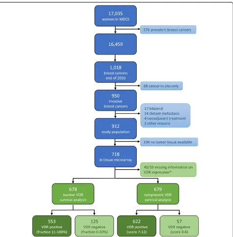
Women included in the MDCS were followed using the Swedish Cancer Registry until December 31, 2010, and the Swedish cause of death registry up until December 31, 2016. Since 576 women out of the 17035 had already been diagnosed with breast cancer prior to baseline examination, these women were excluded from the present study. During the follow-up until December 31, 2010, 1018 women were diagnosed with breast cancer. The intention was to investigate tumor characteristics in relation to breast cancer mortality, and due to this, 68 patients with cancer in situ were excluded, since these tumors are associated with a very low breast cancer mortality, if any. Also, patients who had re-ceived neoadjuvant treatment (n = 4), had distant me-tastases at diagnosis (n = 14), or died from breast cancer-related causes within less than 0.3 years from diagnosis (n = 2) were excluded, as well as one woman who declined treatment for 4 years. Bilateral cases (n = 17) were also excluded due to difficulties in interpreting tumor characteristics. The final study population consisted of 912 patients (Fig. 1).
Histopathological analysis and clinical information
Medical records and pathological reports were used to retrieve information on size and laterality of tumors as well as axillary lymph node involvement. Invasive tu-mors diagnosed during 1991–2004 were pathologically re-evaluated as regards to invasiveness, histological type, grade, estrogen receptor (ER) status, and progesterone receptor (PgR) status by one senior pathologist [17,18]. Such information was readily available from pathological
reports dating 2005 and onwards, with no need for re-evaluation. Information on human epidermal growth factor-2 (HER2) as well as proliferation index (Ki67) on tumors dating 1991–2007 was assessed using tissue mi-croarrays (TMAs) [19]. From 2008 and onwards, infor-mation on HER2 and Ki67 status was also retrieved from the diagnostic pathology report. ER and PgR were considered positive at a cut-off of > 10% positively stained nuclei. Results from in situ hybridization (ISH) were used to define HER2 status when available. When immunohistochemistry (IHC) evaluation was used to de-fine HER2 status, HER2 was considered positive when annotated 3+ and negative for 0 or 1+. IHC scores of 2+ were categorized as missing if ISH was not used to con-firm the result [16]. For Ki67, the distribution was noted to differ between tumors diagnosed at different periods of time. Therefore, tumors were classified into low, intermediate, or high Ki67 expression based on tertiles within the diagnostic period: 1991–2004, 2005–2007, and from 2008 and onwards.
Based on histological grade; ER, PgR, and HER2 status; and Ki67 category, tumors were classified into molecular subtypes: Luminal A-like, Luminal B-like, HER2 positive, and triple negative, according to cri-teria used locally within the south Swedish health care region [20]. Luminal A-like tumors were defined as ER positive, HER2 negative, and either (a) histo-logical grade 1, (b) histohisto-logical grade 2 and low Ki67, or (c) histological grade 2, intermediate Ki67, and positive PgR status. Luminal B-like tumors were also ER positive and HER2 negative but associated with ei-ther (a) histological grade 3, (b) histological grade 2 and high Ki67, or (c) histological grade 2, intermedi-ate Ki67, and negative PgR. Regardless of histological grade and hormone receptor status, all HER2-positive tumors were categorized as HER2 positive. All tumors considered ER negative, PgR negative, and HER2 negative were classified as triple negative (ibid.).
Clinical notes were used to retrieve information on the type of breast surgery, surgery to the axillary lymph nodes, and planned adjuvant therapy, as recommended by a multidisciplinary treatment conference following surgery.
Vitamin D receptor expression
monoclonal D-6 antibody (sc-13133, Santa Cruz Bio-technology) was selected for immunohistochemical ana-lyses of VDR since previous research has found this antibody superior to alternatives as regards to specificity and sensitivity [21, 22]. The antibody was diluted 1:300, and staining was performed automatically in Autostainer
Plus(Agilent/Dako A/S), with visualization kit K801021-2 (Agilent/Dako A/S) and also counterstained with Mayer’s hematoxylin for 2 min. An automated system was used for taking images of the slides, which were
thereafter incorporated in the web-based digital patho-logical platform PathXL Xplore (http://www.pathxl.com, PathXL Ltd., UK). Microscopic evaluations were per-formed using PathXL, consistently on the same com-puter screen.
[image:3.595.61.538.84.570.2]evaluating percentages of positive cells for all loca-tions, 0, 1–10%, 11–50%, 51–75%, and 76–100%. Also, the intensity of staining was evaluated as per-centages of highly intense stain in nuclei and nuclear membranes. The intensity of cytoplasmic stain was evaluated on four levels: no stain or low, moderate, or high intensity of stain (Fig. 2G, H).
Each core was scored twice by the same observer, at least 4 weeks apart, blinded to clinical and patho-logical data but with access to information on inva-siveness from the previous evaluation of hematoxylin/ eosin-stained slides of the cores [16].
Due to considerable differences between the first and second readings of VDR scores regarding nuclear
A)
C)
F) G) H)
D) E)
B)
[image:4.595.60.538.87.604.2]membrane and cellular membrane, it was decided to focus the statistical analysis on the nuclear and cytoplasmic stain. Discordance of nuclear and cytoplasmic stain between core pairs and different readings was treated as described in Appendix 1. The nuclear stain was defined as negative below a cut-off of 10%, positive 11–50%, and strongly posi-tive above 51% of stained nuclei. Cytoplasmic score was cal-culated by multiplying score for fraction, 0 (0%), 1 (1–10%), 2 (11–50%), 3 (51–75%), and 4 (76–100%), by intensity scores, 0 (no stain), 1 (low intensity), 2 (moderate intensity), and 3 (high intensity). Cytoplasmic scores were thereafter subdivided into three groups, 0–6, 7–9, and 10–11.
Endpoint retrieval
All women within the MDCS were followed until December 31, 2016, using the Swedish cause of death registry, which provided information on the date of death, cause of death, and underlying and multiple cause of death. When breast cancer was considered the only cause of death or contributing cause of death, the primary endpoint, breast cancer death, was fulfilled. Other women within the cohort were registered as either dead from other causes, emigrated, or alive. End of follow-up was the date of death, date of emigration, or December 31, 2016.
Statistical analysis
Tumor characteristics and planned treatment were com-pared between categories of VDR expression. Distribu-tions were compared using the χ2 test for categorical factors and Kruskal-Wallis test for continuous variables.
Breast cancer mortality (BCM) was calculated as breast cancer-associated death per 100,000 person-years. BCM in relation to VDR expression was tested using a Cox pro-portional hazards analysis yielding hazard ratios (HRs) with 95% confidence intervals (CI). The assumption of proportional hazards was met as tested by Kaplan-Meier plots, and statistical significance tested by log-rank tests. The crude model was subsequently adjusted in two multi-variate models. Model 2 was adjusted for the season of diagnosis and age at diagnosis which is known to affect levels of vitamin D and might therefore influence the VDR expression. Model 3 was further adjusted for tumor fac-tors known to influence breast cancer prognosis, such as the size of tumor, lymph node status, histological type, and molecular subtypes. As the choice of treatment is heavily dependent on prognostic tumor factors included in the adjusted model, it was decided not to adjust also for treatment variables. All adjusted analyses were first per-formed as complete cases analyses, only including cases with complete information on all included covariates. Missing data on covariates was thereafter included in the adjusted analyses using a multiple imputation model, de-scribed in detail inAppendix 2.
Expression of VDR in association with BCM was tested separately for nuclear fraction and cytoplasmic score, both subdivided in three levels of expression and also two levels of expression (negative vs positive). In a sensitivity analysis, negative nuclear fraction was com-bined with cytoplasmic score 0–6, and this combinatory negative VDR score was tested in a Cox proportional hazards analysis for associations with BCM.
Molecular subtypes showed covariance with VDR ex-pression. To investigate whether or not this covariation explained the association found between VDR expres-sion and BCM, the Cox regresexpres-sion model was repeated stratified by molecular subtypes. Since these groups were small and breast cancer death does not occur in some of the subgroups, the analyses were only performed com-paring nuclear VDR negativity vs positivity.
SPSS 25.0 (IBM) was used for all statistical analyses.
Results
Patterns of VDR expression
VDR was expressed almost exclusively in tumor cells as compared to surrounding cells within the TMA core. When cancer in situ cells were noted in the same core as invasive tumor cells, in situ cells were differently stained compared to invasive cells. Invasive cancer had a more intense stain and a larger fraction of nuclear stain-ing than cancer in situ cells (Fig. 2A). As only invasive cancer cells were scored for this project, such differences were not systematically recorded.
VDR was expressed in all compartments of cancer cells (Fig. 2B). Nuclear VDR expression was assessable in 678 (94.4%) of tissue core pairs, and cytoplasmic VDR expression as regards to fraction and intensity was scored in 679 (94.6%) of available tumors. Due to diffi-culties in distinguishing VDR expression between nu-clear membrane and cellular membrane, there was a high percentage of discordance between the first and second rounds of scoring at these compartments (14.1% of nuclear membrane fraction and 30.1% of cellular membrane fraction).
Staining patterns of nuclear VDR fraction are illus-trated in Fig. 2C–E. Percentages of highly intense stain in nucleus covaried highly with the fraction of nuclear stain and were therefore not included in fur-ther analyses. Distribution of nuclear fraction is pre-sented in Table 1.
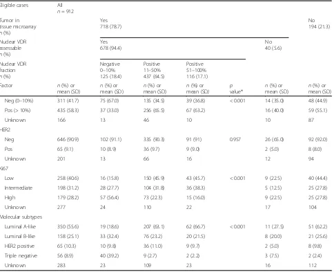
Table 1Distribution of patient and tumor characteristics in relation to nuclear VDR expression
Eligible cases All
n= 912 Tumor in
tissue microarray n(%)
Yes 718 (78.7)
No 194 (21.3)
Nuclear VDR assessable n(%)
Yes 678 (94.4)
No 40 (5.6)
Nuclear VDR fraction n(%)
Negative 0–10% 125 (18.4)
Positive 11–50% 437 (64.5)
Positive 51–100% 116 (17.1)
Factor n(%) or
mean (SD)
n(%) or mean (SD)
n(%) or mean (SD)
n(%) or mean (SD)
p value*
n(%) or mean (SD)
n(%) or mean (SD)
Age at baseline 56.4 (7.2) 55.4 (7.1) 56.6 (7.1) 55.7 (7.4) 0.109** 53.9 (7.5) 57.6 (7.0)
Age at diagnosis 65.4 (8.1) 64.1 (7.7) 66.0 (7.8) 64.8 (8.6) 0.026** 62.1 (9.1) 65.9 (8.3)
Season of diagnosis
Winter 241 (26.4) 35 (28.0) 116 (26.5) 29 (25.0) 0.646 13 (32.5) 48 (24.7)
Spring 221 (24.2) 38 (30.4) 97 (22.2) 27 (23.3) 9 (22.5) 50 (25.8)
Summer 187 (20.5) 19 (15.2) 96 (22.0) 23 (19.8) 6 (15.0) 43 (22.2)
Fall 263 (28.8) 33 (26.4) 128 (29.3) 37 (31.9) 12 (30.0) 53 (27.3)
BMI at baseline
< 25 467 (51.2) 60 (48.0) 215 (49.2) 70 (60.3) 0.253 24 (60) 98 (50.5)
≥25–30 310 (34) 45 (36.0) 150 (34.3) 33 (28.4) 11 (27.5) 71 (36.6)
≥30 135 (14.8) 20 (16.0) 72 (16.5) 13 (11.2) 5 (12.5) 25 (12.9)
Tumor size
1–10 mm 229 (25.8) 14 (11.2) 93 (21.4) 33 (28.7) 0.002 20 (50) 69 (39.7)
11–20 mm 409 (46.1) 57 (45.6) 217 (49.9) 51 (44.3) 13 (32.5) 71 (40.8)
≥21 mm 250 (28.2) 54 (43.2) 125 (28.7) 31 (27.0) 6 (15.0) 34 (19.5)
Unknown 24 0 2 1 1 20
Lymph node status
Positive 262 (31.9) 50 (41.0) 135 (32.8) 36 (32.4) 0.226 9 (22.5) 32 (22.7)
Negative 559 (68.1) 72 (59.0) 276 (67.2) 75 (67.6) 27 (67.5) 109 (77.3)
Unknown 91 3 26 5 4 53
Nottingham grade
I 227 (27.2) 7 (5.9) 119 (27.7) 42 (36.5) < 0.001 12 (30.0) 47 (34.8)
II 393 (47.0) 35 (29.4) 219 (50.9) 61 (53.0) 16 (40.0) 62 (45.9)
III 216 (25.8) 77 (64.7) 92 (21.4) 12 (10.4) 9 (22.5) 26 (19.3)
Unknown 76 6 7 1 3 59
Histological type
Ductal 596 (70.9) 103 (85.1) 290 (67.8) 84 (72.4) < 0.001 24 (60.0) 95 (68.3)
Lobular 166 (19.7) 7 (5.8) 113 (26.4) 20 (17.2) 7 (17.5) 19 (13.7)
Other/mixed 79 (9.4) 11 (9.1) 25 (5.8) 12 (10.3) 6 (15.0) 25 (18.0)
Unknown 71 4 9 0 3 55
ER status
Neg (0–10%) 84 (10.8) 45 (39.5) 25 (6.1) 4 (3.6) < 0.001 3 (7.5) 7 (6.0)
Pos (> 10%) 694 (89.2) 69 (60.5) 382 (93.9) 106 (96.4) 28 (70.0) 109 (94.0)
Unknown 134 11 30 6 9 78
Covariation of VDR expression and tumor characteristics
Distribution of patient and tumor characteristics in rela-tion to nuclear VDR fracrela-tion is presented in Table 1. There was a statistically significant covariation between VDR negativity and many tumor characteristics associ-ated with poor prognosis: large tumor size (p = 0.002), high Nottingham grade (p < 0.001), negative ER status (p < 0.001), negative PgR status (p < 0.001), and high Ki67 expression (p< 0.001). There was also a statistically significant covariation between histological type and VDR expression, where negative tumors more often were considered ductal (p < 0.001). When molecular subtypes were compared, it was noted that only 6.6% of Luminal A-like tumors had a negative VDR expression
in the nuclei as compared to 25.6% among Luminal B-like tumors, and 78.4% among triple-negative tumors.
A similar pattern was observed when the distribution of patient and tumor characteristics in relation to cytoplas-mic VDR score was analyzed (Additional file1: Table S1). One difference was that cytoplasmic VDR score also showed statistically significant covariation with HER2, as no tumors within the group of low cytoplasmic score (0–6) were considered HER2 positive (p = 0.008).
Covariation of VDR expression and breast cancer treatment
[image:7.595.58.542.98.491.2]Mastectomies were performed more often on VDR-nega-tive tumors (55%) compared to VDR-posiVDR-nega-tive tumors Table 1Distribution of patient and tumor characteristics in relation to nuclear VDR expression(Continued)
Eligible cases All
n= 912 Tumor in
tissue microarray n(%)
Yes 718 (78.7)
No 194 (21.3)
Nuclear VDR assessable n(%)
Yes 678 (94.4)
No 40 (5.6)
Nuclear VDR fraction n(%)
Negative 0–10% 125 (18.4)
Positive 11–50% 437 (64.5)
Positive 51–100% 116 (17.1)
Factor n(%) or
mean (SD)
n(%) or mean (SD)
n(%) or mean (SD)
n(%) or mean (SD)
p value*
n(%) or mean (SD)
n(%) or mean (SD)
Neg (0–10%) 311 (41.7) 75 (67.0) 135 (34.5) 39 (36.8) < 0.001 14 (35.0) 48 (44.9)
Pos (> 10%) 435 (58.3) 37 (33.0) 256 (65.5) 67 (63.2) 16 (40.0) 59 (55.1)
Unknown 166 13 46 10 10 87
HER2
Neg 646 (90.9) 102 (91.1) 335 (90.3) 91 (91) 0.957 26 (65.0) 92 (92.0)
Pos 65 (9.1) 10 (8.9) 36 (9.7) 9 (9.0) 2 (5.0) 8 (8.0)
Unknown 201 13 66 16 12 94
Ki67
Low 258 (40.6) 16 (15.8) 150 (45.9) 43 (45.7) < 0.001 9 (22.5) 40 (44.4)
Intermediate 198 (31.2) 28 (27.7) 104 (31.8) 36 (38.3) 5 (12.5) 25 (27.8)
High 179 (28.2) 57 (56.4) 73 (22.3) 15 (16.0) 9 (22.5) 25 (27.8)
Unknown 277 24 110 22 17 104
Molecular subtypes
Luminal A-like 350 (55.6) 19 (18.6) 207 (63.1) 62 (66.7) < 0.001 11 (27.5) 51 (62.2)
Luminal B-like 158 (25.1) 33 (32.4) 76 (23.2) 20 (21.5) 8 (20.0) 21 (25.6)
HER2 positive 65 (10.3) 10 (9.8) 36 (11.0) 9 (9.7) 2 (5.0) 8 (9.8)
Triple negative 56 (8.9) 40 (39.2) 9 (2.7) 2 (2.2) 3 (7.5) 2 (2.4)
Unknown 283 23 109 23 16 112
Percentages do not include missing categories *pvalues calculated withχ2
(41%). The postoperative treatment conference recom-mended adjuvant endocrine therapy for a smaller propor-tion and chemotherapy for a larger proporpropor-tion of patients with VDR-negative tumors compared to VDR-positive tu-mors. A similar pattern was seen when cytoplasmic VDR score was compared to treatment factors.
VDR expression in relation to breast cancer mortality
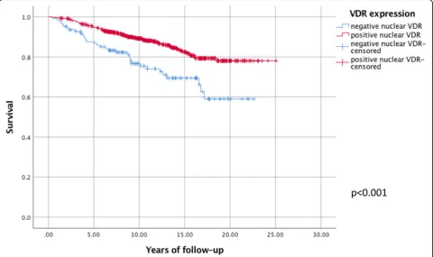
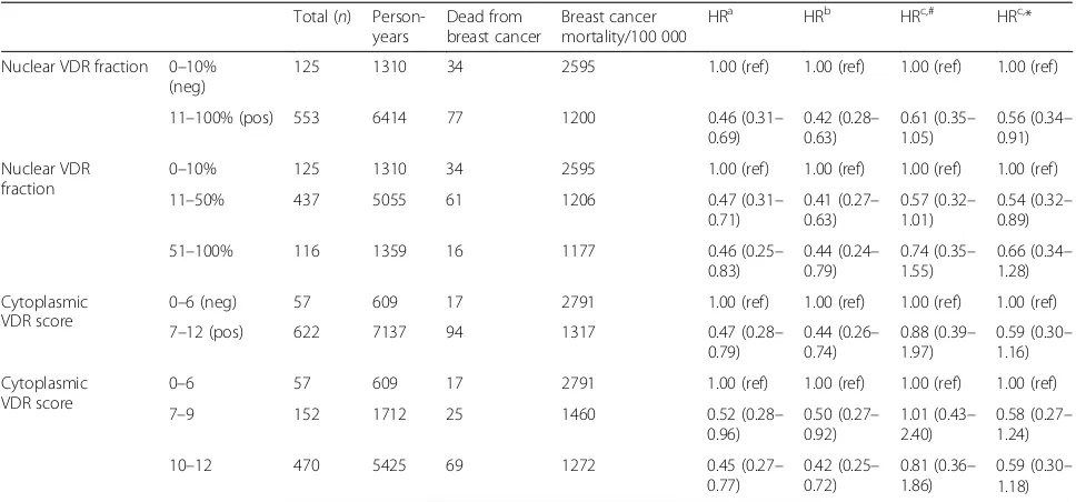
Mean follow-up was 11.5 years with a standard deviation (SD) of 5.2 years. A Kaplan-Meier analysis confirmed proportional hazards as shown in Fig.3. Both crude and adjusted analyses showed a statistically significant associ-ation between nuclear VDR positivity (a fraction above 10% of stained nuclei) and a low risk of breast cancer-as-sociated death (HR = 0.56, 0.34–0.91) adjusted analysis) (Table2). The complete case analysis showed similar but not statistically significant results (0.61, 0.35–1.05). Also, similar but not statistically significant results were seen when nuclear VDR fractions 11–50% (0.54, 0.32–0.89) and nuclear VDR fractions 51–100% (0.66, 0.34–1.28) were compared individually to nuclear VDR fraction below 10%. It was also noted that the difference in HR between nuclear VDR fractions 11–50% and nuclear VDR fractions 51–100% was small.
As regards to HRs calculated for different cytoplasmic VDR scores, they showed similar results as for nuclear VDR expression, i.e., more VDR expression was associated
with decreased risk of breast cancer death, but not statisti-cally significant when adjusted (0.59, 0.30–1.16) (Table2).
VDR expression in relation to breast cancer mortality stratified by molecular subtypes
HRs for breast cancer death calculated in groups strati-fied by molecular subtypes are presented in Table 3. There was a statistically significant association between VDR expression and BCM within the Luminal B-like tu-mors as nuclear VDR positivity was associated with a de-creased risk of breast cancer death (0.37, 0.18–0.77). Also, there seemed to be a possible association within the Luminal A-like molecular subtype and reduced risk of breast cancer death, but this association did not reach statistical significance (0.76, 0.32–2.53). For HER2-posi-tive and triple-negaHER2-posi-tive molecular subtypes, no statisti-cally significant results were observed.
Discussion
In the present study, VDR expression was found to be associated with favorable prognostic characteristics, such as small size, low grade, ER positivity, PgR positivity, low Ki67 expression, and Luminal-like molecular subtypes. This corresponds to the finding that VDR-positive tu-mors were found to be associated with a decreased risk of breast cancer-specific mortality, but this association was also independent of other prognostic factors.
[image:8.595.58.539.430.715.2]Immunohistochemistry and patterns of VDR expression
In the present material, VDR was found almost exclu-sively in tumor cells. As TMA was targeted to evaluate invasive tumors, there was a scarcity of normal breast cells and thorough evaluation of this matter could not be performed. Previous research have found expression of VDR to be higher in in situ and infiltrative carcinoma compared to benign breast disease or normal tissue [23,24], but others argue the opposite [25].
[image:9.595.56.540.98.324.2]When this study was initiated, it was expected to find only nuclear staining concerning VDR, since this is what most previous studies have observed [8, 10, 26]. It was therefore surprising to find VDR staining also in the nu-clear membrane, the cytoplasm, and the cellular mem-brane in our TMA. Early research which concluded VDR to be a primary nuclear receptor used radioactive 1,25-(OH)2 vitamin D3to identify the receptor in mam-mary cells [8]. More recently, it has been shown that Table 2Vitamin D receptor expression in relation to breast cancer mortality
Total (n) Person-years
Dead from breast cancer
Breast cancer mortality/100 000
HRa HRb HRc,# HRc,*
Nuclear VDR fraction 0–10% (neg)
125 1310 34 2595 1.00 (ref) 1.00 (ref) 1.00 (ref) 1.00 (ref)
11–100% (pos) 553 6414 77 1200 0.46 (0.31–
0.69)
0.42 (0.28– 0.63)
0.61 (0.35– 1.05)
0.56 (0.34– 0.91)
Nuclear VDR fraction
0–10% 125 1310 34 2595 1.00 (ref) 1.00 (ref) 1.00 (ref) 1.00 (ref)
11–50% 437 5055 61 1206 0.47 (0.31–
0.71)
0.41 (0.27– 0.63)
0.57 (0.32– 1.01)
0.54 (0.32– 0.89)
51–100% 116 1359 16 1177 0.46 (0.25–
0.83)
0.44 (0.24– 0.79)
0.74 (0.35– 1.55)
0.66 (0.34– 1.28)
Cytoplasmic VDR score
0–6 (neg) 57 609 17 2791 1.00 (ref) 1.00 (ref) 1.00 (ref) 1.00 (ref)
7–12 (pos) 622 7137 94 1317 0.47 (0.28–
0.79)
0.44 (0.26– 0.74)
0.88 (0.39– 1.97)
0.59 (0.30– 1.16)
Cytoplasmic VDR score
0–6 57 609 17 2791 1.00 (ref) 1.00 (ref) 1.00 (ref) 1.00 (ref)
7–9 152 1712 25 1460 0.52 (0.28–
0.96)
0.50 (0.27– 0.92)
1.01 (0.43– 2.40)
0.58 (0.27– 1.24)
10–12 470 5425 69 1272 0.45 (0.27–
0.77)
0.42 (0.25– 0.72)
0.81 (0.36– 1.86)
0.59 (0.30– 1.18) a
Crude analysis
b
Adjusted for age at and season of diagnosis
c
Adjusted for same factors asb
but also for size of tumor, lymph node status, histological type, and molecular subtypes
#
Complete case analysis: analysis including only cases with complete information on all covariates *Multiple imputation performed to include individuals with missing data on covariates in analysis
Table 3Vitamin D receptor expression in relation to breast cancer mortality stratified by surrogate molecular subtypes
Surrogate molecular subtype Nuclear VDR fraction n Person-years Dead from breast cancer Breast cancer mortality/100 000
HRa HRa,* HRb HRb,* HRc HRc,*
Luminal A-like 0–10% 19 275 4 1455 1.00 (ref) 1.00 (ref) 1.00 (ref) 1.00 (ref) 1.00 (ref) 1.00 (ref)
11–100% 269 3132 23 734 0.56 (0.19–
1.62)
0.76 (0.27– 2.14)
0.44 (0.15– 1.31)
0.61 (0.21– 1.76)
0.79 (0.24– 2.63)d
0.76 (0.32– 2.53)d
Luminal B-like 0–10% 33 325 11 3381 1.00 (ref) 1.00 (ref) 1.00 (ref) 1.00 (ref) 1.00 (ref) 1.00 (ref)
11–100% 96 1125 15 1334 0.38 (0.18–
0.84)
0.37 (0.18– 0.76)
0.42 (0.19– 0.93)
0.36 (0.17– 0.73)
0.43 (0.19– 0.94)e
0.37 (0.18– 0.77)e
HER 2 positive 0–10% 10 82 3 3639 1.00 (ref) 1.00 (ref) 1.00 (ref) 1.00 (ref) 1.00 (ref) 1.00 (ref)
11–100% 45 436 12 2751 0.78 (0.22–
2.79)
0.82 (0.25– 2.64)
0.74 (0.20– 2.74)
0.73 (0.22– 2.47)
1.13 (0.31–
4.09)d 1.00 (0.303.33)d –
Triple negative 0–10% 40 419 10 2388 1.00 (ref) 1.00 (ref) 1.00 (ref) 1.00 (ref) 1.00 (ref) 1.00 (ref)
11–100% 11 123 3 2440 1.04 (0.28–
3.77)
0.85 (0.25– 2.86)
0.80 (0.21– 3.00)
0.70 (0.21– 2.37)
0.87 (0.23–
3.19)e 0.78 (0.232.61)e –
a
Crude analysis
b
Adjusted for age at and season of diagnosis
c
Hazard ratio adjusted for covariate most affecting the estimate:d
lymph node status ande
[image:9.595.71.540.517.698.2]cytoplasmic unliganded VDR present in tumor cells of cell lines and mouse models promotes cell growth in contrast to the inhibitory effects of intranuclear VDR which has been activated by vitamin D [27]. An-other recent study also demonstrated VDR to be lo-calized in the cytoplasm of dividing cells [28]. Hence, unliganded VDR can be found on other locations in the tumor cells, and at least one other study on breast cancer survival has also found VDR in the cytoplasm along with the nuclei [12].
When research on which antibody to use in the study, the Santa-Cruz D-6 antibody was preferred over alterna-tives, since validation of this particular antibody for im-munohistochemistry was considered superior to alternatives [21, 22]. This antibody was not used in the previous studies, which is a possible explanation for dif-ferent staining patterns.
Since VDR was found on multiple subcellular locations in cancer cells, and there were no previous records in the literature about this, we concluded it best to evaluate the fraction of stained cancer cells at all sites. In an at-tempt also to score the intensity of nuclear and nuclear membrane VDR expression, an additional score of the highly intensely stained fraction was added. This method was based on previous reports that concluded better re-producibility on very strongly positive scores than in-cluding intermediate intensities [29]. Considering intensity in the cytoplasm, it was easier to distinguish in-tensities of intermediate staining pattern at this location, why it was also included. When evaluating results from scoring, we concluded that scores as regards to nuclear fraction and intensity and cytoplasmic fraction and in-tensity were congruent enough to be valid.
Scores concerning membranous expression had a low reproducibility and were therefore not included in the statistical analyses. It would have been interesting to compare staining in different subcellular compartments in associations to tumor prognostic factors and breast cancer mortality, since VDR activated by vitamin D is translocated into the nucleus and has been shown to re-duce the viability of triple-negative breast cancer cell lines [30], inhibit breast cancer cell line growth [31], and induce autophagy in breast cancer cell lines and in nor-mal breast tissue of mice [32]. Another previous study has shown that unliganded VDR in the cytoplasm pro-motes cell growth in contrast to the inhibitory intranuc-lear ligand-dependent actions of VDR [27]. We suggest that VDR located in the cellular membrane, not acti-vated by vitamin D, hypothetically may be associated with prognostic factors and BCM differently compared to intranuclear VDR. In our study, there were only few individuals with negative nuclear VDR expression and positive cytoplasmic stain, and therefore, we consider that there was not enough power to find any association
between this small group of individuals and a possibly elevated risk of BCM. However, tumors with nuclear VDR expression do probably also express VDR in the cytoplasm.
Methodological considerations
All Swedish residents are given a unique civil registra-tion number at birth or immigraregistra-tion. It is therefore pos-sible to link all women in our cohort to different registries. The Swedish Cause of Death Registry which was used to retrieve information on the cause of death is reported to be virtually complete on the event of death and to 96% complete to cause of death [33]. Deaths caused by a tumor have been found to be correctly regis-tered in 90% of cases [34].
Analyses were performed with nuclear fraction and cytoplasmic score analyzed separately (Table 3). As regards to cytoplasmic score, there were few individuals with negative scores (n = 57), and results from statistical analyses were harder to interpret, although they seemed to be congruent with results from analyzing nuclear VDR fraction. In a sensitivity analysis, a combinatory score of nuclear and cytoplasmic expression was calculated and used to determine HR of BCM. There were very few indi-viduals with both a negative nuclear fraction and negative cytoplasmic score (n= 54), and when this group was ex-panded to contain either a higher cytoplasmic score (7– 10) or larger nuclear fraction (11–50%), any effects of VDR negativity could not be observed. Hence, we con-clude that in our material a dichotomized variable of a nu-clear fraction of VDR expression with a cut-off of 10% is appropriate to use and will be used in our future studies. Another previous study has also noted that the intensity of VDR expression seems to be of less importance [12].
A tissue microarray (TMA) is not an evaluation of a complete tumor, and therefore, results from an immuno-histochemical analysis of TMA are not comparable to a diagnostic immunohistochemical analysis. For research purposes, a TMA is valuable as it makes it possible to evaluate many tumors under a comparably short dur-ation of time. Another weakness of a TMA study is that very small tumors are not represented in the TMA, as seen in Table 1, where small tumors accounted for al-most 40% of tumors not included in TMA but only ap-proximately 20% of tumors evaluated as regards to nuclear VDR were small.
VDR and associations with prognostic factors and breast cancer treatment
statistical significance was seen with most of the prognos-tic factors, except with lymph node involvement and HER2 amplification. A recent study, which used com-puter-assisted image analysis for evaluations of nuclear VDR expression, showed very similar results as regards to covariance with tumor prognostic factors [10], which strengthens our results. Earlier, smaller studies have re-ported divergent results. One study showed associations with tumor size and lymph node involvement, but not with grading, estrogen receptors, progesterone receptors, or HER2 [12]; others reported no associations [13, 14]. The earlier studies were quite small and did not evaluate many of the associations that we found.
As VDR expression covaried with prognostic factors, it was expected to find associations also with the suggested breast cancer treatment. It was noted that VDR-negative tumors more often were surgically treated with mastec-tomy (they were larger), less often suggested endocrine therapy, and more often chemotherapy (they were ER negative to a larger extent).
VDR and breast cancer mortality (BCM)
The present study showed a statistically significant asso-ciation between BCM and VDR tumor expression among breast cancer patients (HR 0.56, 0.34–0.91) (Table2), which has not been reported previously. Previ-ous studies have not used BCM as endpoint when evalu-ating breast cancer prognosis [10–14], and associations found between VDR expression and different endpoints are not conclusive. The most recent and largest study, by Al-Azhri et al., reported very similar results to ours as regards to covariation with tumor prognostic factors, but showed no association between VDR expression and overall survival, progression-free survival, or breast can-cer-specific survival [10]. Differences in study population such as a shorter follow-up time (mean 72 months, com-pared to 137 months in our study) and differing tumor characteristics and that they might have included women with metastatic disease in their study population (not mentioned) may explain some of these discrepancies. The earlier and smaller studies have used different ap-proaches on retrieving information on VDR expression and have not associated VDR expression with any differ-ences in survival [11,13,14] although Berger et al. noted that VDR-positive tumors were associated with a longer disease-free interval [11]. Ditsch et al. showed that VDR expression was associated with a between better progres-sion-free survival, and overall survival in univariate ana-lyses [12], which strengthens our results.
Another study which suggested that phenotype of the normal breast tissue surrounding a breast cancer can predict outcome showed that when VDR was expressed along with androgen receptor (AR) and ER in the sur-rounding breast tissue, patient outcomes were more
favorable than when none of those three was expressed [35]. Their results are also in line with ours, suggesting that VDR expression is associated with a better breast cancer prognosis.
Molecular subtypes were included as a covariate in the adjusted model of BCM, which maintained statistical significance when positive nuclear expression (11–100%) was compared to negative nuclear expression (0–10%), although the confidence interval was widened (0.56, 0.34–0.91) (Table 2). The model stratified on molecular subtypes showed that for tumors classified as Luminal B, VDR positivity was associated with a decreased BCM. Since breast cancer deaths were uncommon for women with tumors classified as Luminal A, the results had a poor precision and wide confidence intervals. Also, small numbers of HER2-positive tumors and triple-negative tumors made the analyses regarding VDR expression and breast cancer mortality inconclusive. Still, we believe that the stratified model confirmed that the positive prognostic effect seen with positive VDR expression was not all due to covariation with molecular subtypes.
Conclusion
The present study indicates that high VDR expression in breast cancer cell nuclei is associated with favorable prognostic factors and a decreased risk of breast cancer death. Women with VDR-positive breast tumors have a better breast cancer-specific survival compared to women with VDR-negative tumors. Future studies ought to investigate the combined effect of VDR expression and serum levels of vitamin D in relation to breast can-cer prognosis.
Appendix 1
Description of handling intraindividual scoring differences
Each tumor was represented by two cores in the TMA, and in order to obtain validity of values of expression, scoring was performed twice. At least several weeks, up to months, went between scoring of the same tumor, de-pending on other obligations of the evaluator (LH). Due to many (seven) scoring categories, an intraindividual discrepancy between the first and second scoring rounds can be expected. Such a discrepancy was treated as follows:
were no differences between core pairs but (only) one score unit between the first and second evaluation, the second score was considered correct, due to the learning curve during the first evaluation. After this procedure, there were still differences as regards to scores for nu-clear fraction (n= 30), cytoplasmic fraction (n= 18), and cytoplasmic intensity (n = 21). These tumors were re-evaluated, with the pair of cores of the same tumor con-sidered as one entity. After this re-evaluation, score for only four tumors could not be concluded, and a second evaluator (AR) was consulted before the score was determined.
Measurement error due to scoring discrepancies was evaluated by performing sensitivity analyses in which all tumors with any discrepancy (either between cores or between first and second evaluation) between negative and positive nuclear VDR expression were excluded (n = 62). These analyses were performed without mul-tiple imputation, and results differed only to a minor ex-tent compared to when the differing cases were included. The adjusted analyses differed the most: 0.65 (0.36–1.15) compared to 0.61 (0.35–1.05).
Appendix 2
Description of the imputation model
Among 718 tumors included in the TMA, there were missing data on the following variables: tumor size (n= 4), lymph node status (n = 38), tumor grade (n = 17), histological type (n= 16), ER status (n = 56), PgR status (n = 79), HER2 expression (n = 107), Ki-67 expression (n= 173), and molecular subtypes (n= 171). Also, it was not possible to evaluate expression of VDR in the cyto-plasm for 39 of tumors and in nuclei for 40 of tumors. All above variables were included in the multiple imput-ation model, except molecular subtypes which were cal-culated based on results from imputation. Since the analysis in mind for the multiple imputation model was a Cox regression, baseline hazards and breast cancer death were also included in the model, as well as age and season of diagnosis (covariates in the analysis). Fur-ther predictors of imputed values and of missingness, such as BMI and year of diagnosis, were also added. SPSS 25.0 (IBM) was used for multiple imputation, and 30 imputations were made. SPSS uses logistic regression for imputation of categorical variables, and linear regres-sion was used for imputation of baseline hazards. Con-vergence was checked, and it appeared after two iterations, although ten iterations were used to impute values.
In the Cox analysis only, imputed values for covariates (tumor size, lymph node status, histological type, and molecular subtypes) were used. Only tumors with values based on defined scores of VDR expression were in-cluded in the final analyses.
Additional file
Additional file 1:Distribution of patient and tumor characteristics in relation to cytoplasmic VDR expression. (DOCX 24 kb)
Abbreviations
BCM:Breast cancer mortality; CI: Confidence interval; ER: Estrogen receptor; HER2: Human epidermal growth factor-2; HR: Hazard ratio; ISH: In situ hybridization; Ki67: Proliferation index factor Ki67; MDCS: Malmö Diet and Cancer Study; PgR: Progesterone receptor; SD: Standard deviation; TMA: Tissue microarray; VDR: Vitamin D receptor
Acknowledgements
The authors wish to thank registered nurse and data manager Anna Hwasser, who prepared the original files and assisted in the data
management; biomedical analysist Björn Nodin, who constructed TMAs; and research engineer Kristina Lövgren, who prepared and stained TMA slides for VDR.
Authors’contributions
LH scored the VDR expression and did all the statistical analyses. LH was also the main author of the manuscript and constructed the tables and figures. STB, AR, and JM supervised the project. SB supervised the pathological evaluation. KE evaluated the inclusion criteria of TMA and revised the manuscript. MS provided the information on molecular subtypes and revised the manuscript. All authors read and approved the final manuscript.
Funding
The present study was supported by the Swedish Cancer Society, The Gunnar Nilsson Cancer Foundation, The Henning and Ida Persson Foundation, The Einar and Inga Nilsson Foundation, The Malmö University Hospital Cancer Research Fund, The Skåne University Hospital Funds and Donations, and The Region Skåne (ALF).
Availability of data and materials
The data that support the findings of this study are available on request from the corresponding author (LH). The data are not publicly available due to Swedish restrictions.
Ethics approval and consent to participate
The MDCS and the present study were approved by the ethics committee in Lund, Sweden, MDCS (LU 51-90) and the present study (Dnr 652/2005 and Dnr 23/2007).
Written informed consent was obtained from all study participants on the inclusion of MDCS. Follow-up studies have been advertised in local media, with information about an option to withdraw.
Consent for publication
Not applicable.
Competing interests
The authors declare that they have no competing interests.
Author details 1
Department of Surgery, Lund University, Skåne University Hospital, SE-205 02 Malmö, Sweden.2Department of Oncology, Aarhus University, Aarhus University Hospital, DE-8000 Aarhus C, Denmark.3Division of Oncology and Pathology, Department of Clinical Sciences, Lund University, Skåne University Hospital, SE-221 85 Lund, Sweden.
Received: 12 February 2019 Accepted: 15 July 2019
References
2. Jacobs ET, Kohler LN, Kunihiro AG, Jurutka PW. Vitamin D and colorectal, breast, and prostate cancers: a review of the epidemiological evidence. J Cancer. 2016;7(3):232–40.
3. Freedman DM, Looker AC, Chang SC, Graubard BI. Prospective study of serum vitamin D and cancer mortality in the United States. J Natl Cancer Inst. 2007;99(21):1594–602.
4. Rose AA, Elser C, Ennis M, Goodwin PJ. Blood levels of vitamin D and early stage breast cancer prognosis: a systematic review and meta-analysis. Breast Cancer Res Treat. 2013;141(3):331–9.
5. Huss L, Butt S, Borgquist S, Almquist M, Malm J, Manjer J. Serum levels of vitamin D, parathyroid hormone and calcium in relation to survival following breast cancer. Cancer Causes Control. 2014;25(9):1131–40. 6. Haussler MR, Whitfield GK, Haussler CA, Hsieh J-C, Thompson PD, Selznick
SH, Dominguez CE, Jurutka PW. The nuclear vitamin D receptor: biological and molecular regulatory properties revealed. J Bone Miner Res. 1998;13(3): 325–49.
7. Berger U, Wilson P, McClelland RA, Colston K, Haussler MR, Pike JW, Coombes RC. Immunocytochemical detection of 1,25-dihydroxyvitamin D receptors in normal human tissues. J Clin Endocrinol Metab. 1988; 67(3):607–13.
8. Narbaitz R, Sar M, Stumpf WE, Huang S, DeLuca HF. 1,25-Dihydroxyvitamin D3 target cells in rat mammary gland. Horm Res. 1981;15(4):263–9. 9. Friedrich M, Axt-Fliedner R, Villena-Heinsen C, Tilgen W, Schmidt W,
Reichrath J. Analysis of vitamin D-receptor (VDR) and retinoid X-receptor alpha in breast cancer. Histochem J. 2002;34(1–2):35–40.
10. Al-Azhri J, Zhang Y, Bshara W, Zirpoli GR, McCann SE, Khoury T, Morrison CD, Edge SB, Ambrosone CB, Yao S. Tumor expression of vitamin D receptor and breast cancer histopathological characteristics and prognosis. Clin Cancer Res. 2017;23(1):97–103.
11. Berger U, McClelland RA, Wilson P, Greene GL, Haussler MR, Pike JW, Colston K, Easton D, Coombes RC. Immunocytochemical determination of estrogen receptor, progesterone receptor, and 1,25-dihydroxyvitamin D3 receptor in breast cancer and relationship to prognosis. Cancer Res. 1991; 51(1):239–44.
12. Ditsch N, Toth B, Mayr D, Lenhard M, Gallwas J, Weissenbacher T, Dannecker C, Friese K, Jeschke U. The association between vitamin D receptor expression and prolonged overall survival in breast cancer. J Histochem Cytochem. 2012;60(2):121–9.
13. Eisman JA, Suva LJ, Martin TJ. Significance of 1,25-dihydroxyvitamin D3 receptor in primary breast cancers. Cancer Res. 1986;46(10):5406–8. 14. Freake HC, Abeyasekera G, Iwasaki J, Marcocci C, MacIntyre I, McClelland RA,
Skilton RA, Easton DF, Coombes RC. Measurement of 1,25-dihydroxyvitamin D3 receptors in breast cancer and their relationship to biochemical and clinical indices. Cancer Res. 1984;44(4):1677–81.
15. Friedrich M, Villena-Heinsen C, Tilgen W, Schmidt W, Reichrat J, Axt-Fliedner R. Vitamin D receptor (VDR) expression is not a prognostic factor in breast cancer. Anticancer Res. 2002;22(3):1919–24.
16. Elebro K, Bendahl PO, Jernstrom H, Borgquist S. Androgen receptor expression and breast cancer mortality in a population-based prospective cohort. Breast Cancer Res Treat. 2017;165(3):645–57.
17. Borgquist S, Anagnostaki L, Jirstrom K, Landberg G, Manjer J. Breast tumours following combined hormone replacement therapy express favourable prognostic factors. Int J Cancer. 2007;120(10):2202–7.
18. Butt S: Reproductive factors and breast cancer - parity, breastfeeding and genetic predisposition in relation to risk and prognosis. Lund University,
Faculty of Medicine Doctoral Dissertation Series, 2011:10:.
19. Borgquist S, Djerbi S, Ponten F, Anagnostaki L, Goldman M, Gaber A, Manjer J, Landberg G, Jirstrom K. HMG-CoA reductase expression in breast cancer is associated with a less aggressive phenotype and influenced by
anthropometric factors. Int J Cancer. 2008;123(5):1146–53.
20. Guidelines for surgical and oncologic treatment of breast cancer - regional adaption of the national treatment guidelines, Region West and South [In Swedish: Lathund för kirurgisk och onkologisk behandling av bröstcancer– Regional anpassning av nationellt vårdprogram, Region Väst och Syd]. [https://www.cancercentrum.se/globalassets/cancerdiagnoser/brost/syd/ sydsvenska-brostcancergruppens-lathund-170401-180331.pdf.]
21. Costa PL, Franca MM, Ferraz-de-Souza B. Nonspecific binding of a frequently used vitamin D receptor (VDR) antibody: important implications for vitamin D research in human health. Endocrine. 2016;54(2):556–9.
22. Wang Y, Becklund BR, DeLuca HF. Identification of a highly specific and versatile vitamin D receptor antibody. Arch Biochem Biophys. 2010; 494(2):166–77.
23. Conde I, Paniagua R, Fraile B, Ruiz A, Arenas MI. Expression of vitamin D3 receptor and retinoid receptors in human breast cancer: identification of potential heterodimeric receptors. Int J Oncol. 2004;25(4):1183–91. 24. Zhalehjoo N, Shakiba Y, Panjehpour M. Alterations of vitamin D receptor
(VDR) expression profile in normal and malignant breast tissues. Clin Lab. 2016;62(8):1461–8.
25. Lopes N, Sousa B, Martins D, Gomes M, Vieira D, Veronese LA, Milanezi F, Paredes J, Costa Jé L, Schmitt F. Alterations in vitamin D signalling and metabolic pathways in breast cancer progression: a study of VDR, CYP27B1 and CYP24A1 expression in benign and malignant breast lesions vitamin D pathways unbalanced in breast lesions. BMC Cancer. 2010;10:483. 26. Berger U, Wilson P, McClelland RA, Colston K, Haussler MR, Pike JW,
Coombes RC. Immunocytochemical detection of 1,25-dihydroxyvitamin D3 receptor in breast cancer. Cancer Res. 1987;47(24 Pt 1):6793–9.
27. Trivedi T, Zheng Y, Fournier PGJ, Murthy S, John S, Schillo S, Dunstan CR, Mohammad KS, Zhou H, Seibel MJ, et al. The vitamin D receptor is involved in the regulation of human breast cancer cell growth via a ligand-independent function in cytoplasm. Oncotarget. 2017;8(16):26687–701. 28. Cui X, Pertile R, Eyles DW. The vitamin D receptor (VDR) binds to the
nuclear matrix via its hinge domain: a potential mechanism for the reduction in VDR mediated transcription in mitotic cells. Mol Cell Endocrinol. 2018;472:18–25.
29. van Diest PJ, van Dam P, Henzen-Logmans SC, Berns E, van der Burg ME, Green J, Vergote I. A scoring system for immunohistochemical staining: consensus report of the task force for basic research of the EORTC-GCCG. European Organization for Research and Treatment of
Cancer-Gynaecological Cancer Cooperative Group. J Clin Pathol. 1997;50(10):801–4. 30. Thakkar A, Wang B, Picon-Ruiz M, Buchwald P, Ince TA. Vitamin D and
androgen receptor-targeted therapy for triple-negative breast cancer. Breast Cancer Res Treat. 2016;157(1):77–90.
31. Murray A, Madden SF, Synnott NC, Klinger R, O'Connor D, O'Donovan N, Gallagher W, Crown J, Duffy MJ. Vitamin D receptor as a target for breast cancer therapy. Endocr Relat Cancer. 2017;24(4):181–95.
32. Tavera-Mendoza LE, Westerling T, Libby E, Marusyk A, Cato L, Cassani R, Cameron LA, Ficarro SB, Marto JA, Klawitter J, et al. Vitamin D receptor regulates autophagy in the normal mammary gland and in luminal breast cancer cells. Proc Natl Acad Sci U S A. 2017;114(11):E2186–e2194. 33. Brooke HL, Talback M, Hornblad J, Johansson LA, Ludvigsson JF, Druid H,
Feychting M, Ljung R. The Swedish cause of death register. Eur J Epidemiol. 2017;32(9):765–73.
34. Johansson LA, Bjorkenstam C, Westerling R. Unexplained differences between hospital and mortality data indicated mistakes in death certification: an investigation of 1,094 deaths in Sweden during 1995. J Clin Epidemiol. 2009;62(11):1202–9.
35. Santagata S, Thakkar A, Ergonul A, Wang B, Woo T, Hu R, Harrell JC, McNamara G, Schwede M, Culhane AC, et al. Taxonomy of breast cancer based on normal cell phenotype predicts outcome. J Clin Invest. 2014; 124(2):859–70.
Publisher’s Note