Open Access
Short Report
Biologically active fibroblast growth factor 1 tagged with various
epitopes
Masahiro Asada*, Emi Honda and Toru Imamura
Address: Signaling Molecules Research Laboratory, National Institute of Advanced Industrial Science and Technology (AIST), Tsukuba Central #6, 1-1-1 Higashi, Tsukuba, Ibaraki 305-8566, Japan
Email: Masahiro Asada* - m.asada@aist.go.jp; Emi Honda - e-honda@aist.go.jp; Toru Imamura - imamura-toru@aist.go.jp * Corresponding author
Abstract
Background: Fibroblast growth factor (FGF) family members are involved in the regulation of a variety of biological phenomena. Because most of their activity is exerted via a signaling complex composed of FGF, heparin/heparan sulfate and FGF receptor tyrosine kinase, it is important to study the dynamic behavior of all the molecules in the complex without disturbing their interaction or activity.
Findings: We used E. coli to express biologically active human FGF1 tagged at its C-terminus with myc-(His)6, V5-(His)6 or 3xFLAG-(His)6. We found that the tagged FGF1s had affinities for heparin
that were similar to that of the native form. The tagged FGF1s also exhibited mitogenic activity similar to that of the native form. Apparently, the tags do not interfere with the formation of the three-member complex involving FGF1, FGF receptor and heparan sulfate/heparin.
Conclusion: Tagged FGF1s should be useful for investigating the dynamic behavior of FGF1 in the context of its three-member signaling complex and other molecular complexes.
Findings
BackgroundEpitope tags are frequently introduced when expressing recombinant proteins, as they make it easy to purify and detect proteins of interest. However, addition of exoge-nous amino acids can alter the properties of proteins, including their affinity for other biomolecules and their biological activities [1]. Nevertheless, the effect of intro-ducing an epitope tag on the activity of a protein of inter-est has rarely been addressed.
Fibroblast growth factors (FGFs) are multifunctional pro-teins that play important roles in many biological phe-nomena. Their effects are elicited mainly through simultaneously binding to various cell surface FGF
recep-tors (FGFRs) and heparan sulfate sugar chains. In order to analyze the structural features of the resultant three-ber complexes, we considered it useful to label each mem-ber with a different epitope tag. Because such analyses would require biologically active FGF1 to be tagged in var-ious formats, we developed those proteins and report some of their characteristics here.
Methods
Construction of cDNAs
The strategy for constructing cDNAs encoding chimeric FGF1s is depicted schematically in Fig. 1, and the primers used are listed in Table 1. Human FGF1 cDNA was cloned into pET-3c vector as described previously [2]. To generate cDNA encoding the chimeric proteins, PCR was carried
Published: 11 July 2008
BMC Research Notes 2008, 1:42 doi:10.1186/1756-0500-1-42
Received: 31 March 2008 Accepted: 11 July 2008
This article is available from: http://www.biomedcentral.com/1756-0500/1/42
© 2008 Asada et al; licensee BioMed Central Ltd.
out using the plasmid as a template with #127 serving as the forward primer and #619 (for V5-(His)6 tag) or #397
(for myc-(His)6 tag) serving as the reverse primer. To
gen-erate the cDNA encoding V5-(His)6 tag, pAc5.1/V5-His
vector (Invitrogen) served as the template with primers
#620 and #301. For cDNA encoding myc-(His)6 tag,
pcDNA3.1(-)-myc-His vector (Invitrogen) served as the template with primers #395 and #396.
To create a cDNA encoding 3xFLAG-(His)6, PCR was
car-ried out with primers #468 and #469 in the presence of two megaprimers, #466 and #467, which served as the template. The product was digested with Xho I and Apa I and cloned into predigested pcDNA3.1(+), yielding
plas-mid 3xFLAG-(His)6/pcDNA3.1(+). The open reading
frame for FGF1 was then amplified using #476 and #477, digested with EcoR I and Xho I, and inserted into
predi-gested 3xFLAG-(His)6/pcDNA3.1(+). Using this plasmid
as a template, PCR was then carried out with primers #117 and #537 to generate cDNA encoding the 3'-part of FGF1-3xFLAG-(His)6. For the 5'-part, PCR was carried out using FGF1/pET-3c as template with primers #127 and #178.
Once the first round PCR products were purified, overlap extension PCR was carried out to generate the intended cDNAs encoding the chimeric FGF1s. For FGF1-V5-(His)6, the PCR products of #127/#619 and #620/#301 were mixed as templates, and second round PCR was carried out using primers #127 and #537. In the same way, for
FGF1-myc-(His)6, the products of #127/#397 and #395/
#396 were mixed as the template, and second round PCR was carried out using primers #127 and #396. For
FGF-1-3xFLAG-(His)6, the PCR products of #127/#178 and
#117/#537 were mixed as the template, and second round PCR was carried out using primers #127 and #537.
[image:2.612.61.291.430.634.2]Once the full-length cDNAs encoding the chimeric pro-teins were generated, the DNA for the open reading frame was excised using Nde I/Bgl II and ligated into pET-3c vec-tor (Novagen) predigested with Nde I/BamH I. The cDNA
Table 1: Primers used in this study
No. for./rev. sequence site
#127 forward 5'-gta ata cga ctc act ata ggg-3' vector primer
#619 reverse 5'-acc ttc atc aga aga gac tgg cag-3' FGF1/3' + V5-(His)6/5'
#397 reverse 5'-cga gct cgg atc aga aga gac tgg cag-3' FGF1/3' + myc-(His)6/5'
#620 forward 5'-tct gat gaa ggt aag cct atc cct-3' FGF1/3' + V5-(His)6/5'
#301 reverse 5'-tag aag gca cag tcg agg-3' vector primer
#395 forward 5'-cca gtc tct tct gat ccg agc tcg gta cca-3' FGF1/3' + myc-(His)6/5' #396 reverse 5'-gct ctA GAT CTt caa tga tga tga tga tga tgg tc-3' myc-(His)6/3' + Bgl II
#468 forward 5'-cag ccg CTC GAG a-3' Xho I + 3xFLAG-(His)6/5'
#469 reverse 5'-tgc GGG CCC tca a-3' 3xFLAG-(His)6/5' + Apa I
#466 forward 5'-ccg CTC GAG act aca aag acc atg acg gtg att ata aag atc atg aca tcg act aca ag-3' megaprimer/5' #467 reverse 5'-tgc GGG CCC tca atg gtg atg gtg atg atg acc ctt gtc atc gtc atc ctt gta gtc ga-3' megaprimer/3'
#476 forward 5'-cgG AAT TCc cac cat gtc ccg ggg agc-3' EcoR I + FGF1/5'
#477 reverse 5'-ccg CTC GAG cat cag aag aga ctg gca g-3' FGF1/3' + Xho I
#117 forward 5'-tct tcc gat aga ctg cgt cg-3' FGF1
#537 reverse 5'-gaA GAT CTc ttc aat ggt gat ggt gat gat gac c-3' (His)6/3' + Bgl II
#178 reverse 5'-agc ccg tcg gtg tcc atg gc-3' FGF1
Note that the capital letters indicate the recognition sequence of restriction enzymes.
Schematic diagram depicting the strategy for plasmid con-struction
Figure 1
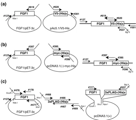
Schematic diagram depicting the strategy for plas-mid construction. Complementary DNAs encoding FGF1 and V5-(His)6 tag (a), myc-(His)6 tag (b) or 3xFLAG-(His)6 tag
(c) were separately amplified, and the products were mixed and used as templates for overlap extension PCR. The sequences of the primers are summarized in Table 1.
V5-(His)6
pAc5.1/V5-His
#301 #620
FGF1
FGF1/pET-3c
#127
#619
Nde I
FGF1 #127
#619
V5-(His)6
#537 #620
#301 Bgl II
FGF1
FGF1/pET-3c
#127
#397
Nde I
FGF1 #127
#397
myc-(His)6
#396 #395
Bgl II myc-(His)6
pcDNA3.1(-)-myc-His
#395
#396
#469 #468
#466
#467 Xho I
Apa I 3xFLAG-(His)6
FGF1
FGF1/pET-3c
#476 EcoR I
#477 Xho I #127
#178
Nde I
3xFLAG-(His)6
pcDNA3.1(+)
Apa I Xho I EcoR I FGF1
#537 Bgl II #117
(a)
(b)
sequences were verified to be the intended sequences using an ABI310 genetic analyzer. The expected numbers of amino acid residues for native FGF1, FGF1-myc-(His)6, FGF1-3xFLAG-(His)6 and FGF1-V5-(His)6 were 136, 165, 167 and 160, respectively.
Expression and semipurification of the recombinant FGF1 Expression vectors generated as described above were used to transform Escherichia coli strain BL21(DE3)/pLysS cells (Novagen), after which positive clones were selected. After preincubating the cells for 3 h at 37°C, protein expression was induced by addition of IPTG (final con-centration: 1 mM), and the incubation was continued for an additional 4 h at 37°C. The cells were then collected by centrifugation and suspended in GET buffer (10 mM glu-cose, 10 mM EDTA, 50 mM Tris-HCl, pH 7.4). The cell walls of the suspended cells were disrupted by freeze/thaw and sonication, after which the resultant lysate was fil-tered (0.45 μm), and the salt concentration was adjusted to 0.5 M. Heparin-Sepharose (Amersham Bioscience) beads were then added, and the mixture was incubated for 18 h at 4°C. The resin was then packed into a small col-umn and washed extensively with wash buffer (10 mM Tris-HCl, pH 7.4, 500 mM NaCl), after which the bound protein was eluted with elution buffer (10 mM Tris-HCl, pH 7.4, 2.0 M NaCl). The peak fractions (judged from the absorbance at 280 nm) were combined and subjected for further analysis.
Gel electrophoresis and immunoblot analysis
Semipurified recombinant FGF1s were resolved by SDS-PAGE (12.5%) and stained with Coomasie brilliant blue (CBB) or transferred to nitrocellulose membranes (Sch-leicher & Schuell BioScience Inc.) for immunostaining. The membranes were probed with monoclonal anti-FGF1 (mAb1; 1 μg/ml) [3], anti-myc (1/5,000 dilution;
Invitro-gen), anti-FLAG (M2; 10 μg/ml; Sigma) or anti-V5 (1/
2,000 dilution; Invitrogen) antibodies in Tris-buffered saline (TBS: 10 mM Tris-HCl, 0.15 M NaCl, pH 7.4) con-taining 5% skim milk (SM), or monoclonal His
anti-body (anti-penta-His; 0.2 μg/ml; QIAGEN) in TBS
containing 3% bovine serum albumin (BSA). HRP-conju-gated goat anti mouse IgG (1/10,000 dilution; Zymed) in TBS with 5% SM served as the secondary antibody for all primary antibodies except anti-His. For anti-His, the same secondary antibody (1/10,000 dilution) was used in TBS with 5% BSA. The blots were visualized using an ECL western blotting detection system (Amersham Bioscience) according to the manufacturer's instructions.
Heparin affinity analyzed with HPLC
The affinities of the semipurified proteins for heparin were analyzed using a Toso-HPLC system equipped with a TSKguardgel Heparin-5PW column (6 mm ID × 1 cm).
Tris-HCl buffer (pH 7.4) containing 0.15 M NaCl (buffer A) or 2.0 M NaCl (buffer B) were used as the effluents, the flow rate was 1.0 ml/min, and the eluate was fractionated into 0.5-ml fractions. The elution profile was monitored based on the absorbance at 280 nm. The salt concentra-tion of each fracconcentra-tion was calibrated based on its conduc-tivity.
Aliquots of the HPLC fractions were subjected to dot-blot analysis. Samples were loaded onto a nitrocellulose filter in a BioRad dot blotting system and then blocked with 5% SM. As the antibody mAb1 preferentially binds to dena-tured FGF1, the filter was first incubated in 2% SDS and 100 mM 2-mercaptoethanol for 30 min at 70°C, after which is was blocked in 5% SM again and immunostained using mAb1 as described above.
Assaying the mitogenic activity of FGF1
Ba/F3 cells were purchased from RIKEN. An expression vector encoding the FGFR1-IIIc was a generous gift from Dr. D. M. Ornitz (Washington Univ., St. Louis, MO). The vector was transfected into Ba/F3 cells by electroporation, after which neo-resistant clones were selected. Ba/F3 cells expressing FGFR1-IIIc (designated FGFR1c-Ba/F3 cells) were cultivated in RPMI1640 supplemented with 10% fetal bovine serum, 50 ng/ml recombinant FGF1, 5 μg/ml heparin and 0.5 mg/ml geneticin. To assay mitogenic activity, FGFR1c-Ba/F3 cells were seeded into a microplate to a density of 6.6 × 10³ cells/well and incubated with the
indicated amount of each tagged FGF1 and 10% FCS in the presence or absence of heparin (10 μg/ml). After 48 h, TetraColor ONE (Seikagaku Kogyo, Tokyo) was added, and the cells were incubated for an additional 4 h, after which the absorbance at 450 nm was measured.
Results and discussion
Construction and expression of tagged FGF1
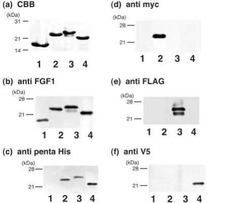
Using overlap extension PCR, we constructed a set of cDNAs encoding FGF1 with various tags. The proteins expressed in E. coli had the expected molecular weights and were semipurified by heparin affinity column chro-matography. Subsequent SDS-PAGE revealed that each construct was purified to homogeneity, as indicated by the single band after CBB staining (Fig. 2a). A monoclonal anti-FGF1 antibody (mAb1) recognized all of the proteins expressed (Fig. 2b). By contrast, monoclonal antibodies against (His)6-tag, myc-tag, FLAG-tag and V5-tag
specifi-cally recognized the correspondingly tagged FGF1, but not FGF1s with different tags (Fig. 2c–f). The minor band
accompanying FGF1-3xFLAG-(His)6 detected with
Heparin-affinity of the tagged FGFs
To determine their affinity for heparin, batch-purified FGF1s were subjected to heparin affinity HPLC and eluted with a linear NaCl gradient. A single absorbance peak at 280 nm was detected for each tagged FGF1 (Fig. 3a), and the FGF1 constructs in the peak fractions were confirmed by dot blot analysis using monoclonal FGF1
anti-body (Fig. 3b). We found that FGF1, FGF1-myc-(His)6,
FGF1-3xFLAG-(His)6 and FGF1-V5-(His)6 were eluted
from heparin Sepharose with 1.24, 1.09, 1.17 and 1.17 M NaCl, respectively, which is in good agreement with ear-lier reports that the NaCl concentration required for FGF1 elution from heparin is 1–1.2 M NaCl [4].
Because Lacy and Sanderson previously reported that
(His)6 tag enhanced the affinity of Sp17 protein for
heparan sulfate [5], we analyzed the effect of various tags on the affinity of FGF1 protein for heparin. FGF1 has a strong affinity for heparin/heparan sulfate, and the inter-action may be critically important for its biological activ-ity. We found that the myc-(His)6, 3xFLAG-(His)6 and V5-(His)6 tags used in this study had little effect on the affin-ity of FGF1 for heparin. Given that the three-dimensional structure of FGF1 indicates that its N- and C-termini are
flexible and outside the β-barrel structure important for receptor binding and heparin affinity [6], it is likely that addition of a tag at the C-terminus has no effect on the protein's affinity to heparin/heparan sulfate.
Mitogenic activity of tagged FGF1
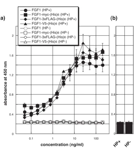
[image:4.612.52.290.89.290.2]We also investigated the ability of tagged FGF1 to stimu-late proliferation of FGFR1c-Ba/F3 cells [7]. As shown in Fig. 4a, the mitogenic activities of FGF1s tagged with myc-(His)6, 3xFLAG-(His)6 or V5-(His)6 were indistinguisha-ble from those of native FGF1. Moreover, statistical anal-ysis revealed no significant difference between any of the FGF1s. These results strongly suggest that in the presence of heparin these tagged FGF1s have affinities for cell sur-face FGFR1-IIIc that are similar to native FGF1. In addi-tion, like native FGF1, they exhibited no activity in the Electrophoresis and immunoblot analysis of the expressed
proteins
Figure 2
Electrophoresis and immunoblot analysis of the expressed proteins. FGF1 (lane 1), FGF1-myc-(His)6 (lane 2), FGF1-3xFLAG-(His)6 (lane 3) and FGF1-V5-(His)6 (lane 4) were expressed by E. coli and semipurified by heparin-Sepha-rose affinity chromatography. The eluate was subjected to SDS-PAGE and stained with CBB (a). After electrophoresis, the separated proteins were transferred to nitrocellulose membranes and immunoblotted using monoclonal anti-FGF1 (b), anti-His (c), anti-myc (d), anti-FLAG (e) or anti-V5 (f) antibodies. Positions of the molecular weight markers are indicated at the left of each panel in kDa.
21 28 (kDa)
1 2 3 4
21 28 (kDa)
1 2 3 4
21 28 (kDa)
1 2 3 4
28
21 (kDa)
1 2 3 4
31
21
14 (kDa)
1 2 3 4
28
21 (kDa)
1 2 3 4
(a) CBB
(b) anti FGF1
(c) anti penta His (f) anti V5
(d) anti myc
(e) anti FLAG
HPLC analysis of the affinity of semipurified FGF1s for heparin
Figure 3
HPLC analysis of the affinity of semipurified FGF1s for heparin. The elution profiles of FGF1, FGF1-myc-(His)6, FGF1-3xFLAG-(His)6 and FGF1-V5-(His)6 are illustrated in (a). NaCl concentration was gradually increased as illustrated at the top, and the absorbance at 280 nm was monitored. An aliquot of the peak fraction was also subjected for dot-blot analysis using anti-FGF1 monoclonal antibody (b).
5 10 15 20 25 30 35
f r a c t i o n n u m b e r
FGF1
a b s o r b a n c e a t 2 8 0 n m
2.0 M 1.5 M
0.15 M
FGF1-myc-(His)6
FGF1-V5-(His)6
FGF1-FLAG-(His)6
14 15 16 17 18 19 20 21 22 13
f r a c t i o n n u m b e r
FGF1 FGF1-myc-(His)6
FGF1-V5-(His)6
FGF1-FLAG-(His)6
(a)
[image:4.612.316.553.91.414.2]Publish with BioMed Central and every scientist can read your work free of charge "BioMed Central will be the most significant development for disseminating the results of biomedical researc h in our lifetime."
Sir Paul Nurse, Cancer Research UK
Your research papers will be:
available free of charge to the entire biomedical community
peer reviewed and published immediately upon acceptance
cited in PubMed and archived on PubMed Central
yours — you keep the copyright
Submit your manuscript here:
http://www.biomedcentral.com/info/publishing_adv.asp
BioMedcentral
absence of heparin, nor was any mitogenic activity detected in the absence of FGF1, despite the presence of heparin (Fig. 4b).
We have shown here that introduction of a myc-(His)6,
3xFLAG-(His)6 or V5-(His)6 tag at the C-terminus of FGF1
had little or no effect on the growth factor's affinity for heparin and its mitogenic activity toward cells expressing FGFR1-IIIc. Apparently, FGF1, FGFR and heparin are able to form an active signaling complex on the cell surface, despite the presence of a C-terminal tag. These tagged FGF1s should be useful for investigating the dynamic behavior of FGF1 in the context of its three-member sign-aling complex and other molecular complexes.
Competing interests
The authors declare that they have no competing interests.
Authors' contributions
TI conceived the study design, was responsible for the data collection, contributed to the data analysis and the writing of the paper. MA completed analyses, interpreted find-ings, and was the principal author of the manuscript. EH was involved in the study design, contributed to the inter-pretation of data, and editing of the paper.
Acknowledgements
The authors thank Dr. Masashi Suzuki for helpful discussion and Ms. Chik-age Aoki for her excellent technical assistance. We appreciate Ms. Junko Oki for her contribution to establishing the FGFR1c-Ba/F3 clone. This work was supported in part by a Grant-in-Aid for Scientific Research (C) 16570128 from Japan Society for the Promotion of Science (JSPS) (to MA), by a National Institute of Advanced Industrial Science and Technology (AIST) grant (to MA and TI) and by the Budget for Nuclear Research of the Ministry of Education, Culture, Sports, Science and Technology (to MA and TI). EH is a recipient of a fellowship provided by AIST.
References
1. Harlow Ed, Lane D: Common problems with peptide tags. In
Using Antibodies: A Laboratory Manual Cold Spring Harbor Laboratory Press, New York; 1999.
2. Forough R, Engleka K, Thompson JA, Jackson A, Imamura T, Maciag T: Differential expression in Escherichia coli of the alpha and beta forms of heparin-binding acidic fibroblast growth fac-tor-1: potential role of RNA secondary structure. Biochim Bio-phys Acta 1991, 1090:293-298.
3. Imamura T, Oka S, Tanahashi T, Okita Y: Cell cycle-dependent nuclear localization of exogenously added fibroblast growth factor-1 in BALB/c 3T3 and human vascular endothelial cells.
Exp Cell Res 1994, 215:363-372.
4. Klagsbrun M: The affinity of fibroblast growth factors (FGFs) for heparin; FGF-heparan sulfate interactions in cells and extracellular matrix. Curr Opin Cell Biol 1990, 2:857-863. 5. Lacy HM, Sanderson RD: 6xHis promotes binding of a
recom-binant protein to heparan sulfate. Biotechniques 2002,
32:254-258.
6. Dubey VK, Lee J, Somasundaram T, Blaber S, Blaber M: Spackling the crack: stabilizing human fibroblast growth factor-1 by targeting the N and C terminus beta-strand interactions. J Mol Biol 2007, 371:256-268.
7. Ornitz DM, Yayon A, Flanagan JG, Svahn CM, Levi E, Leder P:
Heparin is required for cell-free binding of basic fibroblast growth factor to a soluble receptor and for mitogenesis in whole cells. Mol Cell Biol 1992, 12:240-247.
[image:5.612.57.287.101.355.2]Mitogenic activity of FGF1s towards FGFR1c-Ba/F3 cells
Figure 4
Mitogenic activity of FGF1s towards FGFR1c-Ba/F3 cells. (a) FGFR1c-Ba/F3 cells were cultivated for 48 h with the indicated concentrations of FGF1s in the presence of 10 μg/ml heparin (closed symbols). TetraColor ONE was then added, and the cells were incubated for an additional 4 h, after which the absorbance at 450 nm was measured. No mitogenic activity was seen in the absence of heparin (open symbols). (b) In the absence of the FGF1s, no activity was observed, despite the presence of heparin. The data pre-sented are the averages and standard deviations of a tripli-cate experiment. Independent experiments were performed twice, and essentially the same results were obtained.
0 0.4 0.8 1.2 1.6 2
0.1 1 10 100
FGF1 (HP+) FGF1-myc-(His)6 (HP+) FGF1-3xFLAG-(His)6 (HP+) FGF1-V5-(His)6 (HP+) FGF1 (HP-) FGF1-myc-(His)6 (HP-) FGF1-3xFLAG-(His)6 (HP-) FGF1-V5-(His)6 (HP-)
0 0.4 0.8 1.2 1.6 2
absorbance at 450 nm
(a) (b)