JOURNAL OFVIROLOGY, Nov. 1993,p. 6618-6624 Vol.67,No. 11 0022-538X/93/116618-07$02.00/0
Copyright C 1993, American Society for Microbiology
Analysis of the Role of the bel and bet Open Reading Frames
of
Human
Foamy Virus by Using
a
New
Quantitative Assay
SHUYUARN F. YUAND MAXINE L. LINIAL*
DivisionofBasicSciences, Fred Hutchinson Cancer Research Center, 1124 Columbia Street, Seattle, Washington 98104-2092
Received 6July 1993/Accepted5August 1993
We have constructed a BHK-21-derived indicator cell line containing a single integrated copy of the f-galactosidase
(P-Gal)
gene under control of the human foamyvirus (HFV) longterminal repeat promoter (from -533 to+20).Thesefoamyvirus-activated ,8-Gal expression (FAB)cellscanbe used in aquantitative assay to measure theinfectious titer of HFV. Our results show that theFABassay is 50 timesmore sensitive thandetermination ofthe virus titerbytheend-pointdilution method.UsingtheFABassay,wehave found that HFV canproductivelyreplicatein severalerythroblastoidcelllinesaswellasin theJurkatT-cell line. We have alsoexamined the roles ofbel2, bet,and beI3 in viralreplication by constructing proviralHFV clones in which thereading frame ofBel2, Bet,orBel3 isdisrupted by placementof translationstopcodons.Analysisof these mutantsreveals that while thebeI3geneisnotrequiredfor viralreplication invitro,mutations in the beI2or bet gene decrease cell-free viral transmissionapproximately 10-fold.The prototype spumavirus, human foamy virus (HFV) or humanspumaretrovirus,wasoriginally isolated fromapatient with nasopharyngeal carcinoma (1). In culture,spumavirus is highly cytopathic, inducing multinucleated syncytia with a characteristic vacuolated appearance in the cytoplasm. Al-though it has been shown that in vitro HFVreplicates produc-tivelyinfibroblastic diploid cells but poorly inepithelium-like cells(23,24), viral replication in other cell types is still unclear. There have been numerous searches for diseases associated with HFV infection (37). The finding of both virus-related sequences and antigens in patients with Graves' disease sug-gests anassociation with this autoimmune disease (19, 38). It has alsobeen reported that expression of spumaviral genes in neurons and brain cellsin transgenic mice is correlated with neurological disorders (2, 3, 5).
The genome ofHFVcontains gag,pol, and env, as well as several other genes, located between env and the 3' long terminal repeat (LTR) (10, 25). At least four open reading frames (ORFs) are predicted to exist, including bell,bel2,bet, and bel3, which result from singly and/or multiply spliced mRNAs (28). Analysis of viral proteins in infected cells has shown that while Bell accumulates in the nucleus, Bel2 is found in the cytoplasm(11, 21). Bet, an abundant viral protein in the cytoplasm, is encoded by a multiply spliced mRNA in which the N-terminal portion of Bell is spliced in frame with the Bel2ORF. Thusfar, expression of the Bel3 ORF has not been reported, although bel3 transcripts have been found in infected cells (28).
The bell gene, like human immunodeficiency virus (HIV) tat and human T-cell leukemia virus tax, encodes a potent tran-scriptional transactivator of its own LTR promoter (17, 32). Deletion of bell completely abolishes the infectivity of HFV (21). The primary target sequence for Bell within the HFV LTR has been mapped to -130 bp 5' to the cap site (36). A secondregion located -400 bp5' to the cap site also contrib-utes to optimal reactivity to Bell (27). Bell can also trans
*Correspondingauthor.
activate gene expression directed by the HIV type 1 LTR, throughatargetsite different from that of HIV Tat(16, 20). In contrast to bell, the roles of bel2, bet, and bel3 in HFV replication remain unknown.
Titers offoamy virus stocks haveroutinelybeen determined by examination ofcytopathic effects (CPE) by the end-point dilution method. Here, we present a quantitative method for titration ofHFVusing an indicator cell line in which expres-sion of
3-galactosidase
(,B-Gal) can be activated by HFV infection. This foamy virus-activated 1-gal expression (FAB) assayexploits the requirement fortransactivation of theHFV LTR promoterby the viral Bell protein. Itprovidesasimple and rapid method for titration of HFV, which can be com-pleted in 2days and is 50-foldmoresensitive than titration of virus by the end-point dilution method. Using this assay, we have reexamined the spectrum of HFV cell tropism. We also report results of mutationalanalyses of thebel2, bet, and bel3 genes and conclude that while bel3 is dispensable for viral replication, thebel2orbet geneis required for optimal cell-free viraltransmission in vitro.MATERIALSANDMETHODS
Cells. Human embryonic lung (HEL) cells (ATCC CCL 137) and human foreskin primary fibroblastic cells (obtained from Adam Geballe, Fred Hutchinson Cancer Research Cen-ter) were grown in Dulbecco's modified Eagle medium sup-plemented with 10% fetal bovine serum (FBS). BHK-21 (baby hamster kidney cells, ATCC CCL 10) and FAB cells were grown in Dulbecco's modified Eagle medium with 5% FBS. Humancell lines, including B/N (an Epstein-Barr virus-trans-formedBlymphocytic cell line obtained from Mark Groudine, Fred Hutchinson Cancer Research Center), 174xCEM (a T-B lymphoblastic hybrid cell line, NIH AIDS Reagent Program catalogno.272), Jurkat (a CD4+ T-cell line, ATCC TIB 152), U937 (a histiocytic lymphoblastoid cell line, ATCC CRL 1593), K562 (an erythroblastoid cell line, ATCC CCL 243), and H92.1.7 (an erythroblastoid cell line originally called
6618
on November 9, 2019 by guest
http://jvi.asm.org/
QUANTITATIVE ASSAY OF THE ROLE OF HFV bel AND bet ORFs 6619
HEL92.1.7, ATCC TIB 180), were grown in RPMI medium supplementedwith 5% FBS.
Preparation of virus stocks. Virus stocks were originally obtained by transfection of BHK-21 or HEL cells with the full-length HFV proviral DNA. By the modified calcium phosphate method (6), the transfection efficiency of BHK-21 cells was muchhigherthanthat ofHELcells(data not shown). Transfected cells were then scraped from culture dishes and removed along with the culture medium, and virus was re-leased by three cycles of freezingandthawing. These total cell lysates were then added to BHK-21 or HEL cells to obtain high-titer virus stocks. Since the virus titer obtained from propagation of HFVin HEL cells was usually slightly higher than inBHK-21 cells, HELcellswere used in all the infection experiments. To prepare cell-free virus stocks, culture super-natants were cleared by low-speed centrifugation (4,000 x g for 10 min) and filtered through a 0.2-,um-pore-size filter membrane.
Plasmids. The infectious molecular clone of HFV, pHSRV13, and plasmids pdbell and pBCbell have been described previously (21) and were kindly provided by M. Lochelt and R. Flugel (German Cancer Research Center, Heidelberg, Germany). Plasmid pdbell is derived from pHSRV13 but contains a 208-bp deletion in the bell gene. PlasmidpBCbell is a eukaryotic bell expression vectorusing an immediate-early promoter from cytomegalovirus. Plasmid pHSRVAGPE was constructedby insertingthe 2,488-bp ClaI fragment of pHSRV13 into the 2,958-bp ClaI-digested frag-ment of phagemid pBluescriptll SK+ (Stratagene). Plasmid pCMVhph, as described previously (4), encodes resistance to
hygromycin Bdriven bythe cytomegalovirus promoter. Plasmid pEQJK was constructed by
ligating
a2,065-bp
HindlII-SacI fragment of pEQ3
(33),
apromoterless
[3-Gal
vector,withthe5,057-bpHindIII-SacIfragmentofpJK2. pJK2 is an HIV LTR-,B-Gal expressionplasmid in which a nuclear localization signal from simian virus 40 Tantigen
has been inserted upstream of the ,B-Gal gene (18). A DNAfragment
from nucleotides -533to +20 (+1 represents thefirst nucle-otide of the R region) of pHSRV13 was
amplified by
the polymerase chain reactionusing
primers
5'-CTCCCGGGATCAGAACATTGAC-3' and 5'-TGCACTGCAGCGAGTAGT
GAAG-3' and inserted into theSmaI-PstI site of
pEQJK.
The resultant plasmid,pHSRV5LG,
thus contains the HFV LTR promoter, followed by a nuclearlocalizationsignal,
the (-Gal gene,and thepoly(A) signal
from simian virus40(see
Fig.
IA).
Virus assays. For the FAB assay, the indicator cells were
plated in 12-wellplates
(22-mm
diameter)
at4 x 104cellsper well in Dulbecco's modifiedEagle
medium with 5% FBS the day before infection. Thefollowing
day,
the medium wasremoved and the cellswere incubated withdilutions ofavirus stock for 2 hat
37°C.
Viruswas then removed from eachwell and replacedwithgrowth
medium. Twodays
later,
themono-layer was fixed at room temperature for 5 min with 2 ml of fixation solution
(1%
formaldehyde
and 0.2%glutaraldehyde
inphosphate-buffered
saline[PBS]).
Cells weresubsequently
washed three times with PBS and incubatedat
37°C
for 30min with 0.5 mlofstaining
solution(4
mMpotassium
ferrocyanide,
4 mM potassiumferricyanide,
2 mMMgCl2,
and 0.4 mg of5-bromo-4-chloro-3-indolyl-(-D-galactopyranoside
[X-Gal]
per mlinPBS).Thereactionwasstopped
by
removing
thestaining
solution andthenwashing
eachwellwithPBS.Bluecellswerethencounted under a
light
microscope.
To determine the virus titer
by
CPEdevelopment
by
the end-point dilutionmethod,
serial dilutions of the HFV stockwere used to infect
HEL,
BHK-21,
and FAB cells. Cultureswere monitored
daily
for aperiod
of2weeks for theappear-anceof CPE.Incocultivation
experiments,
HELcellswerefirst infected with HFV at amultiplicity
of infection (MOI) of 3. Threedays later,
the cellsweretreated with 10 ,ugof mitomy-cin per ml for 2 h to inhibit cell division. After extensive washes, the treated HEL monolayers were trypsinized and cocultivatedwith differenthematopoietic suspension
cellsat aratio of 1to20.The
suspension
cellsweremaintainedin RPMI mediumcontaining
5% FBS andsubculturedevery3to5days.
After the secondpassage,adherentHELcells wouldno
longer
be observed in the cultures. The virus titer in the culture medium was
periodically
monitoredby
the FAB assay.Site-directed mutagenesis. Translation stop codons were
introduced into the
reading
frame ofbel2, bet,
orbel3 withoutaffecting
thecoding
sequence of the otheroverlapping
genes(see Fig. 3), by using
anoligonucleotide-directed
mutagenesis
kit
(Amersham) according
tothe manufacturer'sprotocol.
Thesingle-stranded
DNAtemplate
derived frompHSRVAGPE
wasused in the
mutagenesis
reaction.A21-mer,
5'-AGGGCT ACTAGAAGAGTCCAG-3', was used to create the Abel2 mutant, in which the Leu codon(TTG)
at residue 34 wasreplaced
with TAG. For theiXbel2C
mutant, theArg
(AGA)
andGln(CAG)
codonsatresidues246and248wereconvertedto TGA andTAG
by
a30-mer,
5'-TGGAATGTCACTQGA
AACTAGGGAAAACAA-3'. A
22-mer,
5'-CATAGAACCATAATGGCAGACT-3',
was used to generate the Aibel3 mu-tant,in which theLeucodon(TTA)
atresidue90wasreplaced
withTAA.The Ser codon
(TCA)
at residue 130waschanged
toTAAinthe
Abel3C
mutantwitha23-mer,
5'-GGTGGAATGTAACTAGAAACCAG-3'. After confirmation
by
DNAse-quencing,
each mutationwas introduced into thefull-length
proviral
cDNAby
inserting
the2,488-bp
ClaIfragment
from eachmutagenized
pHSRVAGPE
into the12,919-bp
ClaIfragment
ofpHSRV13.
Immunoprecipitation.
HEL cellswere infected with eitherwild-type
or mutant virus at an MOIof 20. Fourdays
afterinfection,
cellswerelabeled with 10RI
of[35S]methionine
per mlfor24handlysed
in buffercontaining
20mMTris-HCl(pH
7.5),
0.5%aprotinin,
10 mMiodoacetamide,
0.5% NonidetP-40,
0.5% sodiumdodecyl
sulfate(SDS),
and 0.5% sodiumdesoxycholate.
Two microliters of serum fromchimpanzees
naturally
infected withfoamy
virus(a gift
from KrishnaMurthy,
Southwestern Foundation for BiochemicalResearch,
SanAntonio,
Tex.)
was mixed with the celllysates
at4°C
overnight
and incubated withprotein
A-Sepharose
for 1 h.Samples
were then washed four times with RIPA buffer(10
mM Tris-Cl[pH
7.4],
150 mMNaCl,
1% NonidetP-40,
1% sodiumdesoxycholate,
0.1%SDS,
and 0.5%aprotinin)
andanalyzed
withanSDS-15%polyacrylamide
gel.
Gelswerethendried and
exposed
toKodak XAR-5 film.RESULTS
Constructionofanindicator cell line fortitrationof HFV. In order to
develop
aquantitative
assay for titration ofHFV,
BHK-21 cellswerecotransfectedwith
plasmids
pCMVhph
andpHSRV5LG
(Fig.
IA).
Although
HFV grows to aslightly
higher
titer in HELcells,
HEL cells are not suitable for construction ofan indicatorline sincethey
donot clonewell.Hygromycin-resistant
cloneswererandomly
picked
to screenfor
expression
of(3-Gal.
Aliquots
of each clone were eithermock infectedorinfected withHFV. Two
days
afterinfection,
cultureswerestained with X-Gal andexaminedfor bluecells.
If cells contain an
integrated
LTR-,B-Gal
gene,expression
ofHFV bell after infection would be
expected
totrans activatethe
expression
of(3-Gal,
which could then react with theVot-. 67, 1993
on November 9, 2019 by guest
http://jvi.asm.org/
6620 YU AND LINIAL
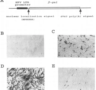
A
HFV LTRpromoter
P-gal
nuclezmar localization-m signal (MPKKKRK)
SV40 poly(A) sigrnal
C
~~~*
a, ' 's..*,j
A...v9
* SX9 ,,X'
*.' *
R~- ,...;:.
E
-0v
FIG. 1. (A) Schematic diagram of plasmid pHSRV5LG.The solid box indicatesthe HFV LTRpromoter(fromnucleotides -533 to +20;+1
representsthe first nucleotide of theRregion),andthe hatched box indicates the [3-Galgene.The nuclearlocalization sequenceconsists of seven
amino acidresidues(MPKKKRK)derived from the Tantigenofsimian virus 40 whicharesufficient to directheterologousfusionproteinstothe nucleus(15).The FAB cells inpanelsBtoEwerefixedandstainedasdescribedin Materials and Methods.(B)Mockinfection of FABcells;(C) transfection of FAB cells with theeukaryoticbellexpressionvectorpBCbell DNA(21);(D)infection of FAB cells with HFVatanMOI of50; (E) infection ofFABcells with HFV atan MOI of 0.005.
specific chromophor, X-Gal. Because a nuclear localization signalwas inserted upstream of the [3-Gal gene, an intensive blue color should be found in cell nuclei where [-Gal was
concentrated.
Of 22 clonesscreened, three expressedahighlevel of [3-Gal activity upon HFVinfection but showed no [-Galexpression
after mock infection. Anintensive blue color could be found in nuclei after 30 min of incubation with X-Gal. One of these clones, termed FAB,wasfurther characterized. The basal level of[-Galactivity in these FAB cellswasextremely low. No cells
turnedblue after mockinfection (Fig. 1B)ortransfectionwith
DNAofabell mutant(pdbell) (datanotshown),evenafter 48
hofincubation with X-Gal. Incontrast, [B-Gal expressionwas
activatedaftertransfectionwith theeukaryotic bell expression vector, pBcbell DNA (Fig. 1C). Results from Southern blot analysisdemonstrated that these cells contained only asingle
copyoftheLTR-[-Galgene(datanotshown). When infected withHFVatahigh MOI, cellsoften formed syncytia
contain-ing more than 10 nuclei (Fig. 1D). At a low MOI, however,
syncytiawere rareandsingly infected cellswereobserved (Fig.
IE).
Sensitivityofthe FAB assay. Experimentswere performed
using the end-point dilution of virus stocks to compare the infectious titer ofHFVdetermined byappearance of CPEto
that obtained by staining infected FAB cells. A series of dilutions of eithertotal HFV-infected cell lysates orcell-free
virus supernatants (prepared as described in Materials and
Methods) were usedto infect FAB, BHK-21, and HELcells. Two days later, FAB cellswere stained with X-Gal and virus
titerswere determined by counting the number of bluecells.
Only those wellscontaining between 20 and 100 blue cellswere
counted. The virus titer of total infected cell lysates deter-minedby the FABassay wasI x
10',
whilethat of thecell-freevirus stock was 7.5 x 10⁶' (Fig. 2A). This difference is not surprising,since it is known that HFV ishighly cell associated (14). However, when virus titers were determined by CPE
developmentonHEL,BHK-21,orFABcells, afteraperiod of
2 weeks, only 0.5 x 10⁷ to 2 x 10⁷ infectious units were
obtained for the total cell lysate and 0.5 x 10⁵ to 2 x 10⁵
infectious unitswereobtained for the cell-free virus stock(Fig.
2B). We thus conclude that not only is the FAB assay more
rapid, but it is more sensitive than the end-point dilution
method. The cell-free virus stock was also serially diluted to determine the linear range of the FAB assay. At a high
dilution, the number of blue cells countedwasproportionalto the amount of virus added and only single blue cells were
found (Fig. IE).
Viral replication in different human hematopoietic cell lines. Determination of the in vivo target cells of HFV is important for understanding the pathogenic potential of
spu-mavirus. It has been shown that inculture HFVgrows
prefer-entially in fibroblastic cells butnotinepithelium-like cells(23, 24). Yet little is known about HFV growth in hematopoietic cells, which are often the primary target cells for the other
B
D
J. VIROL.
Ef~~~~~~~~~~~~~~~~~
on November 9, 2019 by guest
http://jvi.asm.org/
[image:3.612.145.459.82.376.2]QUANTITATIVE ASSAY OF THE ROLE OF HFVbel AND bet ORFs 6621
A
1010
109
E
0
SZ
108
107
106
B
1010
IV
-107
-o6
[image:4.612.329.569.92.206.2]FAB FAB BHK21 HEL
FIG. 2. Comparison of the infectious titers of an HFV stock of either total infected cell lysates (dark greystipple) orcell-free virus
supernatants (black) determined by the FAB assay scored 2 days postinfection (A)orby theappearanceof CPE inFAB, BHK-21, and HELcellsby the end-point dilution method scored 2 weeks postinfec-tion (B).
5 LTR 3
'LTR
gag pol env bell
be7
be2
bell bel2 bet: bel3
hc
FIG. 3. Properties ofthe bel2, bet, and bel3mutants. Lettersatoe
indicate the positions of the translationstopcodons insertedinto each
reading frame of bell genes. a, TAGin Abel2; b, TGAin Abel2C;c,
TAG in Abel2C;d, TAA in Abel3;e,TAAin Abel3C.
(histiocyticlymphoblastoid cells), K562 (erythroblastoidcells), and H92.1.7 (erythroblastoid cells) produced the maximal amountof viruseswithin the 3 weeks of cultivation, adelayed
andprolonged viral replicationwasobserved inJurkat T cells,
peaking at the 4thweek. In contrast, HFVgrowth in B/N (B cells)and174xCEM (T-B hybrid cells)wasrelativelypoor.Itis possible that the virus titer detected at the early time point representsresidualvirus from the mitomycin-treated infected HELcells.
Construction of bel2-betand bel3 mutants.Except for bell, functions of the belgenes arestill unknown. Toexamine the
roles of bel2-betandbel3 in HFVreplication, translation stop codonswereintroduced into the reading frames of thesegenes
by site-directed mutagenesis without affecting the coding se-quences of the other overlapping genes (Fig. 3). Wild-type
proviralDNA aswell asDNAfrom each ofthefourmutants
was transfected into BHK-21 cells. In every case, CPE was
foundasearlyas2to3days posttransfection. Inthewildtype and bel3 mutants (Abel3 orAbel3C), extensive CPE and cell
mock WT s2 A2C A3 A3C
families of retroviruses. Usingthe FABassay, wehave
exam-ined HFVgrowth indifferent human hematopoieticcell lines. Since several attempts to infect these suspension cells with cell-free virus stocks failed,we decided to infect cellsusing a
cocultivation method (described in Materials and Methods). Mitomycin-treated HFV-infected HEL cellswerecocultivated with differenthematopoietic suspensioncells in RPMImedium with 5% FBS. The virus titerin each culturewas periodically
monitoredbythe FABassay.As shownin Table 1,whileU937
TABLE 1. HFVgrowth in humanhematopoietic celllines
measuredbythe FAB assay
BCFU"/mlof culturemedium at wk: Cellline
3 4 5 6
B/N 200 32 7 <0.1
174xCEM 70 7 <0.1 <0.1
Jurkat 1,350 16,000 12,000 12,000
U937 10,000 8,000 1,200 11
K562 5,000 350 60 70
H92.1.7 59,000 4,000 280 <0.1
"BCFU, blue-cell-formingunit.
200-97
6g9
46
46-____E..
30
1 2 3 4 5 6
FIG. 4. Analysisofvirus-specific proteinsinHFV-infected cellsby immunoprecipitation.HELcells
(106)
wereeither mockinfected(lane 1)orinfected withwild-type(lane 2)ormutantHFVvirus(lanes3to6) at an MOI of 20. [35S]methionine-labeled cell lysateswere then immunoprecipitated with chimpanzee serum and analyzed in an
SDS-15% polyacrylamide gel (see Materials and Methods). The positionsof molecularmassmarkers(inkilodaltons)areshownonthe
left,andthe arrowindicates thepositionof 56-kDa viral Betprotein. VOL.67, 1993
104
on November 9, 2019 by guest
http://jvi.asm.org/
[image:4.612.354.542.482.654.2] [image:4.612.68.311.634.722.2]6622 YU AND LINIAL
-t(E-wt
--dbel2
-c- dbeI2C
-4-dbel3
-0-dbec3C
1 2 3
4
days
p.i.
wwt
dbel2
deathwereobservedat5to6
days
posttransfection. However, CPEspread
wasslightly
slower in cells transfected with either of the bel2-bet mutants(Abel2
or Abel2C),peaking
at 7 to 8days.
Thedelay
of viralspread
inbel2-bet mutantswasmoreevident when cell-free rather than cell-associated virus stocks
were usedto infect fresh HEL cells
(data
notshown).
To detect viral proteins in infected cells, labeled HFV-infected celllysates
wereimmunoprecipitated
withserumfrom chimpanzees naturally infected with foamy virus, which is knowntocontainvirus-specific antibody (29).The56-kDa Betprotein,
an abundant viralprotein
in HFV-infected cells(11,
21),
wasdetected in cells infected with the wild type orbel3mutants
(Fig. 4,
lanes2, 5,
and 6)butwasnotpresent in cells infected with either of thebel2-betmutants(Fig. 4,lanes 3 and4),
which contained translation stop codons predicted to disruptthe Bel2 and Bet openreading
frames.Characterization of
beI2-bet
and bel3 mutants. To furtherexplore
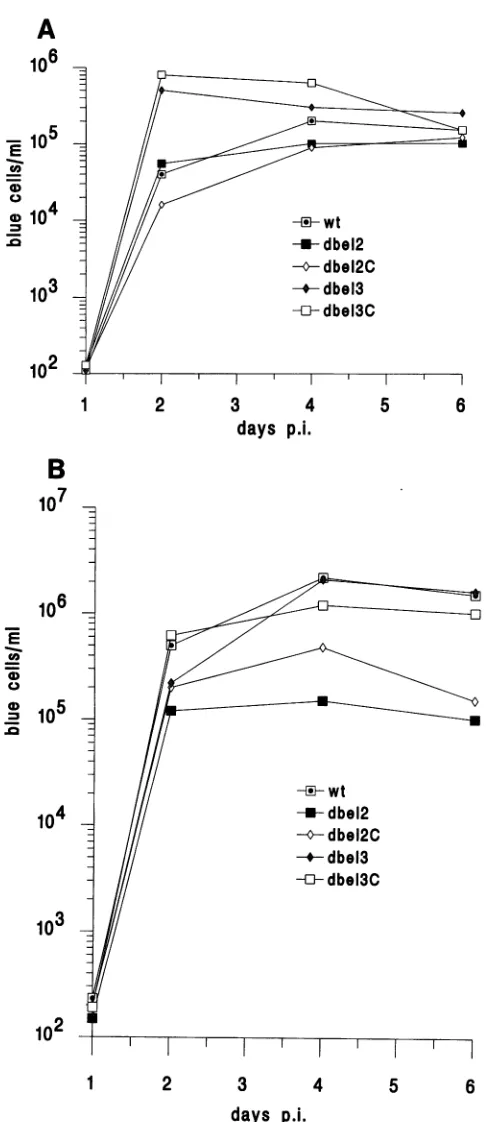
thephenotype
of delayed viral spread observed in bel2-betmutants,growthcurvesofwild-typeHFV and the four mutants were compared. HEL cells were first infected with 5 6wild-type
ormutantvirusesatanMOI of 0.03(as
determinedby
theFABassay).Titers ofcell-associated virus orfree virus released into culture media were measuredseparately by the FAB assay. Interestingly, virus progeny carrying mutations in thebel3 genereplicatedmoreefficiently than wild-type paren-talvirus,
especiallyattheearlyrounds ofinfection (Fig. 5). A plateau in the level of virus titers occurred rapidly with both wild-type and mutant viruses. This isprobably because of the rapid development of cell cytopathicity which prevents further rounds ofinfection and leads to killing of theinfected cells. While no apparent difference was detected in cell-associated~-Z
-° virus titers (Fig.5A),
the titer ofnon-cell-associated virus of bel2-bet mutants wasabout 10 times less than that of the wild type (Fig. 5B). This 10-fold reduction of viral replication in bel2-bet mutants was also observed in human primary fibro-blastic cells(datanotshown). Indeed, the same effect was also found when HEL cells were inoculated with the sameamount ofcell-free virus normalizedby the reverse transcriptase assay (datanot shown).DISCUSSION
._
dbc12C Biologicalstudies of HFV have beenhindered by the lackof-//--
dbel3 asensitive andquantitativeassaysystem.Althoughthereverse-//
{}db*13C
transcriptase
assay measures thephysical
number of virusl//l-
db913C particles, it does not accurately reflect the infectious titer of3 /// viruses (31). Titration of foamy virusesis commonly done by
10 /// either monitoring CPE development or plaque formation by
end-point dilution (22, 30). Both methods rely entirely on
efficient virus spread and require an extended incubation period, ranging from 8 to 14 days. An immunofluorescence assay has been developed as a semiquantitative method to
102
detect
the presence of viralantigens
(8); however,
the resultscan be severelyaffected bythe conditions ofcell growth and
the concentration ofvirus added(12). Therefore, this system 1 2 3 4 5 6 maynotbe reliable for titration ofvery dilute virusstocks.
days p.i. We have constructed an indicator cell line thatprovides a FIG. 5. Growth of
wild-type
andmutantHFV inHELcells. HEL quantitative assay for titration of virus derived from thecellswereinfectedwith wild-type(wt)ormutant(d,A) virusatanMOI proviralclone of HFV. ThissystemiSbasedonactivation ofan
of0.03. Atdifferent time intervals postinfection (p.i.) cells (A)and integrated ,3-Gal gene driven by the HFV LTR in BHK-21
cell-free media (B) from each culture were collected separately and cells, which are permissive for HFV infection. The specific
their virus titerswere measuredby the FAB assay. The data shown response of HFV LTR to its trans activator, Bell, tightly
hereweretaken fromoneof the three repeated experiments.Although controls theexpressionof,B-Galinthese cells.Noactivationof the exact number of titers varied slightly from one experiment to 13-Galwas detected when DNA from abell deletion mutant
another, the slopesand shapesof each growthcurveweresimilar. (pdbel)wastransfectedinto FAB cells, confirmingthe
require-ment foraspecificinteraction ofHFV LTR with Bellin this
A
106
E
1U
c o4
z
rD 104
102
B
107
106
E
1-co
0
a)
J.VIROL.
on November 9, 2019 by guest
http://jvi.asm.org/
[image:5.612.51.294.81.642.2]QUANTITATIVE ASSAY OF THE ROLE OF HFV bel AND bet ORFs 6623 assay. Any HFV virion that has completed the earlysteps of
viral replication and expressed thebell gene should be detect-able by the FAB assay, since this system requires only intra-cellular bell expression. In addition to detecting replication-competent HFV, the FAB assay could also be used for determining the effects of mutations on various stages of the viral life cycle.
Our results have indicated that the FAB assay is about 50-foldmore sensitive thandetermination ofthe titer ofavirus stock by CPE development by end-point dilution. This is not unexpected, as foamy virus often becomes latent in infected cells(14) and formation of CPEdepends heavilyoncontinual productive viral replication. In a routine examination, 1 in-fected cellin 10" uninfected cellscanbeeasilydetectedbyour insitu assayafter 2days.Thisindicatorsystemthus providesa
biological assayfor HFVwhich is simple, rapid, sensitive, and quantitative.
We have examined viral growth in different hematopoietic cell lines to furtherexplore the cell tropism of HFV. Surpris-ingly, HFV grew well in all cell types tested except for the B-cell line. Moreinterestingly, thedelayedandprolongedviral replication inJurkatcells,aCD4+T-cellline,isreminiscent of thepersistent foamy virus infections observed in their natural andexperimentalanimal hosts(35).Itwillbe of interesttotest FAB cells with primary viral isolates from individuals
sus-pected offoamy virus infection.
The roles of the bel2, bet, and bet ORFs inviral
replication
remain unknown. Flugel (9) and others
(13)
have observed a pattern of conserved His andCys
residues in Bel2 similar tothoseseenin HIVVifand
picornaviral
cysteine
proteases and thus suggestedasimilar role forHFV Bel-2 and HIVVif. We haveanalyzed the role of bel2andbetby
constructing
proviral
genomesofHFV in which the
reading
frame ofBel2 and Bet isdisruptedby insertion oftranslationalstop codons.Analysis
of two such mutants has revealed that
although
each clone could produce infectious virusfollowing
transfection,
the bel2 and betmutants are less infectiousthan thewildtype in either fibroblasticdiploid
cell lines orprimary
fibroblastic cells. The 10-fold reduction ofinfectivity
in these mutants is observed onlywhen cell-free virus is used to infect cells.Therefore,
assuggested for HIV
vif (7, 34),
bel2 and bet mayplay
a role inefficient cell-free viral transmission. Our results also indicate that the N-terminal
portion
of the bel2 andbetgeneproducts
is probably notfunctionally
important
since the mutant(zXbel2C)
withthe3-truncatedbel2-bet behavessimilarly
tothe mutant(Abel2)
in which the Bel2-Bet ORF iscompletely
removed.It has been noted that the bel3 gene, whose sequence is found inHFVbutnotinsimian
foamy
virus,
containsaleucineheptad repeated motif
(25).
Interestingly,
ourexperiments
with
site-specific
mutagenesis
have indicated thatinterruption
of the Bel3 ORFactually
enhances viralreplication
in vitro. Althoughanalysis
of the deduced amino acid sequences has revealed somehomology
between Bel3 and HIV type 2 Nef (26), thesignificance
of thissimilarity
is unclear.Despite
the factthat bel3 isnotrequired
for HFVinfection invitro,
the in vivo roles of thebel2, bet,
and bel3 ORFs remain to be determined.ACKNOWLEDGMENTS
We are grateful to R. Flilgel and M.
Lochelt
for theplasmids
pHSRV13, pdbell, and pBCbell; M. Emerman forpJK2;
and A. GeballeforpEO3.
WethankK.Murthy
forproviding
thechimpanzee
serum.Wealsothank D.Miller,M.Emerman,K.Levine,P.Lee,and L. Hajjarforcriticalcommentsonthe manuscript.
This work was supported in part by Public Health Service grant CA-18282(M.L.L.)from the National Institutes of Health. S.F.Y.was
supported by NIH training grant T32 CA09229 and postdoctoral fellowship F32 CA60357.
REFERENCES
1. Achong,B.G.,W. A.Mansell,M. A.Epstein,and P.Clifford.1971. An unusual virus in cultures from a human nasopharyngeal
carcinoma.J. Natl. Cancer Inst.46:299-307.
2. Aguzzi, A.,K.Bothe, I.Anhauser,I.Horak,A.Rethwilm,and E. F.
Wagner. 1992. Expression of human foamyvirus isdifferentially regulated during development in transgenic mice. New Biol. 4:225-237.
3. Aguzzi, A.,E. F.Wagner,K.-O.Netzer,K.Bothe, I.Anhauser,and A. Rethwilm. 1993. Human foamyvirus
proteins
accumulate inneurons and induce multinucleated
giant
cells in the brain oftransgenicmice.Am.J. Pathol. 142:1061-1071.
4. Aronoff,R.,and M. L. Linial. 1991.
Specificity
of retroviralRNA packaging.J. Virol. 65:71-80.5. Bothe, K., A.Aguzzi, H. Lassmann,A. Rethwilm, and I. Horak. 1991.
Progressive encephalopathy
andmyopathy
intransgenic
miceexpressing
humanfoamy
virus genes. Science 253:555-557. 6. Chen, C., and H.Okayama. 1987.High-efficiency
transformationofmammalian cells
by plasmid
DNA. Mol. Cell. Biol. 7:2745-2752.7. Fisher, A. G., B. Ensoli, L. Ivanoff, M. Chamberlain, S. R. Petteway,Jr., L. Ratner, R. C. Gallo, and F. Wong-Staal. 1987. Thesorgeneof HIV-1 is
required
forefficient virus transmissionin vitro. Science 237:888-893.
8. Fleming, W. A., and J. K. Clarke. 1970. Fluorescence assay of
foamy
virus. J. Gen. Virol.6:277-284.9. Flugel,R. M. 1992.
Spumaviruses:
agroupofcomplex
retroviruses.J.
Acquired
Immune Defic.Syndr.
4:739-759.10.
Fluigel,
R. M., A. Rethwilm, B. Maurer, and G. Darai. 1987.Nucleotide sequence
analysis
of the env gene and itsflanking
regions
off the humanspumaretrovirus
reveals twonovel genes. EMBO J. 6:2077-2084.11. Giron, M.-L., F.Rozain,M.-C.Debons-Guillemin,M.Canivet,J. Peries, and R. Emanoil-Ravier. 1993. Human
foamy
viruspolypeptides:
identification ofem'v andbelgeneproducts.
J.Virol. 67:3596-3600.12. Gould,E.A.,andJ.Hartley.1979. Factors
affecting
thegrowth
and titrationby
immunofluorescence of simianfoamy
virus. Arch. Virol. 62:63-70.13. Guy, B., M. Geist, K.Dott, D. Spehner, M. P.Kieny, andJ. P. Lecocq. 1991. A
specific
inhibitor ofcysteine
proteasesimpairs
avif-dependent
modification of humanimmunodeficiency
virustype1
enm
protein.
J. Virol.65:1325-1331.14. Hooks,J.J.,and C.J.Gibbs. 1975.The
foamy
viruses. Bacteriol. Rev.39:169-185.15. Kalderon, D., B. L.Roberts,W. D.Richardson,and A. E. Smith. 1984. A short amino acid sequence able to
specify
nuclear location. Cell 39:499-509.16. Keller,A.,E. D.Garrett,andB. R. Cullen. 1992. The bell
protein
of humanfoamy
virus activates humanimmunodeficiency
virus type 1 geneexpression
via a novel DNA target site.Virology
66:3946-3949.
17. Keller,A.,K.M.Partin,M.
hichelt,
H.Bannert,R. M.Flugel,and B. R. Cullen. 1991. Characterization of thetranscriptional
trans activator of humanfoamy
retrovirus.J. Virol. 65:2589-2594. 18. Kimpton, J., and M. Emerman. 1992. Detection ofreplication-competent and
pseudotyped
humanimmunodeficiency
virus witha sensitive cell line on the basis of activation of an
integrated
3-galactosidase
gene.J.Virol. 66:2232-2239.19. LaGaye, S., P. Vexiau, V. Morozov, V. Guenebaut-Claudet, J.
Tobaly-Tapiero,
M.Canivet,G.Cathelineau,andJ.Peries. 1992. Humanspumavirus-related
sequences in the DNA ofleukocytes
from
patients
with Graves disease. Proc. Natl. Acad. Sci. USA 89:10070-10074.20. Lee,A.H.,K.J.Lee,S.Kim,and Y. C.Sung.1992.Transactivation of human
immunodeficiency
virus type 1long
terminalrepeat-directed gene
expression
by
the humanfoamy
virusbellprotein
requires
aspecific
DNAsequence. J. Virol.66:3236-3240.VOL.67, 1993
on November 9, 2019 by guest
http://jvi.asm.org/
6624 YU AND LINIAL
21. Lichelt, M., H. Zentgraf, and R. M. Flugel. 1991.Construction of aninfectiousDNAclone of thefull-lengthhumanspumaretrovirus genomeandmutagenesisof the bell gene. Virology184:43-54. 22. Loh, P. C., B. C. Achong, and M. A. Epstein. 1977. Further
biological properties of the human syncytial virus. Intervirology 8:204-217.
23. Loh, P. C., and K. S. Ang. 1981.Replicationof humansyncytium formingvirus in human cells: effect of certainbiological factors andselectivechemicals. J. Med. Virol. 7:67-73.
24. Loh, P. C., and F. M. Matsuura. 1984. Human spumavirus replication inhuman cells. J. Med.Virol. 14:247-254.
25. Maurer, B., H. Bannert, G. Darai, and R. M.Flugel.1988.Analysis of theprimarystructure ofthelong terminalrepeatand thegag and polgenes ofthe human spumaretrovirus.J.Virol. 62:1590-1597.
26. Maurer, B., and R. F.Flugel. 1987. The 3'orf protein of human immunodeficiencyvirus 2shows sequence homology with thebel3 geneof the human spumaretrovirus. FEBS Lett.222:286-288. 27. Maurer, B., E. Serfling, V. ter Meulen, and A. Rethwilm. 1991.
Transcription factorAP-1 modulates the activity of the human foamy viruslong terminal repeat. J. Virol. 65:6353-6357. 28. Muranyi, W., and R. M.Flugel.1991.Analysis of splicing patterns
ofhumanspumaretrovirus by polymerase chain reaction reveals complexRNAstructures. J.Virol.65:727-735.
29. Netzer, K. O., A. Rethwilm, B. Maurer, and V. ter Meulen. 1990. Identification of the major immunogenic structural proteins of humanfoamy virus.J. Gen.Virol.71:1237-1241.
30. Parks,W. P., and G.J. Todaro. 1972. Biological properties of syncytium-forming (foamy)viruses.Virology47:673-683. 31. Parks, W.P.,G.J. Todaro, E. M. Scolnick, and S. A. Aaronson.
1971. RNAdependent DNA polymerase in primate syncytium-forming (foamy)viruses. Nature(London)229:258-260. 32. Rethwilm, A., 0. Erlwein, G. Baunach, B. Maurer, and V. ter
Meulen. 1991. Thetranscriptional transactivator of humanfoamy virus maps tothebellgenomic region. Proc. Natl. Acad. Sci.USA 88:941-945.
33. Schleiss, M. R., R. D. Catherine, and A. P. Geballe. 1991. Translational controlofhumancytomegalovirusgp48expression. J. Virol. 65:6782-6789.
34. Strebel, K., D. Daugherty,K. Clouse, D. Cohen, T. Folks, and M.A. Martin. 1987. The HIV 'A'(sor)geneproductisessential for virusinfectivity.Nature(London)328:728-730.
35. Swack, N. S., and G. D. Hsiung. 1975. Pathogenesis ofsimian foamyvirus infection in natural and experimental hosts. Infect. Immun. 12:470-474.
36. Venkatesh,L.K.,P. A.Theodorakis, and G. Chinnadurai. 1991. Distinctcis-acting regionsin U3regulate trans-activation of the human spumaretrovirus long terminalrepeatby the viral bellgene product.NucleicAcids Res. 19:3661-3666.
37. Weiss, R. A. 1988. Foamy retroviruses. A virus in search of a disease.Nature(London) 333:497-498.
38. Wick, G., B. Grubeck-Loebenstein, K. Trieb, G. Kalishnig, and A. Aguzzi. 1992. Human foamyvirus antigens in thyroid tissue of Graves' diseasepatients.Int.Arch.Allergy Immunol. 99:153-156. J.VIROL.