Copyright © 2000, American Society for Microbiology. All Rights Reserved.
Herpesvirus Saimiri vFLIP Provides an Antiapoptotic Function but
Is Not Essential for Viral Replication, Transformation,
or Pathogenicity
DIANA GLYKOFRYDES,1HENK NIPHUIS,2EVA M. KUHN,2BRIGITTE ROSENWIRTH,2
JONATHAN L. HEENEY,2JOSEPH BRUDER,3GERALD NIEDOBITEK,4
INGRID MU¨ LLER-FLECKENSTEIN,1BERNHARD FLECKENSTEIN,1
ANDARMIN ENSSER1*
Institut fu¨r Klinische und Molekulare Virologie der Universita¨t Erlangen-Nu¨rnberg1and Pathologisch-Anatomisches Institut,491054
Erlangen, Germany; Biomedical Primate Research Center, 2288GJ Rijswijk, The Netherlands2;
and GenVec, Inc., Gaithersburg, Maryland 208783
Received 26 June 2000/Accepted 25 September 2000
Apoptosis of infected cells is an important host defense mechanism, and many viruses have exploited anti-apoptotic proteins that interfere with crucial cellular pathways. Viral FLICE inhibitory proteins (vFLIPs) are encoded by rhadinoviruses like herpesvirus saimiri, the related Kaposi’s sarcoma-associated herpesvirus-hu-man herpesvirus 8 (KSHV/HHV8), and the poxvirus responsible for molluscum contagiosum. The vFLIPs can block the interaction of the death receptor-adapter complex with the cellular effector FLICE (caspase-8), and this prevents the initiation of the downstream caspase cascade. KSHV/HHV8 vFLIP overexpression can confer resistance to T-cell-mediated apoptosis and acts as a tumor progression factor in a murine B-cell lymphoma model. To analyze the function of herpesvirus vFLIPs in the genetic background of the virus and in a model for viral pathogenesis, we deleted the vFLIP gene (open reading frame 71) from the genome of herpesvirus saimiri strain C488. The viral deletion mutant was viable and replicated like the wild-type virus. An antiapo-ptotic effect could be attributed to the vFLIP gene, but we also show that the vFLIP gene of herpesvirus saimiri is dispensable for viral transformation of T cells in vitro and for pathogenicity in cottontop tamarins in vivo.
Viruses utilize various strategies that relate to apoptosis of host cells or attacking effector cells of the immune system. Since the induction of programmed cell death in cells infected with various pathogens is an important and common host de-fense mechanism, many viruses have evolved proteins to evade this protective mechanism. On the other hand, apoptosis is used by some viruses to promote the release of progeny from infected cells (47).
Open reading frame 71 (ORF71) of the oncogenic herpes-virus saimiri (saimirine herpesherpes-virus 2) encodes a putative anti-apoptotic protein that is homologous to a family of cellular and viral inhibitory proteins which interfere with apoptosis signaled through the death receptor Fas-CD95 and the tumor necrosis factor receptor 1 (TNFR-1) (5, 30, 31, 54).
The binding of a specific ligand to death receptors expressed on eukaryotic cells induces multimerization of the receptor complex, and this clustering recruits adapter molecules like the FADD (Fas-associated death domain) protein or the TNFR-associated death domain protein via interactions between the death domain of the receptor and the death domain of the adapter. The formation of the death receptor-adapter complex recruits the upstream caspase-8 (FLICE) by interaction be-tween the death effector domain (DED) of the adapter protein and the DED of the caspase. Together they form the death-inducing signaling complex, and caspase-8 is activated by pro-teolytic autocleavage, which initiates the downstream caspase cascade and results in apoptosis (3, 24).
Viral FLICE inhibitory proteins (vFLIPs) are found in the
poxvirus responsible for molluscum contagiosum and in most of theGammaherpesviridaeof the genusRhadinovirus, namely the Kaposi’s sarcoma (KS)-associated herpesvirus-human her-pesvirus 8 (KSHV/HHV8), rhesus rhadinovirus (RRV) (2, 50), herpesvirus saimiri, equine herpesvirus 2 (30), and bovine her-pesvirus 4 (55). The vFLIPs contain two DEDs and have been shown to block the interaction of a death receptor-adapter complex like Fas-FADD with the cellular effector FLICE (cas-pase-8) and prevent its autoactivation (5, 30, 54). A cellular homolog to the vFLIPs has also been identified, although it occurs in two forms: the short cellular form, cFLIP(S), contains two DEDs and acts at the same level as the vFLIPs by pre-venting FADD-FLICE interaction; the long form, cFLIP(L), resembles the caspase-8 structure with two DEDs plus a cas-pase domain. However, the active site of the protease domain is mutated. cFLIP(L) can also interact with the autoactivation step of caspase-8 (31).
Overexpression of KSHV/HHV8 vFLIP by retroviral trans-duction of the murine B cell line A20 has been shown to confer resistance against death receptor-mediated apoptosis. It pro-motes clonal outgrowth in the presence of death stimuli, and caspase activation in the vFLIP-A20 transductants is inhibited. When transferred into syngeneic or semiallogeneic immune competent mice, the vFLIP-transduced B cells induced signif-icantly more and faster-growing B-cell tumors than the mock-transduced line. This difference was not evident in immune response-compromised mice, suggesting that vFLIP is a tumor progression factor that can offer protection from T-cell-medi-ated apoptosis (15). Similarly, the cFLIP has been shown to prevent tumor rejection in a different mouse model, also pre-sumably by escape from cytotoxic T-cell-mediated apoptosis, and lead to tumor progression (40). Additional evidence points to a modulation of the NF-B pathway by the interaction of * Corresponding author. Mailing address: Institut fu¨r Klinische und
Molekulare Virologie der Universita¨t Erlangen-Nu¨rnberg, Schlossgar-ten 4, 91054 Erlangen, Germany. Phone: 9131-8523786. Fax: 49-9131-851002. E-mail: [email protected].
11919
on November 9, 2019 by guest
http://jvi.asm.org/
vFLIPs with signaling proteins (8). However, all these obser-vations are from in vitro overexpression of recombinant pro-teins (5, 30) or transplantation of tumor cell transfectants into mice (15).
Studying the vFLIP function of herpesvirus saimiri provides (i) a permissive cell culture system that allows the construction of recombinant viruses, (ii) in vitro lymphocyte transformation assays in human and simian T cells, and (iii) a meaningful and stringent animal model for pathogenesis in common marmo-sets or cottontop tamarins. A previous study argues for the function of the herpesvirus saimiri vFLIP gene, since it dem-onstrates an antiapoptotic effect during the lytic infection of owl monkey kidney (OMK) cells (54). Although protection from apoptosis was evident in infected cells, the data is weak-ened by the fact that herpesvirus saimiri contains at least one other antiapoptotic protein, a viral Bcl-2 homolog that is func-tional in in vitro models (11, 43). Thus, vFLIP function has been proven neither in the normal genetic background of the virus nor in a model for viral pathogenesis.
Therefore, we generated a vFLIP deletion mutant by remov-ing the vFLIP gene (ORF71) from the genome of herpesvirus saimiri strain C488. The C488⌬FLIP deletion mutant was rep-lication competent, and we demonstrate that the antiapoptotic effect is lost after deletion of vFLIP. However, we also show that the vFLIP gene of herpesvirus saimiri is dispensable for the viral transformation of T cells in vitro and for pathogenicity in cottontop tamarins in vivo.
MATERIALS AND METHODS
Cell culture and virus propagation.OMK cells (ATCC CRL1556), cultivated in Dulbecco’s modified Eagle medium supplemented with glutamine (350g/ ml), gentamicin (100g/ml), and 10% heat-inactivated fetal calf serum, were used for the propagation of herpesvirus saimiri. Virus stocks were generated by the infection of confluent OMK cells seeded in 175-cm2tissue culture flasks at a low multiplicity of infection. When lysis was complete, supernatants were cleared from the cellular debris by centrifugation at 2000⫻gfor 15 min, and cell-free supernatants were stored at⫺80°C. Peripheral blood mononuclear cells (PBMC) ofCallithrix jacchusand human umbilical cord blood lymphocytes (CBL) were isolated by density gradient centrifugation. The cells were cultivated in lympho-cyte growth medium (LGM) (45% RPMI 1640 medium, 45% Panserin [Pansys-tems, Aidenbuch, Germany], 10% fetal calf serum [Pansystems], gentamicin [100
g/ml], and glutamine [350g/ml]). LGM for human CBL was supplemented with 100 U of recombinant human interleukin-2 (IL-2) (aldesleukin; ProleukinR; Chiron, Ratingen, Germany) per ml or 20 U of human IL-2 (Roche Diagnostics, Mannheim-Penzberg, Germany) per ml.
Construction of the viral deletion mutant C488⌬FLIP.ORF71 was deleted from the herpesvirus saimiri strain C488 genome by a cosmid-based approach. All cloning procedures were performed by standard methods. APacI fragment including most of ORF71 was subcloned from cosmid 40 into pNEB193 (New England Biolabs). Most of the ORF71 gene, including the two DEDs, was deleted by digestion withBclI-SalI. A double-stranded oligonucleotide adapter was designed from the oligonucleotides 5⬘-GATCGTTTAAACGTTAATTAATC GA-3⬘ and 5⬘-TCGATCGATTAATTAACGTTTAAAC-3⬘. This adapter con-tained an internalPacI site (bold), as well asBclI-andSalI-compatible 5⬘ over-hangs (underlined) at the ends. It was inserted into the BclI-SalI-digested pNEB193-Pac to replace the deleted ORF71 segment (Fig. 1A). The altered PacI fragment encompassing the deleted ORF71 gene segment was reinserted into the PacI-digested cosmid 40, resulting in cosmid 40⌬FLIP. The correct insertion was verified by sequencing. Recombinant virus was generated by lipo-some-mediated cotransfection of a set of overlapping cosmids, including cosmid 40⌬FLIP, into permissive OMK cells (Fig. 1B). The cosmids were linearized before transfection by restriction withNotI; this also removed the pWE15 clon-ing vector, since twoNotI sites flank theBamHI site that was used to clone the viral DNA. Virus-containing supernatant from completely lysed cultures was harvested by centrifugation, and the pelleted virions were lysed in 100l of PCR buffer containing 100g of proteinase K (Roche Diagnostics) per ml and 0.5% Tween 20 for 1 h at 56°C; then the proteinase K was heat inactivated for 15 min at 95°C. An aliquot of 2 to 4l was used for PCR analysis.
Construction of C488EGFP recombinant.The enhanced green fluorescent protein (EGFP) gene was PCR amplified from plasmid pEGFP-C1 (Clontech). PCR mixtures (50l each) contained 2l of template DNA, 0.2M each deoxynucleoside triphosphates (dNTP), 10M each primer, and 5 U ofPfu polymerase (Stratagene) in 1⫻Pfureaction buffer. The primers 5⬘-GGGCGC GCCGAATGCAGTGAAAAAAATGC-3⬘ and 5⬘-GGCGCGCCATTAATAG
TAATCAATTACG-3⬘were used. After a 1-min initial denaturation step at 95°C, 25 cycles of 10 s at 95°C, 20 s at 55°C, and 3 min at 70°C were performed in an MJ Research PTC-200 thermal cycler, followed by a 4-min final extension step at 70°C. The PCR fragment was then subcloned into the singleSwaI site of cosmid 331 located just upstream of the DHFR gene (ORF2) in a noncoding region. The construct was analyzed by sequencing, and expression of the EGFP was verified by transfection into OMK cells and fluorescence microscopy.
DNA sequence analysis.Nucleotide sequences were determined with an ABI 377A automated sequencer (Applied Biosystems) using the Dye-Deoxy Termi-nator Sequencing kit according to the manufacturer’s instructions (Perkin-Elmer). DNA sequence evaluation was done with XBAP software (10).
Virus stocks and replication studies.For virus titration, OMK cells were grown in 48-well plates (Nalgene Nunc International, Roskilde, Denmark) and infected with serial ten-fold dilutions (10⫺3to 10⫺7) of herpesvirus saimiri C488, C488⌬Flip, and the respective EGFP variants in 400l of Dulbecco’s modified Eagle medium with supplements. A dilution step was defined as positive if the cells in at least 4 of 8 wells were completely lysed. The virus replication kinetics was determined by the infection of OMK cells (3⫻105cells seeded in a 25-cm2 flask 2 days before infection) with 104tissue culture infectious particles (TCIP) in 10 ml of medium. The titers of virus-containing supernatant taken on subse-quent days were determined by limiting dilution as described above.
In vitro transformation of lymphocytes.Lymphocytes were expanded after isolation for 2 to 3 days by stimulation with 0.5 to 1g of phytohemagglutinin A (Murex, Großburgwedel, Germany) per ml. Between 3⫻106and 5⫻106cells were infected with 1 ml of herpesvirus saimiri C488 or mutant virus C488⌬FLIP containing supernatants (titer,⬎106TCIP/ml) and cultivated in LGM as de-scribed above. The transformation of the resulting T-cell lines was assessed microscopically and by the observation of accelerated growth.
Experimental infection of common marmosets.In vivo oncogenicity of the herpesvirus saimiri C488 recombinants was assayed by experimental infection of FIG. 1. Construction of the recombinant viruses herpesvirus saimiri C488⌬FLIP, C488EGFP, and C488⌬FLIP-EGFP. The vFLIP encoding ORF71 of herpesvirus saimiri C488 was deleted from the right terminal cosmid 40. (A) APacI fragment was subcloned into vector pNEB193, and most of the ORF71 gene was deleted by digestion withBclI andSalI and insertion of an adapter containing a PacI site. Then the altered PacI fragment from pNEB193 was reinserted intoPacI-digested cosmid 40 to generate the cosmid 40⌬FLIP. (B) Recombinant virus was generated by the cotransfection of overlapping linearized cosmids 331 or 331EGFP, 261, 291, 336, and 40 or 40⌬FLIP.
on November 9, 2019 by guest
http://jvi.asm.org/
Saguinus oedipustamarins. The study was approved by the Institutional Animal Care and Use Committee and was performed according to governmental regu-lations with purpose-bred, healthy, adult cottontop tamarins at the Biomedical Primate Research Center (Rijswijk, The Netherlands). Two animals were in-fected with the experimental virus C488⌬FLIP, and only one animal was infected with the wild-type control virus C488. Since cottontop tamarins are an endan-gered and protected species, larger numbers of animals could not be justified from an ethical point of view. Two animals per experimental virus are considered necessary to obtain a meaningful result in a primate experiment, and this number is usually used in published studies. Since the wild-type virus C488 is highly pathogenic and generally causes lymphoma in all infected animals (C. jacchusor S. oedipus) (17, 35, 36), only one animal was allowed to obtain control tissues and cell lines. The animals were each intravenously injected with 1 ml of cell-free virus containing supernatant from infected OMK cultures containing 106TCIP of virus. They were housed in separate cages and received a standard monkey diet and drinking water ad libitum. Blood samples were taken prior to infection, at weekly intervals, and before necropsy to expand T lymphoma cells and reiso-late virus by cocultivation on OMK cells. The animals were euthanized as soon as illness became evident. Tissues were fixed in formalin and stained with he-matoxylin and eosin and for immunohistochemistry additionally with antibodies specific for CD3 (Dako, Hamburg, Germany) and CD20 (Dako).
Stable growing transformed T-cell lines were obtained from PBMC, and dif-ferent organs (thymus, spleen, liver, kidney, and lymph nodes) were obtained from each diseased animal. Briefly, tissue obtained at necropsy was cut into pieces with a sterile scalpel blade, and single-cell suspensions were obtained by passage through 70-m nylon sieves (Falcon Cell Strainer, Becton Dickinson, Heidelberg, Germany). Cells were cultivated in LGM without IL-2.
Detection of viral DNA.The status of viral DNA in the transformed cell lines was analyzed by PCR and Southern blotting. PCR analysis was carried out in 25-l reaction mixtures, each containing 2l of template DNA, 0.2M each dNTP, 10M each primer, and 2.5 U of AmpliTaq in 1⫻AmpliTaq buffer (Perkin-Elmer). PCR conditions were as follows: a 5-min denaturation at 95°C; 29 cycles of 30 s at 95°C, 30 s at 56°C, and 1 min at 70°C; a 4-min extension at 70°C; and a 4°C hold. The following primer pairs specific for the respective ORFs were used for the analysis: StpC/Tip (TR1 5⬘-GTAGTAAACTAAGAGCAAA GCAAGC-3⬘and TR2 5⬘-GTACAAGCTGTTCAAGTTTGTTAGC-3⬘), ORF3 (5⬘-CACAACACTGGTATGTACCAATG-3⬘and 5⬘-CTGTGGAGGTAATGC AGATAC-3⬘), ORF75 (5⬘-TGGCTGCTAACAGGCATGG-3⬘and 5⬘-AGCAC GTTGCCCGAGATTG-3⬘), and ORF71/FLIP (5⬘-GGCGCGCCTCGAAATTC TGTAAATGGAC-3⬘and 5⬘-ACAGAAAGAGACACAAGAG-3⬘).
DNA for Southern blotting was prepared by the addition of 400l of extrac-tion buffer (100 mM NaCl, 10 mM Tris-HCl, 25 mM EDTA, 0.5% sodium dodecyl sulfate; proteinase K was added to a final concentration of 1 mg/ml) to 5⫻107cells. After an overnight incubation at 56°C, the solution was digested with 40l of RNase A (5 mg/ml) at 37°C for 30 min. The DNA was extracted with buffer-saturated phenol:chloroform:isoamyl-alcohol (25:24:1), and then a one-half volume of ammonium acetate (7.5 M) was added to the aqueous phase, and the DNA was precipitated with 2 volumes of ethanol. After centrifugation, the pellet was washed with 70% (vol/vol) ethanol, dried briefly, and resuspended in TE buffer (10 mM Tris-HCl, 1 mM EDTA). A total of 20g of cellular DNA was digested withSstI orPstI. The DNA fragments were size fractionated by electrophoresis through a 1% agarose gel. The DNA was then transferred to a nylon membrane (Hybond N; Amersham) and hybridized with a32P-labeled DNA fragment. This 1.9-kb probe specific for ORF71 and -72 was amplified by PCR from cosmid 40 (primers, 5⬘-TGCGTTAGACAAATATCCC-3⬘and 5⬘-C TAAAAATGCAGCATCGTCACC-3⬘; conditions were as above). The DNA fragment was purified from a 1% agarose gel (Qiaquick Gel Extraction kit; Qia-gen, Hilden, Germany), and random labeling with [␣-32P]dATP was performed (20).
RNA and cDNA analysis.Total cellular RNA was prepared by the acidic phenol extraction method (9). Five micrograms of RNA was treated with RNase-free DNase I (Roche Diagnostics) in 1⫻DNAse I buffer for 30 min, followed by heat inactivation at 70°C for 10 min. Then, first-strand cDNA was synthesized with Superscript II reverse transcriptase (Gibco BRL): the RNA was incubated with 500 ng of random hexamer primers for 10 min at 70°C. After a short incubation on ice, 8l of the reaction mixture (4l of 5⫻first-strand buffer, 2
l of 0.1 M dithiothreitol, 1l of 10 mM dNTPs, and 1l of Superscript II) was added, and the synthesis of the cDNA was started (10 min at 25°C and 50 min at 37°C). The enzyme was heat inactivated at 70°C for 15 min. RNA complementary to the cDNA was removed by the addition of 1 U of RNase H (MBI Fermentas) and incubation for 20 min at 37°C. An identical sample was prepared in parallel where the reverse transcriptase was omitted from the reaction mixture (as a control sample). A total of 2l of the reaction mixture was used for reverse transcription (RT)-PCR analysis. PCR conditions were as follows: a 5-min initial denaturation at 95°C; 39 cycles of 20 s at 95°C, 30 s at 52°C, and 1 min at 70°C; a 4-min final extension at 70°C; and a 4°C hold. Primers utilized were specific for the ORF71/FLIP (as above) and ß-actin (5⬘-CGGGAAATCGTGCGTGACA T-3⬘and 5⬘-GAACTTTGGGGGATGCTCGC-3⬘).
Flow cytometry.Transformed human and simian T cells were analyzed by flow cytometry with antibodies for B- and T-cell surface epitopes on a FACS-Calibur flow cytometer (Becton Dickinson). The directly labeled monoclonal antibodies (MAbs) (Cy-Chrome or phycoerythrin conjugated) were specific against CD2
(RPA-2.10; PharMingen, Heidelberg, Germany), CD3ε(SP34; PharMingen), CD3 (Leu-4; Becton Dickinson), CD4 (Leu-3a SK3; Becton Dickinson), CD8 (RPA-T8; PharMingen), CD20 (Leu16 L27; Becton Dickinson), HLA-DR (L243; Becton Dickinson), CD80 (L307.4; PharMingen), and CD86 (IT2.2; PharMingen). Directly labeled isotype-matched control MAbs were used (Bec-ton Dickinson and PharMingen). CD95 surface expression was detected with a MAb directed to CD95 (DX2; PharMingen) and a secondary fluorescein iso-thiocyanate-labeled goat anti-mouse immunoglobin G F(ab⬘)2 fragment (Di-anova, Hamburg, Germany).
Immunofluorescence.A total of 104 OMK cells were seeded in four-well chamber slides (Nalgene Nunc International) and infected with the EGFP vari-ants of C488 and C488⌬FLIP. After 24 h, the cells were overinfected with recombinant adenovirus expressing the soluble Fas ligand (Ad-FasL) (7), or 50
M menadione was added and the cells were incubated for 4 or 8 h at 37°C and fixed with 4% paraformaldehyde (30 min at room temperature). To visualize nuclei and DNA, the cells were stained by the addition of 1g of Hoechst-33342 dye (Sigma) per ml. The apoptotic cells were viewed under a Zeiss Axiovert fluorescence microscope at a magnification of⫻630.
Induction of apoptosis and determination of cell death.A total of 104OMK cells were grown in flat-bottomed 96-well plates (Nalgene Nunc International) overnight and infected with 5⫻104to 10⫻104TCIP of the recombinant viruses C488EGFP and C488⌬FLIP-EGFP (multiplicity of infection, 5 to 10). After 24 h the cells were superinfected with recombinant adenovirus (Ad) Ad-FasL (0.5⫻
106PFU) or the-GAL gene (Ad-Z) as a control (GenVec, Inc., Gaithersburg, Md.). After incubation for 4 h at 37°C, cell death was quantified by an enzyme-linked immunosorbent assay (ELISA) for the detection of histones bound to fragmented DNA released into the cytoplasm of apoptotic cells (Cell Death Detection kit; Roche Molecular Biochemicals, Mannheim, Germany). Cytoplas-matic extracts were normalized to their total protein content that was determined by the BCA-Assay (Pierce, Inc., Rockford, Ill.). Specific protection from apo-ptosis was calculated as follows: 10,000⫻(optical density/protein [g/ml]). Uninfected cells treated with Ad-FasL were assigned a value of 100% apoptosis.
RESULTS
Construction of recombinant viruses.The herpesvirus sai-miri ORF71 encoding the vFLIP was deleted by a cosmid-based approach that allows the construction of recombinant viruses without contaminating wild-type virus. Previously, we had subcloned the genome of herpesvirus saimiri into overlap-ping cosmids (18). The right-terminal cosmid 40 was selected for the deletion of ORF71. In the genome of herpesvirus saimiri strain C488, the methionine start codon of ORF71 is located directly adjacent to the ochrestop codon terminating the ORF72 encoding the vCyclin. The ORF71 contains aPacI restriction site 72 bp downstream of the first ATG codon. A second PacI site is located at a distance of 2.15 kb in the noncoding region between ORF70 (encoding a thymidylate synthase) and ORF71. This 2.15-kb PacI restriction fragment encompassing most of vFLIP was subcloned from cosmid 40 into plasmid pNEB193 to generate pNEB193-Pac. The region including the two DEDs of vFLIP was removed by restriction of pNEB193-Pac withBclI andSalI and replaced by an oligo-nucleotide adapter containing a PacI site. The modifiedPacI fragment was then reinserted into cosmid 40 to generate cos-mid 40⌬FLIP (Fig. 1A). The correct insertion was verified by restriction mapping and sequencing. A human cytomegalovi-rus-promoter driven EGFP expression cassette was inserted into a noncoding region of cosmid 331 to generate cosmid 331EGFP. Recombinant viruses were then constructed by co-transfection of linearized cosmids into OMK cells. The recom-binant virus C488⌬FLIP was generated from cosmids 331, 261, 291, 336, and 40⌬FLIP. We also designed an EGFP-expressing recombinant vFLIP deletion virus and a corresponding control virus. The recombinant EGFP-expressing vFLIP deletion virus C488⌬FLIP-EGFP was generated from cosmids 331EGFP, 261, 291, 336, and 40⌬FLIP; the recombinant EGFP-express-ing control virus C488EGFP was generated from cosmids 331EGFP, 261, 291, 336, and 40 (Fig. 1B).
Replication of recombinant viruses. Recombinant viruses were obtained from cosmid cotransfection, demonstrating that the vFLIP gene of herpesvirus saimiri is not essential for
on November 9, 2019 by guest
http://jvi.asm.org/
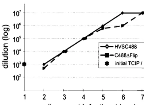
tic replication in OMK cells. No differences in plaque size or morphology in OMK cells were notable (data not shown). Lytic viral replication of C488⌬FLIP on OMK cells was compared to that of C488. Endpoint titers of both viruses were in the range of 106to 107 TCIP per ml of supernatant from several
inde-pendent cultures. The kinetics of viral replication was then compared by infection of OMK cultures with 104TCIP and by
titration of supernatant taken from successive days. There was no apparent difference between the viruses, lysis was complete after 7 days, and similar endpoint titers of 107were observed
(Fig. 2).
Transformation of human and simian T cells in vitro. Her-pesvirus saimiri is able to transform human and simian T cells to permanent antigen-independent growth in vitro (6). Human CBL (13 donors) and PBMC fromC. jacchus(5 donors) andS. oedipus(10 donors) were infected with C488 and C488⌬FLIP in parallel, and the proliferation of the cells was compared to that in uninfected controls. After 4 to 6 weeks of culture, the control cells had stopped growing. Both the deletion mutant virus C488⌬FLIP as well as the wild-type virus C488 trans-formed the T cells. The data are summarized in Table 1. The transformed T cell lines were analyzed by PCR and Southern blot analysis, and the specific viral genotype present in the cells was confirmed. Proliferation tests performed with the estab-lished T-cell lines showed no detectable difference (data not shown). Thus, the vFLIP is not essential for replication and T-cell transformation in vitro.
Pathogenicity in cottontop tamarins.One putative function of vFLIP is the escape of transformed cells from immune surveillance by interference with cytotoxic T-cell-mediated apo-ptosis, which is difficult to study in vitro. Herpesvirus saimiri C488 is able to induce T-cell lymphoma in vivo in several species of New World primates. In vitro transformation is linked to pathogenicity in vivo for most published viral deletion mutants. However, there are reports where efficient in vitro transformation was observed but where the recombinant virus turned out to be apathogenic or less pathogenic than wild-type virus in vivo (16, 28, 38). The relevance of the vFLIP gene for the development of T-cell lymphoma was studied in cottontop tamarins (S. oedipus). Two animals were infected intravenously with the deletion mutant virus C488⌬FLIP (animals B237 and R213) and one was infected with the wild-type virus C488
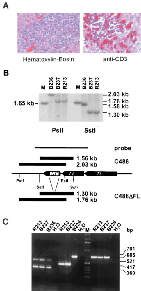
carrying an intact vFLIP (animal B236). All animals developed disease between day 14 and 15 and were euthanized (Table 1). Necropsy was performed, and macroscopic findings were con-sistent with the typical lymphoproliferative disease induced by herpesvirus saimiri. Enlarged lymph nodes and spleen and infiltrations in various organs were recognizable. Immunohis-tochemistry revealed infiltrations of CD3⫹blast-like lymphoid
cells in various tissues of all three animals (Fig. 3A). Infiltra-tion of virus-transformed T cells was widespread in the differ-ent organs or tissues, and the morphology of infiltrations was similar in the wild-type- and deletion virus-infected animals. Continuously growing T-cell lines from thymus, spleen, liver, kidney, and lymph nodes were established. The cultures grew stably over an observation period of more than 10 months, and there was no difference detectable in the proliferation of the cell lines transformed by the recombinant or the wild-type viruses. DNA from the transformed T-cell cultures established from the infected animals was used to confirm the presence of the specific viral genome by Southern blotting and PCR (Fig. 3B and C). Thus, the vFLIP gene is also dispensable for the capacity of herpesvirus saimiri to induce T-cell lymphoma in vivo.
[image:4.612.56.291.72.243.2]Expression of vFLIP and phenotype of transformed cells. The transcription of the mRNA encoding vFLIP was con-firmed by RT-PCR from infected OMK cells, S. oedipus ex vivo-cultured T-cell lines, and transformed human CBL-de-rived T-cell lines. Oligonucleotides flanking the deleted region were chosen that generated PCR amplification products of 266 bp from C488⌬FLIP and 531 bp from C488. Contamination of the reactions resulting from genomic viral DNA was ruled out by DNase I treatment and by analysis of an identically treated parallel sample in which the reverse transcriptase was omitted from the first-strand cDNA synthesis reaction. Fragments of the expected sizes were amplified and confirmed the transcrip-tion of herpesvirus saimiri vFLIP in C488-transformed and -infected cells and the transcript that includes the deletion in C488⌬FLIP-infected cells (Fig. 4). The amplified region also encloses the 3⬘region of the vCyclin gene. However, by using several different sets of oligonucleotide primers, no amplifica-tion products corresponding to transcripts originating at the amino terminus of ORF73 that would be spliced to the vFLIP or vCyclin gene were detected (data not shown). In addition, the surface phenotype of the transformed cells was deter-mined. We analyzed the transformed human CBL-derived and the simian tumor-derived T-cell lines from all animals and could not detect significant and consistent differences in the expression of cell surface markers. An example shows CD3, CD4, and CD8 expression on C488 or the C488⌬FLIP deletion
TABLE 1. Transformation by recombinant viruses in vitro and pathogenesis in infected cottontop tamarins
Virus strain
or mutant Species and/orcell type
In vitro transforma-tion experiments (no. positive/total)a
Pathogenesis (survival time in days)b
C488 Human CBL 8/13 NA
C. jacchusPBMC 9/9 ND
S. oedipusPBMC 10/10 15
C488⌬FLIP Human CBL 7/13 NA
C. jacchusPBMC 9/9 ND
S. oedipusPBMC 10/10 14, 15
aPerformed with human CBL (13 donors) and PBMC fromC. jacchus(5
do-nors) andS. oedipus(10 donors). Parallel cultures of cells were infected with herpesvirus saimiri C488 or C488⌬FLIP.
bNA, not applicable; ND, not done. FIG. 2. Replication of the recombinant viruses. OMK cells were infected
with culture supernatants containing 104infectious particles (determined by
lim-iting dilution) of the EGFP variants of herpesvirus saimiri C488 and C488⌬FLIP. Virus titers were determined from supernatant taken on subsequent days until the cells were completely lysed by cytopathic effect.
on November 9, 2019 by guest
http://jvi.asm.org/
virus-transformed cells (Fig. 5). At least four different lym-phoid cell lines were established from each of the three ani-mals; they showed no significant variation in growth in cell culture, and all were CD3⫹T cells, with some variation in CD4
and CD8 expression also within cell lines established from the same animal. Presumably, during cultivation an initially poly-clonal culture becomes nonhomogenous due to the outgrowth of subclones. All cells expressed surface markers which are typically found on mature activated T cells, like CD2, CD3, CD4 and/or CD8, HLA-DR, CD80, or CD86, but not B-cell markers like CD20 (data not shown).
Protection from apoptosis induced by FasL. The surface expression of the death receptor Fas-CD95 in the different simian and human cell lines was studied by fluorescence-acti-vated cell sorter analysis. The results of an experiment with transformed T cells obtained from the thymus of animals B236 (C488) and R213 (C488⌬FLIP) are shown in Fig. 6, and Jurkat cells served as a positive control. The CD95 surface marker was detectable on both wild-type virus C488 or C488⌬FLIP transformed cells (Fig. 6).
Apoptosis can be detected by morphological criteria. It is characterized by membrane blebbing, nuclear fragmentation, and chromatin condensation that can be observed microscop-ically. We used the EGFP-expressing variants of wild-type (C488EGFP) and deletion (C488⌬FLIP-EGFP) viruses to discriminate infected from uninfected cells. The herpesvirus saimiri-infected cells are easily identified by the green auto-fluorescence of EGFP when examined with a fluorescein iso-thiocyanate-compatible filter set. The FasL-induced apoptosis was detectable in the OMK cells by nuclear fragmentation that was visualized by the DNA binding dye Hoechst-33342. A smaller proportion of green C488-infected cells seemed to show nuclear fragmentation than the C488⌬ FLIP-EGFP-in-fected cells; however, an exact quantification was difficult due to concurrent lytic replication of the virus (data not shown). The antiapoptotic effect of herpesvirus saimiri was then ana-lyzed in a system that quantifies apoptotic events. Endogenous endonucleases are activated in apoptotic cells and cleave the DNA into small oligonucleosomes containing histone H1. These nucleosomal DNA fragments leak through the impaired nuclear membranes into the cytoplasm and are detected in cytoplasmatic extracts by the Cell Death Detection ELISA
[image:5.612.54.297.76.577.2]FIG. 3. Detection of recombinant herpesvirus saimiri in tumor cells. (A) Liver tissue sections from an C488⌬FLIP-infected animal that show infiltration of portal tracts by large blast-like lymphoid cells (left). Immunohistochemistry reveals expression of the CD3 T-cell antigen in these cells (right). (B) DNA was recovered from T-cell lines established from tumorous spleens of infected ani-mals. Total cellular DNA was digested withSstI orPstI and analyzed by Southern blotting for the presence of the ORF71 deletion in the cell lines; the marker is a 1-kb ladder (Gibco BRL Life Technologies, Karlsruhe, Germany). Hybridiza-tion was done with the probe indicated. B236, C488 infected; R213 and B237, C488⌬FLIP infected. (C) PCR was performed with extracts from corresponding transformed T cells. StpC and ORF75 and -3 were not affected by the deletion, whereas the ORF71 deletion was confirmed in the recombinant viral genomes.
FIG. 4. Expression of vFLIP in different cell lines. Total RNA was used for an ORF71-specific RT-PCR. The middle panel shows the transcripts detected from infected OMKs or transformed human and simian T cells. The bottom panel shows the corresponding RNA as detected by RT-PCR for-actin.⫹RT, RT-PCR from first-strand cDNA;⫺RT, PCR from parallel control reaction, where Superscript reverse transcriptase was omitted. The locations of primers are indicated.
on November 9, 2019 by guest
http://jvi.asm.org/
system. When apoptosis was induced by superinfection with a recombinant adenovirus expressing soluble FasL, an anti-apoptotic effect was detectable in C488EGFP-infected OMK cells and not in C488⌬FLIP-EGFP-infected cells. The results of four independent experiments are shown in Fig. 7A. In contrast to the observations of OMK cells, the proliferation of transformed T cells (S. oedipus) was not affected by treat-ment with up to 1g soluble FasL per ml. For comparison, Jurkat T cells are highly susceptible to 20-fold-lower doses of soluble FasL. Thus, a protective effect was not detectable, presumably due to overall resistance of the transformed T cells. Only the Fas-independent proapoptotic substance men-adione induced a decrease in proliferation in both cells trans-formed with the wild-type virus C488 and those transtrans-formed with the deletion mutant virus C488⌬FLIP (Fig. 7B).
DISCUSSION
Herpesvirus saimiri is the prototype of the gamma-2 herpes-viruses orRhadinoviridae (1). It is apathogenic in the per-sistently infected natural host, the squirrel monkey (Saimiri sciureus), but causes rapidly progressive T-cell leukemia and lymphoma in several species of New World primates (22). Strains of the highly oncogenic subgroup C are capable of transforming primary human T cells to permanent antigen-independent growth (6), while subgroup A and B strains do not transform human cells. The herpesvirus saimiri genome (M-DNA) has a size of about 155 to 160 kb; it consists of a 113-kb AT-rich unique region (L-DNA) that is flanked by about 20 to 25 GC-rich, tandem repeats of 1.4 kb (H-DNA). This principal genome structure is shared by all rhadinoviruses except equine herpesvirus 2. Within the L-DNA region, herpesvirus saimiri has at least 76 ORFs plus genes for small nuclear URNAs, termed HSURs. The H-DNA terminal repeats of herpesvirus saimiri do not encode any known viral proteins (1). At the very left end of the L-DNA, all herpesvirus saimiri subgroups en-code the saimiri transformation-associated proteins (STP-A, -B, or -C), a family of weakly conserved oncoproteins that are essential for the transformation of T cells and pathogenicity (12, 16, 33). The subgroup C strains additionally encode the tyrosine kinase-interacting protein, TIP, in this region, which is also essential for transformation (17). The HSURs are en-coded in the region adjacent to the transforming genes; they are expressed in transformed cells but are dispensable for transformation in vitro (18). In addition to conserved gamma-herpesvirus genes, gamma-herpesvirus saimiri shares several ORFs carrying sequestered cellular genes with the other rhadino-viruses KSHV/HHV8 and RRV, namely the vFLIP gene (ORF71), a vCyclin gene (ORF72), and a viral G protein-coupled receptor gene (ORF74, IL-8 receptor) (44). Recom-binant herpesvirus saimiri has further been used to study the putative transforming protein K1 of KSHV/HHV8 (38) and is a useful model for gammaherpesvirus oncogenesis (32). RRVs are the closest relatives of KSHV/HHV8 (2, 50). Like herpes-virus saimiri, the RRVs do not seem to be tumorigenic in the natural host (39), and a lytic cell culture system in primary rhesus fibroblasts would allow the construction of recombinant viruses (13). However, no RRV-associated tumor or in vitro transformation model has been described so far.
Epidemiological data as well as the consistent detection of viral DNA in diseased tissue suggest that KSHV/HHV8 is most likely the major cofactor for the development of both classical KS and KS in immune response-compromised patients. In addition, KSHV/HHV8 is linked to several rare B-cell lympho-proliferative diseases, like primary effusion lymphoma (PEL) and multicentric Castleman’s disease (27, 41). Although sev-eral reports hint at the possibility for the propagation of
[image:6.612.54.294.71.467.2]FIG. 6. Expression of the Fas/CD95 on transformed T cells. Expression of CD95 on the surface of Jurkat cells (positive control), C488⌬FLIP-transformed T cells (S. oedipusanimal R213), and herpesvirus saimiri C488-transformed T cells (S. oedipusanimal B236) derived from infected animals. The open graph represents the negative isotype control, and the solid graph represents the spe-cific staining of Fas/CD95.
FIG. 5. Surface phenotype of transformed simian and human T cells. A typ-ical example of the expression of the T-cell markers CD3, CD4, and CD8 is shown for the spleen-derived T-cell lines B236 (herpesvirus saimiri C488 in-fected) and R213 (C488⌬FLIP infected) (A) and for T-cell lines derived from human CBL transformed by C488 or C488⌬FLIP (B). The histograms show fluorescence intensity in logarithmic scale on thexaxis and cell numbers in linear scale on theyaxis. Open graphs represent negative isotype controls, and solid graphs represent specific staining.
on November 9, 2019 by guest
http://jvi.asm.org/
KSHV/HHV8 in cells derived from human endothelium or kidney (23, 42, 46), there is no accessible experimental model for the generation of recombinant viruses. Furthermore, there exist neither in vitro transformation models that involve the complete KSHV/HHV8 virus nor animal models for pathogen-esis, e.g., for the development of KS- or PEL-like disease after viral infection.
Interestingly, the vFLIP- and vCyclin-encoding mRNA is found latently transcribed in KS in situ (51) and in KS and PEL by Northern blotting (14, 48, 49, 52, 53). The presence of a latent bicistronic mRNA encoding vFLIP and vCyclin, as well as a latent tricistronic mRNA encoding vFLIP, vCyclin, and the latency-associated nuclear antigen LANA in diseased tis-sue from KS and MCD hint at a role in progression or main-tenance of the disease state by the inhibition of tumor cell apoptosis. vFLIP may also be required to counteract putative proapoptotic functions of the vCyclin gene under specific cir-cumstances where high levels of cyclin-dependent kinase 6 are present in infected cells (45). Similarly, the KSHV/HHV8 LANA has been shown to exert an antiapoptotic function by inhibition of p53 transcriptional activity and thus promote cell survival (25). In contrast to the cultures established from KSHV/HHV8-associated PEL, where a small proportion of cells always produce viral particles and where the lytic cycle can be induced by stimulation with phorbol esters or sodium butyrate, the herpesvirus saimiri-transformed human T cells harbor the viral episomes in a latent state, and no infectious virus is produced even upon stimulation. We demonstrate that the ORF encoding vFLIP is transcribed in simian and human T cells transformed by herpesvirus saimiri (Fig. 4). However,
vFLIP protein expression has not been published for KSHV/ HHV8-associated tumors, and the protein was not detectable from our herpesvirus saimiri-transformed cell lines or other infected cells by immunoblot analysis with specific rabbit anti-sera (data not shown).
The antiapoptotic effect observed in the C488 wild-type virus-infected OMK cells was no longer detectable in the C488⌬FLIP deletion virus-infected cells (Fig. 7B). However, the fact that the herpesvirus saimiri vFLIP gene deletion re-verses the protective effect of herpesvirus saimiri C488 in OMK cells does not rule out a relevant antiapoptotic function of the vBcl2 homolog. It may be that a missing balance to a potentially proapoptotic vCyclin masks vBcl2 effects. However, this could only be studied effectively if vBcl2 deletion viruses were available as well.
On one hand, induction of apoptosis by herpesvirus proteins, including homologs of cellular regulators of apoptosis, has been postulated as a way for more-efficient release of progeny virions from the infected cell and for the elimination of im-mune cells attacking the virus host cell. An example for her-pesvirus proapoptotic proteins is the induction of apoptosis in mononuclear cells and bovine B-lymphoma BL-3 cells after binding of bovine herpesvirus 1 glycoprotein D (29). Herpes simplex virus type 1 (HSV-1) induces and blocks apoptosis at multiple steps during infection and protects cells from exoge-nous inducers in a cell type-dependent manner (26), and ICP27-deleted HSV-1 induces apoptosis in epithelial cells (4). HSV-2 induces apoptosis of macrophages in a Fas- and TNFR-independent manner (21). On the other hand, premature host cell destruction may be induced by the activation of cellular signaling and apoptotic pathways. Such signals could result from upregulation of surface receptor molecules, cytokine se-cretion by infected cells, increased production of death recep-tor ligands, or from endogenous events, e.g., those related to the p53 or NF-B pathways. vFLIP genes could interfere with some of these events, and antiapoptotic gene expression in lytically infected cells could result in a more-efficient produc-tion of progeny virions. Although we speculated that a vFLIP deletion virus would be impaired during lytic replication, no significant difference in virus endpoint titer or viral replication kinetics was detectable (Fig. 2). Thus, the herpesvirus saimiri vFLIP does not increase viral particle yield under the condi-tions of lytic tissue culture in OMK cells, the standard cell line for propagation of herpesvirus saimiri (19).
Inhibition of death receptor-mediated apoptosis is a com-mon mechanism exploited by several DNA viruses to protect infected cells from immune system attack by cytotoxic T cells. Adenovirus E3-14.5 and E3-10.4 proteins are inhibitors of death receptor internalization. The vFLIP proteins interfere with death receptor signal transduction, and cowpox virus CrmA, baculovirus p35, and Ad E3-14.7 proteins have been identified as downstream inhibitors of cellular caspases (re-viewed by Roulston and colleagues [47]). Several studies sug-gest that the herpesvirus and cellular FLIPs act as putative tumor progression factors in vitro and in tumor models based on the introduction of FLIP-transduced cell lines in mice; they provide evidence that tumor progression is in fact mediated by resistance to T-cell-induced death receptor-mediated apopto-sis (15, 40). Our data obtained with the C488⌬FLIP deletion mutant cannot support the view that the herpesvirus saimiri vFLIP is a cofactor for oncogenesis. There was no difference in the efficiency of viral transformation in vitro; nor was there a significantly different incubation period of herpesvirus saimiri disease in cottontop tamarins (Table 1). There may be small differences in incubation time or disease progression that FIG. 7. Protection from apoptosis by herpesvirus saimiri. (A) Infection of the
permissive cell line OMK with C488 and C488⌬FLIP. Cells were infected with herpesvirus saimiri and superinfected with recombinant Ads. Four hours after superinfection with FasL- or lacZ-expressing adenovirus (0.5⫻106PFU), the resulting apoptotic effect was assayed by the Cell Death Detection ELISA. The results are mean values from four normalized experiments. (Left) negative trol (cells not superinfected); (middle) control cells superinfected with the con-trol Ad-Z; (right) cells superinfected with Ad-FasL. The protective effect from apoptosis provided by herpesvirus saimiri C488 was no longer detectable in the C488⌬FLIP infected cells. (B) Proliferation of C488- and C488⌬ FLIP-trans-formed T-cell lines fromS. oedipustamarins. Proliferation was assayed by [3H] thymidine incorporation. Cells were studied for Fas-independent cell death by incubation with 50M menadione for 8 h, for susceptibility to Fas-mediated apoptosis with 1g of soluble FasL (cross-linked with anti-FLAG M2 antibody) per ml or by study of control cells in the presence of medium alone. Mean values
from three transformed cell lines and two experiments are shown.
on November 9, 2019 by guest
http://jvi.asm.org/
might be detectable by titration of the pathogenic viruses in the highly susceptible tamarins.
We also could not detect a significant difference in prolifer-ation between T cells transformed by herpesvirus saimiri C488 or C488⌬FLIP (Fig. 7B). The postulated protective effect of vFLIP may be too discrete to be detected in transformed T cells, since herpesvirus saimiri-transformed human and simian T cells are rather resistant to Fas-mediated apoptosis (37). However, this resistance is not due to lack of death receptor expression, since significant levels of Fas-CD95 are detectable on the transformed T cells (Fig. 6). Moreover, a balanced ex-pression and/or interaction of Fas-FasL may promote T-cell growth, resulting in a net growth of the culture, along with some apoptosis in fewer cells than those replenished by expan-sion (34).
The ORF carrying the vFLIP gene of herpesvirus saimiri is transcribed in virus-infected and -transformed cells, and the antiapoptotic effect found after infection of OMK cells by herpesvirus saimiri C488 is reversed by deletion of the vFLIP gene. Thus, an antiapoptotic effect can be attributed to the vFLIP gene expression in virus-infected cells. However, in the herpesvirus saimiri system which allows testing of rhadinovirus transformation and pathogenesis, the deletion of the vFLIP gene did not affect transformation or oncogenicity. This may have implications for KSHV/HHV8-associated disease, since the vFLIP gene is also latently transcribed in cells persistently infected by KSHV/HHV8.
In principle, the vFLIPs may have evolved to counteract T-cell-mediated apoptosis of persistently infected cells in the natural host or to compensate for proapoptotic signals provided by other viral proteins like envelope glycoproteins or vCyclin. The latter is an attractive hypothesis: the growth-promoting vCyclin is expressed from the same bi- or poly-cistronic message that also encodes vFLIP, and the vCyclin of KSHV/HHV8 has been shown to promote apoptosis in trans-fected cells that have entered S phase (45). High levels of the vFLIP and vCyclin message are detectable in advanced le-sions of KS (51). In this model, vFLIP would not offer protection from externally induced apoptosis but would bal-ance proapoptotic stimuli associated with the growth promot-ing functions of the virus itself. Although the similar domain structure and conserved genomic context, as well as in vitro data obtained from overexpression studies in transfected cell lines, suggest an analogous function, it still may be that the vFLIPs of KSHV/HHV8 and herpesvirus saimiri play different roles in their respective target tissues in vivo, and the vFLIP of KSHV may be essential for the transformation by KSHV, in contrast to herpesvirus saimiri. However, rhadinoviruses usu-ally cause either minor or no relevant pathology in their nat-ural host. Since we do not find herpesvirus saimiri vFLIP rel-evant to viral transformation or pathogenesis, we speculate that vFLIP expression may result in improved survival of acute-ly or persistentacute-ly infected or transformed cells and may conse-quently enlarge or maintain the cellular reservoir for viruses in the persistently infected natural host.
ACKNOWLEDGMENTS
We thank Martina Go¨en and Monika Schmidt for their excellent technical assistance and Helmut Fickenscher and Ju¨rg Tschopp for reagents.
This work was supported by the Deutsche Forschungsgemeinschaft, Sonderforschungsbereich 466, Lymphoproliferation und Immundefi-zienz.
REFERENCES
1.Albrecht, J. C., J. Nicholas, D. Biller, K. R. Cameron, B. Biesinger, C. Newman, S. Wittmann, M. A. Craxton, H. Coleman, B. Fleckenstein, and
R. W. Honess.1992. Primary structure of the herpesvirus saimiri genome. J. Virol.66:5047–5058.
2.Alexander, L., L. Denekamp, A. Knapp, M. R. Auerbach, B. Damania, and R. C. Desrosiers.2000. The primary sequence of rhesus monkey rhadinovirus isolate 26-95: sequence similarities to Kaposi’s sarcoma-associated herpes-virus and rhesus monkey rhadinoherpes-virus isolate 17577. J. Virol.74:3388–3398. 3.Ashkenazi, A., and V. M. Dixit.1998. Death receptors: signaling and
mod-ulation. Science281:1305–1308.
4.Aubert, M., J. O’Toole, and J. A. Blaho.1999. Induction and prevention of apoptosis in human HEp-2 cells by herpes simplex virus type 1. J. Virol.73: 10359–10370.
5.Bertin, J., R. C. Armstrong, S. Ottilie, D. A. Martin, Y. Wang, S. Banks, G. H. Wang, T. G. Senkevich, E. S. Alnemri, B. Moss, M. J. Lenardo, K. J. Tomaselli, and J. I. Cohen.1997. Death effector domain-containing herpes-virus and poxherpes-virus proteins inhibit both Fas- and TNFR1-induced apoptosis. Proc. Natl. Acad. Sci. USA94:1172–1176.
6.Biesinger, B., I. Mu¨ller-Fleckenstein, B. Simmer, G. Lang, S. Wittmann, E. Platzer, R. C. Desrosiers, and B. Fleckenstein.1992. Stable growth trans-formation of human T lymphocytes by herpesvirus saimiri. Proc. Natl. Acad. Sci. USA89:3116–3119.
7.Bruder, J. T., A. Appiah, W. M. Kirkman III, P. Chen, J. Tian, D. Reddy, D. E. Brough, A. Lizonova, and I. Kovesdi.2000. Improved production of adenovirus vectors expressing apoptotic transgenes. Hum. Gene Ther.11: 139–149.
8.Chaudhary, P. M., A. Jasmin, M. T. Eby, and L. Hood.1999. Modulation of the NF-kappa B pathway by virally encoded death effector domains-contain-ing proteins. Oncogene18:5738–5746.
9.Chomczynski, P., and N. Sacchi.1987. Single-step method of RNA isolation by acid guanidinium thiocyanate-phenol-chloroform extraction. Anal. Bio-chem.162:156–159.
10. Dear, S., and R. Staden.1991. A sequence assembly and editing program for efficient management of large projects. Nucleic Acids Res.19:3907–3911. 11. Derfuss, T., H. Fickenscher, M. S. Kraft, G. Henning, D. Lengenfelder, B.
Fleckenstein, and E. Meinl.1998. Antiapoptotic activity of the herpesvirus saimiri-encoded Bcl-2 homolog: stabilization of mitochondria and inhibition of caspase-3-like activity. J. Virol.72:5897–5904.
12. Desrosiers, R. C., R. L. Burghoff, A. Bakker, and J. Kamine.1984. Construc-tion of replicaConstruc-tion-competent herpesvirus saimiri deleConstruc-tion mutants. J. Virol. 49:343–348.
13. Desrosiers, R. C., V. G. Sasseville, S. C. Czajak, X. Zhang, K. G. Mansfield, A. Kaur, R. P. Johnson, A. A. Lackner, and J. U. Jung.1997. A herpesvirus of rhesus monkeys related to the human Kaposi’s sarcoma-associated her-pesvirus. J. Virol.71:9764–9769.
14. Dittmer, D., M. Lagunoff, R. Renne, K. Staskus, A. Haase, and D. Ganem. 1998. A cluster of latently expressed genes in Kaposi’s sarcoma-associated herpesvirus. J. Virol.72:8309–8315.
15. Djerbi, M., V. Screpanti, A. I. Catrina, B. Bogen, P. Biberfeld, and A. Grandien.1999. The inhibitor of death receptor signaling, FLICE-inhibitory protein defines a new class of tumor progression factors. J. Exp. Med.190: 1025–1032.
16. Duboise, M., J. Guo, S. Czajak, H. Lee, R. Veazey, R. C. Desrosiers, and J. U. Jung.1998. A role for herpesvirus saimiri orf14 in transformation and per-sistent infection. J. Virol.72:6770–6776.
17. Duboise, S. M., J. Guo, S. Czajak, R. C. Desrosiers, and J. U. Jung.1998. STP and Tip are essential for herpesvirus saimiri oncogenicity. J. Virol.72: 1308–1313.
18. Ensser, A., A. Pfinder, I. Mu¨ller-Fleckenstein, and B. Fleckenstein.1999. The URNA genes of herpesvirus saimiri (strain C488) are dispensable for transformation of human T cells in vitro. J. Virol.73:10551–10555. 19. Falk, L. A.1980. Biology of herpesvirus saimiri and herpesvirus ateles, p.
813–833.InG. Klein (ed.), Viral oncology. Raven Press, New York, N.Y. 20. Feinberg, A. P., and B. Vogelstein.1983. A technique for radiolabeling DNA
restriction endonuclease fragments to high specific activity. Anal. Biochem. 132:6–13.
21. Fleck, M., J. D. Mountz, H. C. Hsu, J. Wu, C. K. Edwards III, and E. R. Kern.1999. Herpes simplex virus type 2 infection induced apoptosis in peritoneal macrophages independent of Fas and tumor necrosis factor-re-ceptor signaling. Viral Immunol.12:263–275.
22. Fleckenstein, B., and R. C. Desrosiers.1982. Herpesvirus saimiri and her-pesvirus ateles, p. 253–332. In B. Roizman (ed.), The herher-pesviruses, Vol. 1. Plenum Press, New York, N.Y.
23. Flore, O., S. Rafii, S. Ely, J. J. O’Leary, E. M. Hyjek, and E. Cesarman.1998. Transformation of primary human endothelial cells by Kaposi’s sarcoma-associated herpesvirus. Nature394:588–592.
24. French, L. E., and J. Tschopp.1999. Inhibition of death receptor signaling by FLICE-inhibitory protein as a mechanism for immune escape of tumors. J. Exp. Med.190:891–894.
25. Friborg, J., Jr., W. Kong, M. O. Hottiger, and G. J. Nabel.1999. p53 inhibition by the LANA protein of KSHV protects against cell death. Nature 402:889–894.
26. Galvan, V., and B. Roizman.1998. Herpes simplex virus 1 induces and blocks apoptosis at multiple steps during infection and protects cells from
on November 9, 2019 by guest
http://jvi.asm.org/
nous inducers in a cell-type-dependent manner. Proc. Natl. Acad. Sci. USA 95:3931–3936.
27.Ganem, D.1998. Human herpesvirus 8 and its role in the genesis of Kaposi’s sarcoma. Curr. Clin. Top. Infect. Dis.18:237–251.
28.Guo, J., K. Williams, S. M. Duboise, L. Alexander, R. Veazey, and J. U. Jung. 1998. Substitution ofrasfor the herpesvirus saimiri STP oncogene in lym-phocyte transformation. J. Virol.72:3698–3704.
29.Hanon, E., G. Keil, S. Drunen Littel-van den Hurk, P. Griebel, A. Vander-plasschen, F. A. Rijsewijk, L. Babiuk, and P. P. Pastoret.1999. Bovine herpesvirus 1-induced apoptotic cell death: role of glycoprotein D. Virology 257:191–197.
30.Hu, S., C. Vincenz, M. Buller, and V. M. Dixit.1997. A novel family of viral death effector domain-containing molecules that inhibit both CD-95- and tumor necrosis factor receptor-1-induced apoptosis. J. Biol. Chem.272: 9621–9624.
31. Irmler, M., M. Thome, M. Hahne, P. Schneider, K. Hofmann, V. Steiner, J. L. Bodmer, M. Schroter, K. Burns, C. Mattmann, D. Rimoldi, L. E. French, and J. Tschopp.1997. Inhibition of death receptor signals by cellular FLIP. Nature388:190–195.
32. Jung, J. U., J. K. Choi, A. Ensser, and B. Biesinger.1999. Herpesvirus saimiri as a model for gammaherpesvirus oncogenesis. Semin. Cancer Biol.9:231– 239.
33. Jung, J. U., J. J. Trimble, N. W. King, B. Biesinger, B. W. Fleckenstein, and R. C. Desrosiers.1991. Identification of transforming genes of subgroup A and C strains of herpesvirus saimiri. Proc. Natl. Acad. Sci. USA88:7051– 7055.
34. Kennedy, N. J., T. Kataoka, J. Tschopp, and R. C. Budd.1999. Caspase activation is required for T cell proliferation. J. Exp. Med.190:1891–1896. 35. Knappe, A., C. Hiller, H. Niphuis, F. Fossiez, M. Thurau, S. Wittmann,
E. M. Kuhn, S. Lebecque, J. Banchereau, B. Rosenwirth, B. Fleckenstein, J. Heeney, and H. Fickenscher.1998. The interleukin-17 gene of herpesvirus saimiri. J. Virol.72:5797–5801.
36. Knappe, A., M. Thurau, H. Niphuis, C. Hiller, S. Wittmann, E. M. Kuhn, B. Rosenwirth, B. Fleckenstein, J. Heeney, and H. Fickenscher.1998. T-cell lymphoma caused by herpesvirus saimiri C488 independently ofie14/vsag, a viral gene with superantigen homology. J. Virol.72:3469–3471.
37. Kraft, M. S., G. Henning, H. Fickenscher, D. Lengenfelder, J. Tschopp, B. Fleckenstein, and E. Meinl.1998. Herpesvirus saimiri transforms human T-cell clones to stable growth without inducing resistance to apoptosis. J. Virol.72:3138–3145.
38. Lee, H., R. Veazey, K. Williams, M. Li, J. Guo, F. Neipel, B. Fleckenstein, A. Lackner, R. C. Desrosiers, and J. U. Jung.1998. Deregulation of cell growth by the K1 gene of Kaposi’s sarcoma-associated herpesvirus. Nat. Med.4:435– 440.
39. Mansfield, K. G., S. V. Westmoreland, C. D. DeBakker, S. Czajak, A. A. Lackner, and R. C. Desrosiers.1999. Experimental infection of rhesus and pig-tailed macaques with macaque rhadinoviruses. J. Virol.73:10320–10328. 40. Medema, J. P., J. de Jong, T. van Hall, C. J. Melief, and R. Offringa.1999. Immune escape of tumors in vivo by expression of cellular FLICE-inhibitory protein. J. Exp. Med.190:1033–1038.
41. Moore, P. S., and Y. Chang.1998. Antiviral activity of tumor-suppressor
pathways: clues from molecular piracy by KSHV. Trends Genet.14:144–150. 42. Moses, A. V., K. N. Fish, R. Ruhl, P. P. Smith, J. G. Strussenberg, L. Zhu, B. Chandran, and J. A. Nelson.1999. Long-term infection and transforma-tion of dermal microvascular endothelial cells by human herpesvirus 8. J. Vi-rol.73:6892–6902.
43. Nava, V. E., E. H. Y. Cheng, M. Veliuona, S. Zou, R. J. Clem, M. L. Mayer, and J. M. Hardwick.1997. Herpesvirus saimiri encodes a functional homolog of the humanbcl-2oncogene. J. Virol.71:4118–4122.
44. Neipel, F., J. C. Albrecht, and B. Fleckenstein.1998. Human herpesvirus 8—the first human rhadinovirus. J. Natl. Cancer Inst. Monogr.23:73–77. 45. Ojala, P. M., M. Tiainen, P. Salven, T. Veikkola, V. E. Castanos, R. Sarid, P.
Biberfeld, and T. P. Makela.1999. Kaposi’s sarcoma-associated herpesvirus-encoded v-cyclin triggers apoptosis in cells with high levels of cyclin-depen-dent kinase 6. Cancer Res.59:4984–4989.
46. Renne, R., D. Blackbourn, D. Whitby, J. Levy, and D. Ganem.1998. Limited transmission of Kaposi’s sarcoma-associated herpesvirus in cultured cells. J. Virol.72:5182–5188.
47. Roulston, A., R. C. Marcellus, and P. E. Branton.1999. Viruses and apo-ptosis. Annu. Rev. Microbiol.53:577–628.
48. Sarid, R., O. Flore, R. A. Bohenzky, Y. Chang, and P. S. Moore.1998. Transcription mapping of the Kaposi’s sarcoma-associated herpesvirus (hu-man herpesvirus 8) genome in a body cavity-based lymphoma cell line (BC-1). J. Virol.72:1005–1012.
49. Sarid, R., J. S. Wiezorek, P. S. Moore, and Y. Chang.1999. Characterization and cell cycle regulation of the major Kaposi’s sarcoma-associated herpes-virus (human herpesherpes-virus 8) latent genes and their promoter. J. Virol.73: 1438–1446.
50. Searles, R. P., E. P. Bergquam, M. K. Axthelm, and S. W. Wong.1999. Sequence and genomic analysis of a rhesus macaque rhadinovirus with sim-ilarity to Kaposi’s sarcoma-associated herpesvirus/human herpesvirus 8. J. Virol.73:3040–3053.
51. Stu¨rzl, M., C. Hohenadl, C. Zietz, V. E. Castanos, A. Wunderlich, G. Ascherl, P. Biberfeld, P. Monini, P. J. Browning, and B. Ensoli.1999. Expression of K13/v-FLIP gene of human herpesvirus 8 and apoptosis in Kaposi’s sarcoma spindle cells. J. Natl. Cancer Inst.91:1725–1733.
52. Sun, R., S. F. Lin, K. Staskus, L. Gradoville, E. Grogan, A. Haase, and G. Miller.1999. Kinetics of Kaposi’s sarcoma-associated herpesvirus gene ex-pression. J. Virol.73:2232–2242.
53. Talbot, S. J., R. A. Weiss, P. Kellam, and C. Boshoff.1999. Transcriptional analysis of human herpesvirus-8 open reading frames 71, 72, 73, K14, and 74 in a primary effusion lymphoma cell line. Virology257:84–94.
54. Thome, M., P. Schneider, K. Hofmann, H. Fickenscher, E. Meinl, F. Neipel, C. Mattmann, K. Burns, J. L. Bodmer, M. Schroter, C. Scaffidi, P. H. Krammer, M. E. Peter, and J. Tschopp.1997. Viral FLICE-inhibitory pro-teins (FLIPs) prevent apoptosis induced by death receptors. Nature386:517– 521.
55. Wang, G. H., J. Bertin, Y. Wang, D. A. Martin, J. Wang, K. J. Tomaselli, R. C. Armstrong, and J. I. Cohen.1997. Bovine herpesvirus 4 BORFE2 protein inhibits Fas- and tumor necrosis factor receptor 1-induced apoptosis and contains death effector domains shared with other gamma-2 herpesvi-ruses. J. Virol.71:8928–8932.