A COMPARATIVE STUDY ON VARIOUS TREATMENT
PROCEDURES IN THE MANAGEMENT OF
HEMORRHOIDS - BANDING, SCLEROTHERAPY AND
HEMORRHOIDECTOMY
DISSERTATION SUBMITTED FOR THE DEGREE OF
M.S .GENERAL SURGERY (BRANCH – I)
APRIL 2013
THE TAMILNADU
BONAFIDE CERTIFICATE
This is to certify that the Dissertation entitled “A COMPARATIVE STUDY ON VARIOUS TREATMENT PROCEDURES IN THE MANAGEMENT OF HEMORRHOIDS – BANDING, SCLEROTHERAPY, AND HEMORRHOIDECTOMY“ is a bonafide Record work done By DR. R. KALPANA under my direct supervision
and guidance, submitted to The Tamilnadu DR. M.G.R. Medical University in partial fulfillment of University regulation for M.S.General Surgery , Branch I.
DR. S.MEENAKSHISUNDARAM, M.S., DR. SOUNDARARAJAN M.S.,
UNIT CHIEF PROFESSOR
PROFESSOR OF SURGERY HEAD OF THE DEPARTMENT
MADURAI MEDICAL COLLEGE MADURAI MEDICAL COLLEGE
DECLARATION
I, Dr.R.Kalpana solemnly declare that the dissertation Titled “ A
COMPARATIVE STUDY ON VARIOUS TREATMENT
PROCEDURES IN THE MANAGEMENT OF HEMORRHOIDS –
BANDING , SCLEROTHERAPY , AND HEMORRHOIDECTOMY “
has been prepared by me. I also declare that this bonafide work or a part
of this work was not submitted by me or any other for any award, degree, diploma to any other university board either in India or abroad.
This is submitted to the Tamilnadu DR.M.G.R. Medical University, Chennai in partial fulfilment of the rules and regulation for the award of
M.S. (General Surgery ) Branch – I to be held in April 2013.
PLACE: Madurai . DR. R. KALPANA.
ACKNOWLEDGEMENT
I am greatly indebted to our DEAN, PROF.DR.MOHAN M.S., Government Rajaji Hospital , Madurai for his kind permission to allow me
to utilize the clinical material from the hospital.
I wish to express my sincere gratitude to PROF.DR. SOUNDARARAJAN M.S., Head of the Department of Surgery,
Government rajaji hospital, Madurai for his expert supervision due to which , i could complete this study successfully.
At the outset I wish to thank our Unit chief
PROF.DR.S.MEENAKSHI SUNDARAM. M.S., and my Assistant Professors DR.S.DHAMOTHARAN M.S., DR.P.AMUTHA.M.S., DR.M.LAKSHMI NARAYANAN. M.S., and DR.J.AMUTHAN M.S., for their valuable guidance & advice.
CONTENTS
S.No TITLE PAGE .N0
1. INTRODUCTION 1
2. AIM OF THE STUDY 3
3. REVIEW OF LITERATURE 4
4. MATERIALS & METHODS 41
5. RESULTS OF THE STUDY 58
6. DISCUSSION & ANALYSIS 83
7. SUMMARY & CONCLUSION 94
8. BIBLIOGRAPHY
9. PROFORMA FOR DATA COLLECTION
10. MASTER CHART
11. ETHICAL CLEARANCE APPROVAL
INTRODUCTION
A large proportion of world’s population are troubled by
haemorrhoids related perhaps to the inconsistency of the human diet and to
the social obligations demanded by civilisation.
The term haemorrhoid is derived from the Greek word meaning bleeding (HAEMA- BLOOD, RHOOS – FLOW) which emphasizes the most prominent symptom in majority of the cases. It was probably
Hippocrates (460 BC) who was the first to apply this name to the flow of blood from the veins of the anus. The term piles ( LATIN –PILA., means “A BALL” indicating the most common sign hemorrhoidal disease, was
widely used by the public till today.
Hemorrhoidal disease is treated by many modalities. Earlier days with the insertion of suppositories & application of leeches, and topical application of nitric acid(Riviere 1657)
Surgery has also been practised from the earliest time. Hippocrates recommended surgery in his writings.
All patients with hemorrhoids cannot be treated with only one
In this study, we are evaluating patients with haemorrhoids , treated with various treatment modalities like banding, sclerotherapy and surgery ( open & closed hemorhoidectomy) and comparing efficacy of each of
AIM OF THE STUDY
In our study, the various presentation of haemorrhoids , their treatment and complications of various modalities of treatment for
haemorrhoids ( like banding, sclerotherapy, and hemorhoidectomy) are studied and compared.
To study the age incidence of haemorrhoids.
To study the sex ratio of haemorrhoids.
To analyse the incidence of various clinical presentation of
haemorrhoids.
To study the suitable method of treatment for each degree of
haemorrhoids.
To study the complication for each method of treatment.
To analyse the efficacy & outcome of each mode of treatment.
Techniques analysed in this study:
Injection sclerotherapy
Rubber band ligation.
REVIEW OF LITERATURE
ANAL CANAL ANATOMY:
Anal canal is the distal most portion of the alimentary canal
which extends for a distance of about 4 cm from the anorectal junction to the hairy skin of the anal verge. The surgical anal canal is longer in men than women, which is directed downwards and backwards, situated below the level of pelvic diaphragm. (1)
Its epithelial lining and musculature along with the pelvic floor apparatus contributes significantly to the regulation of defecation and continence. Its borders include the coccyx posteriorly , on either side being
the ischiorectal fossa and its contents, perineal body and vagina in women and urethra in men anteriorly. Anus is the surface opening of anal canal situated 4 cm below and in front of tip of coccyx. The surrounding skin is pigmented and thrown into radiating folds and contains a ring of large
apocrine glands.(1)
The epithelium that lines the anal canal differs at various levels. The
morgagni with corresponding anal crypts emptying its secretions in between each fold. (6)
Proximal to the dentate line, the anal mucosa is pinkish in color
which is lined by columnar epithelium. Distally, it is lined by squamous epithelium with hair and glands incorporated in it similar to skin in any other part of the body.(6)
There is a gradual change between columnar to squamous epithelium extending for about 1-1.5 cm forming the transitional zone which lies at the level of dentate line . It looks purplish in color and consists of layers of cuboidal cells interspersed with tongues of columnar epithelium.
Embryologically, the rectum and upper part of anal canal is formed from the endoderm, the dorsal portion of cloaca. The lower part of anal canal is formed from ectoderm of anal pit or proctodaeum formed by
heaping up of mesoderm around anal membrane. The dentate line represents the junction between ectoderm and endoderm.(2)
ANAL CANAL MUSCULATURE:
Anal canal is surrounded by its specialised musculature acting as sphincters which keep the lumen of anal canal closed in the form of anteroposterior slit. It is arranged as two tubular structures overlying each
ANAL CANAL ANATOMY:
rectum forming the internal sphincter that ends 1.5 cm below the dentate line, slightly above the external sphincter.
The outer component is a continuous sheet of striated muscle
constituting the pelvic floor which comprises the levator ani muscle ,external sphincter and puborectalis muscle. The external sphincter is divided into subcutaneous, superficial and deep portions.
External sphincter is elliptical in shape surrounding the whole length of anal canal and terminates as subcutaneous portion. The superficial and deep portion of the external sphincter is continuous above with puborectalis & levator ani. Perineal body is formed by the confluence
of external sphincter, bulbospongiosus and transverse perineal muscles at the centre of perineum. (1)
The internal sphincter, which is innervated by the autonomic
nervous system is independent of voluntary control, where as the external sphincter is supplied by the inferior rectal branch of internal pudental nerve and the perineal branch of fourth sacral nerve is under voluntary
control.
anteriorly where the fibres of puborectalis is absent. Surgical division of this ring results in anal incontinence.(1)
BLOOD SUPPLY:
The anal canal is supplied by branches from the superior , middle
and inferior hemorrhoidal arteries. The most important is the superior hemorrhoidal artery whose left branch supplies the left half of the canal by a single terminal branch, while its right branch has two terminal branches.
All the arteries contribute to a rich submucous and intramural plexus, so that interruption of the arterial supply from above by division of the superior and middle rectal arteries does not deprive the anus of its blood
supply.(1)
VENOUS SUPPLY:
The anal veins are distributed in similar fashion to the arterial supply. Superior and middle hemorrhoidal veins drains the upper part of
the anal canal empties into superior rectal vein which finally ends in portal circulation via inferior mesenteric vein. Inferior hemorrhoidal veins drain the lower half of anal canal and the subcutaneous perianal plexus of veins,
LYMPHATIC DRAINAGE:
Lymph from the upper half of the anal canal flows upwards to drain into the pararectal lymphnodes and through inferior mesenteric chain in to the para-aortic nodes. Lymph from the lower half of the anal canal drains
on each side first in to the superficial and then in to the deep inguinal group of lymph nodes.
PHYSIOLOGY OF THE ANAL CANAL:
The function of the anal canal and pelvic floor muscles are, in addition to containing the loads of the rectum, it is also essential for effortless unimpeded voiding at defecation. Interference with the integrity
of the anatomy and physiology of the muscles of the anus and pelvic floor can lead to extremes of intractable constipation or incontinence.(4)
The physiology of the anal canal is a highly complex mechanism, but with the advent of sophisticated investigations like manometry,
defecography, electromyography, evacuability testing has improved our understanding. Under normal situation, anal canal allows the person to control the retention and evacuation of gaseous, liquid, solid fecal material.
parasympathetic nerves, striated and smooth muscle, and environmental factors.(4)
When fecal matter enters the rectum, there occurs three phases:
ACCOMMODATION:
The rectum slowly expands but both the internal and external sphincter retains their tone. Rectal distention and electrical stimuli induce
relaxation of the internal anal sphincter with a resultant lowering of the resting pressure. The internal anal sphincter recovers within one minute from small volumes of air or stool.
A reflex contraction of the external anal sphincter (inflation
reflex or rectoanal excitatory reflex) is seen at the time of the internal anal sphincter relaxation except during sleep. This inflation reflex is lost at high rectal volumes (400 ml).(4)
SAMPLING:
Rectal contents come into contact with the sensory lining of the anal canal after temporary relaxation of the internal sphincter. Stool or flatus
entering into the rectum causes a reflex relaxation of the internal anal sphincter.
sensitive anal canal. A deficit in this sensory function can be a contributing factor in patients with fecal incontinence in the absence of motor deficits. Biofeedback can improve rectal balloon distention thresholds.(3)
DEFECATION:
The awareness of the need to defecate or urinate occurs in the superior frontal gyrus and anterior cingulate gyrus. Rectal distention
stimulates internal anal sphincter relaxation and the sampling reflex. If defecation is to be deferred, voluntary contraction of the external anal sphincter and levator ani muscles occurs.
Accommodation refers to the relaxation of the rectal ampulla after
an initial increase in pressure. At the appropriate time for defecation or when rectal pressure is high, the levator ani muscle, puborectalis muscle and external anal sphincter relaxes. Rectal anal inhibitory reflex involves
inhibitory neurons in myentric plexus which innervates the internal sphincter, is responsible for relaxation of internal sphincter when the rectum contracts. Pelvic floor relaxation, along with a squatting position,
VASCULAR SUPPLY OF ANAL CANAL:
Although this is under voluntary control to a certain extent, when the volume of the rectal contents reaches a critical point, the urge to defecate becomes over powering and the tone of the external sphincter is inhibited.
The anal canal with the mean length of 4 cm lengthens when external sphincter contracts and shortens with straining. The tone of the internal sphincter at rest is about 90 cm of water. This is called the resting pressure , which exerts resistance to leakage of stools at rest. It is comparatively lower in women and older patients.(8)
Squeeze pressure is the maximum voluntary pressure which the patient can exert to close the anal canal. It is generated by the contraction
of the external anal sphincter, puborectalis muscle along with gluteal muscles, and more than doubles the intra anal canal resting pressure, prevents leakage of stools when it enters anal canal at inappropriate times.
The main mechanism that provides continence is difference in the pressure between rectum(6 cm H2O) and anal canal (90 cm H2O). Further , the normally located anal vascular cushions contribute to continence by
mechanically blocking the anal canal.(8)
The anorectal angle measuring about 80 degree is produced by the anterior pull of the puborectalis muscle. Manoeuvres that sharpen this angle augment continence where as those that straighten it, favours
EXAMINATION OF THE ANAL CANAL:
A good couch and a good light in a private place is mandatory. It should be adequately equipped with instruments needed for complete
pelvic examination. Essential things are head light / torch, gloves, lignocaine jelly, cotton wipes, proctoscope and minor instruments. General and systemic examination should always be done before this to rule out
secondary treatable causes of haemorrhoids.
Examination can be done in either left lateral, lithotomy, knee-elbow position. Lithotomy position is less satisfactory for an elderly patient and can cause social embarrassment for a young woman. The sims
left lateral position is the simplest position and does not require special couch.(9)
INSPECTION:
With the buttocks opened, the anal canal opening and surrounding region is inspected. Note is made on any lesions like inflammatory skin changes, skin tags, external pile masses, fissures, or
DIGITAL RECTAL EXAMINATION:
For most of the patients, the digital rectal examination remains the least tolerable segment of the complete physical examination. It may
cause discomfort and embarrassment to the patient, but if done properly, it should not be truly painful in most circumstances. Omission of this examination carries a risk of missing an asymptomatic carcinoma. It is
important that the procedure should be properly explained to the patient and gentle slow movement of the finger inside the anal canal is the key for successful examination. (9)
The patient is asked to lie on his left side with his buttocks close to
the edge of the examining table, and should flex his legs at hips and knees. Appropriate draping and light adjustments are essential for good visualization of the anus and surrounding area.
After explaining the patient about the procedure, the gloved index finger is well lubricated . As the patient strains, the finger is kept over the anal opening and as the sphincter relaxes, the fingertip is gently inserted
into the anal canal, in a direction pointing towards the umbilicus.(10)
INSTRUMENTS USED FOR RECTAL EXAMINATION:
COLONOSCOPE:
acutely inflamed anus like fissure / abscess, it is advised not to force it and can be postponed once the acute inflammation subsides.
It is difficult to feel an uncomplicated early hemorrhoidal tissue
because, they are soft vascular cushions which usually collapse easily under pressure of the examining finger.(10)
The lithotomy position may be helpful to reach any suspicious
lesion high in the rectum. By placing the other hand on the patient’s abdomen, a bimanual examination can be done , thus delineating a pelvic mass causing secondary anal canal changes.(9)
Irregularities or Nodules.
By inserting the finger farther into the rectum ,it is advised to palpate all the surfaces of rectum, the right lateral, posterior, and left lateral surfaces, for any nodules or irregularities. Then the anterior surface
of anal canal with the underlying prostate gland is palpated for the size, shape, and consistency of the prostate, any nodules or tenderness.
Rectal lesions just beyond the fingertip can be made felt by asking
After completing thorough examination, the finger is gently withdrawn and the anus is wiped off with cotton. Any altered color of the fecal matter on the stained glove is noted and stool for occult blood is
asked for if necessary.
In a female, it should not be forgotten to do vaginal examination and it is done before inserting finger into the rectum.
ANOSCOPE:
It is an instrument used in visualisation of anal canal and minor procedures can be done. It has 2 cm diameter, and should never be inserted without obturator in place. The lubricated instrument is inserted into the
anal canal, the obturator is withdrawn and rotated 90 degress to allow proper inspection of all quadrants of anal canal.(9)
PROCTOSCOPY:
It can be done in the sims or lithotomy position .Rigid proctoscope is 25 cm long and 10-20mm diameter attached with fibreoptic light for illumination. It has an obturator and a sheath , good lubricant is necessary.
The instrument is introduced at first in the direction of axis of anal canal, upwards & forwards towards the umbilicus and then directed to enter into rectum. The obturator is withdrawn, lower third of the rectum , the
withdrawn slowly with pumping small amount of air to keep the rectum from collapsing, any pile mass can prodrude out . The patient should be asked to strain during withdrawal. As by doing so, an internal
intussusceptions may be made visible.(12)
SIGMOIDOSCOPY:
In an elderly patient, sigmodoscopy is done to rule out any other
cause for secondary hemorrhoidal masses. The instrument is 35 cm long and allows surgeon to examine the distal colon. Both the rigid and flexible scopes are used for the same, has its own advantages and disadvantages . After introducing the lubricated sigmoidoscope along the curvature of anal
canal, the rectum entered and examined thoroughly by circumduction movement. The entry into pelvic colon is more difficult and by gentle inflation of the bowel under direct vision, the lumen can be opened out in
advance of the instrument.(14)
COLONOSCOPY:
Complete colon examination should be performed in all patients
HEMORRHOIDS:
Hemorrhoids are a very common anorectal condition, defined as the symptomatic enlargement and distal displacement of the normal anal
cushions, clinically manifesting as swelling around the anus or in the lower rectum. The abnormal dilatation and distortion of the vascular channel in anal cushion, together with destructive changes in the supporting
connective tissue, is a classical finding of hemorrhoidal disease.(11)
It is one of the most misunderstood problem by all people , because patients attribute almost any of their anorectal problem from pruritis ani to malignancy as haemorrhoids only. They affect millions of
people around the world, and represent a major medical and socioeconomic problem. Multiple factors have been claimed to be the etiologies of hemorrhoidal development, including constipation and
prolonged straining.
Although the exact incidence of haemorrhoids is not known, because patients with such complaints have a tendency to use
self-medication rather than to seek proper medical attention, about 90% of population will show evidence of hemorrhoids at some point of their lives. In United kingdom, it is estimated that hemorrhoidal disease has a prevalence rate of 4.4%. In both sexes, peak occurs between 45-65 years of
years. Higher socioeconomic status individuals were affected more frequently than those of lower socioeconomic status, may be attributed to their diet habits.(4)
PATHOLOGICAL ANATOMY:
Haemorrhoids are vascular cushions that are located in the submucosa covered with mucosa, bulge in to the lumen of the anal canal .
They contain tissues with high number of arterio venous anastomoses without interposed capillary networks, and connective tissue rich in elastic fibres & collagen , lined by cylindrical epithelium. They are present from birth and forms part of normal anatomy which contributes upto 20%
resting anal canal pressure , playing a major role in continence.(9)
PATHOPHYSIOLOGY:
Hemorrhoidal disease is the pathological condition when these
anal cushions containing vascular plexus are excessively distended, stretching the overlying mucosa and anoderm, loosening their attachments to the muscle of the anal canal, causing discomfort and other problems to
straining can cause small tears and ulcerations of the insensitive mucosa leading to occasional bleeding per rectum. Over time, with continuing altered physiological mechanisms, chronically enlarged hemorrhoidal
cushions becomes lax, leading to prolapse.(9)
The submucosal hemorrhoidal plexus of the superior rectal vein is involved as the main content and also include a small branch of superior
rectal artery plus areolar tissue. Microscopically, haemorrhoids are sinusoids i.e., vascular structures without muscular walls. Bleeding from the hemorrhoidal tissue is mainly arterial, arising from the presinusoidal arterial plexus as evidenced by its bright red colour and having arterial
pH.(9)
Several enzymes or mediators that are responsible for the degradation of supporting connective tissues in the anal cushions have been studied
recently. Among these, matrix metalloproteinase (MMP), a zinc-dependent proteinase, is one of the most potent enzymes, that is capable of degrading extracellular proteins such as elastin, fibronectin, and collagen. MMP-9
was found to be over-expressed in hemorrhoids. Activation of MMP-2 and MMP-9 resulted in the disruption of the capillary bed and promotion of angioproliferative activity of transforming growth factor β (TGF-β).(4)
Recently, hemorrhoidal tissue was found to contain increased
important phenomenon of hemorrhoidal disease. In 2004, Chung et al reported that endoglin, one of the binding sites of TGF-β and a proliferative marker for neovascularization, was expressed in more than
half of hemorrhoidal tissue specimens compared to none taken from the normal anorectal mucosa. (4)
AETIOLOGY:
The following are attributed as the causative factors in the development of haemorrhoids..(16)
1) With a definite organic obstruction to the venous return from the
superior hemorrhoidal veins .,
a) Abdominal tumors, pregnancy. – pregnancy cause increased
pelvic vasculature, laxity of tissue and increased venous congestion from the fetal pressure, haemorrhoids are common.
b) Carcinoma middle 1/3rd of rectum – congestion of the superior
hemorrhoidal veins may occur , a feature of an encircling mass of mid- rectum.
2) Obesity.
3) Cirrhosis of liver with portal hypertension
5) Ageing and hereditary.
6) Idiopathic haemorrhoids where no evidence of organic venous
obstruction is present.
CLASSIFICATION OF HEMORRHOIDS:
Hemorrhoids may be internal or external to the anal orifice.(14)
EXTERNAL HEMORRHOIDS:
External hemorrhoids are located distal to the dentate line under the skin around the anus, arising from the squamous epithelium lining the lower 1/3rd anal canal or at the anal orifice itself. They may swell causing discomfort and poor hygiene, pain occurs when it is thrombosed.
INTERNAL HEMORRHOIDS:
Internal hemorrhoids develop in the lower rectum, arising in the upper 2/3rd of the anal canal which is lined by columnar epithelium.
Internal hemorrhoids cause painless bright red bleeding during defecation, can sometimes protrude or prolapse outside through the anal opening. Most of the prolapsed pile mass shrink back into the rectum on their own
INTERNO-EXTERNAL HEMORRHOIDS:
When the above two varieties are associated , they are known as interno- external haemorrhoids. Because of the communications between
internal & external plexuses, if the former become engorged , the later is liable to become involved.
NUMBER & POSITION OF INTERNAL HAEMORRHOIDS:
Three main hemorrhoidal mass is situated at the right anterior, right posterior, and left lateral position at 11,7,3,o’clock position corresponding to normal position of anal cushions. Additional haemorrhoids may be
present between these pile masses. The arrangement of piles was due to the difference in the termination of the right & left main branches of the superior rectal artery, the left branch continuing essentially as a single
vessel, while the right branch splits in to an anterior & posterior branch. Consequently, when the associated radicles of the superior rectal vein become varicose, two sets of haemorrhoids from the right side and one on
PARTS OF HEMORRHOIDS:
Each principal haemorrhoids can be divided in to three parts:
PEDICLE: is situated at the anorectal ring. It is covered with pink mucosa
when seen through a proctoscope.
INTERNAL HEMORRHOID: commences just below the anorectal ring. It is bright red in colour and covered by mucous membrane.
EXTERNAL HEMORRHOID: lies between the dentate line and the anal margin , covered by skin.
DEGREES OF HEMORRHOID FORMATION (14)
I DEGREE:
Only bleeding is present. Dilated anal cushion does not descend below the dentate line on straining.
II DEGREE:
DEGREES OF HAEMORRHOIDS:
III DEGREE:
Mass descends to the exterior on straining and remain outside unless they are digitally replaced into the anal canal where they remain until the
next act of straining.
IV DEGREE:
Mucosa covered internal cushions that are remaining permanently
outside the anal verge , because of inability to get back inside anal canal either due to edema/ thrombosis.
CLINICAL PRESENTATION:
There are two cardinal features of haemorrhoids – bleeding and prolapse. Pain is not usually the symptom, however the h/o of minimal pain during defecation can be elicited.(9)
BLEEDING:
Painless bleeding is usually the first symptom and occurs initially as a slight streak of blood on the motion or toilet paper , especially when the
haemorrhoids . Sometimes , patients may suffer severe haemorrhages emphasized the blood leak from haemorrhoidal mass is bright red in colour and therefore it is arterial , rather than venous bleed. On the other hand,
some large prolapsing pile mass do not bleed because their exposed mucosa becomes thick and callosed. Bleeding unrelated to defecation should always raise the possibility of some other pathology.
PROLAPSE:
As a rule, onset of prolapse is late in haemorrhoids. It occurs initially during defecation, the pile mass appears at the anal orifice during the height of expulsive effect and slip back to its position immediately
afterwards. Later, the pile mass lead in to a prolapsed position after passing motion, and patient finds it has to be replaced manually . Later, it become permanently prolapsed outside due to edema and may get
ulcerated due to friction & contact with undergarments .
CONSTIPATION:
It is the most common non- specific symptom associated with
DISCHARGE:
Mucus is produced by the secretory columnar epithelium above the dentate line. A mucoid discharge from the rectum can occur in any case
with prolapsing piles, because the columnar mucosa gets exposed to outside and irritated , thus secreting mucus. But, it is more severe in permanently prolapsed piles. Soiling of the undergarments becomes a
troublesome symptom.
ANAL IRRITATION:
Irritation of the perianal region due to its becoming moist and sodden from discharge is almost an accompaniment of III & IVth degree
haemorrhoids.
SECONDARY ANAEMIA:
It is important to remember that bleeding from haemorrhoids can
MANAGEMENT:
Therapeutic treatment of hemorrhoids ranges from lifestyle and dietary modification to radical surgery, depending on degree and severity
of symptoms. Various modalities of treatments are available with medical & surgical management. Many patients with minimally symptomatic haemorrhoids require no intervention except for dietary and life style
modifications. The objective of any form of treatment to haemorrhoids include that it must either reduce the size and vascularity of the hemorrhoidal cushions or to reduce the laxity between anal canal epithelium and the muscle to prevent the mucosal prolapse. These
objectives can be achieved by most of the recently available treatment options for haemorrhoids.(20)
AT-HOME TREATMENT:
Diet and lifestyle changes can relieve hemorrhoidal symptoms in early cases. Consumption of high-fiber diet makes the stools softer and easier to pass, thus reducing the pressure on hemorrhoids during
straining.(11)
are good sources of fibre. The American Dietetic Association recommends 25 grams of fiber per day for women and 38 grams of fiber per day for
men. In some clinical studies ,it is found that fiber supplements reduced the
risk of bleeding by approximately 50%, but did not improve the symptoms of prolapse, pain, and itching. Fiber supplement is therefore regarded as an effective treatment in non-prolapsing haemorrhoids, but it is important to wait for atleast 6 wks for a significant improvement to be manifest.(4)
A bulk stool softener or a fiber supplement like psyllium (Metamucil) or methylcellulose (Citrucel) can be taken as a routine to prevent constipation.
Other changes that may help relieve hemorrhoid symptoms include
drinking six to eight 8 glasses of water or other nonalcoholic fluids
each day.
sitting in a tub of warm water for 10 minutes several times a day
after passing stools.
exercising to prevent constipation
not straining during bowel movements
Over-the-counter creams and suppositories may temporarily relieve the pain and itching of hemorrhoids. These treatments should only be used for a short time because long-term use can damage the skin.(24)
Examples of foods that have fiber include.,
Breads, cereals, and beans
Whole-grain cereal,
Fruits & Vegetables
Here the medical management was not included in this study. The surgical management was divided into non-surgical and surgical
management.
NON- SURGICAL METHODS:
1) Injection treatment (sclerotherapy).
2) Rubber band ligation.
3) Manual dilatation.
4) Cryotherapy.
SURGICAL METHODS:
1) Milligan – morgan open hemorrhoidectomy.
2) Ferguson closed hemorrhoidectomy.
3) Stapler hemorrhoidectomy.
NON- SURGICAL METHODS:
A)INJECTION TREATMENT (SCLEROTHERAPY):
The first person to practice injection of haemorrhoids was morgan of Dublin in 1869, who treated the symptomatic internal pile mass with an injection of persulphate of iron.
Mitchell of Illinois treated piles with an injection consists of one part
carbolic acid and two parts olive oil, in 1871.
The objective of injection treatment of haemorrhoids is quite different from that of similar treatment for varicose veins of the lower
limbs, where the aim of the injection is to damage the tunica intima and produce intravascular thrombosis.
No such effect is sought with injection of haemorrhoids, one reason
TRADITIONAL GABRIEL SYRINGE AND NEEDLE:
of the pile , the point usually ends up between the veins , not in their lumen.
In practice, the injection is given in to the areolar tissue of submucosa at
the base of the hemorrhoid, in which the dilated veins lie and the effect of the irritant solution is to produce an inflammatory reaction leading to fibrosis and contraction of the submucosal anal cushion fixing it in its
normal position ,by relieving the congestion of venous plexus.(9)
Injection sclerotherapy can be done in single or multiple sessions as preferred by patient requirements and practitioner decision . It is advisable to re-examine patients after 3-6 weeks after the injection or earlier when
symptoms persists. There is no evidence to support the importance of single or multiple session injection. Further multiple injections do not prove to be significantly increase the cure rate after the first one which is
approximately 60%.(27)
HISTOLOGICAL FEATURES:
After 24 hrs of injection, there appears marked edema of the
thrombosis of the vessels, but clotting became increasingly evident after 5th day.
RUBBER BAND LIGATION:
This procedure was developed by Barron. The objective of the band ligation is to promote fibrosis of the submucosa and subsequent fixation of the anal epithelium to the underlying sphincter, thus impeding
the downward displacement of the anal cushions. The principle of the method is to apply a rubber ring ligature through a proctoscope to the mucosa covered part of the internal pile mass. Over the period of seven to ten days, this elastic band gradually cuts through the tissues and the pile
sloughs off spontaneously.(9)
An assistant is required to hold the proctoscope, while the surgeon is holding the tissue forceps and ligator. To obviate this, two recent
instrumental innovations designed. One is the VAN HOORN LIGATING PROCTOSCOPE , which is capable of applying the rubber band to the pile mass, without the need to introduce a separate ligator.
INSTRUMENTS FOR RUBBER BAND LIGATION:
1.8cm., and conventional ligator has 1.1cm with which the surgeon can perform a ligation with out an assistant.
DISADVANTAGE:
Little tougher and uncomfortable to the patient. The other new device is the Thompson modification of the conventional barron ligator. No anaesthetic is required for rubber band ligation. Virtually it is a painless
procedure. More than one pile mass can be dealt at the same time.(30)
Next procedure done after an interval of three weeks or so.
ANAL DILATATION:
It is a initial form of treatment reserved for patients with
symptomatic haemorrhoids that would justify hemorrhoidectomy, developed by Lord in 1969. Stretching the fibrotic bands in the anal sphincter muscle reduces the ano-rectal pressure that would relieve the
venous engorgement and help in improvement of symptoms. Anal dilatation is done at the sides , 3 & 9 ‘o clock position because the anal
sphincter is thinner and weaker in front and back. (17)
percentage people. One study of the Lord procedure to treat second- and third-degree internal haemorrhoids with a median follow-up of 17 years found a nearly 40% recurrence rate and a 52% rate of incontinence.(5)
CRYOTHERAPY
In this technique, the cryoprobe which is a device used to apply extreme cold to contacting tissues with a coolant like liquid nitrogen or
carbondioxide , is applied to the pile mass for about 3 minutes which causes liquefaction of frozen tissue, over the ensuing 2-3 weeks. Cryotherapy freezes all tissue it comes into contact, thereby destroying the hemorrhoidal plexus. Once a popular treatment, its use has declined
because this painful procedure can cause foul smelling profuse discharge due to the necrosis of tissue and healing may be also be prolonged.(19)
INFRARED PHOTOCOAGULATION
This technique uses the principle of application of infrared rays to coagulate the contacting tissue, leading to fibrosis. The device is applied to the desirable area of mucosa for about 1.5 seconds in two or three sites
proximal to hemorrhoidal plexus. Reported results for first- and second-degree haemorrhoids are comparable to those for RBL or sclerotherapy.A study showed a 10% relapse rate at 3 years in patients with third-degree
SURGERY
Lateral internal sphincterotomy occasionally is performed, if patients have an associated fissure with hemorrhoids at the time of surgery to
decrease the anal tone, which also treats the associated constipation.
Hemorrhoidectomy still remains the gold standard surgical procedure for fourth-degree and some third-degree hemorrhoids and
occasionally done in cases of first- or second-degree haemorrhoids. Hemorrhoidectomy should be done under general or regional anesthesia. After excision ,the cut edges of the mucosa are either closed or left open is a matter of preference, as results in terms of postoperative pain and
outcome are similar with both approach. One study reported after hemorrhoidectomy, recurrence were found in 26% , but only 11% of patients needed a secondary procedure.(5)
Postoperative pain is the major drawback of hemorrhoidectomy. In an effort to reduce postoperative pain, topical and oral metronidazole have been used with success, although the mechanism of this action is not
MILIGAN MORGAN OPEN HEMORRHOIDECTOMY:
procedure is called the procedure for prolapse of hemorrhoids(PPH). It is usually used for third- and fourth-degree haemorrhoids.
Results of the randomized multicenter U.S. experience, which
compared PPH with the traditional excisional hemorrhoidectomy, showed that PPH-treated patients experienced significantly less pain and perhaps had better overall long-term results. Another study comparing PPH with
RBL found patients reported more pain and an increased risk of postoperative bleeding with PPH. However, more patients in the RBL group required excisional hemorrhoidectomy for persistent symptoms.
PPH can have significant postoperative complications, of which
bleeding and urinary retention are the most common. Severe persistent postoperative pain can occur in one third of patients and may be related to placing the staple line too close to the dentate line.Additionally, defecation
urgency can be persistent in up to 28%. Perhaps the most feared complication is pelvic sepsis leading to death. In summary, more data may be needed, but it appears that in some individuals PPH is superior to traditional hemorrhoidectomy in limiting postoperative pain.(5)
WHITE HEAD HEMORRHOIDECTOMY:
that are formed between the dentate line and anal epithelial-cutaneous junction were excised in the past which is called Whitehead hemorrhoidectomy. But, this procedure is no longer used because, serious
complications such as fecal incontinence, anal stenosis and mucosal eversion can occur. Later, this procedure is modified to avoid these complications, where only submucosal excision of secondary
haemorrhoids was done, The conventional hemorrhoidectomy is accepted as a gold standard treatment by most surgeons but it is essential to remember that the excessive excision of hemorrhoids to prevent recurrence may lead to complications such as anal stricture or secondary bleeding.(8)
EXTERNAL HEMORRHOIDS:
Reassurance and proper anal hygiene are generally adequate for external haemorrhoids. Resection of the external hemorrhoid is rarely
required , because of the swelling that follows any surgical excision, redundant tissue may persist after wound healing. Patient may feel the redundant tissue and become upset thinking about the “recurrence.” When
surgical excision is undertaken for internal haemorrhoids as discussed earlier, any external component is usually is excised at the same time.(5)
Thrombosed hemorrhoids are treated when they are acutely
time. Sitz baths and analgesics are prescribed for the moderate pain . Because of the high rate of recurrence with simple enucleation alone, some authorities recommend excising the entire thrombosis and overlying skin.(5).The skin edges may be left open to heal by secondary intention.
Another successful therapy has been found to be topical application of 0.3% nifedipine cream. (5)It is speculated that the success of
this cream in reducing pain from thrombosed haemorrhoids results from anti-inflammatory and smooth muscle relaxing properties. It is used to relieve the initial pain and preclude the need for surgery.
ANAL SKIN TAGS:
These may cause irritation and difficulties with hygiene and patient may request excision. The removal of small tags can be disappointing as a new tag sometimes forms on healing. Narrow stalked
tags can be simply excised under local anaesthesia, but careful preoperative assessment is essential. The tag may be a sentinel pile marking an underlying anal fissure or it may be the broad based external component of an associated internal haemorrhoid. Treatment should then
DOPPLER-GUIDED HEMORRHOIDAL ARTERY LIGATION:
This new technique is based on doppler-guided ligation of the terminal branches of the superior hemorrhoidal artery it was first introduced in 1995 as an alternative to hemorrhoidectomy. Doppler-guided
hemorrhoidal artery ligation (DGHAL) has become increasingly popular in Europe. The rationale of this treatment include that in patients with haemorrhoids, the terminal branch of the superior rectal arteries found to
have increased caliber and arterial blood flow. Hence, ligating the arterial supply to hemorrhoidal tissue by suture ligation may improve symptoms. DGHAL is most effective for second- or third-degree haemorrhoids, but
may not improve prolapsing symptoms in advanced hemorrhoids. Studies show that short-term outcomes and 1-year recurrence rates of DGHAL did not differ from those of conventional hemorrhoidectomy. Given the fact
that there is the possibility of revascularization later and recurrence of symptoms, further studies on the long-term outcomes of DGHAL are still required.(4)
STAPLED HEMORRHOIDOPEXY:
the prolapsing hemorrhoids, it also helps in interrupting blood supply to hemorrhoidal tissue. A recent meta-analysis comparing outcomes between SH and hemorrhoidectomy, showed that SH was associated with less pain,
earlier return of bowel function, earlier return to normal activities, and better wound healing. Considering the higher recurrence rate, cost of stapling device and possible serious complications including rectovaginal
fistula and rectal stricture, SH is reserved for patients with circumferential prolapsing hemorrhoids and having more than 3 lesions of advanced internal hemorrhoids.(5)
MATERIALS & METHODS
This study was conducted at government rajaji hospital , Madurai between the period October 2010 to October 2012. About 120 patients
presented with the clinical features of primary haemorrhoids to the department of General surgery , were included in the study. It includes data of all patients with haemorrhoids treated with some form of treatment
under study and their effects on their symptoms and outcome are analysed.
An informed written consent was obtained from all the patients before being included in the study. Proper pre operative counselling was done regarding the nature of their illness, disease complications, and the
necessity for treatment . Various treatment options for haemorrhoids were discussed elaborately with the patients including the effectiveness and chances of failure of each form of treatment. Patient general health
condition was thoroughly examined before subjecting to the study to avoid untoward complications.
Equal number of cases (40) are allotted to each form of treatment under study and the data were recorded in a master chart. Analysis of the
STATISTICAL TOOLS :
The information collected regarding all the selected cases were recorded in a Master Chart. Data analysis was done with the help of
computer using Epidemiological Information Package (EPI 2010)
developed by Centre for Disease Control, Atlanta.
Using this software range, frequencies, percentages, means, standard
deviations, chi square and 'p' values were calculated. Kruskul Wallis chi-square test was used to test the significance of difference between quantitative variables and Yate’s chi square test for qualitative variables. A
'p' value less than 0.05 is taken to denote significant relationship.
SELECTION OF PATIENTS:
Proper selection of patients is very important to avoid any untoward complications during the study. After thorough pre-operative
work- up and ruling out secondary causes of haemorrhoids, patients are allotted to each study group.
CONTRAINDICATIONS:
CONTRAINDICATION FOR RBL:
Patients on anticoagulants.
Very large painful prolapsed haemorrhoids.
Hypertrophied anal papilla.
Chronic anal fissure, any anorectal sepsis.
CONTRAINDICATIONS FOR SCL:
Inflammatory bowel disease with anorectal sepsis.
Immunocompromised states.
Large grade III & IV haemorrhoids.
Thrombosed and external piles.
CONTRAINDICATION FOR HEMORRHOIDECTOMY:
Very minimal haemorrhoid.
Excess scar from previous disease or surgery.
Unusually thin tight anoderm.
Anal incontinence.
PERI-OPERATIVE PREPARATION:
Stool softeners and bulk agents are given to patients preoperatively so that patient avoids straining at bowel movements. Pre-operative enema on
the day before is usually given to keep the rectum empty during the procedure. However, these are essential for hemorrhoidectomy but not so in cases of band ligation and sclerotherapy, but can be given for ease of the
procedure. After the procedure, sitz bath is advised to keep the anal area clean and hygienic to prevent infections and reduce pain. The patient should also advised to avoid heavy weight lifting or strenuous activities for 3-4 days.
INJECTION TREATMENT: (SCLEROTHERAPY):
In this study, cases are allotted to sclerotherapy group includes grade I, II and early small grade III & IV are also included for effective
comparison. Eventhough, Third and fourth degree haemorrhoids cannot be completely cured by injection treatment, it is included for the study to assess the outcome.
Practical details of injection sclerotherapy:
Instruments used:
1) Proctoscope
2) IM disposable syringe
3) Inj.Sodium tetradecyl sulphate 1% ( 2-4 ml.)
Other sclerosing solutions that can be used are,(9)
- 5%phenol in almond oil.
- 5%quinine &urea hydrochloride.
- 23.4% hypertonic saline.
- Sodium morrhuate.
- Aluminium potassium sulphate.
TECHNIQUE OF INJECTION:
Injection treatment is an outpatient procedure. It requires no special
preparation of the patient or the bowel and can be carried out even when the rectum is loaded. But if possible, patient should be encouraged to have a bowel movement or be given an enema prior to the procedure , thus
The traditional Gabriel syringe is probably best suited for sclerotherapy, as the three ring construction allows for injection of viscous oil- based solutions against resistance. The important principle is to ensure
the needle is of adequate length and calibre to facilitate injection. The sclerosing solution should be packaged in sterile individual container and drawn up in to the syringe in sufficient quantity just prior to beginning of
the procedure.
The patient is positioned in the left decubitus or modified prone position. Adequate lighting for visualisation of the entire anal canal is essential. After sterile preparation of the local area, Well lubricated
proctoscope is introduced & anorectal ring is identified, easily by projection of puborectalis posteriorly.
The next step is to bring the proposed site of injection at or just
above the anorectal ring clearly into view. To do this, side-viewing proctoscope is brought to bear more directly on the bowel wall in the appropriate quadrant and the pile mass is allowed to fall in to the open slot.
The mucosa at risk of injection is swabbed clear with a pledget of cotton wool and then the needle is inserted obliquely through the mucosa. The base of the haemorrhoid is identified and the needle is advanced in to the
During the withdrawal, a very small amount of fluid about 3- 5 ml is injected cautiously and the reaction is noted. If the injection is too superficial, the mucosa will become tense and blanched, hence remove and
re insert in different plane to avoid mucosal necrosis. If the mucosa immediately balloons up in an oedematous wheal with vessels, so called ‘
striation sign” the surgeon knows that the fluid is spreading in the sub
mucosal plane. If no wheal is produced, the injection needle might be placed so deeply. Cautious attention to the depth of the needle advancement and angle of approach will help to ensure proper depth . Particular attention is recommended when injecting anterior wall
haemorrhoids , because of the adjacent urologic structures in male patients and the female vagina.
On withdrawal of the needle, there is sometimes a little bleeding
and escape of the solution from the punctured mucosa which invariably stops on its own accord. Very rarely, it is necessary to touch the bleeding point with a stick of silver nitrate to arrest bleeding.
The amount of fluid to be injected at each site depends upon the submucosa, but will not be less than 3-5 ml in a new case. In patients who have had previous injections, the submucosa becomes so much fibrosed,
As a rule, no special treatment is required after injections. Patient may go to their normal activities, but for mild discomfort in the anal region/ rectum that evening. After each defecation for a few days, the
patient should take particular care to replace any prolapsing piles.
IMMEDIATE EFFECTS OF INJECTION:
A) PAIN & DISCOMFORT DURING INJECTION:
High injection of haemorrhoids are quite painless. Usually pain is absent in competent hands. Pain most commonly occurs due to abrupt tissue expansion or infiltration of the solution towards the dentate line. A slow and consistent injection of appropriate volume will usually help
to avoid this discomfort. If pain persists , it indicates fault in technique or low injection. But dull pain and discomfort is usual for about 2 days ,due to quantity of the sclerosant.
B) FAINTNESS & COLLAPSE:
This is usually due to psychological and physical make up of the patient. Accidental injection of sclerosant into a hemorrhoidal vein
C) RAPID CESSATION OF SYMPTOMS:
Within 24-48 hours, bleeding stops. But improvement in prolapse of pile mass takes time.
D) REACTION TO INJECTION IN RECTAL WALL:
Within 2-3 days, the areas of injections becomes indurated above the anorectal ring. Induration is more prominent in 2-3 weeks but
gradually it fades. The mucosa is adherent to the underlying fibrosed submucosa. The induration feels like malignant tumour spreading to submucosa. So elicitation of previous h/o of sclerotherapy in suspected cases of carcinoma rectum is essential.
E) PELVIC SEPSIS:
A rare but severe life-threatening complication , which can occur 3 to 5 days after injection and usually is manifested by any
combination of perianal pain or swelling, watery anal discharge, urinary retention, fever, leukocytosis, and other signs of sepsis. Prompt surgical intervention and intravenous antibiotics are mandatory.
of patients with first and second-degree haemorrhoids improve after injection therapy. (5)
RUBBER BAND LIGATION:
In this study, we selected cases for rubber band ligation including all degree of haemorrhoids and the results are analysed.
Instruments used:
1) Proctoscope
2) Haemorrhoid grasping forceps.
3) Barron’s banding apparatus
4) Rubber bands.
5) Light source and instruments
Barron special instruments:
Barron’s instrument consists of hollow drum of 11mm in diameter.
Two black rubber bands of 2-3 mm in diameter are placed over the drum by means of a loading cone.
A second drum moves over the outer surface of the first drum and
pile mass into the hollow drum. The trigger is pulled slowly & steadily. As the outer drum moves, the rubber bands are pushed to the base of the pile mass. (9)
Technique of ligation:
It is an outpatient procedure. No special preparation of the patient or bowel is required.
It can done in left lateral (sim’s) position or lithotomy position. Properly performed ligation is painless, but for convenience, it is advised to inject 1 ml of lidocaine at the submucosa of the anal canal with a fine needle on the area where banding is to be applied, to facilitate grasping the
mucosa. A proctoscope is passed inside gently, and the patient is asked to strain a little to display the pile masses. An assistant is asked to hold the handle of the proctoscope . The loaded ligator is taken in the left hand and
sizing forceps taken in the right hand. The forceps passed through the hollow drums and jaws are fastened on the haemorrhoid. The base of the pile mass is drawn in to the hollow drum. Grasping or suctioning the pile
mass by its middle point can produce laceration and bleeding .A check ensures that the rubber ring will not grip the sensitive anal skin below the dentate line.
By closing the handles of the ligator, the ring pushes off the drum and instantly close on the base of the pile mass. The resulting nub of the strangulated tissue is usually about the size of the small cherry. It is
preferable to treat all the haemorrhoids in one single session. Sequential single banding can be done but atleast 21 days should elapse between the sessions.
The procedure is painless, but it may cause mild discomfort. The patient is advised to take bedrest for a day and warned about the possibility of bleeding after one or two weeks of ligation.It is important to avoid putting the bands very close to one another when banding all the piles in
single session because this can produce bleeding or ulcer when the rubber ring is eliminated. Patient can be reviewed after a week.
EFFECTS OF LIGATION:(9)
1) DISCOMFORT AND TENESMUS: It is common to be
noticed in 27% of patients after banding because of some tight sensation. A little enema of 10ml lidocaine after finishing all
banding procedures avoids this side effect.
2) PAIN: can occur in 14% of patients ,which is usually relieved by
3) BLEEDING: minimal early bleeding is common to observe in
13% of patients but severe bleeding occurs in 0.1% of patients due to mucosal necrosis and ulceration at the site of bleeding.
4) PERIANAL SEPSIS: can occur in minority of patients which
needs to be attended immediately by surgical debridement to prevent fatal sepsis.
HEMORRHOIDECTOMY:
We have selected patients for this procedure, with 2nd degree haemorrhoids with skin tags and most of the patients with 3rd & 4th degree haemorrhoids. Since, hemorrhoidectomy is difficult in very minimal
haemorrhoid like grade 1 hemorrhoid, it is excluded for this group. Surgical procedure can be accomplished readily with spinal anaesthesia.
TECHNICAL DETAILS:
MILLIGAN MORGAN OPEN HEMORRHOIDECTOMY:
This is the method developed by Dr. Miligan and morgan in united kingdom in 1937 which is the most commonly used technique and is
and the underlying hemorrhoidal plexus. Adequate bridges of skin and mucosa must be left intact between the excisions to prevent stenosis developing during healing. The dissection is in the deep submucosal plane
between the hemorrhoidal plexus and the internal sphincter muscle which must be identified and preserved.(8)
The procedure is done under regional anaesthesia.With the patient
in lithotomy position and buttocks projecting well beyond the end of the table, the anal region is cleaned and draped with sterile towels & instrument table are arranged. A proctoscope is inserted and when slowly withdrawn allows the haemorrhoids to prolapse.
Application of skin forceps:
The skin component of the main piles are seized with forceps and retracted outwards. This causes the pile mass to protrude out to carrying
extent.
Application of mucosa forceps:
The purple anal mucosa is seized and drawn outwards. This pulls
the piles well out of the anus and brings into view of the pink rectal mucosa at its upper pole. A crown which is put in the apex of the pile mass using 2-0 chromic catgut is kept as stay suture.
It is often easier to mark all the incisions in the anal canal to ensure adequate skin and mucosal bridges between the excisions, before proceeding with the removal of first haemorrhoid. After traction of the skin
& mucosa forceps, a V- shaped cut is made in the perianal skin. The point of V being 2.5 – 3 cm away from the muco-cutaneous junction. The dissection is deepened under the V-cut to develop the plane outside the
hemorrhoidal tissue , but great care must be exercised to ensure that this dissection is inside the internal sphincter. The muscle fibres should be clearly visualised and preserved.
The hemorrhoidal tissue is dissected off the underlying internal
sphincter up in to the anal canal until it is attached by its pedicle of mucosa and the feeding vessels of plexus. Pedicle is transfixed & the pile mass is cut 2-3mm distal to the ligature. Hemostasis is achieved with diathermy
coagulation and so painful anal pack is avoided. The other primary & secondary pile masses are ligated & excised in the same manner. The surgeon is often concerned by the residual hemorrhoidal tissue left in situ under the bridges of stretched and prolapsed mucosa and anoderm. In
Assess the size of the anal canal:
After the excision and ligation, finger is passed to determine the size of the anal canal and any tightness can be stretched adequately.
To reduce the pile pedicles completely, a dry gauze is inserted. After that loose skin edges are trimmed to have better wound healing. There is no pain immediately after surgery because of anaesthesia. As the drug
effect wears off, pain starts requiring sedation. Patient allowed to take normal but high fibre diet on the same evening.
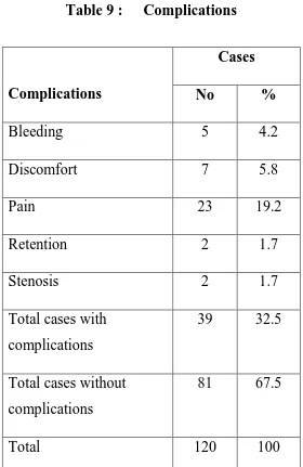
COMPLICATIONS:
1) Hemorrhage.
2) hematoma
3) Pain.
4) Retention of urine.
5) Wound infection.
6) Stenosis
7) Fistula formation.
CLOSED FERGUSON HEMORRHOIDECTOMY:
This is a surgical method developed by Drs. Ferguson and Heaton in 1952, which is a modification of Milligan-Morgan method
described above. On completion of excision of pile mass, the cut edges of the skin and mucosa are approximated with absorbable sutures. It has no advantage in terms of wound healing because of the high rates of suture
RESULTS OF THE STUDY
In this study, we have included 120 cases of haemorrhoids who attended government rajaji hospital, Madurai during the two year period
2010-2012.
THE STUDY GROUPS:
Group I : Sclerotherapy
Group II : Rubber band ligation
Group III : Hemorrhoidectomy
AGE INCIDENCE:
In this study the range of age included are 17-70 yrs and the majority
A : CHARACTERISTICS OF CASES STUDIED
Table 1 : Age distribution
Age group Cases
No %
Upto 20 years 7 5.8
20- 29 years 16 13.3
30-39 years 33 27.5
40- 49 years 30 25
50 – 59 yrs 22 18.3
60 yrs & above 12 10
Total 120 100
Range 17 - 70 years
Mean 40.8 years
SEX INCIDENCE:
It is found in this study, that the incidence is higher in males than females.
[image:79.595.112.484.483.739.2]
Table – 2: Sex Distribution
Sex
Cases
No %
Male 79 65.8
Female 41 34.2
Total 120 100
79 41
FAMILIAL PREDISPOSITION:
In this study group, only three patients had family h/o of haemorrhoids. In those cases, their father had h/o surgical treatment taken
for haemorrhoids. It is not usually hereditary, may be related to the common diet habits.
CLINICAL PRESENTATION:
In this study, the symptoms more prevalent are bleeding , prolapse and constipation..
BLEEDING: It is the most common presenting feature in most of the
cases. Among 120 cases, 70 had presented with bleeding.
CONSTIPATION: It is common associated symptom in patient presenting with haemorrhoids. In this study, 69 cases had this symptom.
PROLAPSE: This is the annoying symptom for which most of the patients
turn up for treatment . It is present in 42 no of cases.
MUCOUS DISCHARGE : This is often associated with prolapsed of pile
mass. It is noted in 20 no of cases.
Table – 3
[image:81.595.124.500.368.784.2]CLINICAL FEATURES.
Table 3 : Clinical presentations
Clinical presentations
Yes No
No % No %
Bleeding 70 58.3 50 41.7
Prolapse 42 35 78 65
Constipation 69 57.5 51 42.5
Anal Pain 26 21.7 94 78.3
Mucous 20 16.7 100 83.3
70 42
69 26
20
0 10 20 30 40 50 60 70
Number of cases