COMPARATIVE STUDY OF LIQUID BASED CYTOLOGY
AND PAP SMEAR IN CANCER CERVIX SCREENING
Dissertation submitted to
THE TAMILNADU DR. M.G.R. MEDICAL UNIVERSITY
in partial fulfilment of the requirements for the award of
M.D. DEGREE (BRANCH-II) IN
OBSTETRICS & GYNAECOLOGY
DECLARATION
I hereby declare that the dissertation entitled “COMPARATIVE STUDY OF LIQUID BASED CYTOLOGY AND PAP SMEAR IN CANCER CERVIX SCREENING” is a bonafide research work done by me at Coimbatore Medical College and Hospital, Coimbatore during the period from June 2011 - June 2012 under the guidance and supervision of
Dr.MURUGALAKSHMI,M.D.,DGO., Assistant Professor, Coimbatore Medical College and Hospital, Coimbatore. This dissertation is submitted to
The Tamilnadu Dr. M.G.R. Medical University, towards partial fulfilment of the requirement for the award of M.D Degree (BRANCH-II) in
OBSTETRICS & GYNAECOLOGY
I have not submitted this dissertation on any previous occasion to any University for the award of any Degree.
Place: Coimbatore Dr. S. RAJESHWARI
CERTIFICATE
This is to certify that the dissertation entitled “COMPARATIVE STUDY OF LIQUID BASED CYTOLOGY AND PAP SMEAR IN CANCER CERVIX SCREENING” is a record of bonafide work done by
DR. S. RAJESHWARI, Post Graduate Student in the Department of
Obstetrics & Gynaecology Coimbatore Medical College and Hospital, Coimbatore, under the supervision of Dr. M. SWATHANDRADEVI, M.D., DGO., Professor and Head, Department of Obstetrics & Gynaecology, Coimbatore Medical College and Hospital, and under the guidance of Dr. MURUGALAKSHMI M.D., DGO., Assistant Professor, Coimbatore Medical College and Hospital, in partial fulfilment of the requirement of the regulations of the Tamilnadu Dr. M.G.R Medical University towards the award of M.D. Degree (Branch II) in Obstetrics & Gynaecology.
Dr. R. VIMALA., M.D., (Path) Dr. SWATHANDRADEVI M.D., DGO
The Dean, Professor, & Head Coimbatore Medical College, Department of O &G
Coimbatore . Coimbatore Medical College
Guide
Dr. Murugalakshmi M.D.,DGO
Assistant Professor Department of O &G
ACKNOWLEDGEMENT
I sincerely thank Prof. Dr.Vimala M.D., Dean, Coimbatore Medical College - Coimbatore for allowing me to conduct this study.
I express my sincere and deep sense of gratitude to Dr.M.SwathandraDevi M.D., D.G.O., Professor and Head, Department of Obstetrics and Gynaecology, Coimbatore Medical College, Coimbatore for the valuable guidance, expert advice, and constant encouragement, given to me in the preparation of this dissertation.
I am extremely grateful to Dr.Murthy M.D., Professor and Head, Department of Pathology for his support and guidance in doing this dissertation. I am also grateful and thankful to Dr.Sundari, Dr.Revathy, Dr.Sumathy, Dr.VatsalaDevi and Dr.Usha, Professors, Department of Obstetrics and Gynaecology for their constant support.
I extend my heartfelt gratitude to my guide and teacher Dr.Murugalakshmi, M.D., D.G.O., for her guidance in completing this dissertation.
I thank all the Assistant Professors of the Department of Obstetrics and Gynaecology for their support throughout this study without which this work would never have been possible.
CONTENTS
PAGE NO.
1.
INTRODUCTION
1
2.
AIM OF THE STUDY
4
3.
MATERIALS AND METHODS
5
4.
REVIEW OF LITERATURE
10
5.
RESULTS
40
6.
DISCUSSION
63
7.
SUMMARY
72
8.
CONCLUSION
75
9.
BIBLIOGRAPHY
10.
ANNEXURE
•
PROFORMA
i
•
CONSENT FORM ii
•
KEY TO MASTERCHART
iii
•
MASTER CHART
iv
LIST OF ABBREVIATIONS
ACOG American College of Obstetricians and Gynaecologists
ACS
American Cancer SocietyAGC
Atypical Glandular CellAHRQ
Agency for Health Care Research and QualityAIS
Adenocarcinoma in situASC
Atypical Squamous CellASC-H
Atypical Squamous Cell –cannot exclude High grade lesionASC-US
Atypical Squamous Cell – Undetermined SignificanceAUB
Abnormal Uterine BleedingCIN
Cervical Intraepithelial NeoplasiaDNA
Deoxy ribonucleic acidFDA
Food and Drug AdministrationHIV
Human Immuno Deficiency VirusHPV
Human Papilloma VirusHSIL
High Grade Squamous Intraepithelial lesionLSIL
Low Grade Squamous Intraepithelial lesionRb
Retinoblastoma geneSCJ
Squamo Columnar JunctionSCC
Squamous Cell CarcinomaSTD
Sexually Transmitted DiseaseTZ
Transformation ZoneUS
United StatesVIA
Visual Inspection after Acetic acidVIAM
Visual Inspection after Acetic acid with MagnificationVILI
Visual Inspection after Lugol’s IodineWHO
World Health OrganisationLIST OF TABLES
Table no.
Title
Page no.
1. HPV prevalence among women 14 2. Comparison of cervical cytology screening 28 3. Comparison of cytology classification system 32 4. The 2001 Bethesda system: Epithelial cell abnormalities 34
5. Age wise cases 40
1
ABSTRACT Objective:
Cervical cancer is a preventable disease. Screening for precancerous
lesion of cancer cervix is one of the secondary prevention activities. In 1940’s
Pap smear was introduced for cancer cervix screening. The imperfect sensitivity
and variable smear quality of Conventional Pap collection lead to the
development of Liquid Based Cytology (LBC). The aim of this study is to
evaluate liquid based cytology and to compare the sensitivity of liquid based
cytology with conventional Pap smear.
Methodology:
This study was carried out on 100 randomly selected women of aged
21-60 years who presented with symptoms of vaginal discharge, lower abdomen
pain, post coital bleeding, irregular periods and with cervical lesion from the
Gynaecology outpatient department at CMCH between June 2011 – June
2012.Specimen was collected for both conventional pap smear and liquid based
cytology then colposcopic guided biopsy was performed in all 100 cases. By
using biopsy proven CIN cases as the gold standard the sensitivity of both the
cytology method was obtained.
2
Results:
In our study LBC showed abnormality in 32 cases and in Pap smear it
was in 17 cases. The rate of Unsatisfactory smear was low in LBC than Pap
smear (1% vs 6%). The rate of prediction of infectious agent was more with
LBC (14%) than Pap smear (3%). It is noted that Low grade squamous
intraepithelial lesion (16%) and High grade squamous intraepithelial lesion
(10%) prediction was more with LBC than Pap smear (LSIL 5%, HSIL 1%) and
it was statistically significant. The prediction of Atypical Squamous cell (ASC)
category was more with Pap smear than LBC (8% vs 2%).The false negative
rate of Pap smear was more than LBC (49% vs 3%).The sensitivity for detection
of a histologically proven lesion is significantly higher in LBC compared to
Conventional Pap smear(96% vs 51%).
Conclusion:
It is inferred from this study on comparing LBC with Conventional Pap
smear relative to histology, Liquid Based Cytology is better than Conventional
Pap test in detecting preinvasive cervical lesions. Liquid Based Cytology
showed increased sensitivity for the prediction of cytologic abnormality and
decreased false negative rate compared to Pap tests.
KEY WORDS:Cancer cervix, Liquid Based Cytology, Conventional Pap
INTRODUCTION
Cervical cancer is the most common gynaecologic neoplasm. It is the leading death causing cancer among women of developing country. Recent estimates point out that every year 134420 women are diagnosed with cervical cancer and 72825 die from the disease in India (24). Cervical cancer is an important public health problem because of its high mortality.
Cervical cancer is a preventable disease. Prevention lies mainly in early detection. The risk of cervical cancer is mainly due to persistent HPV infection, older age, and other demographic, behavioural, and medical risk factors, that vary among population worldwide.
Screening is a secondary level of prevention which is done to detect or rule out disorders at an early stage in healthy person. Screening and treatment of Precancerous lesion of cervical cancer is one of the secondary prevention activities.
In 1940s, cervical cancer screening was first made possible by the introduction of the Papanicolaou (Pap) smear. Population – based screening with Pap smear has been used to detect precancerous lesions since 1960 which leads to a decline in the number of invasive cervical cancer and also higher rate of detection of pre invasive lesions of cervix.
The Pap smear technique is simple and inexpensive but the sensitivity of a single Pap test is limited which varies between 50 – 70%. The Pap test has high false–negative rate which is associated with several factors of both sampling and interpretation. Uneven cell distribution, overlapping cells, blood, or inflammation in conventional Pap smear makes its interpretation difficult.
The imperfect sensitivity and variable smear quality of conventional Pap collection have driven the development of Liquid – Based Cytology (LBC) as a substitute to conventional Pap smear.
The most important benefit of LBC consist of, a probable increase in prediction of high – grade cervical intra epithelial neoplasia (CIN), reduction in the number of unsatisfactory smears, most of the collected cellular material is available for laboratory processing, and the residual specimens can be used for HPV DNA testing .
The expenditure of LBC method is significantly more when compared to conventional Pap method. In developing countries where the resources are inadequate the usage of LBC method for cervical cancer screening programs has a particular concern due to its high price. While comparing LBC and Pap method, if considerable improvement in the performance of LBC method is not achieved, its usage for cervical cancer screening program may not be justified.
The aim of this study is evaluation of liquid based cytology and comparison of sensitivity between conventional Pap smear and Liquid Based Cytology. The performance of both techniques were compared and validated by a histological follow up.
Aim of the Study
Aim of the Study
Aim of the Study
AIM OF THE STUDY
1. To evaluate Liquid Based Cytology.
2. To compare the sensitivity of Liquid Based Cytology with Conventional Pap smear.
Materials and Methods
Materials and Methods
Materials and Methods
Materials and Methods
MATERIALS AND METHODS
DESIGN OF THE STUDY
Prospective randomised evaluation of diagnostic tests
METHODOLOGY AND TECHNIQUES
Sample size – 100 subjects
Subjects are randomly selected from the out patients attending the Gynaecology department of Coimbatore Medical College Hospital between June 2011 and June 2012.
Inclusion criteria
Women aged 21 to 60 years presenting with complaints of abnormal vaginal discharge, irregular periods, lower abdomen pain, post coital bleeding, or abnormal cervical findings on per speculum examination were studied randomly.
Exclusion criteria
Women aged less than 21 years, pregnant women, women who underwent hysterectomy or prior treatment for CIN, and proved cancer cervix cases.
After a detailed history and getting informed consent, thorough clinical examination was done, specimen was collected for cytology, and colposcopic guided biopsy was performed in all 100 cases.
LABORATORY TESTING
By putting the patient in dorsal position, per speculum examination was done, after visualisation of the cervix. Conventional Pap smear was collected by using the Ayer’s spatula and the cells were smeared on the glass slide. The glass slide was fixed immediately with 1:1 mixture of 95% ethyl alcohol and ether, and then the slide was sent to the laboratory for processing, where the smear was stained with papanicolaou stain and was reported.
Then another cervical sample was taken with a cytobrush .The head of the brush was detached and placed into the liquid – Prep collection vial which was capped, labelled, and sent to the laboratory for processing and analysis. In the lab the specimen was mixed well using a vortex for 5 to 20 seconds and the entire contents were poured into a clean 15 ml centrifuge tube and it was centrifuged for 10 minutes at 1500 RPM. After centrifugation supernatant fluid was discarded and to the residual pellet cellular base (this component is for encapsulation and adherence of processed cells on a clean glass slide) was added .The specimen was mixed using vortex and pipette 50micro litre onto glass slide. The slide was allowed to dry, stained and was analysed.
In Liquid prep slide the cells are distributed over a small circular area which is 13 mm. In Pap method, as the smear is manually prepared, the thickness of the smear varies. In LBC method, debris, mucus, blood and cell overlap are largely eliminated. The infectious organisms are clearly visible, and air – drying artefacts are not seen. The number of leucocytes is low in LBC method. Hence the epithelial cells and the abnormal cells are easily visualised.
After collecting cytology specimens, colposcopic guided biopsy was performed in all women.
COLPOSCOPY
Colposcopy is an outpatient procedure. It allows examination of the lower genital tract with a microscope. The colposcope consists of a stereoscopic viewing system with magnification settings ranging from 3 to 40 fold attached to a freely movable stand. A high –intensity halogen light provides illumination. Use of a green (red- free) light filter emphasizes contrast by causing the colour red to appear black aiding the examination of vascular patterns.
Steps of Colposcopic examination
Patient was put in dorsal or lithotomy position. Colposcopy was placed one foot from vulva. Cervix was visualised by inserting a Bivalve speculum and colposcope was focused on the cervix. Cervix was cleaned with saline to remove the mucous, 3% acetic acid and lugol’s iodine were applied one after
the other, and findings were noted. After documentation of colposcopic findings punch biopsies were obtained from acetowhite and iodine negative areas. When there was no abnormality, cervical biopsy was taken from the anterior cervical lip close to the squamocolumnar junction.
Cytology reporting
Reporting of cytology examination in our study was done using the Bethesda
S
ystem’s 2001 modification. The abnormality in cytology specimen is due to infection, inflammation, reparative changes, and epithelial cell abnormality. The epithelial cell abnormality includes low –grade (LSIL) squamous intra epithelial lesion, high- grade (HSIL) squamous intraepithelial lesion, and atypical squamous intraepithelial lesion.All cytology specimens were classified either as satisfactory or unsatisfactory. Unsatisfactory specimen was due to scanty cells, improper fixation of slide, debris, mucus, blood cells, inflammation, and other factors which obscure 75% or more of epithelial cells. The specimen was considered to be satisfactory if they contain 5000-10000 well preserved and well visualised squamous cells along with cells from transformation zone.
For the purpose of tabulation, cases whose smears showed infective organisms and all unsatisfactory smears in both cytology methods were considered as cases with normal reports.
In our study cervical biopsy reporting was given using the CIN terms. By using biopsy proven CIN as the “gold standard”, the sensitivity of both the cytology methods were obtained. The cut off points used for the cytology results were ASC-US or higher and for the histology CIN1or higher.
REVIEW OF LITERATURE
CRITERIA FOR SCREENING OF DISEASE (1)
The disease to be screened should fulfil the following criteria before it is considered suitable for screening:
The disease should be an important health problem.
The disease should have a latent asymptomatic stage.
There is a test that can detect the disease prior to its onset.
Facilities should be available for confirmation of the diagnosis.
There is good evidence that early detection and treatment reduces morbidity and mortality.
Cervical cancer screening matches these criteria quite well.
Compelling Reasons for Cervical Cancer Screening
Cervical cancer is the most common malignancy worldwide. Its high mortality makes cervical cancer an important public health problem. Early detection of cervical cellular changes and Cervical Intraepithelial Neoplasia (CIN) followed by treatment will reduce the risk of developing cancer. Epidemiological data show that organized screening with Pap smear has had a major impact on both morbidity and mortality from cervical cancer (2)
Cervical Anatomy
The lower part of uterus is cervix. The lining epithelium of the endocervix is columnar epithelium, and the ectocervix is lined by squamous
epithelium (3). The point where they join is called the Squamo Columnar Junction (SCJ).
The Squamo Columnar Junction
The SCJ is a dynamic point that changes in response to puberty, pregnancy, menopause, and hormonal stimulation. At menarche, the production of estrogen causes the vaginal epithelium to fill with glycogen. Lactobacilli act on the glycogen to lower the pH, stimulating the sub columnar reserve cells to undergo metaplasia (3).
Squamous metaplasia is a normal process and occurs adjacent to the original SCJ, creating a zone of metaplastic epithelium termed the Transformation zone (TZ). It extends from the original SCJ to the physiologically active SCJ. Gland openings and nabothian cysts mark the original SCJ. Nearly all cervical neoplasia, develops within the TZ, usually adjacent to the new SCJ (4).
Normal Transformation Zone
The original squamous epithelium of the vagina and ectocervix has four layers - the basal layer, the parabasal layer, intermediate layer and the superficial layer. The superficial layer includes five to eight rows of flattened cells with small nuclei and a cytoplasm filled with glycogen. The nucleus becomes pyknotic, and the cells detach from the surface (exfoliation). These cells form the basis for Papanicolaou (Pap) testing (5).
Columnar Epithelium
It has a single layer of columnar cells with mucus at the top and a round nucleus at the base.
Metaplastic Epithelium
Metaplastic cells are normal cells without nuclear atypia. Atypical metaplasia with abnormal nuclear changes is a precursor of dysplasia and malignancy. PH changes, hormonal effect, infection and certain mutagens cause
atypical metaplasia (6).
Pre invasive cervical cancer or CIN
In 1947 the perception of preinvasive cervical lesion was introduced. The epithelial cell abnormalities which were similar to invasive cervical lesion but restricted to the epithelial cellular layer were identified (7).
Cervical dysplasia is a cytological term to describe cells resembling cancer cells. Cervical intraepithelial neoplasia (CIN) refers to the histopathological description in which a part or the full thickness of the stratified squamous epithelium is replaced by cells showing varying degrees of dysplasia, with intact basement membrane (6)
Dysplasia
Mild dysplasia (CIN-1)
The undifferentiated cells are confined to lower one - third of the epithelium. Mild dysplasia due to infection is often seen in sexually active
young women. CIN-1 is lately described as low grade squamous intraepithelial lesion (LSIL) according to Bethesda classification.
Moderate dysplasia (CIN –II)
The undifferentiated cells occupy lower 50-75% of the epithelial thickness. The cells are mostly intermediate with moderate nuclear enlargement, hyper chromasia, irregular chromatin and multiple nucleations.
Severe dysplasia and carcinoma in situ (CIN –III)
In this grade of dysplasia, the entire thickness of epithelium is replaced by abnormal cells but the basement membrane is intact and there is no stromal infiltration. The cells are mostly parabasal with increased nuclear – cytoplasmic ratio.
CIN II and CIN III are described as high grade squamous intraepithelial lesions (HSIL) according to latest Bethesda classification (6).
Cancer cervix
If the dysplastic cells occupy the entire thickness of the epithelium and the basement membrane is not intact, invading the stromal tissues or the adjacent organs, then the disease is called invasive cervical cancer or cervical cancer.
Squamous cell carcinoma of cervix (SCC)
SCC is the most common variety of invasive cancer in the cervix. Histologically, variants of SCC include large cell keratinizing, large cell nonkeratinizing and small cell types.
Adenocarcinoma of cervix
About 80% of cervical adenocarcinomas are made up of cells of the endocervical type with mucin production.
Aetiology and Pathogenesis
Human Papilloma Virus (HPV)
The most important initiating factor in cervical dysplasia and carcinogenesis is infection with HPV. It infects epithelial cells exclusively; infectiousness depends upon desquamation of infected cells (8). HPV infection is sexually transmitted (10).
There are 14 high risk HPV subtypes; two of the high risk subtypes, 16 and 18, are found in up to 70% of cervical carcinoma (9).
Table – 1: HPV prevalence among women (22)
HPV prevalence among women with Percent Normal cytology HSIL/CIN2/CIN3 LSIL/CIN1 Cervical cancer 6 56 29.4 82.5
The mechanism by which HPV affects cellular growth and differentiation is through the interaction of viral E6 and E7 proteins with tumour suppressor
genes p53 and Rb, respectively, resulting in unregulated cellular proliferation(11).
Risk factors for Cervical Neoplasia
Mean age for cervical cancer in the United States is 47 years, cervical cancer peaks at 35 to 39 years and 60 to 64 years of age (5).
Coitus before the age of 18 years
Multiple sexual partners.
Delivery of the first baby before the age of 20 years.
Multiparity
Poor personal hygiene
Poor socio economic status.
Sexual contact with uncircumcised partners as the incidence of HPV is low in circumcised men.
Smoking and drug abuse.
Women with STD, HIV infection, herpes simplex virus – 2 infection, HPV infection have a high predisposition to cancer
Immunosuppressed individuals.
Women with pre invasive lesions.
Women who do not come for regular health check-up and Pap tests. Combined oral contraceptive pills use over 8 year period can cause adenocarcinoma of the endocervix (6).
Evaluation
(5)Vaginal bleeding is the most common symptom occurring in patients with cancer cervix. Most often, this is post coital bleeding, but it may occur as irregular or post menopausal bleeding as well. Patients with advanced disease may present with a malodorous vaginal discharge, weight loss, or obstructive uropathy. In asymptomatic women, cervical cancer is most commonly identified through evaluation of abnormal cytologic screening tests.
All women should have a general physical examination including evaluation of the supraclavicular, axillary and inguinofemoral lymph nodes to exclude the presence of metastatic disease. On pelvic examination, cervix and vaginal fornices should be visualised for doubtful areas.
Rectal examination should be done to found out cervical consistency, size, and paramatrial extension of disease (5).
Prevention and Early detection of cancer cervix (12).
Prevention of cervical cancer consisting of
Creating awareness about the risk factors among women.
Promoting practice of safe sex.
Use of condoms to prevent STDs.
Life style modification.
Screening for and early treatment of premalignant lesions
HPV vaccines.
HPV vaccination
HPV infection is mostly asymptomatic; hence its identification is not easy. HPV vaccination leads to prevention of cervical carcinoma and cervical precursor lesions. Bivalent vaccines against HPV – 16 and 18 and quadrivalent vaccines (HPV-6, 11, 16, 18) have been developed to protect against HPV infection. Duration of efficacy is 3.5- 5 years. Age of administration is between 9-13 years, must be given before the onset of sexual activity. Three doses at 0, 2 and 6 months should be given. Screening with cytology must be continued after vaccination as per schedule (12).
Screening for Cervical Intraepithelial Neoplasia
It is technically easy to screen for cervical cancer since.
The cervix can be easily visualized and tests performed.
Cervical cancer goes through premalignant changes, and if the diagnosis is made at this stage, cancer can be prevented.
There is a lag time of 10-20 years before the disease progresses from pre invasive to invasive diseases.
Several screening modalities are available, but cytology is most widely used. In resource – poor settings where cytological screening is not easily available other methods are used (12).
Methods used for Cervical Cancer screening (12)
Cytology
o Conventional cytology (Pap smear)
o Liquid – based cytology (LBC)
Naked eye visual inspection or Down staging
Visual Inspection after acetic acid (VIA)
Visual Inspection after acetic acid with magnification (VIAM)
Visual Inspection after lugols iodine (VILI)
Cervicography
HPV DNA testing
Investigational strategies
o Polar probe
o Laser – induced fluorescence
Cervical Cytology
The abnormal cells of cervical neoplasia exfoliate and can be collected by scraping the cervix and staining the smear. The Pap test detects most cervical neoplasia during premalignant phases when treatment outcome is optimized. Pap test was used for cervical cancer screening in the 1950s. Its usage resulted in major decline in the incidence and mortality from cancer cervix (14). In the developed world cytology based method of cervical cancer screening has been used, which has resulted in the decline of incidence and mortality from cervical cancer (25). In developing countries due to inadequate financial and human resources alternative methods of cervical screening which is more cost effective is being used.
Efficacy of Cytology based Screening
The Pap tests specificity is consistently high, approximating 98 percent. However, estimates of its sensitivity are lower and more variable. A recent meta-analysis found a sensitivity of 51 percent for detection of any grade of CIN by a single Pap test (15).
Women should be aware of the imperfect sensitivity of the Pap test and the need for periodic screening to compensate.
False negative Pap tests may result from
Sampling error – in which abnormal cells are not present in the Pap test.
Screening error – in which the cells are present but missed by the screener.
Interpretation error – in which abnormal cells are misclassified as benign(16).
Preparation for Cytological Screening
Pap tests should be scheduled to avoid mensturation. Patient should abstain from vaginal intercourse, douching, and medicinal or contraceptive cream preparation for a minimum of 24 to 48 hours before a test. Treatment of cervicitis or vaginitis prior to Pap testing is optimal.
Complete clinical information includes documenting the date of last menstural period or pregnancy, exogenous hormone use, menopausal status, and past history of abnormal bleeding or abnormal Pap test results, dysplasia or cancer.
Intra uterine devices can cause reactive cellular changes and their use should be noted. Adequate visualization of the cervix is essential for detection of gross lesions and identification of the SCJ.
Sampling of the transformation zone is paramount to the sensitivity of the Pap test (16).
Methods of Cytological screening
Conventional cytology (Pap smear)
The cervix is visualized after placing a bivalve speculum, and the ectocervix is scraped using an Ayer’s spatula and endocervix with a cytobrush. The cells are smeared on a glass slide and fixed in a 1:1 mixture of 95% ethanol and ether.
They are stained using papanicolaou’s stain and examined.
Efficacy of Pap smear
The Pap smear has been found to have a poor sensitivity and high false negative rate though the specificity of the test is high (12).The main drawback of Pap smear was inadequate shift of cells from the collecting device to the slide, obscuring blood, mucus and variable thickness of smear. The occurrence of false-negative and unsatisfactory Pap smear has prompted the development of LBC and automated screening devices (18).
Liquid – Based Cytology (LBC)
LBC has been developed to address the limitation of Pap smear and represents the first major change in preparation method for cervical screening
samples. The LBC corresponds to a sampling where cells are put in suspension in a liquid transport medium. The cytology specimen is taken either with a cyto brush or by using both the spatula and endocervical brush. The specimen is then rinsed in the bottle containing a fixative fluid, or the tip of the cyto brush is cut and left in the bottle. In LBC the smearing is done in the laboratory and not manually as in Pap smear where the physician prepares the smear. At present, two methods of LBC – Thin Prep and Sure Path, which use automats, were validated by Food and Drug Administration and are, used frequently (18). Although LBC has been available for less than a decade, researchers estimate that approximately 80% of Pap tests now performed in the United States is LBC.
CURRENT METHODS USING LBC TECHNOLOGY:
• Thin prep
• Sure path (Auto Cyte PREP or CytoRich LBC)
Sure path allows for the use of all three collection devices; the spatula, the broom, and the endocervical brush (14) but with modified device tips that can be broken off and sent to the laboratory in the liquid medium. The liquid medium is ethanol – based fluid which maintains cell morphology and nuclear features. In the Sure path method the tip of the endocervical brush is cut and put in the collection bottle containing the liquid medium. By doing so all the scraped cervical cells attached to the endocervical brush has been used for processing and smearing. In the laboratory the bottles are
centrifuged, processed with automated Prep stain machine and smears are prepared.
Thin prep requires immediate and vigorous agitation of the chosen collection device in the liquid medium, after which the device is discarded. In this method by means of T2000 or T3000 machine processing of liquid medium is
done. The slides are processed independently with the T2000 device whereas with
the totally automated T3000 device around 80 slides can be processed in a single
rotation. The slides are then stained and microscopically assessed the same way as in Pap method (17).
In LBC the area of the slide covered with cells are less than with conventional smear. However obscuring blood, mucus, debris and cellular overlap are largely eliminated. The abnormal cells that might be few in number clustered and obscured on a conventional smear will be randomly and evenly distributed over the area of the LBC slide and thus be more visible for detection(16).Thin Prep and AutoCytePrep specimens have been examined independently, from the same specimens, and there was a high degree of concordance between the two techniques (23, 24).
Advantages of LBC
• Better sensitivity and specificity in view of the fact that sampling error is reduced.
• Lower rate of unsatisfactory cervical samples.
• Lesser time for interpretation
• HPV DNA testing is done from the remaining liquid medium (18).
• Enhanced cytoplasmic and nuclear details.
Comparison of LBC & Conventional Pap smear
Liquid based cytology now accounts for most of the Pap test performed in the US. Both LBC products are FDA – approved to claim a 65- percent increased detection rate of HSIL compared to conventional smear
LBC is more sensitive and accurate for the detection of both squamous lesions and adenocarcinomas of the cervix (20, 21).
Since 1950 the Pap method is being used for cancer cervix screening. Its usage resulted in decreasing the occurrence of cancer cervix to 79% and death due to cervical cancer was reduced to 70% (5).The Agency for Health Care Research And Quality(AHRQ), undertook a literature review of conventional cytology testing techniques and compared them with newer technologies designed to reduce the false-negative rate (26). For this project, five reports were analyzed, and found out that the sensitivity of conventional Pap method in predicting preinvasive lesions was 51%.
The accurateness of cervical cytology evaluation was reviewed in three reports. According to those reports, sensitivity of the Pap method in predicting CIN 2 or higher lesions was between 47% and 62% and the specificity was between 60% and 95 %( 27, 28, and 29). Previously it had been widely believed that sensitivity of the Pap test was in the 80% range (31).Recommendations for Pap test screening was based on this perceived 80% sensitivity. Every year about
30% of fresh carcinoma cervix cases occur in women who underwent screening with Pap method, mainly because of sampling, fixation and interpretation error(30).
It is obvious that improvement in the Conventional Pap test techniques necessary. False-negative errors occur in sampling, preparation, and interpretation. Sampling errors occur because a lesion is too small to exfoliate cells or the device used did not pick up the cells and transfer them to the glass slide. Preparation errors may occur because of poor fixation on the glass slide, leading to air – drying and an inability to interpret the results. The slide may also be too thick and obscured by vaginal discharge, blood, or mucus. The thick slide also leads to poor fixation because the fixative does not penetrate the cell sample. Interpretive errors occur when the slide contains diagnostic cells that the screening technician did not identify.
Using a Liquid-Based medium to collect the cytologic sample and preserve the collected cervical cells can alleviate sampling and preparation errors. In LBC the cells are distributed uniformly in a smaller area of the slide.
The AHRQ reported that liquid-based cytology assessment improved the sensitivity of the Pap test to the stated goal of 80%.With this technique,80% to 90% of cells are transferred to the liquid media, as compared with the only 10%-20% transferred to the slide with conventional cytologic testing. In addition, using liquid-based media eliminates air- drying. The cells are retrieved from the vial by passing the liquid through a filter, which traps the larger
epithelial cells, separating them from the small blood and inflammatory cells. This process leads to a thin layer of diagnostic cells properly preserved and more easily interpreted by the cytologist. This technique reduces by 70% to 90% the rate of unsatisfactory samples encountered with conventional cytologic testing (32).
Down staging
Down staging of cervical cancer is defined as a process of using clinical approaches for early detection of the diseases. In this process, trained paramedical personnel do a speculum examination, identify any visible abnormalities, and refer cases with suspicious appearance to centres with facilities for diagnosis and management. The WHO has introduced down staging for developing countries where screening methods are not universally available (13). But the sensitivity and specificity of this method is poor (33).
Visual Inspection after Acetic Acid (VIA)
As it is not easy to practise cytology screening tests in a set up with inadequate resources, other methods of screening have been introduced. Acetic acid when applied to the cervix coagulates the mucus and the areas of cells with increased chromatin density. Areas of CIN, neoplasia, HPV- related changes are aceto white. This method has been extensively studied in developing countries and is currently recommended by the WHO as the primary screening test for developing countries. The disadvantage of this test is the high false –positive rate, resulting in large number of referral (12).
VIA with Magnification
After application of acetic acid, cervix is visualized under magnification using a hand held magnifying lens. Number of false positives can be reduced with this technique.
Visual Inspection after Lugol’s Iodine (VILI)
On application of iodine, normal cervical epithelium which is rich in glycogen stains mahogany brown (Schillers test).The abnormal areas, columnar epithelium and areas lined by immature metaplastic epithelium do not contain glycogen and appear mustard yellow. This method has similar advantages and disadvantages as the VIA.
Cervicography
Photograph of the cervix are taken under magnification before and after application of acetic acid and are read by colposcopist.
HPV – DNA Testing
Use of HPV DNA testing for primary screening, instead of Pap smear, is under study. The test is expensive and currently not recommended for routine primary screening in place of cytology. HPV DNA testing is highly sensitive for diagnosis of High grade squamous intraepithelial lesion (HSIL) (12).
Polar Probe
The normal and abnormal tissues differ in its tissue impedance to electrical stimulation (18). Polar probe technique is based on this information.
Laser Induced Fluorescence
It is shown that, the chemical and morphological composition of individual tissues can provoke endogenous tissue fluorescence on low powered illumination. The detection of this spectroscopic difference can be used to differentiate normal and abnormal tissues (18).
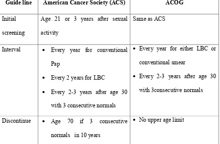
Screening Guide Lines
Recommendations for cervical cancer screening are slightly conflicting. The American Cancer Society (ACS) updated recommendations of 2002 states that
- By using Pap method screening is done every year. On using LBC two yearly screening should be done.
- Screening has to be done either at 21years of age or in 3 years of the beginning of sexual action. At 70 years of age, if the Pap reports were normal for the precedent 10 years screening has to be stopped.
- Screening after hysterectomy for benign disease is not necessary (14). The American College of Obstetricians and Gynaecologists (ACOG) differs from the ACS recommendations by stating that
- Women younger than the age of 30 should undergo cervical cytology screening yearly and in those older than the age 30 if there is no associated risk factors screening should be done once in 2 to 3 years using either LBC or conventional Pap testing furthermore. According to ACOG, women who are 30 and above ideally should undergo both
cytology and HPV DNA testing. If results are negative using this combination, a woman can be retested at only 3 year intervals (19)
[image:45.595.68.512.271.562.2](Table-2). The negative predictive value of this combination test exceeds 99% (5). In women younger than 30 years, the high prevalence of high risk HPV infection makes this strategy too nonspecific for use.
Table – 2: Comparison of Cervical Cytology Screening
Guide line American Cancer Society (ACS) ACOG
Initial
screening
Age 21 or 3 years after sexual
activity
Same as ACS
Interval • Every year for conventional
Pap
• Every 2 years for LBC
• Every 2-3 years after age 30
with 3 consecutive normals
• Every year for either LBC or
conventional smear
• Every 2-3 years after age 30
with 3consecutive normals
Discontinue • Age 70 if 3 consecutive
normals in 10 years
• No upper age limit
Screening guide lines for countries with inadequate resources
The median age of cervical cancer diagnosis is in the mid to late forties approximately a decade later than CIN (16), carcinoma cervix mostly occurs at the age of 35 to 39 years and 60 to 64 years. Hence in developing countries
where resources are inadequate screening programs should involve women in fourth decade of life.
Pre invasive cervical lesions like CIN predispose to cancer cervix; therefore, compared to frequent screening as per protocol, occasional screening also be able to decrease the prevalence, sufferings and death from carcinoma cervix. Cervical cancer screening done once in 10 years or once in a patient’s life time has a major effect in reducing its incidence. Hence screening programmes should focus on screening women of high risk category than on regularity (34). In developing countries, with inadequate funds, the aim of screening should be to include all women of high risk category once in her life time around her 40 years of age (13).
REPORTING OF CERVICAL CYTOLOGY TESTING
Bethesda System -2001
In the year 1988, in Bethesda, Maryland, the first National Cancer Institute (NCI) workshop was conducted, where the Bethesda system for the reporting of cytology testing was proposed (5). Standardization of cervical cytology reporting took place with the development of the Bethesda system. The terminology was refined in the Bethesda III system 2001.
According to this system, potentially premalignant squamous lesion falls into three categories (16).
• Atypical squamous cells (ASC)
• Low - grade squamous intraepithelial lesion (LSIL)
• High – grade squamous intraepithelial lesion (HSIL) The ASC category is subdivided into two categories
• those of undetermined significance (ASC-US)
• those in which high-grade lesion must be excluded (ASC-H) A comparison of the various terms as they relate is shown in Table – 3
The 2001 Bethesda System Cytology Report Components (16)
Specimen type
Conventional Pap test
Thin-layer liquid-based cytology
Specimen adequacy
Satisfactory for evaluation Unsatisfactory for evaluation
General categorization (optional)
Negative for intraepithelial lesion or malignancy Epithelial cell abnormality
Other findings that may indicate increased risk
Interpretation of results
Negative for intraepithelial lesion of malignancy
Organisms:
Trichomonas vaginalis
Fungal organisms consistent with Candida species Shift in flora suggestive of bacterial vaginosis
Cellular change consistent with herpes simplex virus Bacteria consistent with Actinomyces species
Other non-neoplastic findings (optional)
Reactive cellular changes (inflammation, repair, and radiation) Glandular cells post hysterectomy
Atrophy
Epithelial cell abnormalities
Squamous cell Glandular cell
Other:
Endometrial cells in a woman 40 years of age.
Table –3: Comparison of Cytology Classification System (5)
Bethesda System Dysplastic / CIN System Papanicolaou System
Within normal limits Normal I
Infection (organism should be
specified)
Reactive and Reparative changes
Squamous cell abnormalities Atypical squamous cells
1.of undetermined significance
(ASC-US)
2.exclude high – grade lesions
(ASC-H)
Inflammatory atypia (organism)
Squamous atypia, HPV atypia,
exclude LSIL, exclude HSIL
HPV atypia
II
II
Low grade Squamous intra
epithelial lesion (LSIL)
Mild dysplasia CIN I
High grade Squamous
intraepithelial lesion (HSIL)
Moderate dysplasia CIN 2
severe dysplasia CIN 3
Carcinoma in situ
III
IV
Squamous cell carcinoma Squamous cell Carcinoma V
Clinically, the key elements in Bethesda system are assessment of specimen adequacy and epithelial cell abnormality (16) (Table 4).
Specimen Adequacy
The significant and the quality assurance part of the Bethesda System is its assessment of specimen adequacy. Earlier versions of Bethesda included three categories of adequacy: Satisfactory, Unsatisfactory and a borderline category initially termed “Satisfactory but limited by” in 1991.
The 2001 Bethesda System eliminates the borderline category because of confusion among clinicians as to the appropriate follow-up for such findings and also due to variability in reporting “Satisfactory but limited by” among laboratories. To provide a clearer indication of adequacy, specimens are now designated as “Satisfactory” or “Unsatisfactory”.
The satisfactory smear is one which shows abnormal glandular and epithelial cells. In an adequate Pap specimen at least 8000 to 12000 well-preserved and well visualized squamous epithelial cells should be there. In liquid based preparation 5000 well – visualized /well preserved squamous cells will do. For both conventional smears and liquid based preparations adequacy of Transformation zone part requires a minimum of 10 well-preserved endocervical or squamous metaplastic cells. If more than 75% of squamous cells in the smear is masked by blood and mucus the specimen is labelled as unsatisfactory, as the cells are not identified (35).
Table – 4: The 2001 Bethesda System: Epithelial Cell Abnormalities
Squamous cell
Atypical squamous cells (ASC)
o of undetermined significance (ASC-US) o cannot exclude HSIL (ASC-H)
Low-grade squamous intraepithelial lesion (LSIL)
High -grade squamous intraepithelial lesion (HSIL)
Squamous cell carcinoma
Glandular cell
Atypical glandular cells (AGC)
o Endocervical, endometrial, or not otherwise specified
Atypical glandular cells, favour neoplastic
o Endocervical or not otherwise specified
Endocervical adenocarcinoma in situ (AIS)
o Adenocarcinoma
Epithelial cell abnormalities: Significance
A cytology report is a medical consultation that interprets a screening test and is not a diagnosis. Thus a final diagnosis is determined clinically, often with results from histological evaluation (16).
Atypical Squamous Cells of Undetermined Significance
The most common cytologic abnormality ASC – US precedes the diagnosis of CIN 2 or 3, this risk approximates 5 percent. Three options for evaluation of ASC-US are HPV – DNA testing, colposcopy or repeat cytologies at 6 and 12 months, with referral to colposcopy if either of these is abnormal(20). Here cells are found in sheets or singly, nuclei 2 ½ - 3 times the size of an intermediate nucleus with uniform chromatin distribution. The frequency of ASC-US diagnoses should not exceed 2-3- times the rate of SIL (35).
Atypical Squamous Cells cannot exclude HSIL
10 percent of ASC is ASC – H. This describes cellular changes that do not fulfil criteria for HSIL cytology, histologic HSIL is found in 25 percent of these cases hence colposcopy is indicated for evaluation (20). Atypical squamous cells cannot exclude HSIL includes immature metaplastic cells with increased nucleo cytoplasmic ratios, slight nuclear membrane irregularities and nuclear size variability, suggestive of but not definitive for HSIL (35).
Low Grade Squamous Intraepithelial Lesion
This cytology result indicates the likely presence of HPV infection or low-grade neoplasia (CIN I), carries a 15 to 30 percent risk of CIN 2 or 3, LSIL encompasses the cytologic features of HPV infection and CIN I.
Colposcopy is indicated for evaluation. Increased nuclear detail, irregular nuclear membrane, and nuclei 3-4 times the size of intermediate nucleus, sharp irregular cytoplasmic cavitation (HPV effect) are characteristic of LSIL (35).
High–grade Squamous Intraepithelial Lesion and Glandular Abnormalities
HSIL, all glandular epithelial cell abnormality and suspicion of carcinoma should be evaluated by colposcopy. HSIL encompasses features of CIN 2 and CIN 3. Management of HSIL includes immediate diagnostic loop excision because colposcopy may miss a high grade lesion. Immature metaplastic type squamous cells showing increased nucleo cytoplasmic ratios (in excess of 50%) nuclear irregularities and coarse chromatin result in a diagnosis of HSIL (35).
Glandular Cell
Squamous neoplasia is the most common diagnosis found upon evaluation of Atypical Glandular Cell (AGC) cytology, but there is also a high risk of both endocervical, endometrial and other reproductive tract cancer (16).
Non Neoplastic Findings
These findings include Trichomonasvaginalis, candida species, Actinomyces species, herpes simplex virus, or flora consistent with bacterial vaginosis, and the reactive changes due to inflammation, repair and radiation(16).
Colposcopy
Colposcopy allows examination of the lower genital tract with a microscope to further evaluate abnormal Pap test results and visible epithelial abnormalities.
COLPOSCOPIC FINDINGS AND TERMINOLOGY
Colposcopically, the squamous epithelium of the cervix appears as a featureless, smooth, pale pink surface. The single cell layer columnar epithelium of the endocervix appears red and velvety due to the proximity of blood vessels.
Adequate visualization of the entire cervical SCJ ad upper limits of all lesions defines whether a colposcopic examination is termed satisfactory or unsatisfactory.
Aceto white Epithelium
Epithelium that turns white after application of 3 percent acetic acid is called aceto white epithelium. The components of the cell which contain proteins are coagulated on applying acetic acid and become white. The areas of immature squamous metaplasia, transformation zone, inflammatory, healing, and regenerative epithelium also become acetowhite on applying acetic acid (36). Low grade lesions are thin, less intense, and narrow. The margins are asymmetrical, geographic, fluffy, or angular with fine punctuation or mosaic; occasionally they are separated from the squamo columnar junction; and atypical vessels are not present in low-grade lesions.
In high-grade lesions thick, deep, opaque, grey white, acetowhite areas are present, coarse punctuation or mosaicism are seen in high grade lesions. The borders are regular and well demarcated; both cervical lips are involved in these
lesions. Atypical vessels are seen here. In high-grade lesions, CIN-3 is complex and occupies the cervical os (36).
On applying Lugol’s Iodine, normal glycogen rich squamous epithelium and mature metaplastic epithelium present in vagina and cervix take up the iodine and become brown. Dysplastic epitheliums do not have glycogen, and they do not take up iodine and they look mustard or saffron yellow in colour. Based on the colour of the epithelium, normal and abnormal areas in the transformation zone are clearly identified when the aceto white areas are not easily made out (36).
Lesion vascular patterns
The vascular patterns associated with abnormal epithelium include punctuation, mosaicism and atypical vessels.
Fine punctuation and mosaicism, which are created by narrow vessels and uniform intercapillary distances, typify low – grade lesions. A coarse pattern results from wider vessels, variable intercapillary distance, vessel diameter, and spacing. This indicates high grade lesions (16).
Atypical vascular patterns are characteristic of invasive cervical cancer and include looped vessels, branching vessels, and reticular vessels (5).
Several colposcopy grading systems that quantify various colposcopic signs have been developed to improve accuracy. Best known, the Reid Colposcopic Index (RCI) has a reported histologic correlation of 97 percent.
The RCI is based on four colposcopic lesions features (16). Peripheral margin, color, vascular patterns, and iodine staining. Each category is scored from zero to two and the summation provides a numeric index which correlates with histology.
FIG 1: Slide of Conventional Pap smear (Macroscopic)
FIG 2: Slide of LBC (Macroscopic)
FIG 4: Same Patient - LBC smear
FIG 5: Conventional PAP – Smear Pattern
FIG 7: LBC smear - containing 40 squamous cells. Unsatisfactory field
FIG 8: Trichomonas vaginalis in LBC smear
FIG 10: ASC-US in LBC
FIG 11: LSIL in LBC
FIG 13: Squamous
FIG 14: N
FIG 15: NORMAL COLPOSCOPIC PICTURE showing 1.External os 2.columnar epithelium 3. New SCJ 4.original SCJ
FIG 13: Squamous Cell Carcinoma in LBC
FIG 14: Normal Colposcopic picture
FIG 16: Invasive cancer on colposcopy
FIG 18: LOW GRADE LESION in colposcopy
vasive cancer on colposcopy FIG 17: Abnormal vascular pattern
FIG 18: LOW GRADE LESION in colposcopy FIG 19: HIGH GRADE LESION
FIG 17: Abnormal vascular pattern
FIG – 20: CERVICAL INTRAEPITHELIAL NEOPLASIA
CIN 1 - Undifferentiated cells occupy lower one third of the epithelium
CIN2 - Undifferentiated cells occupy lower two third of the epithelium
Results
Results
Results
RESULTS
The present study was carried out on 100 patients who are randomly selected from the Out Patient Department of Gynaecology at CMCH during the period of June 2011 to June 2012.After taking history and clinical examination cytology specimen was collected for both Pap test and Liquid Based Cytology, then colposcopic guided cervical biopsy was taken for all patients.
Table -5: Age wise cases
Age (years) Cases Percentage
21 – 30 35 35
31 – 40 40 40
41 – 50 17 17
51 – 60 8 8
It is noted in our study that out of total 100 cases, 35 cases
(35%) studied belonged to third decade of life, 40 cases (40%)
belonged to fourth decade of life, 17 cases (17%) belonged to fifth
decade of life, and 8 cases (8%) belonged to sixth decade of life.
Figure – 21: Age wise cases
17%
21 - 30 (yrs)
21: Age wise cases
40%
8%
30 (yrs) 31 - 40(yrs) 41 - 50(yrs) 51 - 60(yrs)
Table – 6: Age wise case distribution
Age(years) Normal
Abnormal
CIN SCC
21 – 30 31 4 -
31 – 40 26 13 1
41 – 50 8 7 2
51 – 60 2 5 1
In current study , 4 (13%) abnormal cases belonged to third decade of life, 13 (44%) abnormal cases belonged to fourth decade of life, 7 (24%) abnormal cases belonged to fifth decade of life and 5 (17%) abnormal cases belonged to sixth decade of life. Out of 4 squamous cell carcinoma cases, 1 case belonged to fourth decade of life, 2 cases belonged to fifth decade of life, and 1 case belonged to sixth decade of life. In our study 44% of abnormal cases and the earliest age of occurrence of squamous cell carcinoma belonged to fourth decade of life.
Figure-22: Age wise case distribution. 0 5 10 15 20 25 30 35
21 - 30(yrs) 31
4
0
22: Age wise case distribution.
31 - 40(yrs) 41 - 50(yrs) 51 - 60(yrs) 26 8 2 13 7 5 1 2
Normal CIN SCC
60(yrs) 5
Table-
7
: Place wise case distribution
Place Total cases Normal Abnormal
Rural 24 18 6
Urban 76 49 27
In the present study out of 100 cases, 24 cases were from rural area and 76 cases were from urban area. Of the abnormal cases 6 cases (18%) belonged to rural area and 27 cases (82%) belonged to urban area.
Table-8: Socio economic class wise case distribution
SE class Total cases Normal Abnormal
I - - -
II 1 1 -
III 29 19 10
IV 70 47 23
It is noted in our study that out of 100 cases, 70 cases belonged to socio economic class IV (Modified Kuppusamy’s Scale), 29 cases belonged to socio economic class III, 1 case from socio economic class II.
Of the abnormal cases 10 cases (30%) belonged to socio economic class III and 23 cases (70%) cases belonged to socio economic class IV.
Figure 23: Socio economic class wise case distribution 0 10 20 30 40 50 60 70
SE class I 0 0 0
Figure 23: Socio economic class wise case distribution
SE class II SE class III SE class IV 1 29 70 1 19 0 10
Total Normal Abnormal
SE class IV 47
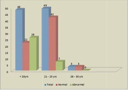
Table-9: Case distribution in relation to age at first coitus
Age at first coitus(years) Total cases Normal Abnormal
Less than 20 48 22 26 21-25 49 42 7 26-30 3 3 -
In our study 48 cases started sexual activity less than 20 years of age,49 cases had first coitus between 21-25 years of age and 3 cases between 26-30 years of age.
Of the abnormal cases 26(78%) cases started sexual activity less than 20 years of age,7cases (22%) started sexual activity between 21-25 years of age and no CIN cases were detected in women who had first coitus between 26-30 years of age.
Figure 24: Case distribution in relation to Age at first coitus 0 5 10 15 20 25 30 35 40 45 50 < 20yrs 48 22 26
Figure 24: Case distribution in relation to Age at first coitus
21 - 25 yrs 26 - 30 yrs 49 3 42 3 7 0
Table-10: Case distribution in relation to Parity index
Obstetric score Total cases Normal Abnormal
Para 1 15 14 1
Para 2 58 47 11
Para 3 & above 27 6 21
In the present study out of 100 cases, 15 cases belonged to Para 1, 58 cases belonged to Para 2 and 27 cases belonged to Para 3 and above.
21 (64%) out of 33 abnormal cases had Para 3 and above, 11 cases (33%) belonged to Para 2, and 1 case (3%) belonged to Para 1.
Figure 25: Case distribution in relation to parity index
0 10 20 30 40 50 60
Para 1 15 14
1
Figure 25: Case distribution in relation to parity index
Para 2 Para 3 & above 58
27 47
6 11
21
Table-11: Case distribution in relation to presenting symptoms
Symptoms Total cases Normal Abnormal
AUB 6 6 - Discharge P/V 66 45 21 Lower abdomen pain 20 16 4
Post coital bleed 8 - 8
In our study out of 100 cases, 66 cases presented with symptoms of vaginal discharge, 20 cases with lower abdomen pain, 8 cases with post coital bleed and 6 cases with irregular periods (Abnormal uterine bleeding).
21 (64%) out of 33 abnormal cases presented with discharge P/V, 4 cases (12%) with lower abdomen pain and 8 cases (24%) presented with post coital bleeding. All the cases with post coital bleeding had biopsy abnormality.
Table – 12: Case distribution in relation to clinical findings
P/S findings Total cases Normal Abnormal
Normal 87 67 20
Erosion Cx 3 - 3
Eversion Cx 2 - 2 Hypertrophied Cx 2 - 2
Ulcer Cx 4 - 4
Papillary growth 2 - 2
In current study, 87 cases had clinically normal cervix, 13 cases had lesion on cervix. 20 (61%) out of 87 normal cases and all 13(39%) cases who had clinically detectable lesion on cervix showed HPE abnormality. Of the 13 abnormal cases 3 cases had erosion cervix, 2 had eversion cervix, 2 had hypertrophied cervix, 4 had ulcerative lesion on cervix, and 2 patients had papillary growth on cervix.
Table-13: Case distribution of LBC and Conventional Pap smear
Cytology Normal Abnormal
LBC 68 32
PAP 83 17
In our study out of 100 cases, 68 (68%) cases in LBC were normal and 32cases (32%) showed abnormality. In Conventional Pap smear 83 (83%) cases were normal and17 cases (17%) showed abnormality. LBC predicted 15 extra abnormal cases than Pap smear.
Figure 26: Case distribution of LBC and PAP smear
0 10 20 30 40 50 60 70 80 90 100
LBC 100
68
32
Figure 26: Case distribution of LBC and PAP smear
PAP 100
83
3
Table-14: Comparison of Specimen adequacy in LBC and PAP
Cytology Satisfactory smear Unsatisfactory smear
LBC 99 1
PAP 94 6
In the present study out of 100 cases, 99 smears of LBC were satisfactory for evaluation and 1 smear was unsatisfactory for evaluation. In PAP 94 smears were satisfactory for evaluation and 6 smears were unsatisfactory for evaluation. Unsatisfactory smear in LBC was due to scant cellularity and in Pap smear it is due to thick smear caused by obscuring mucus.
Figure 27: Comparison of Specimen adequacy in LBC and PAP smear
0 10 20 30 40 50 60 70 80 90 100
LBC 100 99
1
Total
Figure 27: Comparison of Specimen adequacy in LBC and PAP smear
PAP 100
94
6
Satisfactory smear Unsatisfactory smear
Table-15: Comparison of Infectious agent prediction in LBC and PAP
Infectious agent LBC PAP
Total cases 14 3 Bacterial Vaginosis 2 1
Candida 5 -
Trichomonas vaginalis 7 2
In the present study infectious agents were detected in 14 cases (14%) of LBC of these 2 cases showed features of Bacterial vaginosis , 5 cases showed features of Candida and TV in 7 cases.
Pap smear showed infectious agents in 3 cases (3%) of these 2 cases showed features of Trichomonas vaginalis and 1 case showed features of Bacterial vaginosis.
Figure 28: Comparison of Infectious agent prediction in
0 2 4 6 8 10 12 14
Total Bacterial vaginosis 14
3
Figure 28: Comparison of Infectious agent prediction in LBC and PAP
Bacterial vaginosis Candida Trichomonas 2
5
7
1
0
LBC PAP
LBC and PAP
Table – 16: Distribution of cases in LBC
Bethesda category LBC Percentage
Normal 68 68
LSIL 16 16
HSIL 10 10
ASC(ASC-US&ASC-H) 2 2
SCC 4 4
In the present study out of 100 cases, LBC showed LSIL in 16 cases (16%), 10 cases (10%) showed features of HSIL. ASC was seen in 2 cases (2%) and Squamous cell carcinoma in 4 cases (4%).
Figure 29: Distribution of cases in LBC
LSIL 16%
HSIL 10%
: Distribution of cases in LBC
ASC 2%
SCC 4%
LBC
Table – 17: Distribution of cases in PAP