Copyright © 2000, American Society for Microbiology. All Rights Reserved.
Characterization of a Baculovirus Alkaline Nuclease†
LULIN LI
ANDGEORGE F. ROHRMANN*
Department of Microbiology, Oregon State University, Corvallis, Oregon 97331-3804
Received 10 February 2000/Accepted 17 April 2000
All baculovirus genomes sequenced to date encode a homolog of an alkaline nuclease that has been
char-acterized in the
Herpesviridae
. In this report we describe the characterization of the alkaline nuclease (AN)
homolog of the
Autographa californica
multinucleocapsid nucleopolyhedrovirus (Ac
M
NPV) (open reading frame
133). His-tagged AN constructs were expressed in recombinant baculoviruses and affinity purified, and then
their enzymatic activity was characterized. AN was found to degrade linear DNA at alkaline pH, preferred Mg
2ⴙover Mn
2ⴙ, had optimal activity at 35°C, and did not appear to have a salt requirement. To rule out
contam-ination by the endogenous baculovirus gene product or a cellular enzyme, point mutations were introduced into
a highly conserved domain of the gene. These mutations were found to markedly reduce or eliminate most of
the activity of the affinity-purified enzyme. An antibody generated against the protein was used to analyze its
expression by Western blot analysis. AN was found to be expressed at low levels by 12 h postinfection, with
maximal expression at 24 h postinfection. Attempts to generate a virus with this gene inactivated were
un-successful, suggesting that AN may be encoded by an essential gene.
Baculoviruses are a large family of viruses that infect
inver-tebrates, particularly insects of the order
Lepidoptera
. They
contain circular, supercoiled, double-stranded DNA genomes
of 100 to 180 kb. These genomes are punctuated by repeated
sequences called homologous regions that function as origins
of DNA replication in transient assays (18, 26). Evidence
sug-gests that genome replication occurs through a rolling-circle
intermediate, resulting in large concatemers that are resolved
into unit-length molecules during virion maturation (21, 25,
34). Although a number of genes have been identified that are
involved in DNA replication (17, 23), neither
cis
-acting
ge-nome sequences nor the genes required for proper gege-nome
processing have been identified.
The complete sequences of a number of baculovirus
ge-nomes have recently been reported (1, 2, 12, 15, 19). All
en-code a homolog of an alkaline nuclease present in members of
the
Herpesviridae
(reviewed in reference 9). In herpes simplex
virus type 1 (HSV-1), the alkaline nuclease (AN) has the
prop-erties of an exonuclease and functions optimally at pH 9 on
linear DNA (4, 9, 10, 16). Recombinant cell lines expressing
AN have been used to produce HSV-1 AN deletion mutants.
These virus synthesize wild-type (wt) levels of DNA and
pro-duce encapsidated genomes. However, the virions are of low
infectivity in certain cell lines and are not detected in the
infected cell cytoplasm, suggesting that AN is required for the
production of viable nucleocapsids that are capable of exit
from the nucleus into the cytoplasm (30). This may involve the
processing of branched replication intermediates into genomic
DNA that can be encapsidated (10, 24). It has also been
sug-gested that it may play a role in the generation of 3
⬘
OH-terminal single-stranded DNA tails that are thought to be
involved in repair of breaks in homologous DNA regions as
part of a DNA recombination system (16).
Because AN homologs appear to be universally present in
baculovirus genomes, are likely to play an essential role in
bac-ulovirus replication, and may be involved in the final
process-ing steps leadprocess-ing to the production of mature genomes, we
ini-tiated investigations of this enzyme. In this report, we describe
the purification and characterization of the
Autographa
califor-nica
multinucleocapsid nucleopolyhedrovirus (Ac
M
NPV) AN
and compare it properties to those of the herpesvirus enzyme.
MATERIALS AND METHODS
Virus and cell lines.Spodoptera frugiperda(Sf-9) cells (32) were cultured in TNM-FH medium (14) supplemented with 10% fetal bovine serum, penicillin G (50 U/ml), streptomycin (50g/ml; Whittaker Bioproducts), and amphotericin B (Fungizone; 375 ng/ml; Flow Laboratories). Cell culture maintenance was car-ried out according to published procedures (31). Sf-9 cells were also cultured in Sf-900 II medium (Gibco-BRL) as previously described (11). AcMNPV (strain E-2) was used for wt infections.
Enzymes, radioisotopes, DNA purification, PCR, and DNA sequencing. Re-striction and DNA-modifying enzymes were purchased from Life Technologies and New England Biolabs and were used according to the manufacturer’s in-structions. Isotopes were purchased from New England Nuclear, Inc. DNA sequence analysis and PCR were carried out as described previously (22). DNA was purified using Qiagen columns (Qiagen, Inc.).
Recombinant baculovirus and construction of mutants.Recombinant baculo-viruses were produced using pBlueBacHis2B vector and BacNBlue linear DNA (Invitrogen) as instructed by the manufacturer. To remove theBamHI site in pBlueBacHis2B, the plasmid was digested withBamHI, blunted with T4 DNA polymerase, and religated. The vector was then digested withXhoI andPstI and ligated to aSalI (nucleotide [nt] 112551)-to-NsiI (nt 114840) (2) fragment con-taining the AcMNPV AN homolog. TheSalI site is 9 nt upstream of the pre-dicted translational initiation codon of AcMNPV AN, whereas theNsiI site is about 1,000 nt downstream of the stop codon. This resulted in a His6-tagged
fusion protein with a predicted mass of 52.6 kDa and the sequence MPRGSH HHHHHGMASMTGGQQMGRDLYDDDDKDASELDIM upstream of the wt ATG.
Mutant AN construction took advantage of unique StyI (nt 112975) and
BamHI (nt 113032) (2) sites that flanked a motif (motif II) which is predicted to encode a metal binding domain (30) and is conserved between baculoviruses and herpesviruses. Two oligomers were synthesized for each mutant so that they could be annealed and inserted into DNA cut with these two enzymes. The double-stranded oligomers (translation products are indicated above the nucle-otide sequences; mutant nuclenucle-otide and amino acids are underlined) were
AN-G141A
L A L H A A S P D A . . .
⫹5⬘CTTGGCTTTGCACGCCGCTTCGCCCGATGCGTATTTTTCTCTCGCCGACGGAACGTG3⬘ ⫺3⬘ CGAAACGTGCGGCGAAGCGGGCTACGCATAAAAAGAGAGCGGCTGCCTTGCACCTAG5⬘
and
AN-S146A
L G L H A A A P D A . . .
⫹5⬘CTTGGGTTTGCACGCCGCTGCGCCCGATGCGTATTTTTCTCTCGCCGACGGAACGTG3⬘ ⫺3⬘ CCAAACGTGCGGCGACGCGGGCTACGCATAAAAAGAGAGCGGCTGCCTTGCACCTAG5⬘
* Corresponding author. Mailing address: Nash Hall 220,
Depart-ment of Microbiology, Oregon State University, Corvallis, OR
97331-3804. Phone: (541) 737-1793. Fax: (541) 737-0496. E-mail: rohrmann
@bcc.orst.edu.
† Technical Report no. 11656 from the Oregon State University
Agricultural Experiment Station.
6401
on November 9, 2019 by guest
http://jvi.asm.org/
After cloning of each mutant sequence, the alteration was confirmed by DNA sequence analysis (1).
Deletion of AN.We attempted to delete the AN gene from AcMNPV. To accomplish this, the-galactosidase gene under theDrosophilaheat shock pro-moter (hsp) (pAcDZ1 [33]) was cut withXbaI andSmaI to isolate thehsp-lacZ -containing fragment, theXbaI end was blunted with T4 DNA polymerase, and the fragment was inserted into pAN1 (see below) between theStyI (nt 112975) andHpaI (nt 113148) sites such that about 175 nt, including the highly conserved motif II, were deleted. The resultant plasmid, pANlacZ, was linearized and trans-fected with wt AcMNPV DNA into Sf-9 cells to construct recombinant virus.
Preparation of extracts of Sf-9 cells.The His-tagged enzyme expressed in Sf-9 cells was purified using a modification of a published procedure (30). Log-phase Sf-9 cells (about 2⫻106/ml) in about 100 ml of Sf-900 II medium (Life
Tech-nologies) in a 2-liter tissue culture flask were infected at a multiplicity of infec-tion of about 10 and incubated on a shaker at 27°C. After 1 h of incubainfec-tion, the volume was increased to 200 ml with Sf-900 II and incubated at 27°C for 48 h. Cells were then harvested by centrifugation (3,000 rpm for 15 min), resuspended in 10 ml of phosphate-buffered saline, pH 7.4 (Sigma Chemical Co.), and cen-trifuged again; the pellet was stored at⫺80°C. For enzyme purification, the frozen pellets were resuspended in 8 ml of buffer A (20 mM Tris-HCl [pH 7.5], 1 mM MgCl2, 5 mM-mercaptoethanol, 80 mM KCl, 0.2% NP-40),
freeze-thawed twice, and then incubated on ice for 20 min in the presence of 1 mM phenylmethylsulfonyl fluoride (PMSF) and the following proteinase inhibitors from Life Technologies: aprotinin (10g/ml), leupeptin (7g/ml), and pepstatin (7g/ml). Cells were homogenized 20 to 25 times in a Dounce homogenizer with a type B pestle and centrifuged at 10,000⫻gin a Sorvall GSA rotor, and the pellet and supernatant were saved. The pellet was treated again as described above, and the supernatants were combined and then centrifuged at 100,000⫻ gin an SW28 rotor. The supernatant (about 16 ml) was precipitated with 3.2 g of ammonium sulfate (20%) for 45 min at 10°C. The precipitate was pelleted by centrifugation at 17,000⫻gin a Sorvall SS-34 rotor for 30 min. An additional 5.6 g of ammonium sulfate was added to the supernatant (to about 55%) and incubated for 1 h at 10°C. This preparation was centrifuged as described above, and the two pellets were suspended in 5 ml of buffer B (20 mM Tris-HCl, 150 mM NaCl, 5 mM -mercaptoethanol, 20% glycerol [pH 8.0]) and dialyzed overnight against buffer B. The dialysate was then centrifuged at 17,000⫻gto remove insoluble material, and then the preparation was affinity purified on TALON resin (Clontech, Inc.) essentially as recommended by the manufacturer. The resin (150l) that had been washed with 10 ml of wash buffer (buffer B containing 0.1% Triton X-100) was mixed with the dialysate and rotated for 30 min at 4°C. The resin was centrifuged at 1,000 rpm in an International centrifuge for 5 min. The supernatant was removed and designated the flowthrough. The resin was then treated with four 1-ml aliquots of wash buffer (wash fractions 1 to 4 [W1 to W4] and 1-ml solutions of wash buffer containing the following imida-zole concentrations: four times, 10 mM (W5 to W8); four times, 30 mM (elution fractions 1 to 4 [E1 to E4]), four times, 50 mM (E5 to E8); and two applications, 100 mM (E9 and E10). Protein concentration was determined by Coomassie blue staining, Western analysis, and spectrophotometric quantification using a Coo-massie Plus protein assay kit (Pierce, Inc.). Fractions E6 and E7 were used for the assays described below.
Cloning, expression, and antibody production against bacterially expressed His-tagged AN.The AN open reading frame (ORF) was cloned as aSalI (nt 112551)-NsiI (114840) (2) fragment inserted into the XhoI andPstI sites of pKS(⫺) to construct pAN1. pAN1 was cut withKpnI and XbaI; the insert containing the AN gene was gel purified and inserted into pHT4 cut with the same enzymes. pHT4 was constructed by cloning AcMNPV DNA polymerase gene, with aNcoI site at the initial ATG and extended to the downstreamSacI site, intoNcoI andSacI sites of pTrc-7Hpro (7). The resultant plasmid was digested with NcoI andKpnI, blunted with T4 DNA polymerase, and then religated to produce pHT-AN. This His7-tagged fusion contained the sequence
MMHHHHHHHAMGPPLDIM upstream of the AN ATG and has a predicted molecular size of about 50.4 kDa. For protein production,Escherichia coliBL21 cells transformed with pHT-AN were inoculated into 2 ml of Luria-Bertani (LB) broth (29) containing 50l of ampicillin per ml. After 3 h at 37°C, this culture was used to seed a 100-ml LB culture and incubated for 3 to 4 h until the cells reached an optical density at 600 nm of 0.6 to 1.0. Isopropyl--D
-thiogalactopy-ranoside (IPTG) (dissolved in 2% ethanol) was added to a final concentration of 1 mM, and the culture was incubated overnight at 22°C. Cells were harvested and washed with 10 ml of phosphate-buffered saline and the pellet was stored at
⫺80°C. Protein purification was adapted from reference 4. The pellet was re-suspended in 20 ml of lysis buffer (20 mM Tris-HCl [pH 8.0], 100 mM NaCl, 5 mM-mercaptoethanol, 1 mM PMSF, 20% sucrose) containing 1 mg of ly-sozyme per ml, incubated on ice for 20 min, subjected to two freeze-thaw cycles, and then sonicated for 30 s. This sample was centrifuged at 20,000⫻gfor 15 min, and the supernatant was transferred to a fresh tube. The pellet was resuspended in 20 ml of lysis buffer and centrifuged as above. The two supernatants were combined and mixed with 150l of TALON (Clontech) resin previously treated with wash buffer (20 mM Tris-HCl [pH 8.0], 100 mM NaCl, 5 mM -mercapto-ethanol, 1 mM PMSF, 0.1% Triton X-100, 10 mM imidazole) and rotated for 1 h at 4°C. The suspension was centrifuged at 1,000 rpm and washed sequentially with 2-ml aliquots of wash buffer containing the following concentrations of imidazole: 25 mM, four times; 100 mM, twice; and 200 mM, once. The 100 and
200 mM eluates were pooled, and the protein content and purity were assayed by Coomassie blue staining of polyacrylamide gels after polyacrylamide gel electro-phoresis (PAGE) and spectrophotometrically using a Coomassie Plus protein assay kit (Pierce). A rabbit was injected with 100g of this preparation in complete Freund’s adjuvant. Beginning 3 weeks after the first inoculation, the animal was subjected to three boosts of 30g at 14-day intervals in incomplete Freund’s adjuvant. One week after the final boost, the animal was bled and the serum was prepared for use in this study.
Assay for AN activity using [3H]DNA.E. coliDNA was labeled with [3
H]thy-midine as described elsewhere (13), and assays for AN activity were similar to the procedures of Goldstein and Weller (9). Two micrograms (about 240,000 cpm) of labeled single-stranded DNA plus 4g of single-stranded salmon sperm DNA were mixed with 10 ng of affinity-purified AN. Standard conditions used were 200
l of 50 mM Tris (pH 9.0) per ml–5 mM Mg2⫹–0.1 mg of bovine serum albumin
(BSA) per ml at 37°C for 20 min. The digested DNA was mixed with 0.25 mg of BSA, precipitated with 5% trichloroacetic acid and microcentrifuged for 5 min. The supernatant was mixed with 3.5 ml of scintillation cocktail (formula A-989; Packard, Inc.) and counted.
RESULTS AND DISCUSSION
Baculovirus AN homologs.
Five baculovirus homologs of
the herpesvirus AN are aligned in Fig. 1A. These ORFs are
from Ac
M
NPV (orf133) (2),
Orgyia pseudotsugata M
NPV
(Op
M
NPV) (orf131) (1),
Lymantria dispar M
NPV (Ld
M
NPV)
(orf157) (19),
Choristoneura fumiferana M
NPV (Cf
M
NPV) (A.
Poloumienko and P. Krell, unpublished data [GenBank
ac-cession no. AAB53344]), and a granulovirus of
Xestia c-nigrum
(XcGV) (orf145) (12). The following amino acid sequence
identities of the predicted baculovirus ORFs were observed:
Op
M
NPV and Cf
M
NPV, 80%; Op
M
NPV and Cf
M
NPV to
Ac
M
NPV, 53%; Op
M
NPV, Cf
M
NPV, and Ac
M
NPV to
Ld
M
NPV, 37 to 40%; and XcGV to the NPVs, about 30%. The
sequence identity is concentrated in the N-terminal 240 amino
acids with six regions of limited sequence variation. Several of
these regions are related to conserved motifs found in
herpes-virus AN (9, 16). The baculoherpes-virus motif I, Ia, II, and III
se-quences are 15/16, 3/3, 5/11, and 6/11 identical to the
cor-responding alphaherpesvirus consensus sequence (Fig. 1B).
There is a highly conserved domain at amino acids 190 to 200
that does not appear to correspond to a herpesvirus motif. In
addition, there is a conserved region that shows limited
ho-mology to motif VI. The baculovirus predicted proteins are
about 200 amino acids shorter than those from HSV-1 (Fig.
1B). This is evident in a significantly truncated baculovirus
amino-terminal region upstream of motif I (218 amino acids in
HSV-1, versus 56 in Ac
M
NPV). A form of the HSV-1 AN has
been described that results from initiation at an internal ATG
codon such that it too lacks a significant portion (126 amino
acids) of this N-terminal region (5). It has been found to retain
its enzymatic activity and is capsid associated. However, the
predicted baculovirus AN sequences all also lack motifs IV,
V, and VII. In addition, the region between motifs III and
VI contains 151 amino acids in HSV-1, versus about 40 in
Ac
M
NPV (Fig. 1B), but the distance from motif VI to the C
terminus is longer in Ac
M
NPV (180 amino acids, versus 112 in
HSV-1).
Expression and purification of Ac
M
NPV orf133.
Since
ami-no-terminal regions of the homologs of Ac
M
NPV orf133 are
highly variable (Fig. 1), we reasoned that alteration of
amino-terminal amino acids would be unlikely to affect the activity of
the enzyme. We used a
Sal
I site at nt 112551 on the Ac
M
NPV
genome (2) which is 9 nt upstream of the orf133 ATG and an
Nsi
I site at nt 114845 that is about 1,000 nt downstream of the
stop codon in our cloning protocols. A His
7-tagged construct
was expressed in bacteria, and the protein was processed using
a renaturing protocol that was successfully used to generate
active HSV-1 AN (4). However, we were unsuccessful in
gen-erating an active protein using this technique. Therefore, we
on November 9, 2019 by guest
http://jvi.asm.org/
FIG. 1. Comparison of baculovirus AN sequences. (A) Alignment of bac-ulovirus sequences. Indicated are: AcMNPV (ac133) (2), CfMNPV (cfexo) (Poloumienko and Krell, unpublished), OpMNPV (op131) (1), LdMNPV (ld157) (19), and XcGV (xc145) (12). The domains conserved with theHerpesviridaeAN are boxed. Dots indicate positions where all amino acids are identical, and dashes indicate gaps in the alignment. (B) Comparison of conserved domains between HSV-1 (9) and baculovirus AN. Below are shown the sequences with the con-sensusBaculoviridae(bac) (four out of five identical) andHerpesviridae(hpv) sequences indicated. Asterisks indicate nonconsensus amino acids. The muta-tions within motif II that were constructed and tested are also shown.
on November 9, 2019 by guest
http://jvi.asm.org/
constructed a recombinant baculovirus expressing a His
6-tagged version of orf133.
[image:4.612.55.293.69.314.2]Sf-9 cells infected with the recombinant Ac
M
NPV
express-ing orf133 were processed usexpress-ing a combination of ammonium
sulfate precipitation and affinity chromatography (Fig. 2A).
The resuspended ammonium sulfate precipitate is shown in
Fig. 2A, lane 1. After binding of this extract to the affinity resin,
the unbound material looked similar to the starting material
(lane 2). The resin was washed initially without imidazole (lane
3) and then with buffer containing 10 mM imidazole (lane 4).
A slight amount of protein was evident in the latter wash (lane
4). When treated with 30 mM imidazole, two major Coomassie
blue-stained bands of about 43 and 53 kDa were eluted (lane
5). Subsequent washes with 30 and 50 mM imidazole showed
similar elution profiles (lanes 5 to 12). Elution with 100 mM
imidazole yielded only a small amount of these two bands.
To determine whether these bands were composed of
His-tagged recombinant AN, we used a reagent that specifically
stains His-tagged proteins (Fig. 2B). We found that extracts
from uninfected Sf-9 cells (lane 1) or wt Ac
M
NPV-infected
cells (lane 2) showed no evidence of a reactive His-tagged
protein. However, when fractions described above were
exam-ined, we found that the starting material before affinity
puri-fication contained a His-tagged protein (lane 3), as did
frac-tions E2, E6, and E9. This demonstrated that the two major
bands of 43 and 53 kDa were His-tagged molecules and
indi-cated that the His-tagged An was present as two species; one
full length (53 kDa) and a shorter (43-kDa) form that
appar-ently retained the N-terminal His tag and therefore likely
lacked the carboxyl terminus.
Nuclease activity of affinity-purified AN.
To determine if the
purified His-tagged protein had nuclease activity, we tested it
with both linear and supercoiled DNA templates at neutral
and alkaline pH (Fig. 3). We found that under the conditions
that we examined, most of the linear template was degraded in
20 min at pH 9.0, whereas at pH 7.0 most of the starting
material remained at 60 min (Fig. 3A). In contrast, the effect
on supercoiled templates was not nearly so dramatic. At pH 9,
there appeared to be an initial conversion of some DNA to
linear-sized fragments at 1 min (Fig. 3B). Although this linear
DNA was subsequently degraded, substantial amounts of the
supercoils remained after 20 min. The effect at pH 7 was even
less than at pH 9, with some conversion to linear-sized DNA at
1 min; however, by 60 min almost all of the supercoiled DNA
remained. Therefore, the purified AN has a strong preference
for linear over supercoiled DNA and showed the highest levels
of activity at an alkaline pH, suggesting that the
baculovirus-encoded protein is an alkaline exonuclease.
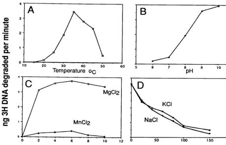
Characterization of the Ac
M
NPV alkaline exonuclease.
We
characterized the properties of the baculovirus AN in more
detail by quantifying its ability to hydrolyze
3H-labeled linear
DNA (Fig. 4). We found that the optimal temperature was
35°C (Fig. 4A), and as expected from Fig. 3, the highest activity
was observed in an alkaline pH range of 9 to 10 (Fig. 4B).
Divalent cations were required for activity, and the enzyme
showed optimal activity over a broad range (2 to 10 mM);
Mg
2⫹gave about a 10-fold-higher level of activity than Mn
2⫹ [image:4.612.314.551.429.654.2](Fig. 4C). The enzyme did not appear to require salt, and
FIG. 2. Purification and characterization of His-tagged alkaline exonuclease. Sf-9 cells were infected with baculovirus expressing the His-tagged AN gene and affinity purified as described in Materials and Methods. (A) PAGE analysis of affinity purification. Lane 1, infected cell extract (CE) after dialysis; lane 2, flowthrough (FT) from the TALON resin; lane 3, W4 without imidazole; lanes 4 to 14, W8 with 10 mM imidazole and elution with imidazole at 30 mM (lanes 5 to 8; E1 to E4), 50 mM (lanes 9 to 12; E5 to E8), and 100 mM (lanes 13 and 14; E9 and E10). Lanes 1 and 2 represent 5l of the 5.0-ml dialysate before and after binding the affinity resin; lanes 3 to 14 represent 15 l from 1-ml fractions. Samples were analyzed by PAGE through a 10% gel and stained with Coomassie brilliant blue. The positions of selected size standards (Life Technologies 10-kDa ladder) are shown on the left, and the estimated masses (in kilodaltons) of the polypeptides binding to the affinity resin are shown at the right. (B) Identification of His-tagged polypeptides. The enzyme was characterized using the INDIA HisProbe-HRP reagent (Pierce) according to the manufacturer’s instructions. Lane 1, uninfected Sf-9 cells (Sf); lane 2, wt AcMNPV-infected Sf-9 cells (Wt). Lanes 3 to 8 are from the samples described for panel A.
FIG. 3. Characterization of AN activity on DNA at pH 7.0 and 9.0. (A) Time course of digestion of linear pKS(⫺) DNA at the times and pH indicated. Qiagen column-purified pKS(⫺) was linearized withEcoRI. DNA (0.2g) was mixed with 10 ng of affinity-purified enzyme and incubated for various times in 200l of the standard buffer (50 mM Tris-HCl, 5 mM MgCl2) at 37°C and the indicated
pH. The digests were then electrophoresed on a 1.0% agarose gel. (B) Time course of digestion of supercoiled pKS DNA at the times and pH indicated. Supercoiled DNA (0.2g) for each sample was processed as described above. Positions of markers (M) are indicated in kilobases.
on November 9, 2019 by guest
http://jvi.asm.org/
increasing concentrations were inhibitory (Fig. 4D). These
op-timal conditions are similar to those reported for HSV-1 AN
with the exception of salt concentration, for which the HSV-1
enzyme showed a broad optimum of up to 40 mM (4).
The fact that the baculovirus-encoded enzyme has
proper-ties similar to those of the enzyme characterized from HSV-1
suggests that the shared motifs (I, Ia, II, III, and VI) may
contribute to this activity, but the other motifs common to the
Herpesviridae
but lacking in the
Baculoviridae
(IV, V, and VII)
(Fig. 1) may be involved in some other function.
Mutations in a highly conserved domain.
To ensure that the
enzyme activity that we observed was due to the
affinity-puri-fied recombinant AN (rAN) and not contamination from the
endogenous AN encoded by the virus or from a cellular
en-zyme, we constructed a number of mutants with single amino
acids altered at positions in motif II that are conserved in all
baculovirus and herpesvirus sequences (Fig. 1B). Selected
mu-tations in this region of the HSV-1 AN led to inactivation of
the enzyme (9). We produced three mutations in this region:
amino acid mutations G141A and S146A and deletion of
amino acids 142 to 148. The His-tagged rAN and two of the
mutants were expressed and could be detected by Western blot
analysis using a rabbit antiserum that we generated against a
bacterially expressed form of the protein (Fig. 5, lanes 1 to 3).
However, the mutant with amino acids 142 to 148 deleted was
apparently unstable and was not evident in extracts of cells
infected with recombinant viruses expressing this construct. An
immunoreactive band was not observed in affinity-purified
extracts from uninfected Sf-9 cells or cells infected with wt
Ac
M
NPV (lane 4 or 5, respectively). In contrast, the antiserum
reacted with an appropriate-sized band in extracts that had not
been affinity purified from cells infected with either wt virus or
the recombinant virus expressing His-tagged rAN (lane 6 or 7,
respectively).
We then examined the ability of the affinity-purified mutant
protein to hydrolyze linear [
3H]DNA (Fig. 6). We found that
G141A showed about 35% the activity of the rAN, whereas
S146A showed less than 10% the activity of the nonmutant
enzyme. No activity was evident from affinity-purified extracts
of cells that had been infected with wt virus indicating that, as
expected, the native enzyme which would lack the His tag was
not affinity purified by our protocol (Fig. 6, lane 4). These data
indicated that the wt enzyme was not a major contaminant of
our affinity-purified preparations, and as with HSV-1 AN (9),
motif II is critical for the activity of the baculovirus enzyme.
[image:5.612.70.529.72.366.2]Construction of a mutant of Ac
M
NPV with AN deleted.
In an
attempt to construct a mutant of Ac
M
NPV with the AN gene
inactivated, we constructed a plasmid with the

-galactosidase
gene under the control of the
Drosophila
heat shock promoter
inserted within the AN gene such that the gene was disrupted
and a portion was deleted. We used cotransfection with the wt
virus and linearized plasmid DNA to produce the deletion
mutants. We isolated and extensively plaque purified a number
of isolates expressing

-galactosidase. However, upon PCR
analysis, we found that although the

-galactosidase-express-ing construct was present in the genome, the wt gene was also
FIG. 4. Effects of temperature (A), pH (B), divalent cations Mg2⫹and Mn2⫹(C), and salt concentration (D) on purified AcMNPV alkaline exonuclease activity.
For these experiments, single-stranded DNA (2g of labeled DNA plus 4g of salmon sperm DNA) was mixed with 10 ng of purified AN. Samples were prepared for each condition tested and then incubated for 20 min. Standard conditions were 37°C, 50 mM Tris (pH 9.0), 5 mM MgCl, and 0.1 mg of BSA per ml in 200l unless otherwise indicated. The digested DNA was mixed with 0.25 mg of BSA, then precipitated with 5% trichloroacetic acid, and centrifuged for 5 min in a microcentrifuge; the supernatants were mixed with 3.5 ml of scintillation cocktail (formula A-989; Packard) and counted. All samples were done in triplicate, and each point represents an average of the values. Very little deviation from the average was observed.
on November 9, 2019 by guest
http://jvi.asm.org/
present. In addition, attempts to delete the AN homolog from
the
Bombyx mori
NPV genome have been unsuccessful (S. J.
Gomi, personal communication). The data from both of these
viruses suggest that this enzyme plays a vital role in the
bacu-lovirus replication cycle.
Characterization of AN expression in Ac
M
NPV-infected
cells.
The anti-AN antiserum that we prepared against
bacte-rially expressed, His-tagged AN was used to examine the time
course of infection of AN in Ac
M
NPV-infected cells (Fig. 7).
As a control we used the affinity-purified recombinant AN
(Fig. 7, lane 1). We found that a polypeptide of the predicted
molecular mass (48 kDa) was first observed at about 12 h
postinfection (p.i.) and the peak level of expression occurred at
24 h p.i., consistent with late gene expression. There is a
pos-sible RNA polymerase II promoter (TATTT) and mRNA start
site consensus sequence (CAGT) (3) starting about 140 nt
upstream of the predicted initiation codon. There is also a late
promoter element (GTAAG) located 22 nt upstream of the
ATG (2). This suggests that Ac
M
NPV AN may be expressed as
both an early and a late gene. There was also a smaller, 38-kDa
band present at a lower concentration that may represent a
breakdown product similar to that observed with rAN (Fig. 2);
however, an immunoreactive band of this size was also
ob-served in extracts of uninfected cells.
We previously reported the presence of a nuclease active
against linear DNA templates in nuclear extracts of Ac
M
NPV-infected cells that were used for in vitro transcription assays
(8). Although the extracts from uninfected or early infected
cells lacked the nuclease activity, those at 24 h p.i. showed a
high level of activity. The reaction conditions for these
inves-tigations were buffered at pH 8.4, which is well within the
active range of the AN we have described in this report. A
nuclease activity associated with baculovirus infection was also
reported by others (6). It is likely that the Ac
M
NPV AN that
we have characterized is responsible for the nuclease activities
described in these reports.
ACKNOWLEDGMENTS
We thank Doug Leisy for reviewing the manuscript and Doug
Gros-senbach and Dennis Hruby for assistance in characterization of
His-tagged proteins.
This project was supported by a grant from the NSF
(MCB-9630769).
REFERENCES
1.Ahrens, C. H., R. Russell, C. J. Funk, J. T. Evans, S. H. Harwood, and G. F. Rohrmann.1997. The sequence of theOrgyia pseudotsugata multinucleocap-sid nuclear polyhedrosis virus genome. Virology229:381–399.
2.Ayres, M. D., S. C. Howard, J. Kuzio, M. Lopez-Ferber, and R. D. Possee.
1994. The complete DNA sequence ofAutographa californicanuclear poly-hedrosis virus. Virology202:586–605.
[image:6.612.316.546.71.256.2]3.Blissard, G. W., and G. F. Rohrmann.1989. Location, sequence, transcrip-tional mapping, and temporal expression of the gp64 envelope glycoprotein FIG. 5. Western blot analysis of wt and mutant AN. AN was purified as
[image:6.612.58.288.71.235.2]described in Materials and Methods. Samples include His-tagged rAN (lane 1) and the mutants indicated (lanes 2 and 3). Controls show that there is no material binding to the affinity resin from extracts of uninfected Sf-9 cells (lane 4) or wt AcMNPV-infected cells (lane 5). For these control assays (lanes 4 and 5), the cells were carried through the purification protocol, fractions E6 and E7 (Fig. 2) were pooled, and about three times the volume used for the His-tagged enzyme-containing extracts (about 24l) was loaded onto the gel. Lanes 6 and 7 contain 10l of dialysate from cells infected with wt AcMNPV and recom-binant AcMNPV expressing AN, respectively. Samples were electrophoresed through sodium dodecyl sulfate–10% polyacrylamide gels (20) and electroblotted onto polyvinylidene difluoride membranes (Micron Separations, Inc.) for 2 h at 185 mA; then, Western blot analyses were carried out as previously described (27). Samples were treated with 1:1,000 dilution of the antiserum, and the second antibody (goat anti-rabbit conjugated to horseradish peroxidase; Promega) was used at 1:2,500. The positions of selected size standards are shown in kilodaltons on the left. The estimated values for the major immunoreactive bands are shown on the right.
FIG. 6. Quantification of activity of mutant and wt AN. For details, see Materials and Methods. All samples were done in triplicate, and the error bars represent 1 standard deviation. The experiment is representative of at least two different infection and extract preparations for each mutant virus. Lane 4 is a sample from wt AcMNPV-infected cells processed as described in the legend to Fig. 5.
FIG. 7. Western blot analysis of AN expression in extracts of infected insect cells. Monolayers of Sf-9 cells were infected with AcMNPV at a multiplicity of infection of 10 and prepared as previously described (28). Lane 1, rAN (300 ng); lanes 3 to 10, time course of wt AN expression in AcMNPV-infected Sf-9 cells. The hour postinfection is indicated above the lanes; sizes are indicated in kilo-daltons.
on November 9, 2019 by guest
http://jvi.asm.org/
gene of theOrgyia pseudotsugatamulticapsid nuclear polyhedrosis virus. Virology170:537–555.
4.Bronstein, J. C., and P. C. Weber.1996. Purification and characterization of herpes simplex virus type 1 alkaline exonuclease expressed inEscherichia coli. J. Virol.70:2008–2013.
5.Bronstein, J. C., S. K. Weller, and P. C. Weber.1997. The product of the UL12.5 gene of herpes simplex virus type 1 is a capsid-associated nuclease. J. Virol.71:3039–3047.
6.Davidoff, A. N., and B. V. Mendelow.1997. Identification of endonuclease activity in HIV-1 gp120 preparations produced using baculovirus expression systems. BioTechniques23:296–299.
7.Evans, J. T., and G. F. Rohrmann.1997. The baculovirus single-stranded DNA binding protein, LEF-3, forms a homotrimer in solution. J. Virol.71:
3574–3579.
8.Glocker, B., R. R. Hoopes, and G. F. Rohrmann.1992. In vitro transactiva-tion of baculovirus early genes by nuclear extracts fromAutographa califor-nicanuclear polyhedrosis virus-infectedSpodoptera frugiperdacells. J. Virol.
66:3476–3484.
9.Goldstein, J. N., and S. K. Weller.1998. The exonuclease activity of HSV-1 UL12 is required for in vivo function. Virology244:442–457.
10. Goldstein, J. N., and S. K. Weller.1998. In vitro processing of herpes simplex virus type 1 DNA replication intermediates by the viral alkaline nuclease, UL12. J. Virol.72:8772–8781.
11. Harwood, S. H., L. Li, P. S. Ho, A. K. Preston, and G. F. Rohrmann.1998. AcMNPV late expression factor-5 interacts with itself and contains a zinc ribbon domain that is required for maximal late transcription activity and is homologous to elongation factor TFIIS. Virology250:118–134.
12. Hayakawa, T., R. Ko, K. Okano, S. Seong, C. Goto, and S. Maeda.1999. Sequence analysis of theXestia c-nigrumgranulovirus genome. Virology262:
277–297.
13. Hays, J. B., S. J. Martin, and K. Bhatia.1985. Repair of nonreplicating UV-irradiated DNA: cooperative dark repair byEscherichia coli uvrandphr
functions. J. Bacteriol.161:602–608.
14. Hink, W. F.1970. Established insect cell line from the cabbage looper,
Trichoplusia ni. Nature226:466–467.
15. Ijkel, W. F. J., E. A. van Strien, J. G. M. Jeldens, R. Broer, D. Zuidema, R. W. Goldbach, and J. M. Vlak.1999. Sequence and organization of the Spodo-ptera exiguamulticapsid nucleopolyhedrovirus genome. J. Gen. Virol.80:
3289–3304.
16. Kehm, E., M. Goksu, S. Bayer, and C. W. Knopf.1998. Herpes simplex virus type 1 DNase: functional analysis of the enzyme expressed by recombinant baculovirus. Intervirology41:110–119.
17. Kool, M., C. Ahrens, R. W. Goldbach, G. F. Rohrmann, and J. M. Vlak.1994. Identification of genes involved in DNA replication of theAutographa cali-fornicabaculovirus. Proc. Natl. Acad. Sci. USA91:11212–11216. 18. Kool, M., P. M. M. M. Van Den Berg, J. Tramper, R. W. Goldbach, and J. M.
Vlak.1993. Location of two putative origins of DNA replication of Autog-rapha californicanuclear polyhedrosis virus. Virology192:94–101. 19. Kuzio, J., M. N. Pearson, S. H. Harwood, C. J. Funk, J. T. Evans, J. Slavicek,
and G. F. Rohrmann.1999. Sequence and analysis of the genome of a baculovirus pathogenic forLymantria dispar. Virology253:17–34. 20. Laemmli, U. K.1970. Cleavage of structural proteins during the assembly of
the head of bacteriophage T4. Nature227:680–685.
21. Leisy, D. J., and G. F. Rohrmann.1993. Characterization of the replication of plasmids containinghrsequences in baculovirus-infectedSpodoptera fru-giperdacells. Virology196:722–730.
22. Li, L., S. H. Harwood, and G. F. Rohrmann.1999. Identification of addi-tional genes that influence baculovirus late gene expression. Virology255:
9–19.
23. Lu, A., and L. K. Miller.1995. The roles of eighteen baculovirus late ex-pression factor genes in transcription and DNA replication. J. Virol.69:
975–982.
24. Martinez, R., R. T. Sarisky, P. C. Weber, and S. K. Weller.1996. Herpes simplex virus type 1 alkaline nuclease is required for efficient processing of viral DNA replication intermediates. J. Virol.70:2075–2085.
25. Oppenheimer, D. I., and L. E. Volkman.1997. Evidence for rolling circle replication of Autographa californica M nucleopolyhedrovirus genomic DNA. Arch. Virol.142:2107–2113.
26. Pearson, M. N., R. M. Bjornson, G. D. Pearson, and G. F. Rohrmann.1992. TheAutographa californicabaculovirus genome: evidence for multiple rep-lication origins. Science257:1382–1384.
27. Quant-Russell, R. L., M. N. Pearson, G. F. Rohrmann, and G. S. Beaudreau.
1987. Characterization of baculovirus p10 synthesis using monoclonal anti-bodies. Virology160:9–19.
28. Rasmussen, C., and G. F. Rohrmann.1994. Characterization of the Spo-doptera frugiperdaTATA-binding protein: nucleotide sequence and response to baculovirus infection. Insect Biochem. Mol. Biol.7:699–708.
29. Sambrook, J., E. F. Fritsch, and T. Maniatis.1989. Molecular cloning: a laboratory manual, 2nd ed. Cold Spring Harbor Laboratory, Cold Spring Harbor, N.Y.
30. Shao, L., L. M. Rapp, and S. K. Weller.1993. Herpes simplex virus 1 alkaline nuclease is required for efficient egress of capsids from the nucleus. Virology
196:146–162.
31. Smith, G. E., and M. D. Summers.1978. Analysis of baculovirus genomes with restriction endonucleases. Virology89:517–527.
32. Summers, M. D., and G. E. Smith.1987. A manual of methods for baculo-virus vectors and insect cell culture procedures. Bulletin no. 1555. Texas Agricultural Experiment Station, College Station, Tex.
33. Vaughn, J. L., R. H. Goodwin, G. J. Tompkins, and P. McCawley.1977. The establishment of two cell lines from the insectSpodoptera frugiperda (Lepi-doptera: Noctuidae). In Vitro13:213–217.
34. Vlak, J. M., A. Schouten, M. Usmany, G. J. Belsham, E. C. Klinge-Roode, A. J. Maule, J. W. Van Lent, and D. Zuidema.1990. Expression of cauli-flower mosaic virus gene I using a baculovirus vector based upon the p10 gene and a novel selection method. Virology179:312–320.
35. Wu, Y., G. Liu, and E. B. Carstens.1999. Replication, integration, and packaging of plasmid DNA following cotransfection with baculovirus viral DNA. J. Virol.73:5473–5480.
on November 9, 2019 by guest
http://jvi.asm.org/