INTERCUSPAL POSITION AND
CENTRIC RELATION
Dissertation submitted to
THE TAMILNADU DR. M.G.R.MEDICAL UNIVERSITY
In partial fulfillment for the degree of
MASTER OF DENTAL SURGERY
BRANCH V
ORTHODONTICS AND DENTOFACIAL
ORTHOPEDICS
I would like to take this opportunity to express my gratitude to
everyone who has helped me through this journey.
I would like to start with my very respected and beloved professor,
Dr. N.R. KRISHNASWAMY, M.D.S., M.Ortho RCS. (Edin), Diplomat of
Indian board of Orthodontics, Professor and Head, Department of
Orthodontics, Ragas Dental College and Hospital, Chennai. I consider myself
extremely lucky to have had the opportunity to study under him. He has
always been a source of inspiration to perform better not only in academics
but also in life. I would like to thank him for having taken interest in my
study and providing his valuable insight.
My
sincere
thanks
also
go
out
to
my
Professor
Dr.S. VENKATESWARAN, M.D.S for his undying enthusiasm and
guidance which helped me complete this study. He has been an integral part
of my post graduate life and I want to take this opportunity to acknowledge
and thank him for his help and support.
with an opportunity to utilize the facilities available in this institution in
order to conduct this study.
I would also like to acknowledge Dr. SHAHUL (Associate Professor)
Dr. JAYAKUMAR (Reader), Dr. ANAND (Reader), Dr. SHAKEEL
(Reader ),
Dr. Rekha (Sr. Lecturer ) Dr. RAJAN(Sr. Lecturer),
Dr. SHOBANA (Sr. Lecturer), Dr. PRABHU (Sr. Lecturer) and Dr. BIJU
(Sr. Lecturer) for their support, enthusiasm & professional assistance
throughout my post graduate course.
My heartfelt thanks to my wonderful batch mates, Dr.Amey,
Dr.Gautham, Dr.Kavitha, Dr.Subu, Dr.Geetha, Dr.Fayyaz, and Dr.Ritika
who were cheerfully available at all times to help me. I wish them a
successful career ahead.
I also extend my gratitude to my juniors Dr. Sheel, Dr. Mahalaxmi,
Dr.Ayush, Dr. Ashwin, Dr. Saravanan, Dr. Sabitha, Dr. Sreesan, Dr. Vinod
Dr. Deepak, Dr. Noopur Arthi, Dr. Vijayashri Shakthi, Dr. Ashwin, Dr.
Ravanth Kumar, Dr. Siva Subramanian, Dr. Manikandan, and Dr. Vijay
Anand for all their support and for cooperating with me to conduct this
study on their patients.
I thank Mr. Bhupati for helping me with the statistical analysis for
the study.
co-operation and help during my post-graduate course.
Title
Page Number
1.
Introduction
1
2.
Review of Literature
6
3.
Materials and Methods
48
4.
Results
59
5.
Discussion
64
6.
Summary and Conclusion
73
INTRODUCTION
The search for the optimal and preferred types of static and functional
occlusions has occupied the minds of dentists for more than a century. The
possible role of occlusion in the aetiology of temporomandibular disorders
(TMD) also has been the subject of debate.
Much of the occlusion/TMD debate involves issues surrounding
centric relation (CR), including definition, recording and measurement, use of
articulators and deprogramming splints, and possible relationship to either
stomatognathic health or disease.
Over the years, many methods have been developed to study theTMJ and the condyle position in the joint space. Some of the conventionally used
methods are radiographs, laminographs, tomograms, and magnetic resonance
imagings. However, these are not without their limitations as they present only
anatomical parameters and ignore the extremely dynamic functions of this
complex joint system.
The centric jaw relation (CR) position, in the naturally dentulous state
does not usually coincide with the position the mandible assumes when the
teeth are in centric occlusion (CO).
Historically, centric relation jaw position has been defined as the most
"retruded” contact position15, but current research indicates that the correct
condyles articulate with the thinnest avascular portion of the respective discs
in their most anterior superior position against the slopes of the articular
eminences regardless of tooth contact143”.
"Centric occlusion jaw position "is defined as the most closed position
that the mandible assumes, determined by the full intercuspation of opposing
teeth, irrespective of condylar position 143”.This potential difference is
associated with contradictory theories and is therefore an important topic for
further study and understanding.
Since the monograph was introduced byPosselt100 in1952 concerning the range of the motion of the human mandible, a number of studies have described the movement of the mandible from retruded contact position to
maximum intercuspal position.
In 1952Sears129 studiedsagittal,vertical and horizontal changes with the condyle migration recorder.
Possellt101 used the gnathothesiometer in analyzing the condylar path whereas Long74 used the Buhnergraph to locate the hinge axis and verify the centric jaw relation. Hoffman, Silverman and Ganfinkel61 used a modified articulator to measure differences in condyle position between CR and CO.
Slavicek130 described the use of the SAM articulator with Mandibular Position Indicator (MPI) to quantify differences between joint dominated
recorded condyle position and the tooth dominated position of maximum
intercuspation.
Utt141 used the Mandibular Position Indicator for 3 dimensional comparison of condyle position between CR and CO. Hinge axis refers to the
arbitrary hinge axis and not the true hinge axis.
In the 1970s, Roth120, a gnathologic orthodontist, suggested that orthodontists should embrace the principles of gnathology that had long been
held by eminent prosthodontists and restorative dentists. He reasoned that
orthodontic treatment is analogous to doing full-mouth occlusal rehabilitation,
with the difference being that orthodontics did not “cut” or modify the natural
tooth structure. Purveyors of this view were critical of nongnathologic
orthodontists for what they saw as their lack of concern about establishing an
“optimal” functional occlusion in addition to attaining the long-held traditional
goals of static occlusion.
Today’s gnathologically oriented orthodontists advocate the use of
articulators with dental casts mounted in anterior-superior CR, with the major
goal of orthodontic treatment being to establish coincidence of MI-CR.
Accordingly, they believe that the tolerance for MI-CR discrepancies is 1.5
(T) plane (average: Utt and colleagues,141 2.0 mm H and V, 0.5 mm T; Crawford,88 1.0 mm H and V, 0.5 mm T).
They further contend that articulator mounted casts, instead of
hand-held dental casts, are the only way to discern the MI-CR discrepancies.
For instance, using articulator-mounted dental casts, Klar and colleagues93found a statistically, but perhaps not clinically, significant change in the pre– versus post–MI-CR recordings (differences of no more than 0.39
mm in any of the three spatial planes) among 200 consecutively
gnathologically treated orthodontic patients.
Lastly, gnathologically oriented orthodontists advocate the use of the
terminal hinge axis position, the need for pretreatment CR-MI–converted
lateral cephalograms and the placement of gnathologic positioners
immediately after orthodontic appliances are removed115,120,121.
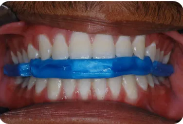
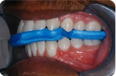
A two-piece bite registration technique by Roth121 called the “power centric bite registration” is believed to seat the condyles in the optimal,
anterior-superior CR position—or as Utt and colleagues141 wrote, “condyles centered transversely and seated against the articular disk at the posterior slope
of the articular eminences without dental interferences.”
Harmony of form is a prerequisite for harmony of function. There is no
of the masticatory musculature depends on the harmonious relationship
between the occlusion and the TMJ.
Stability is one of the treatment goal of orthodontic treatment, and to
get to that, a gnathological approach is necessary. Establishing equilibrium
between the teeth and the neuromusculature is critical because whenever there
is disequilibrium, the muscles will attempt to regain it. The evidence of this is
excessive tooth wear, tooth hypermobility, periodontal break down in the
presence of good oral hygiene.
If treatment goals include condyles seated in the fossa and an occlusion
that will not interfere with condylar border movements, then it is essential to
assess the occlusion with the condyles in centric relation.
This study was thus conducted with the aim of establishing the
discrepancy between Maximum Intercuspation (MI) and Centric Relation
(CR) in post orthodontic patients and to statistically evaluate the range of
REVIEW OF LITERATURE Literature has been reviewed under the following headings:
- Centric relation, Centric occlusion, maximum intercuspation and
concept of Functional occlusion
- Occlusal Interferences and TMD
- Malocclusion and TMD
- Orthodontic treatment and TMD
- Condyle and Disc position
- Methods of evaluating TMJ
Centric relation, Centric occlusion, maximum intercuspation and concept of Functional occlusion
Dentistry has not arrived at a consensus definition and concept of
CR. In 2004,Christensen20said that he and most practitioners “accept the concept that CR is the most comfortable posterior location of the mandible
when it is bilaterally manipulated gently backward and upward into a
retrusive position.” However, CR has not been recognized as a posterior,
retruded condyle position for almost 20 years41.
In 2000, Jasinevicius and colleagues63 found that faculty and students at seven dental schools could not agree on a unified definition of
CR. The definition of centric relation has changed numerous times over
the years. The definition of CR has evolved over the past Half-century
from being a posterior and superior position of the condyle in relation to
The glenoid fossa to an anteriorsuperior position. Glossary of
Prosthodontic terms published in the Journal of prosthetic dentistry has
Definition for CR and CO in 1st, 3rd, 5thand 6thedition of the glossary is as
follows:
1st Edition (1956)15
CR - Most retruded relation of the mandible to the maxilla when
the condyles are in the most posterior unrestrained position in the glenoid
fossa from which lateral movements can be made, at any given degree of
jaw separation.
CO - Not defined
3rd Edition (1969)56
CR - Most retruded physiologic relation of the mandible to the
maxilla to and from which the individual can make lateral movements. It is
a condition, which can exist at various degrees of jaw separation. It occurs
around the terminal hinge axis. The most posterior relation of the mandible
to maxilla at established vertical relation.
CO - Centered contact position of the lower occlusal surfaces against
the upper ones, a reference position from which all other horizontal positions
are eccentric.
5thEdition (1987)41:
CR - A maxillo mandibular relation in which condyles articulate with
anterior superior position against the slopes of the articular eminences. This
position is independent of tooth contact. This position is clinically discernible
when the mandible is directed superiorly and anteriorly and restricted to a
purely rotary movement about a transverse horizontal axis.
CO - Occlusion of opposing teeth when the mandible is in CR. This may or may not coincide with maximum intercuspation position.
Maximum intercuspation (MI) : the complete intercuspation of opposing teeth independent of condylar position143.
6thEdition (1994)143: Same as the 5thedition.
Parker (1978)98 states that contacts, which occur on the non working -side are not only damaging to the periodontium but probably also contribute to
the TMJ syndrome.
Roth (1981)120 has advocated the importance of treating patients in centric relation emphasizing the. relationship between temporomandibular
dysfunction and occlusal interferences since the 1970s.
Williamson (1981)151 in an interview on occlusion and TMJ dysfunction states that in centric relation both mandibular condyles are
simultaneously seated more superiorly on the posterior slope of the articular
eminences, with the menisci interposed properly in between. They are placed
in that position by the patient's own healthy musculature, which is contracting
Centric relation at first tooth contact is referred to as centric relation occlusion
(CRO) or retruded contact position (RCP). Williamson defines CO as that
position of the mandibular condyles when the teeth are in maximum
intercuspation.
However the glossary of prosthetic dentistry defined maximum
intercuspation as "the complete intercuspation of the opposing teeth
independent of the condylar position".
Gelb's (1985)50 concept for preferred CR position was one in which the condyle translated approximately half way down the posterior slope of the
articular eminence i.e. anterior mid condylar position.
Okeson (1989)95advocated an anterior superior condylar position and believed it to be the most stable joint position and also the musculoskeletally
stable position.
In the past, gnathologists suggested ' rearmost, uppermost and
midmost" to describe the condyle position in centric relation.
Dawson (1989)26 has stated that"rearmost and uppermost' is an incorrect description because the condyles cannot be in the rearmost
position when they are in the "uppermost" position and vice versa. He has
suggested that the condyles should be positioned most superiorly and
anteriorly against the posterior slope of the eminence when in centric
Pfeiffer - Flor and Pancherz (1991)99 in an extensive review concluded that canine guidance occlusion is just as acceptable as group
functioning occlusion. In addition, a natural dentition with canine guidance
occlusion will tend to become group functioning with time, due to wear of
the maxillary canine.
Roth (1995)123 has suggested that the diagnosis and treatment planning of orthodontic patients must be made with the mandible in centric
relation because orthodontic treatment is essentially full mouth reconstruction
of the patients own dentition.
Ramfjord and Ash (1995)108 Centric relation (CR) is a defined mandibular position from which interocclusal relationships are analyzed.
Cecere, Reef, and Pancherz (1996)19 investigated EMG recordings (of the anterior temporal and masseter muscles) when several factors were
varied: relocation of the electrodes between readings, the effect of not
removing electrodes and the use of new electrodes were compared following
various chewing / biting activities. They found that, depending on the time interval between recordings, the muscles considered, and the function
performed, individual errors ranged from 5% to 63%. The method error
increased significantly with a time interval between recordings. They stated
that quantitative electromyography of the masticatory muscles seems to have
Truitt, Strauss and Best (2009)140 conducted a study to determine whether there is a consensus among oral and maxillofacial surgeons and
orthodontists as to the definition of centric relation. There was no difference
between the 2 groups on the need for mounting models in centric relation for
use in orthognathic surgery. Regarding the definition of centric occlusion,
there was a significant difference between orthodontists and surgeons. The
results of this study show that there is a statistical lack of consistency among
practitioners regarding an absolute definition of centric relation as it relates to
orthognathic surgery. The inconsistency exists between specialties and within
practitioners in each specialty
Weffor and Solange Mongelli de Fantini (2010)158 conducted a study to measure condylar displacement between centric relation (CR) and
maximum intercuspation (MIC) in symptomatic and asymptomatic subjects.
And concluded that
- When the plane and the direction of the displacement were considered,
statistically significant differences between CR and MIC were
quantifiable at the condylar level in symptomatic and asymptomatic
individuals.
Occlusal interferences and TMD:
Ziebert and Donegan (1979)160 investigated 10 subjects (age range 20 to 64) who required occlusal adjustment to eliminate non
working - side contacts (among other occlusal features). Study models
were taken before and after occlusal grinding and 6 weeks later. Silicone
putty interocclusal records (taken in ICP and RCP) were taken and
repeated until identical records were obtained at each stage. Six patients,
each with a full compliment of teeth (excluding third molars), showed no
clinically perceptible slide from CR to CO 6 weeks after the occlusal
adjustment, but four patients (those who had a missing tooth but with
spaces closed) had occlusions that had relapsed after adjustment.
Agersberg and Sandstrom (1988)2 found that 75% of 15 and 22 years old subjects had unilateral tooth contacts in retruded position. 88%
and 89% respectively, of the individuals in both age groups had at least
one occlusal contact, usually defined as an interference in one or more of
the nine registered positions of the mandible. None had TMD.
Mohl and Ohrback (1992)87 suggested that occlusal adjustments should no longer be used in the management of
TMDs. Their arguments include the irreversibility and invasiveness
of occlusal adjustment, the lack of evidence for the causal role of occlusal
factors, the reported good short- term results of reversible therapies, and
Kirveskari (1997)71showed that several controlled clinical trials have failed to disprove the etiologic role of occlusion in TMDs. These
trials also suggest an effect for occlusal adjustment on chronic
headaches and on chronic neck and shoulder pain in comparison with
conventional treatments. Long-term studies have disclosed no adverse
effects of occlusal adjustment apart from transient tooth sensitivity in a
very small number of cases. In view of the possibility that occlusal
factors have a causal role in TMDs, research efforts on the role of
occlusion should be intensified, and teaching should be revised
accordingly.
Luther (1998)77 in his extensive review has questioned the validity of the results obtained by a few researchers to assess the effect of
an artificially introduced interference, often using electromyography. He
states that a number of weaknesses are present, and whether they actually
reflect the natural situation is debatable. Not all studies provide details of
the size of the interference and where this is described, the magnitudes are
large (0.05mm high, Riise and Sheikholesam, 0.05 to 0.75 mm,
Christensen and Rassouli). Some of the other weaknesses are a small
sample size and their background (student nurses and hygienists) might
haveinfluenced the subjects.
Luther concluded in his review that there are no long-term studies investigating the longevity of a functional occlusion following orthodontic
for all cases one must question the stability of this goal and address the
problem of whether repeated grinding to maintain it is a suitable form of
treatment. There is little evidence to show whether functional occlusion IS
always stable. Aubrey despite advocating a functional occlusion as a
treatment aim suggested that teeth move despite this and may need repeated grinding. The works of Forsell et al, and Kirverskari et al also
acknowledged this where repeated occlusal adjustments were needed.
Pahkala and Laine (2002)97conducted a study to focus on if early signs of different orofacial dysfunctions, e.g. misarticulations of speech,
problems in oral motor skills and TMD, malocclusions or occlusal
interferences could predict the development of temporomandibular
disorders (TMD) in adolescence. Altogether there were 94 children
referred for speech therapy and 93 controls who participated in all three
stages of this longitudinal study. In the whole sample the mean age during
the first examination was 7.6 years, during the second examination 10.6
years, and during the third one 15.4 years.Deviation on opening was
associated with problems in oral motor skills, and some signs of TMD
seemed to be related to each other. In addition, girls had a higher risk of
having several signs of TMD than boys did. In conclusion, tendency to
open bite, both mesial and distal molar occlusion and increased and
decreased overjet were occlusal anomalies associated with TMD.
Altogether, among 15-year-olds there seems to be both local and central
Barker (2004)9 in his study sought to determine how a balanced occlusion, providing uniform contact in centric relation, would affect signs
and symptoms of TMD. A randomly chosen group of 60 patients with
occlusal interferences and signs and symptoms of TMD used a mandibular
orthotic to balance their occlusions at centric relation (CR). When the
occlusions of symptomatic patients were balanced in CR, there was a
significant reduction or elimination of TMD complaints, suggesting a
relationship between balancing occlusion in CR and optimum
management of TMD.
Bonjardim and Lopes-Filho (2009)14 conducted a study was to find out the prevalence of temporomandibular disorder (TMD) in a sample
of university students and its relationship to gender, occlusion, and
psychological factors. According to our results, 50% of the subjects had
TMD, but it was of moderate or severe degree in only 9.18% of them. No
statistically significant association could be found between TMD and
gender or occlusion. TMD was found to have statistically significant
association with HADSa (anxiety) but not with HADSd (depression).
Malocclusion and TMD:
Mohlin and Kopp (1978)88 conducted a study on 56 patients with TMD between the age group of 16 to 62 years and noted that 16.1 % of the
sample had anterior open bite, 5.4% of the sample had deep overbite and
patients had unilateral crossbite and 16% of the patients had bilateral
crossbite. Mohlin and Kopp reported a positive correlation between
crossbite and interferences between RCP and ICP and mediotrusion
interferences. He also found that 8.5% had Class I occlusion, 19.7% had
Class II occlusion and only 1.8% had Class III occlusion.
Pullinger et at (1987)105 investigated 44 young adults (with a variety of malocclusions) with no history (or signs or symptoms) of
TMD and no orthodontic or other occlusal therapy. Using corrected
lateral tomograms, they found that 25% of the adults with Class I
malocclusion had posteriorly positioned condyles, 18% had anteriorly
positioned condyles, and 57% had concentrically placed condyles.
Egermark - Eriksson et at (1990)34 followed 238 subjects on a longitudinal basis for 4 to 5 years. Three different age groups were
involved (7, 11 and 15 years old). Few significant correlations were
found in any age group between morphological and functional
malocclusions, but the oldest group showed a significant positive
correlation between postnormal occlusions and large anteroposterior
distance between RCP and ICP. In the different age groups, non
working-side interferences were significantly correlated with and
number of malocclusions associated with anterior open bite, but most
of the correlations lay for such features as lateral open bite and
weak and only a few were significant (the strongest included unilateral
crossbite and extreme maxillary overjet).
Cohlmia, Ghosh, and Nanda (1996)22 evaluated the morphologic relationship of the condyle and fossa in patients with
different malocclusions and skeletal relationships. Pretreatment records of
232 orthodontic patients ranging in age from 9 to 42 years were examined.
Records included dental casts, lateral cephalometric radiographs, hand
wrist radiographs and corrected tomograms of right and left TM joints.
Non concentricity and mild asymmetry of the condyle fossa
relationship were commonly observed
Left condyle was found to be more anteriorly positioned than the
right with he mean joint space being 6.93% on the left and -1.24%
on the right
Skeletal and dental Class III patients demonstrated significantly
more anteriorly positioned condyles
No significant difference in condylar position between Class I and
Class II groups based on ANB or Angles classification
No significant difference in condylar position between group based on
Thor, Ekberg, and Nilner (1998)139 evaluated the masticatory efficiency and mandibular dysfunction in a total of 183 girls, aged 11 to 15
years. Six subjects had normal occlusion and 123 subjects had Class II
malocclusion. Examination included registration of signs and symptoms of
TMD. Subjects with normal occlusion presented significantly better
masticatory efficiency and ability than subjects with Class II
malocclusion. Few occlusal contacts and a large overjet predicted a
reduced masticatory efficiency. Subjects who reported frequent TMJ
clicking and subjects who estimated their overall symptoms of TMD as
moderate or severe also had reduced masticatory efficiency. The authors
concluded that masticatory efficiency and ability was partly dependent on
the occlusion and those symptoms of TMD influenced the masticatory
efficiency and ability.
Mohlin and Pilley (2004)90 performed a study in which A total of 1018 subjects were examined at the age of 11 years, 791 were reexamined at
15 years, 456 at 19 years, and 337 at 30 years. Anamnestic and clinical
recordings of temporomandibular disorder (TMD) were made. Morphology,
including calculation of peer assessment rating (PAR) scores, was recorded.
Previous history of orthodontic treatment was assessed. Muscular endurance
was recorded. The subjects completed four psychological measures. The
malocclusion prevalence, occlusal contacts, psychological factors, and
muscular endurance in subjects with no recorded signs and symptoms of TMD
age. The further development of TMD to 30 years of age was followed. PAR
scores were significantly higher in the subjects with the most severe
dysfunction. Apart from crowding of teeth, no other significant differences
were found between the groups with regard to separate malocclusions, tooth
contact pattern, orthodontic treatment, or extractions. A greater proportion of
subjects with low endurance were found in those with TMD. Significant
associations between TMD and general health and psychological well-being
as well as the personality dimension of neuroticism and self-esteem were
found.
Mackie and Lyons (2008)80 conducted a review of literature about the role of occlusion on TMD. Unfortunately, there appears to be no consensus
regarding the definition of a temporomandibular disorder within the literature
(Mohlin and Thilander, 1984; Okeson, 2003a), and there is considerable
variation among epidemiological studies. These studies report that between 5
and 50% of individuals experience TMD pain (Dworkin and Massoth, 1994),
with females comprising 75% to 84% of those affected (Dworkin et al., 1990).
This may be related to differences in pain measurement criteria or study
design, and women tending to present for treatment more readily than men.
Jerjes and Upile (2008)65conducted a study to explore the etiology of temporomandibular disorders and discusses the controversies in variable
treatment modalities.Pathologies of the temporomandibular joint (TMJ) and
its' associated muscles of mastication are jointly termed temporomandibular
pain in the joint and its surrounding area, jaw clicking, limited jaw opening
and headaches. It is mainly reported by middle aged females who tend to
recognize the symptoms more readily than males and therefore more
commonly seek professional help. Several etiological factors have been
acknowledged including local trauma, bruxism, malocclusion, stress and
psychiatric illnesses. The Research Diagnostic Criteria of the
Temporomandibular Disorders (RDC/TMD) is advanced to other criteria as it
takes into consideration the socio-psychological status of the patient. Several
treatment modalities have been recommended including homecare practices,
splint therapy, occlusal adjustment, analgesics and the use of psychotropic
medication; as well as surgery, supplementary therapy and cognitive
behavioral therapy. Although splint therapy and occlusal adjustment have
been extensively used, there is no evidence to suggest that they can be
curative; a number of evidence-based trials have concluded that these
appliances should not be suggested as part of the routine care. Surgery, except
in very rare cases, is discouraged since it is the most invasive alternative;
recent studies have shown healthier outcome with cognitive behavioural
Orthodontic treatment and TMD:
Sadowsky and Begole (1980)124 evaluated the status of TMJ function and functional occlusion by means of a questionnaire
and a detailed clinical examination in a group of 75 subjects. These
subjects were between 25 and 55 years of age who had been treated
orthodontically with full fixed appliances at least 10 to 35 years
previously, during adolescence. The findings were compared with those of
the control group adults with untreated malocclusion. Findings indicate
that in patients who underwent orthodontic treatment many years
previously the prevalence of TMJ signs and symptoms similar to that of
control group of adults with untreated malocclusions. However a trend
exists which suggests that subjects who had undergone extensive fixed
appliance orthodontic treatment many years previously may possibly have
a lower prevalence of TMJ problem than a similar group of adults with
untreated malocclusions.
Sadowsky and Polsen (1984)125 studied the relation between TMD and functional occlusion after orthJdontic treatment. The findings
from the total sample of 96 orthodontically treated subjects as compared
with 103 controls 'rom the Illinois study group was contrasted to the
findings from an independent study on 111 subjects who received
orthodontic treatment at .east 10 years previously and were compared with
111 adults with untreated malocclusion. Non extraction and extraction
similar in both studies with the prevalence of symptoms varying between
15% to 21 % and 29% to 42% for signs Ooint sounds), there being no
statistically significant differences between treated and untreated subjects
in either of the studies. The conclusion from the above two studies was
that orthodontic treatment performed during adolescence did not generally
increase the risk of developing TMD in later life.
Wyatt (1987)156recommemded the following for the diagnosis and treatment planning of orthodontic patients.
1) Etiologic factors that might cause upward and backward pressures on
the mandible should be reduced as much as possible.
2) Mechanotherapy that may cause upward and backward pressures on
the condyles is not recommended. Final detailed correction of dental
abnormalities should always consider optimal temporomandibular
health and function.
Gianelly, and Hughes (1988)52 evaluated the condylar position with corrected tomogram before orthodontic treatment in 37 consecutive
patients between the ages 10 and 18 yaars and compared them with 30
consecutively treated four premolar extraction cases at the completion of
treatment. All patients were treated with fixed appliances, 23 with
Edgewise and 7 with Begg technique. They could find no difference in
condylar position between the extraction and the untreated groups. It was
of distally positioned condyles. Condylar position tended to be centered on
average; however a wide variation in position was noted.
Heikinheimo, Salmi, Myllarniemi, and Kirveskari (1989)45
followed 167 subjects from ages 12 to 15 years and found that symptoms
of craniomandibular disorders (CMD) did not change in 38% of subjects,
increased in 32%, and decreased in 31 %. Half of those exhibiting
clicking at the age of 12 years lost the clicking by the age of 15 years.
Gianelly (1989)40 summarized the problem said to arise following certain forms of orthodontic treatment. According to him, an iatrogenic
cause of posterior condylar position is premolar extraction in orthodontic
treatment. Moreover, posterior condylar position within the fossa is
associated with an anteriorly displaced disc. He pointed out that it is not
clear whether posterior positioning of the mandible leads to internal
derangement or vice versa. Gianelly cited the work of Farrar and
McCarty (1983) who proposed that a space of less than 2.4mm posterior
to the condyle on a transcranial x-ray suggested an internal derangement.
Sadowsky and Theison (1991)126 reported on their prospective longitudinal study of 160 patients with an average age of 14 years, treated
with full fixed appliances for an average of 35 months. Of the 160 patients,
54% were treated with an extraction treatment strategy and 42.5% were
treated with nonextraction. In addition to recording symptoms, joint sounds,
treatment 25% of the patients had joint sounds, whereas 16.2% had sounds
after treatment. In 27 patients the sounds were not evident after treatment, in
3 patients there was no change in occurrence, and sounds developed in 13
patients by the end of treatment. Before treatment 14% of the patients had
reciprocal clicking, where as only 8% had reciprocal clicking after treatment.
Sadowsky (1992)127 studied the risk of orthodontic treatment producing TMD. His findings represented approximately 1300 previously
treated orthodontic patients from different regions of the world, treated
with varying strategies including extraction and nonextraction approaches,
and various appliance systems both fixed and removable. While most were
cross sectional studies some prospective longitudinal studies existed. The
overwhelming evidence supported the conclusion that orthodontic
treatment performed on children and adolescents was generally not a risk
for the development of TMD years later.
Morrant and Taylor (1996)92 studied the prevalence of TMD in patients referred for orthodontic assessments. Three hundred and one
unselected orthodontic referrals were assessed for TMD, using a
standardized questionnaire and clinical examination protocol. The mean
age of the patients was 13.4 years. Over one third of the 301 patients were
found to exhibit at least one sign of TMD, and two thirds had a
mandibular dysfunction index (MDI) score of 1, 2 or 3, indicating mild to
temporomandibular dysfunction. Statistically significant relationship was
found between patient age and mandibular opening, and TMJ noises. No
relationship was found between signs detected by clinical examination and
symptoms reported by the patients.
Williams (1998)147 in his study determined pretreatment and posttreatment condylar stability on forty TMD patients with symptoms of
pain in the muscles of mastication, TMJ sounds, attrition, interceptive
occlusal contacts and restricted range of motion. Axial corrected midcut
sagittal tomograms were made for the 80 temporomandibular joints before
treatment. Tracings from the tomograms. were used to measure and analyze
pretreatment position and posttreatment stability. Results showed that
pretreatment condyle fossa position was not concentric in 26 to 80 patients
(32.5%). Posttreatment condylar position showed no change and was
statistically stable
Kim and Graber (2002)68conducted a meta-analysis to investigate the relationship between traditional orthodontic treatment, including the specific
type of appliance used and whether extractions were performed, and the
prevalence of temporomandibular disorders (TMD) was investigated. After an
exhaustive literature search of 960 articles, we found 31 that met the inclusion
criteria (18 cross-sectional studies or surveys and 13 longitudinal studies). We
divided and extracted data from the 31 articles according to study designs,
summarized without further statistical analysis. The heterogeneous result
might originate from lack of a universal diagnostic system and the variability
of TMD. Because of heterogeneity, a definitive conclusion cannot be drawn.
The data included in this comprehensive meta-analysis do not indicate that
traditional orthodontic treatment increased the prevalence of TMD. It is
apparent that a reliable and valid diagnostic classification system for TMD is
needed for future research.
Henrikson and Nilner ( 2003)55conducted a study to To prospectively and longitudinally study symptoms and signs of temporomandibular disorders
(TMD) and occlusal changes in girls with Class II malocclusion receiving
orthodontic fixed appliance treatment in comparison with untreated Class II
malocclusions and with normal occlusion subjects. Prospective observational
cohortSixty-five girls with Class II malocclusion who received orthodontic
treatment, 58 girls with no treatment, and 60 girls with normal occlusion. The
girls were examined for symptoms and signs of TMD and re-examined 2 years
later. Additional records were taken in the orthodontic group during active
treatment and 1 year after treatment. All three groups included subjects with
more or less pronounced TMD, which showed individual fluctuation during
the ongoing study. In the orthodontic group, the prevalence of muscular signs
of TMD was significantly less common post-treatment. Temporomandibular
joint clicking increased in all three groups over the 2 years, but was less
prevalence of TMD than the orthodontic and the Class II group at both
registrations. Functional occlusal interferences decreased in the orthodontic
group, but remained the same in the othother groups over the 2 years.
Abrahamsson and Ekberg (2007)1 conducted a study to answer the question whether orthognathic surgery does affect the prevalence of signs and
symptoms of temporomandibular disorders (TMDs). The search strategy
resulted in 467 articles, of which 3 met the inclusion criteria. Because of few
studies with unambiguous results and heterogeneity in study design, the
scientific evidence was insufficient to evaluate the effects that orthognathic
surgery had on TMD. Moreover, the studies had problems with inadequate
selection description, confounding factors, and lack of method error
analysis.To obtain reliable scientific evidence, additional well-controlled and
well-designed studies are needed to determine how and if orthognathic
surgery alters signs and symptoms of TMD.
Slade and Diatchenko (2008)132 published in seminars in orthodontics, genetic markers can be of additional value in identifying
gene-environment interactions, that is, isolating population sub-groups, defined by
genotype in which environmental influences play a relatively greater or lesser
etiological role. This paper reviews concepts and study design requirements
for epidemiological investigations into TMD etiology. Findings are presented
from a prospective cohort study of 186 females that illustrate an example of
the gene encoding catechol-O-methyl-transferase, an enzyme associated with
pain responsiveness, risk of developing TMD was significantly greater for
subjects who reported a history of orthodontic treatment compared with
subjects who did not (P=0.04). While further studies are needed to investigate
TMD etiology, this genetic variant potentially could help to identify patients
whose risk of developing TMD is heightened following orthodontic treatment,
hence serving as a risk marker useful in planning orthodontic care.
MacFarlane, Kenealy and Kingdon (2009)79 conducted a study to investigate the relationship between orthodontic treatment and TMD with a
longitudinal study design.TMD prevalence was higher in females at all
follow-up points, except the baseline. Overall, incidences of TMD were
11.9%, 11.5%, and 6.0% at the first, second, and last follow-ups, respectively.
Females were more likely to develop TMD than males (hazard ratio [HR], 2.1;
95% CI, 1.3 and 3.3), and those with high self-esteem were less likely to
develop TMD (HR, 0.6; 95% CI, 0.4 and 0.8). There was no association
between orthodontic treatment and new TMD onset. The incidences of
persistent TMD were 20.0%, 34.9%, and 28.0% at the first, second, and last
follow-ups, respectively. Females were more likely to have persistent TMD
than males (HR, 2.5; 95% CI, 1.0 and 6.1). There was no association between
orthodontic treatment and persistent TMD. The only significant predictors of
TMD in adults aged 30 to 31 were female sex (odd ratio, 3.0; 95% CI, 1.1 and
CONCLUSIONS: Orthodontic treatment neither causes nor prevents TMD.
Female sex and TMD in adolescence were the only predictors of TMD in
young adulthood.
Hirsch (2009)60 conducted a study to find out whether orthodontic therapy is a risk factor for temporomandibular disorders (TMD) or
parafunctional habits such as bruxism is a question that has long been
discussed. The issue is highly relevant to public health due to the frequency of
these functional disorders in the general population and the sheer number of
orthodontic treatments. The study revealed no increased risk of TMD in
children and adolescents during orthodontic therapy, which seems to reduce
parafunctional activities and thus the likelihood of noncarious dental damage.
Condyle and disc position:
Pringle (1919)102 was the first to recognize and describe the condition of disc displacement.
Ricketts (1969)115 in a viewpoint article has described types of "improper occlusion" of which two are said to result in
posterior displacement of the condyle.
Weinberg (1970)145 concluded that bilateral asymmetric temporomandibular joint spaces are considered to be radiographic
evidence of dysfunction, with rare exceptions. Unilateral or bilateral
palpable muscle spasm. In his sample of 67 patients, only 10 were
asymptomatic, and these had concentrically positioned condyles.
Hoffman and Silverman (1973)61 conducted a study to relate condylar position in centric relation and in centric occlusion in dentulous
subjects. He found that 1) the condyle centers in a chin point guided CR
average 0.28 mm posterior to their position in CO. 2) in this CR position,
the condyles are sometimes superior to and sometimes inferior to their
position in maximum intercuspation in about equal numbers. The further
posterior the guided position is, the more likely the condyles are to be
inferior, and the less posterior it is, the more likely they are superior. 3)
Many displacement patterns were quite asymmetrical, exhibiting both AP
torquing and S-I torquing of the mandible.) Some subjects CR's were
slightly to the left and some were slightly to the right of their CO's.
Weinberg (1979)146concluded that condylar position in the fossa is a significant factor in TMJ dysfunction pain syndrome. Condylar
retrusion occurs much more (71 %) than other types of displacement in
acute TMJ dysfunction pain syndrome. Condylar retrusion also occurs
with enough frequency in the control group to indicate that retruded
mandibular position of centric relation does not necessarily orient the
condyle correctly in the fossa. Condylar concentricity was 6.4 times more
prevalent in the control group, this confirms that it is the optimum position
Sakuda and Tanne (1992)128 constructed a three dimensional configuration of the TMJ by 108 triangles for the condyle and glenoid fossa. The shortest distance between the condyle and
glenoid fossa was calculated inthe model along a line perpendicular to the center of gravity of a triangle on the condyle. The condyle and glenoid
fossa was determined in the anterior, posterior, middle, lateral and medial
areas of the condyle. Preliminary investigations revealed that the
technique was accurate, regardless of condylar rotations and inclinations
to the tomographic table. This approach provided a method for evaluating
the positional relationship between the mandibular condyle and glenoid
fossa in patients with TMD.
Braun (1996)16 studied a method of describing the geometric relationship of the condyle within the glenoid fossa on a sagittal
cephalometric radiograph. Improved glenoid fossa and condyle visualization
was achieved by adapting the Denar TMJ orthoceph Slimline cassette, which
contains rare earth intensifying screens to enhance the TMJ. A plastic
template was used to locate condyleI fossa relationship in habitual occlusion
and it was found that 89% of the subjects exhibited noncentric condyle
positions, while free of TMJ symptoms.
Tasaki and Westesson (1996)137 developed a classification system for disk displacement in the TMJ and studied the prevalence of the various
types of TMJ disk displacement in patients and symptom free volunteers. The
volunteers. Eight different types of disk displacement were identified in
addition to the superior disk position and a tenth indeterminate category.
Superior disk position was observed bilaterally in 18% of the patients and
bilaterally in 70% of the symptom free volunteers.
Hickman and Cramer (1998)57 evaluated twenty normal adults to determine masseter and temporal is activity in maximum static clench
with mandibular condyles in different therapeutic positions. Bimanually
manipulated, leaf gauge, centric occlusion and neuromuscular condylar
positions were studied. Results showed that when mandibular condyles
were placed anteroinferiorly in a neuromuscular position, total masticatory
muscle recruitment was greatest. In a bimanually manipulated or a leaf gauge
position, mandibular condyles were positioned superiorly, producing the least
amount of muscle recruitment.
Crawford (1999)23 conducted a study to determine if there is a relationship between condylar axis position as determined by the occlusion
and signs and symptoms of TMD, using the condylar position indicator (CPI).
A sample of subjects with ideal occlusions, defined as centric relation
approximating centric occlusion, was compared with a control sample of
untreated subjects. The comparison was based on written patient histories,
clinical exams, and CPI measurements. The ideal sample of 30 subjects was
selected from a population that had undergone full-mouth reconstruction using
gnathologic principles that included centric relation (CR) being coincident
subjects from the general population and was matched with the ideal sample
with regard to sex. A duplicate written exam was given to the subjects in the
ideal sample to assess symptoms prior to treatment. The CR bite registration
technique developed by Roth was used. When the pre- and posttreatment
examination scores of the ideal sample were compared, an 84% reduction in
symptoms was found after treatment. A high correlation (p<.001) between
signs and symptoms of TMD and CPI values was documented. Since condylar
axis position is dictated upon closure of the dentition into maximum
intercuspation and since condylar axis position was shown in this study to be
strongly correlated with TMD symptomatology, it can be concluded that a
statistically significant relationship exists between occlusion-dictated condylar
position and symptoms of TMD.
Vasconcelos and Menezes (2007)144 conducted a study in subjects who tested free of psychological stress to determine the position of the
condyle and whether that position was related to signs and symptoms of
temporomandibular disorders (TMDs).Forty subjects underwent psychological
evaluation to ensure freedom from psychological stress. The authors evaluated
tenderness of the masticatory muscles and temporomandibular joints (TMJs)
by means of bimanual digital palpation, and they determined the positions of
the condyle and disk by using magnetic resonance imaging.A total of 23.75
percent of the condyles were displaced away from the centric position either
anteriorly (3.75 percent) or posteriorly (20.00 percent). chi(2) analysis showed
disk, as well as a relationship between the position of the condyle and
tenderness of the TMJs. Although these relationships proved significant, it
cannot be assumed that displacement of the condyle away from the centric
position is predictive of TMD.Only two subjects were judged to have had
TMJ internal derangement. Thus, the absence of psychological stress seems to
have played a role in this finding.
Robinson and Marques (2009)118conducted a study to assess the disk-condyle-fossa relationship through magnetic resonance imaging and
determine its association with clinical signs and symptoms of
temporomandibular disorder in patients with myofascial pain and disk
displacement (with and without reduction).No significant association was
found between the independent variables (condylar position, disk position,
and condylar excursion) and the dependent variables (pain, maximal opening
of the mouth, maximal lateral movement). However, there was a significant
association between increased condyle excursion and pain (P = .035) and also
maximum mouth opening movement was associated with lateral movement (P
= .01; r = 0.31). Increase in condyle excursion may significantly influence
pain perception in TMD patients. The type of dysfunction and severity of
alterations on the imaging exams were not related to the severity of pain or
range of motion of the mandible.
Maizlin ZV, and Nutiu N (2010)162evaluated whether MRI findings of various degrees of disk displacement could be correlated with the presence
Concluded that Disk displacement on MRI correlated well with clinical
symptoms in cases of significant disk displacement and in cases of disk
displacement without reduction. When disk displacement with reduction was
mild, there was no statistically significant difference between symptomatic
and asymptomatic joints, which suggests that other causes should be
considered.
Methods of evaluating the TMJ:
Dawson (1971)24 believes that if severe symptoms can be triggered by tooth interferences, which deviate the condyle position less than the thickness
of thin cellophane, it is unrealistic to think that such minute deviations can be
detected by any existing radiographic technique.
Mikhail (1979)86 showed that TMJ radiographs made using the head positioner provided a valuable adjunct to diagnosis and treatment planning for
patients with mandibular pain dysfunction syndrome (MPDS) syndrome. It
was found that radiographic retrusion was more frequently accompanied by
signs and symptoms than bilateral condyle symmetry and protrusion.
Franco Mongini (1981)91 studied 8 men and 22 women with TMJ pain dysfunction syndrome. After clinical examination, transcranial
radiographs (TR) and serial tomography were performed in 5 to 7 different
planes. In 27 patients TR showed a condylar displacement. This was confirmed by serial tomography, which also showed that the apparent position
might vary from the medial to the lateral aspect due to rotation in same
Blaschke and Blaschke (1981)12 investigated the condylar position in 25 symptomatic patients using corrected lateral tomograms. They found that
mandibular condyles assumed widely varying positions within their respective
joints when teeth were in centric occlusion. Some of the normal subjects
presented joints in which the condyles could subjectively be classified as
severely retruded or protruded.
Dixon et al (1984)31 investigated the validity of transcranial radiographs in the diagnosis of anterior disc displacement in the TMJ. They
examined 34 patients who had sufficiently severe symptoms to warrant
arthrographic examination of one or both joints. Bilateral standardized
transcranial radiographs were also obtained in the open, closed, and rest
positions. The researchers found that transcranial radiographs were unreliable
in predicting the presence of anterior disc displacement. The method was more
valuable in correctly identifying disease free joints, but even then, the
radiographs were not totally accurate.
Juniper (1994)66 reported severe changes in condylar shape and size in the joints of 105 patients whose treatment involved arthrotomy for the
surgical treatment of their TMD. Some of the changes reported would be
hidden on standard radiographic views: 24% had medial bony excavaton, and
15% had lost part of their anterior surface and had an oblique shape.
Admittedly, these patients may have suffered quite severe symptoms, but this
Reef and Pancherz (1995)110 in a dried skull study investigating the use of orthopantomography for TMD diagnosis questioned the reliability
of such radiographs for TMD diagnosis. They reported simulation of condylar
flattening, osteophytes, joint space narrowing, and similar artifacts due to
changes in skull positions.
Ramfjord and Ash (1995)108 believe that TMJ radiographs are useful for differential diagnosis but are valueless in diagnosis and treatment of
TMJ dysfunction.
Emshoff and Bertram (1997)35 investigated the value of ultrasonography to determine the TMJ disc position in 17 patients. 100 TMJ
positions were studied by static and dynamic ultrasonography to analyze the
disk- condyle relationship. To compare the respective findings with those of a
diagnostic method offering high accuracy, coronal and sagittal magnetic
resonance imaging was carried out immediately afterwards. Results revealed
that static and dynamic ultrasonography are marginal in detecting the presence
of disk displacement, but dynamic ultrasonography is sensitive in detecting
the absence of disk displacement. The results indicate that both modalities are
insufficient in establishing a correct diagnosis for the presence or absence of
disk displacement.
Landes and Walendzik (2000)72 conducted a study in which sonographic examination was compared with MRI and axiography in
assessing temporomandibular joint (TMJ) function in 55 patients. Fifty-five
axiography, sonography and some also by MRI. The range of motion was
measured by sonography and axiography and the results compared using
Student's t-test. Anatomical details diagnostic for disc-displacement were
tested by sonography and MRI.The average time required for sonography was
2 min and for axiography 20 min. The mean measurement differences for
condylar movement in maximal mouth opening was 1.7 mm, for protrusion
1.6 mm and for mediotrusion 2.5 mm. The range of condylar movement as
measured by sonography and axiography coincided for opening and for
protrusion (statistically significant). No significance was found for lateral
excursions. The concordance in diagnosis of disc dislocation, hypermobility
and impaired range of motion when comparing ultrasound with MRI was
83%. All sonographic examinations were performed by one person only. Sixty
repeat examinations in patients produced no complaints and showed an
absolute range of difference of 0.6 mm, with a relative range of 7%. Student's
t-test was significant (p<0.05) (two repetitive measurements). Sonography
proved to be a fast and reliable method for evaluating the range of movement
of the TMJ. The lateral joint capsule, lateral disc, and upper condyle could be
demonstrated. Pathological processes such as anterior or lateral disc
displacement, disc perforation, seroma following contusion, capsular fibrosis,
crystalline structures in the synovia and fracture dislocation of the condyle
could be diagnosed with considerable reliability when compared with MRI.
However, the medial aspect of the joint, medial disc dislocation and the
Gomi and Yokoi (2007)54 conducted a study to evaluate the potential clinical application of digital linear tomosynthesis systems in imaging of the
temporomandibular joint (TMJ).A volumetric X-ray digital linear
tomosynthesis system (Sonialvision Safire; Shimadzu Co., Kyoto, Japan) was
used for TMJ imaging. Images were reconstructed with a modified
three-dimensional (3D) filtered backprojection (FBP) algorithm on this device. Our
modified 3D FBP was first evaluated using simulated images of numerical
phantoms. Next, patients with TMJ disease were evaluated with X-ray digital
linear tomosynthesis. The results indicate that numerical phantom and TMJ
visualization can be improved by the ability to produce sectional images that
blur overlying structures and yield 3D information. The flexibility of digital
linear tomosynthesis, as well as the fact that through an appropriate choice of
modified FBP algorithms it can suppress streak artefacts, makes it a
potentially appropriate approach for evaluating the TMJ. The utility of digital
linear tomosynthesis for the evaluation of TMJ was demonstrated. Digital
linear tomosynthesis may be considered as the imaging technique of choice in
the investigation of bony changes of the TMJ.
Ottl and Hardenacke (2008)96 conducted a retrospective study to systematically assess the temporomandibular joint (TMJ) using a newly
developed standardized evaluation form with 16 parameters based on MRI
diagnostics and to verify the reliability of the MRI diagnoses. One hundred
fifty-four (154) TMJs of 77 patients with arthrogenic complaints were
closed-mouth and open-closed-mouth positions. The sequences used were intermediary
FLASH and spin echo sequences using T1 or T2 weighting with fat
suppression. Examination of the reliability of the MRI evaluations of three
independent observers evaluating 60 randomly selected TMJ from among the
overall sample using the new evaluation form yielded an average Pearson
contingency coefficient of between 0.64 and 0.70 with regard to the 16
parameters studied. In the evaluation of the 77 left (L) and 77 right (R) joints,
the biplanar morphology of the disk was the most frequent with 24.7% (L) and
32.5% (R). In paracoronal projection, medial displacement of the disk was
seen in 7.9% (L, R) of the cases and lateral displacement in 6.4% (L) and
3.2% (R). The use of the new evaluation form, in combination with MRI of
the TMJ, demonstrated a substantial reliability of the diagnoses. In TMD
patients, the biconcave disk shape cannot be considered the sole normal,
standard situation. The presence of lateral and medial disk displacement
should be given more diagnostic consideration.
Guarda and Manfredini (2009)44studied the use of hyaluronic acid in temporomandibular joints with inflammatory/degenerative processes. This
investigation aimed at evaluating retrospectively the efficacy of intra-articular
injections of hyaluronic acid in elderly patients (aged >65 years) with
osteoarthritis of the temporomandibular joint as compared with those of a
group of adult non-elderly patients.At the end of the treatment period,
improvements in the elderly group were significant with respect to baseline
values, and functional limitation scores. In the non-elderly group, significant
improvements at the end of treatment were showed in all treatment outcome
variables, except than minimum pain at rest values. All improvements were
maintained over the six-month span of the follow-up period, and no significant
differences were showed between groups for any of the outcome variables,
except than functional limitation scores, which improved more in the elderly
group.These findings are not supportive for a difference in efficacy between
the elderly patients and the other subjects, even though further works on
different age groups are needed before generalization of results.
ZK Ahlers and Jakstat HA (2010)161 describes Computer-aided examination reports in clinical functional analysis of TMD. Clinical functional
analysis is the first step in the diagnostic cascade in the diagnosis of
craniomandibular dysfunction (CMD). This examination comprises palpation
of the muscles, auscultation of noises in the temporomandibular joint,
registration of jaw mobility as well as the search for dysfunctional contact
relations in static and dynamic occlusion. The structured evaluation of these
findings enables an initial diagnosis to be made as the basis for further
diagnostics and therapy. Because of the complexity of the material, both the
referring dentists and the patients expect a meaningful examination report.
However, writing it is extremely time-consuming. New software for the
semi-automatic generation of such illustrated medical reports has therefore been
developed to reduce internal costs and provide quality assurance. The CMD
initial diagnoses through a standardized interface from the dentaConcept
diagnostic software CMDfact. The CMD medical report assistant assigns
matching texts to the findings on the basis of random number generators and
combines these findings-dependent texts with illustrations of the most
important findings as well as standard texts. The software transfers the
multi-page examination report to the standard Microsoft Word for Windows word
processing program, saves the data by means of an individualizable file name
generator and uses the Word functions for the color printout.
Recording maxillomandibular relations:
Long (1973)75 was the first to introduce the leaf gauge in 1973. It consists of a number of shims usually made from acetate or plastic such as
exposed radiographic film cut into 1 by 5 em. pieces. These are then
connected at one end by means of a staple. Use of the leaf gauge is made by
adding individual leaves between maxillary and mandibular incisors and
instructing the patient to "bite on the back teeth, both sides at the same time".
If any posterior teeth begin to touch another leaf is added and the instructions
to the patient are repeated. When the patient is able to close on the leaf gauge
for approximately five minutes without posterior tooth contact, the mandibular
condyles are considered to be in a muscle dictated CR. Condyles are
positioned by the patients own muscular force, and not by the operator. The
leaf gauge permits this accomplishment without any tooth contact influencing
patterns have been shown to be affected by aberrant tooth contact or
deflections.
Williamson et al (1978)149 used the "centric ceph" technique on a sample of 46 patients divided into groups of Angle Class I and Angle Class II
cases. They concluded that there were differences in cephalometric
measurements with respect to the mandibular position, though most
differences were slight. They found that Class II patients exhibited the largest
discrepancies.
Williamson (1980)150 used the" veri check" to analyze the variability of CR records or to compare mandibular condyle position in the glenoid fossa
by using different types of interocclusal records. The Vericheck instrument
offers one method of testing sequential records for reproducibility. It
resembles an articulator in that models previously mounted by means of a face
bow recording and CR interocclusal records may be transferred to the
vericheck simply by attaching them to the upper and lower members by means
of the mounting plates.
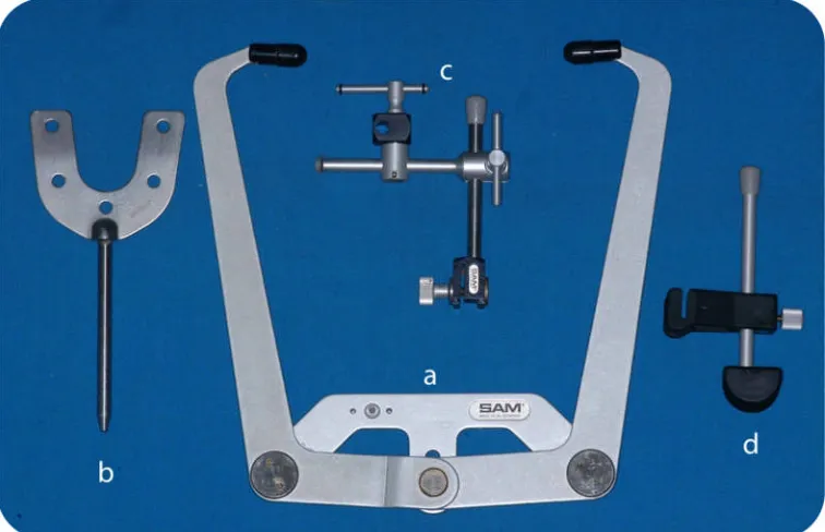
Slavicek (1988)130 described the use of the SAM articulator with the MPI to quantify differences between the joint dominated recorded condylar
position and the tooth dominated position of maximum intercuspal position.
The MPI is an instrument that allows the clinician or researcher to evaluate the
magnitude and directional displacement that occurs in the condylar axis from
CR to CO. The nature of the slide at the level of the occlusion most often does
member of the SAM2 articulator in which the condylar housings have been
replaced with laterally sliding cubes that contact the medial poles of the
condylar elements when related to the lower member of the articulator.
Yasuo and Kolling (1989)157 described a method in which mandibular border movements of a subject can be compared with the
movements generated by various articulators (fully adjustable Denar and
semiadjustable Denar) by using an electronic pantograph. Pantronic
pantograph detected differences between human border movements and those
generated by each articulator and method of adjusting it. In the horizontal
table, the semiadjustable articulator without immediate side shift always
showed the potential of greatest errors, especially as excursions started. When
the semiadjustable instrument was programmed with immediate side shift, its
movements were comparable with the fully adjustable articulator. Neither
articulator exactly simulated the subject's movements.
Wood (1994)154 studied "centrically related cephalometrics" with a sample of 30 patients whose casts were mounted on a whip mix articulator
(using Face bow at centric bite) His "shadowgraph technique permitted the
comparison between CO and CR. Limitations due to the radiographic
enlargement factor allowed the measurement of only a small number of
cephalometric angles. Wood stated that "although the statistical analysis
suggests the accuracy of the shadowgraph, it by no means renders the
technique clinically applicable". He did however conclude that mounted casts
hand held casts.
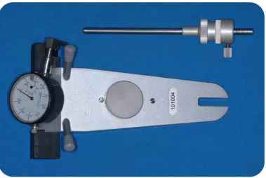
Klar and Kulbersh (2003)93conducted a study to examine the condylar position indicator (CPI, Panadent Corp, Grand Terrace, CA) readings of 200
consecutively finished patients in a gnathologically oriented practice to
determine the nature of the centric relation (CR)-maximum intercuspation
(MI) discrepancy pretreatment and posttreatment, in extraction and
nonextraction cases as well as to examine the possible effect of skeletal
morphologic parameters on treatment outcome. The study consisted of 200
patients, 127 women and 73 men, whose average age was 14 years and 2
months and ranged from 9 years to 55 years old. These patients were treated
using the Roth gnathologic treatment philosophy and straight-wire appliance.
Finished cases were defined as patients who completed treatment with a
gnathologic positioner. Initial records included upper and lower alginate
impressions, an estimated face-bow transfer, a maximum Intercuspation wax
bite using Moyco 10× pink wax (Moyco Industries Inc, Philadelphia, PA) and
a preliminary 2-piece Roth power centric CR bite registration using Delar blue
wax (Delar Corp, Lake Oswego, Or.). CPI measurements were made on all
casts, pretreatment and posttreatme