JOURNAL OFVIROLOGY,
0022-538X/97/$04.0010 May 1997, p. 3375–3384 Vol. 71, No. 5
Copyrightq1997, American Society for Microbiology
Bipartite Structure and Functional Independence of Adenovirus
Type 5 Packaging Elements
SUSANNE I. SCHMIDANDPATRICK HEARING*
Department of Molecular Genetics and Microbiology, State University of New York at Stony Brook, Stony Brook, New York 11794
Received 14 November 1996/Accepted 24 January 1997
Selectivity and polarity of adenovirus type 5 DNA packaging are believed to be directed by an interaction of putative packaging factors with the cis-acting adenovirus packaging domain located within the genomic left end (nucleotides 194 to 380). In previous studies, this packaging domain was mutationally dissected into at least seven functional elements called A repeats. These elements, albeit redundant in function, exhibit differences in the ability to support viral packaging, with elements I, II, V, and VI as the most critical repeats. Viral packaging was shown to be sensitive to spatial changes between individual A repeats. To study the importance of spatial constraints in more detail, we performed site-directed mutagenesis of the 21-bp linker regions separating A repeats I and II, as well as A repeats V and VI. The results of our mutational analysis reveal previously unrecognized sequences that are critical for DNA encapsidation in vivo. On the basis of these results, we present a more complex consensus motif for the adenovirus packaging elements which is bipartite in structure. DNA encapsidation is compromised by changes in spacing between the two conserved parts of the consensus motif, rather than between different A repeats. Genetic evidence implicating packaging elements as indepen-dent units in viral DNA packaging is derived from the selection of revertants from a packaging-deficient adenovirus: multimerization of packaging repeats is sufficient for the evolution of packaging-competent viruses. Finally, we identify minimally sized segments of the adenovirus packaging domain that can confer viability and packaging activity to viruses carrying gross truncations within their left-end sequences. Coinfec-tion experiments using the revertant as well as the minimal-packaging-domain mutant viruses strengthen existing arguments for the involvement of limiting, trans-acting components in viral DNA packaging.
Increasing the usefulness of adenovirus as a vector for the delivery of foreign genes into mammalian cells calls for the development of an in vitro packaging system to assemble re-combinant viruses carrying a packaging signal fused to heter-ologous sequences. Such a packaging system could entail a number of advantages. First, the size of the foreign gene, which is restricted to about 10 kbp in order to propagate the virus in tissue culture (3), could be expanded substantially. Second, with all viral genes absent, the overwhelming immune response of the host to adenovirus infection potentially could be mini-mized. This response, both humoral and cytotoxic T-cell me-diated, has mainly been ascribed to various viral gene products produced even in the context of replication-deficient second-generation adenovirus vectors (12, 32, 33). Little information is presently available about the basic mechanism of encapsida-tion of adenovirus DNA.
The assembly of adenovirus particles has been studied ex-tensively by using a large number of viral temperature-sensitive mutants blocked at different stages of assembly at the restric-tive temperature and by pulse-chase kinetic analyses (6–8, 10, 11). These early studies clearly established that adenovirus DNA is inserted into preformed, empty capsids late in the viral life cycle. Whether viral genomes enter the prohead in associ-ation with core proteins or as a separate entity is unclear, as is the exact structure of the viral DNA before and during entry.
cis-acting packaging sequences in the adenovirus genome are
required to direct selective encapsidation from the left end of the viral DNA. Polarity of adenovirus DNA packaging was
initially demonstrated in studies on viral incomplete particles containing viral DNA molecules of subgenomic length (5, 31), where a striking overrepresentation of left-end sequences was revealed, suggesting that DNA packaging occurs in a polar fashion from left to right. It was subsequently shown for ade-novirus type 16 (Ad16) and Ad3 that a cis-acting packaging domain is located within the left 390 bp (19, 29). Sequence alignments of Ad3 with Ad5 and Ad12, representatives of adenovirus subgroups A, B, and C, revealed a large degree of sequence conservation within the interval between nucleotides (nt) 237 and 491 (25). This conservation is likely due to a conserved packaging signal in addition to a number of tran-scription factor binding sites located within this region. Con-servation of the packaging sequences may reflect the depen-dence of all adenoviruses on a cis-acting encapsidation signal, suggestive of a similar mechanism of selective and polar DNA packaging for all adenovirus subgroups.
The cis-acting packaging domain in Ad5 is located in the left end 380 bp (16, 17, 20) and overlaps two distinct enhancer elements (Fig. 1A). Enhancer element I consists of a repeated sequence motif (21) and stimulates E1A transcription specifi-cally upon binding of a cellular nuclear factor, EF-1A (4). Mutations in element I affecting its function can be efficiently complemented by propagation of the virus in 293 cells, a cell line that constitutively expresses the viral E1A and E1B gene products (18). Element II enhances transcription in cis from all early transcription units by an unknown mechanism (22). Ele-ment II mutations result in a decrease in early transcription and, since some of the early gene products are required for DNA replication, in a corresponding reduction in virus growth. This cis-acting defect can be efficiently complemented in trans by providing all of the early gene products in a mixed infection with wild-type virus (22). Deletion of the Ad5 packaging do-* Corresponding author. Mailing address: Department of Molecular
Genetics and Microbiology, Health Sciences Center, State University of New York at Stony Brook, Stony Brook, NY 11794. Phone: (516) 632-8813. Fax: (516) 632-8891.
3375
on November 9, 2019 by guest
http://jvi.asm.org/
main resulted in nonviability, but wild-type growth could be restored by the substitution of the left-end 353 bp at the right end of the genome (21). The Ad5 packaging domain was shown to be flexible with respect to its position as well as orientation within certain boundaries. However, it must be located within 600 bp of the inverted terminal repeat (ITR) (20). Whether ITR sequences themselves are required for ad-enovirus DNA packaging has not been determined yet due to a cis requirement for DNA replication.
Detailed analysis of the Ad5 packaging domain by deletion as well as linker scanning mutagenesis (16, 17) revealed that it consists of at least seven functional units called A repeats due to their AT-rich character (Fig. 1B). These elements are func-tionally redundant, but in spite of their redundancy, they fol-low a hierarchy of importance, with elements I, II, V, and VI as the functionally most dominant repeats. In support of a model implicating the A repeats as binding sites for limiting,
trans-acting packaging components, cotransfection of an excess
of packaging-domain sequences and wild-type adenovirus ge-nomes resulted in a dramatic decrease in virus yield while DNA replication as well as late transcription were unaffected (17). This result is thought to reflect a competition event on-going between unlinked packaging sequences and the viral genomes for the recognition of limiting packaging compo-nents. Consistent with this model is the fact that there are spatial constraints between individual packaging elements (17), presumably because the protein components involved have to interact with the packaging elements in a coordinate fashion. Notably, elements I and II as well as elements V and VI are separated by a spacing of 21 bp, or two helical turns of the DNA. Factors bound to these repeats would come to locate to the same side of the DNA double helix to interact with each other and/or components outside the packaging domain to mediate recognition of the prohead followed by DNA encap-sidation.
Here we focus on an analysis of the 21-bp region separating A repeats I and II (AI and AII) and A repeats V and VI (AV and AVI). Mutagenesis within this region identifies additional sequence determinants as part of the packaging consensus motif. Of special importance is the presence of a CG
dinucle-otide located downstream of each of the four A repeats. Spac-ing between the consensus 59-TTTG-39 and the CG located downstream, rather than spacing between individual A repeats, appears critical. We propose an extended consensus motif for the packaging elements which could represent a binding site for one or several packaging factors. In addition, we define minimal packaging sequences that exhibit maximal packaging activity in functional assays when introduced into a packaging domain-minus background. These minimal packaging domains may prove useful for the construction of new adenoviruses in the field of gene therapy. Finally, we selected revertants from a packaging-deficient adenovirus and show that in each case, A repeats were amplified, providing genetic evidence that these elements act as independent units in viral DNA packaging. The revertants exhibit a phenotype in which viruses containing more A repeats have a competitive advantage over viruses with fewer A repeats in coinfections with either wild-type virus or each other. These results again support an argument for the existence of limiting protein components as part of the pack-aging machinery.
MATERIALS AND METHODS
Plasmids and virus constructions.Ad5 dl309, the parent for all viruses de-scribed in this report, is a phenotypically wild-type virus that contains a unique
XbaI cleavage site at 3.8 map units (24). dl309-194/316 and dl309-274/358,
de-scribed previously (16, 17), carry deletions of sequences between nt 194 and 316 and nt 274 and 358, respectively. dl309-274/376 carries a deletion between nt 274 and 376. Mutations were constructed originally in plasmids pKS-194/316 and pKS-274/358, which contain the left-end XbaI fragment (nt 1 to 1339) of each mutant virus cloned into the polylinker region of the pBS-KS (1) Bluescript vector (Stratagene). They were used as parents for the generation of a series of linker scanning and insertion mutations by the method of Kunkel (26). Plasmid pBR-53/322 comprises the left-end Ad5 XbaI fragment containing a deletion between nt 53 and 322 cloned into pBR322. It served as a parent for further deletion of sequences between nt 640 and 814 as well as nt 455 and 814, using
RsaI/NaeI and PvuII/NaeI restriction digestion, respectively. pBR-194/814 and
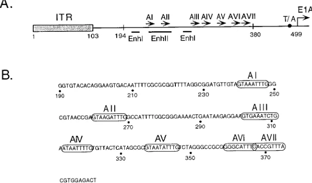
pBR-53/814 have sequences between nt 194 and 814 and nt 53 and 814 deleted. A monomer and dimer of viral sequences located between nt 334 and 385 which contain AV, AVI, and AVII was cloned into the 194/814 deletion. A dimer of the nt 334 to 385 fragment as well as 12 head-to-tail copies of an oligonucleotide containing AVI ((59-TCGACCGCGGGGACTTTGACC-39:59-TCGAGGT CAAAGTCCCCGCGG-39) were cloned into the 53/814 deletion in either ori-entation. All mutations were verified by nucleotide sequence analysis. The mu-tations were subsequently rebuilt into intact viruses by the method of Stow (30). FIG. 1. (A) The Ad5 genomic left end and packaging domain. The schematic diagram shows the ITR, the packaging/enhancer region (nt 194 to 380), and the E1A 59flanking region. Numbers indicate nucleotide positions relative to the left-end terminus. The packaging repeats (AI through AVII) are represented by arrows. The ITR is represented by a shaded box, and the E1A transcriptional start site is indicated by an arrow at nt 499. Transcriptional elements include a TATA box motif (T/A; black circle) and enhancer elements I and II (EnhI and EnhII; indicated by lines). (B) Nucleotide sequence of the Ad5 packaging domain. Numbers correspond to nucleotides relative to the left-end terminus. AI through AVII are circled.
on November 9, 2019 by guest
http://jvi.asm.org/
[image:2.612.149.463.76.260.2]Virus was amplified and titers were determined on 293 cells. Mutant viruses were screened by restriction analysis of viral DNA obtained from infected 293 cells by the Hirt procedure (23). The authenticity of all linker scanning mutations was confirmed by amplification of viral left-end sequences spanning nt 1 to 1347 by PCR; the left-end fragments were gel purified and used as templates for the Double Stranded DNA Cycle Sequencing System (GIBCO BRL).
Cell lines and infections.293 cells (18) and A549 cells, a human lung epithe-lium cell line, were maintained as monolayers in Dulbecco modified Eagle medium containing 10% bovine calf serum (HyClone). Virus stocks were gen-erated by three freeze-thaw cycles of infected cell lysates, and titers were deter-mined by plaque assays on 293 cells. Alternatively, virus particles were purified by CsCl equilibrium gradient sedimentation and titers were determined by op-tical density, whereby 1 opop-tical density unit at A260equals 1012particles. Virus
infections were performed at a multiplicity of infection of 3 PFU per cell or 200 particles per cell for 1 h at 378C. Cells were then washed twice with Tris-buffered saline solution and overlaid with fresh medium.
Selection of revertants of a packaging-deficient adenovirus.The parent virus for the selection of revertants was constructed as follows. Plasmid pBR-194/ 4551AVI6ENH2contains the Ad5 left-end Xba fragment carrying a deletion between nt 194 and 455, with six copies of an oligonucleotide containing AVI (see above) introduced at the deletion junction. Insertion of the AVI head-to-tail multimer created an Xho site at the right junction, which was used for the insertion of four copies of the E1A core enhancer element binding sites to ensure efficient E1A expression (2, 4). An EcoRI site separates the hexamer of AVI from the E1A core tetramer in the resulting plasmid, pBR-194/ 4551AVI6ENH1. The construct was then rebuilt into a dl309 virus background. The resulting virus had acquired two additional A repeats during the first growth cycle (see Results). To avoid recombination events between adenovirus left-end sequences integrated in the genome of 293 cells and parental genomes, the virus was subsequently propagated on A549 cells after initial recovery of plaques from 293 cells. After 12 passages of a number of independent isolates, an accelerated onset of cytopathic effect was evident. Putative revertants were plaque purified and amplified, and the revertant phenotype of each of the viruses relative to the parent was confirmed in single-infection and coinfection experiments. Left-end sequences of parent and revertant viral DNAs were compared by restriction and nucleotide sequence analysis as described above.
Determination of virus yield and packaging efficiency.To determine virus yield in a single infection, infected cell lysates were prepared 48 h postinfection, and the amount of infectious virus was determined by plaque assays on 293 cells. Packaging efficiency of the mutant viruses was determined in a coinfection of 293 cells with both mutant and wild-type dl309 virus. Forty-eight hours postinfection, one half of the cells was used to isolate total nuclear DNA; the other half was used for the preparation of viral DNA from purified virions as previously de-scribed (17). Briefly, total nuclear DNA was isolated from infected cells following the addition of Nonidet P-40 to 0.6% and precipitation of the nuclei by centrif-ugation. Viral DNA was prepared after lysis of infected cells in 0.2% deoxy-cholate and 10% ethanol at pH 9. Following treatment with RNase A and DNase, virions were lysed in 0.5% sarcosyl, and viral DNA was digested with proteinase K, phenol-chloroform extracted, and precipitated with ethanol. Both DNA preparations were digested with XbaI (XbaI and EcoRI for the revertant viruses and their parent) to distinguish between mutant and wild-type DNAs and quantitated by Southern blot hybridization (27) using pE1A-WT (21),32P
la-beled by the random primer method (13), as a probe. The relative intensities of the bands in autoradiograms were determined by densitometric scanning, using blots that were exposed to X-ray film without an intensifying screen. Quantita-tion of the data was performed by using the public domain NIH Image program (written by Wayne Rasband at the National Institutes of Health and available from the Internet by anonymous ftp from zippy.nimh.nih.gov or on floppy disk from NTIS, 5285 Port Royal Rd., Springfield, VA 22161, part no. PB93-504868). The data presented for virus yield in the single infections and the data for packaging efficiency based on coinfection experiments represent the averages of three to five independent experiments.
RESULTS
Spatial and sequence determinants important for viral DNA
packaging in the spacer region between A repeats.As
previ-ously reported, changes of spacing between different A re-peats, as well as between A repeats and regions outside the packaging domain, can have dramatic effects on viral packag-ing efficiency (16, 17). In this study, we focused on potential spacing requirements as well as sequence requirements within the two 21-bp spacer regions separating AI and AII, and AV and AVI. These four packaging elements have previously been implicated as the most relevant A repeats within the functional hierarchy of packaging elements (17). It was hypothesized that the separation of AI from AII and of AV from AVI by 21 bp, or two turns of the DNA double helix, may reflect a
require-ment for an interaction of trans-acting packaging components with packaging repeats on the same face of the DNA.
We constructed a panel of mutant viruses carrying linker scanning and insertion mutations within the 21-bp spacer re-gions between AI and AII as well as between AV and AVI. In view of the functional redundancy of the A repeats, we intro-duced all mutations between AI and AII in the context of a deletion of AIII, AIV, and AV (dl309-274/358). Mutations in the spacer region between AV and AVI were constructed in a mutant background with a deletion of AI, AII, and AIII (dl309-194/316). We tested all recombinant viruses in two indepen-dent assays. In the first assay, infectious virus yield was deter-mined by plaque assays after a 48-h single infection of 293 cells. The adenovirus DNA packaging domain overlaps an enhancer element (element II) which stimulates transcription from all viral early transcription units in cis by an unknown mechanism. Mutations affecting the function of this element result in a decrease in viral DNA replication and consequently in a re-duction of overall viral growth (22). This rere-duction (element II phenotype) can be complemented in a coinfection with wild-type virus providing all viral gene products in trans. To deter-mine what portion of the reduction in overall growth as ob-served in the single infection is caused by a packaging defect, we based our second assay on a coinfection of 293 cells with the mutant and wild-type viruses. Total replicated DNA and pack-aged DNA were isolated from 293 cells after a 48-h infection. Mutant and wild-type DNAs were distinguished by restriction digestion, and their relative amounts were quantitated by Southern blot analysis. That way, the amount of packaged mutant virus DNA relative to the coinfecting wild-type DNA could be normalized to the levels of replicated DNA of each mutant and wild-type virus. This quantitation of the data en-abled us to determine the reduction of viral DNA packaging independent of an element II phenotype whereby the coinfect-ing wild-type virus served as an internal control.
Figure 2A shows the mutations introduced into the spacer between AI and AII and summarizes the results obtained with these mutants. Figure 2C shows a Southern blot of a represen-tative coinfection experiment. The parent virus, dl309-274/358, displayed a sixfold reduction in a single infection and a twofold decrease in packaging efficiency in a coinfection. A 4-bp inser-tion between AI and AII (IN4 251/252) resulted in a dramatic decrease in virus growth in a single infection, and packaged mutant viral DNA was not detectable in the coinfection. A 3-bp substitution overlapping the site of this insertion (LS 250–252) had no effect on virus growth compared to the parent virus. Mutant LS 255–260, carrying a 6-bp linker scanning mutation within the spacer, was dramatically defective in the single-infection and coinfection experiments. This result could reflect the fact that the first two nucleotides (GT) of the AII consensus sequence (17) were mutated; alternatively, it could implicate previously unrecognized sequences as part of the
cis-acting packaging consensus motif. To distinguish between
the two possibilities, 3-bp substitutions were introduced across the region between nt 249 to 261. Mutants LS 250–252, LS 253–255, and LS 259–261 grew and were packaged at the levels of the parent virus, whereas LS 256–258 displayed a 68-fold reduction in growth and packaged DNA was not detectable in a coinfection. This finding suggested that the defect observed with the mutant virus LS 255–260 was due to mutation of the three nucleotides CCG. The dinucleotide CG within this motif is located 11 nt downstream of the central thymidine in the thymidine triplet of AI. A CG dinucleotide is conserved in the same position relative to AI, AII, AV, and AVI (Fig. 1B), indicating that it may function in conjunction with the previ-ously defined consensus packaging motif. The mutant virus LS
VOL. 71, 1997 ADENOVIRUS PACKAGING 3377
on November 9, 2019 by guest
http://jvi.asm.org/
CG emphasizes the importance of the CG located downstream of AII. During the construction of the parent virus dl309-274/ 358, the CG dinucleotide downstream of AII had originally been deleted but was coincidentally replaced by an Xho linker (16). Transversion of the dinucleotide in the mutant LS CG resulted in a dramatically defective phenotype of the mutant virus in single-infection and coinfection experiments, suggest-ing that even in the context of nonviral sequences, the presence of the dinucleotide CG downstream of AII is critical for viral DNA packaging.
Insertion of four nucleotides between AI and AII in mutant
IN4 251/252 dramatically affected viral growth and packaging efficiency. This mutation, however, alters both spacing between AI and AII and the spacing between AI and the CG dinucle-otide downstream of it. To distinguish between these two pos-sibilities, we rebuilt IN4 251/252 as well as a 4-bp mutation to
FIG. 2. Mutations surrounding AI and AII. (A) The sequence of the parent virus dl309-274/358 between nt 231 and 361 is shown at the top. AI and AII are boxed; an Xho linker present at the 274/358 deletion junction is underlined. A schematic of the dl309-274/358 deletion is shown just below, with the deletion indicated by a dark bar (nt 275 to 357) and the sequences between nt 249 to 261 indicated. The mutant viruses described in the text are depicted underneath and are named according to the nucleotides mutated. The site of a 4-bp insertion or the nucleotides mutated are shown below the parent virus nucleotide sequence, with the corresponding nucleotide changes indicated. All mutant viruses except for LS CG carry mutations within the region between nt 249 and 261. Mutant LS CG has a mutation in the CG sequence in the Xho linker (represented by a bracket). Mutant virus yields in the single infections are expressed as fold re-duction in yield relative to that of the wild-type virus. The results from the coinfection experiments and Southern blot analysis (Coinf.) are expressed as fold reduction in packaged mutant DNA relative to packaged wild-type DNA. These data were normalized to the levels of viral DNA (mutant and wild type) present in total nuclear DNA. ND, packaged viral DNA was below the level of accurate quantitation. (B) Viral mutants with 4-bp insertions in the spacer region between AI and AII in the background of the parent virus 274/376. The nomenclature and results of viral infection are as described for panel A. NV*, transfection exper-iments yielded plaques which could not be amplified without reversion to wild-type virus. (C) Southern blot analysis of viral DNA represented either in total nuclear DNA or in virion particles isolated from 293 cells coinfected with wild-type virus and the mutant viruses depicted in panel A. Total nuclear DNA or virion DNA was digested with XbaI and subjected to Southern blot analysis using an Ad5 left-end fragment as a32P-labeled probe. The corresponding left-end
fragments of mutant (M) and wild-type (WT) genomes are indicated on the left. The mutant viruses tested were dl309-274/358 (lanes 1), LS 250–252 (lanes 2), LS 253–255 (lanes 3), LS 256–258 (lanes 4), LS 259–261 (lanes 5), LS 255–260 (lanes 6), IN4 251/252 (lanes 7), and LS CG (lanes 8).
on November 9, 2019 by guest
http://jvi.asm.org/
the right of the CG dinucleotide, IN4 258/259, into a mutant virus background lacking AIII, AIV, AV, AVI, and AVII (dl309-274/376 [Fig. 2B]). This mutant virus background was selected to exclude the possibility that effects on viral DNA packaging are not due to changes of spacing between AI and AII but instead are due to changes of spacing between AI and the remaining repeats to the right of AII (AVI and AVII) present in the dl309-274/358 parent virus. The results of single and coinfections with these viruses are displayed in Fig. 2B. The parent virus dl309-274/376 is 165-fold reduced in the single infection, and packaged mutant virus DNA is not detectable in the coinfection due to the large truncation of the packaging domain. Mutant 274/376:IN4 258/259 displayed a very similar phenotype, whereas mutant 274/376:IN4 251/252 was nonvia-ble. These data emphasize that rather than a 21-bp spacing between AI and AII, the spacing between AI and the CG dinucleotide located 11 bp to the right appear to be critical for viral DNA packaging.
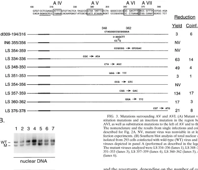
We introduced a similar set of mutations into the spacer region between AV and AVI in the context of a mutant virus that carries a deletion of AI through AIII (dl309-194/316). The results are summarized in Fig. 3A, and a representative South-ern blot of a coinfection experiment is shown in Fig. 3B. The parent virus was reduced threefold in overall growth in a single infection, and its packaging efficiency was sixfold less than that of the coinfecting wild-type virus. Insertion of 6 nt into the CG located 11 bp to the right of AV resulted in a nonviable virus (IN6 355/356). Two mutant viruses, LS 354–359 and LS 354– 356, with 6- and 3-bp substitutions overlapping the CG dinu-cleotide, respectively, also were nonviable. Their defective phenotypes implicate the CG dinucleotide downstream of AV as an important determinant for adenovirus DNA packaging in accordance with the previous set of mutants. In contrast to the previous results, however; mutation of the CG dinucleotide downstream of AVI (mutant LS 376–378) did not significantly affect viral packaging efficiency; overall growth in the single infection was reduced 21-fold, but virus growth in a coinfection was not affected. Most of the remaining 3-bp substitution mu-tants displayed a phenotype that is reminiscent of the element II phenotype described above. That is, defects observed in a single infection could be fully or largely complemented in a coinfection with a wild-type virus. The two most dramatic ex-amples of this phenotype were caused by the mutations in LS 348–350 and LS 357–359. These mutants exhibited a small-plaque phenotype, which could, at least partially, account for the incongruities between the data from the single-infection and coinfection experiments. In spite of the generally more complex phenotypes observed with this panel of mutants, the presence of the dinucleotide CG downstream of AV appears to be necessary for viral DNA packaging as shown above for AI and AII. We suggest an extended consensus packaging motif for AI, AII, and AV, which in addition to the previously de-fined AT-rich motif contains a dinucleotide CG in position 11 and 12 downstream of the central thymidine of the invariable thymidine triplet (Fig. 4). This dinucleotide is also conserved downstream of AVI, but our mutational studies do not confirm its significance with respect to viral DNA packaging.
Defining minimal adenovirus packaging domains with
max-imal function in vivo.The adenovirus packaging repeats are
functionally redundant, and in addition to the seven known A repeats, the presence of other packaging elements located out-side the previously defined packaging domain has been sug-gested (17). For these reasons, it has been difficult to define a packaging domain of minimal size which is necessary and suf-ficient for adenovirus DNA packaging in vivo. We began our search for such a minimal packaging domain by deleting
re-gions flanking the packaging domain in the context of a mutant virus 53/322, which carries only AV, AVI, and AVII. Figure 5 shows the parent virus 53/322 and two viruses derived from it, 53/322:640/814 and 53/322:455/814, which combine the 53/322 deletion with deletions extending from nt 640 to 814 and from nt 455 to 814, respectively. None of these viruses show signif-icantly reduced abilities to package their DNA in a coinfection with wild-type virus. We conclude from these data that the regions between nt 53 to 322 and between nt 455 and 814 do not contain sequences with a critical function for viral DNA packaging in the context of a packaging domain that consists of AV, AVI, and AVII.
It has previously been demonstrated that the insertion of multimeric A repeats can rescue the nonviable phenotype of a virus lacking AI through AV (17). In a similar approach, we used a deletion of the entire packaging domain between nt 194 and 814 for the reinsertion of sequences that may function as minimal packaging domains (Fig. 5). The nt 814 position was chosen since previous results demonstrated that the Ad5 pack-aging sequences must be positioned within 600 bp of the left terminus (20). Thus functional packaging elements to the right of nt 800 appeared unlikely. Based on the results just de-scribed, we first introduced a monomer and dimer of a frag-ment containing AV, AVI, and AVII, which we will refer to as A(V-VII) and A(V-VII)2, respectively. The fragments were inserted in both orientations. The parent virus 194/814 was nonviable, but viability could be restored by a monomeric and dimeric insert of AV, AVI, and AVII. The dimeric insert was significantly more effective at directing packaging than the monomeric insert, and packaging of the monomer was favored in the forward orientation relative to the reverse orientation. Importantly, the insertion of a forward dimer resulted in full restoration of packaging efficiency to wild-type levels. These data suggest that a dimer of AV, AVI, and AVII can function as a minimally sized, yet maximally functional, packaging do-main in an orientation independent manner.
We next introduced (AV-VII)2into an even larger deletion background, eliminating sequences between nt 53 and 814. The nonviable phenotype of the parent virus 53/814 was also res-cued by insertion of (AV-VII)2in either orientation. Packaging efficiency was three- to fourfold less than that of the coinfect-ing wild-type virus and was independent of the orientation of the insert. Overall growth in a single infection was reduced 82-fold in the forward, and 16-fold in the reverse, orientation. Again, the plaque size with these mutants was small, which may partly explain the large difference between single and coinfec-tion results.
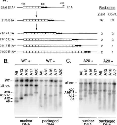
Selection of revertants from a packaging-deficient adenovi-rus.To generate a packaging-deficient virus as a parent for the selection of revertants, we constructed a virus with a deletion of sequences between nt 194 to 458 which eliminated the viral packaging domain (Fig. 1A). Six copies of AVI (21-bp oligo-nucleotide [see Materials and Methods]) were introduced at the site of the deletion, as well as four E1A enhancer repeats (2, 4) to ensure efficient E1A transcription (Fig. 6A). The parent virus was plaque purified from 293 cells, and a number of independent isolates were passaged on A549 cells to mini-mize the chance of recombination events between adenovirus left-end sequences integrated in the genome of 293 cells and parental genomes. During the first growth cycle on 293 cells, the parent virus had already acquired two additional copies of AVI (Fig. 6A); we therefore refer to it as 21/8 E1A1. After 12 passages on A549 cells, an accelerated onset of cytopathic effect was evident. Viruses were plaque purified and amplified, and growth and packaging efficiencies of several independent isolates were determined on 293 cells. Figure 6A shows the
VOL. 71, 1997 ADENOVIRUS PACKAGING 3379
on November 9, 2019 by guest
http://jvi.asm.org/
results of single infections and coinfections of the parent virus and four revertants. The parent virus 21/8 E1A1was reduced 32-fold in growth in a single infection relative to the wild type. Packaging efficiency was reduced correspondingly in a coinfec-tion with wild-type virus. Left-end sequences of the revertants were analyzed by restriction digestion and sequencing and were found to harbor precise amplifications of AVI, with 12, 16, 17, and 20 elements present, compared to 8 elements in the parent virus. All revertant viruses exhibited a significant (11- to 16-fold) increase of viral growth in the single infection and a comparable increase of packaging efficiency in the coinfection relative to the parent virus, indicating that amplification of AVI provided the viruses with a competitive advantage. The fact that revertants of 21/8 E1A1evolved through the ampli-fication of A repeats presents genetic evidence that these se-quences function as independent units in viral DNA packaging. An example of a Southern blot prepared from 293 cells coinfected with wild-type virus and the parent virus (21/8 E1A1) or its revertants is shown in Fig. 6B. The relative mo-bility of the left-end fragment varies between the parent virus
and the revertants, depending on the number of copies of AVI present. Each of the revertants packaged viral DNA in the coinfection experiment with increased efficiency relative to the parent virus. The faint band above the AVI fragment with the parental virus (Fig. 6B, lane 1) presumably represents genomes with already multimerized packaging repeats, suggesting a strong evolutionary pressure for the presence of more than eight copies of AVI. We also tested the packaging abilities of the parent virus and of the revertant viruses carrying 12, 16, and 17 copies of AVI in a coinfection with the revertant
[image:6.612.106.509.84.431.2]car-FIG. 3. Mutations surrounding AV and AVI. (A) Mutant viruses with sub-stitution mutations and an insertion mutation in the region between AV and AVI, as well as substitution mutations to the left of AV and to the right of AVII. The nomenclature and the results from single infections and coinfections are as described for Fig. 2A. NV, mutant virus was nonviable in at least three trans-fection experiments. (B) Southern blot analysis of total nuclear and virion DNA isolated from 293 cells coinfected with wild-type (WT) virus and the mutant (M) viruses depicted in panel A (performed as described in the legend to Fig. 2C). The mutant viruses analyzed were LS 334–336 (lanes 1), LS 348–350 (lanes 2), LS 351–353 (lanes 3), LS 357–359 (lanes 4), LS 360–362 (lanes 5), and LS 376–378 (lanes 6).
FIG. 4. Alignment of AI, AII, AV, and AVI. The packaging repeats are boxed; invariable nucleotides are indicated by lines between the sequences. Shown below is an alignment of the individual A repeats. Invariant nucleotides between these repeats are indicated by large letters. A new consensus motif for the Ad5 packaging elements is shown at the bottom.
on November 9, 2019 by guest
http://jvi.asm.org/
[image:6.612.317.556.549.682.2]rying 20 A repeats. These viruses differ from each other only by the copy number of AVI present in place of the viral packaging domain. Figure 6C shows Southern blots prepared from total nuclear DNA as well as packaged DNA from these coinfec-tions. The uppermost band is shared by all viruses and is composed of the four E1A enhancer repeats in conjunction with the region between nt 458 and 1339. The lower fragments contain nt 1 to 194 as well as the AVI multimers. All viruses replicated to equal levels in a coinfection with the 21/20 E1A1 virus (lanes 1 to 4). When packaged DNA was analyzed, how-ever, packaging of the parent virus as well as the revertant virus with 12 A repeats was substantially reduced (lanes 5 and 6). Viruses harboring 16 and 17 copies of AVI packaged their DNA as efficiently as the 21/20 E1A1revertant (lanes 7 and 8). These data strongly support the notion that viruses with more packaging elements have a competitive advantage over viruses with fewer elements when they are challenged to compete with each other. All four revertants, when tested in a single infec-tion, exhibited similar growth properties (Fig. 6A), which strengthens our hypothesis that trans-acting packaging compo-nents are required to bind the packaging repeats before DNA encapsidation can ensue. Such packaging factors may be lim-iting, which would result in preferential encapsidation of ge-nomes containing more packaging elements in a coinfection experiment.
We also inserted 12 head-to-tail copies of AVI in either orientation into the 53/814 deletion as shown in Fig. 7. The resulting mutant viruses, 194/814:AVI/12F and 194/814:AVI/ 12R, were 22- and 48-fold reduced in the single infection, with a corresponding decrease in the coinfection of 17- and 21-fold. Sequencing of viral left-end fragments revealed more than 12 copies of AVI in both viruses, indicating that an amplification
of packaging elements had taken place. At least 18 copies were present in mutant 194/814:AVI/12F, and at least 15 copies were present in mutant 194/814:AVI/12R. This amplification event is reminiscent of the multimerization of A repeats in revertants selected from a packaging-deficient mutant virus described above. These data confirm the results obtained with the selection of revertants carrying amplified packaging re-peats.
DISCUSSION
Adenovirus DNA packaging is often likened to DNA pack-aging in double-stranded DNA bacteriophage systems such as phage lambda orf29. Viral DNA is specifically selected from the pool of total DNA for its packaging, and insertion of the genome into preformed, empty capsids proceeds in a polar fashion. For these two phages, it has clearly been shown that phage-encoded packaging factors selectively bind packaging sequences located within the genomic left-end terminus. For-mation of a nucleoprotein complex on the packaging domain then marks the respective molecule as a bone fide packaging substrate followed by recognition of the empty capsid and insertion of the DNA with left-to-right polarity (1, 9).
The identities of such packaging factors in the case of ade-novirus are still unknown, but several lines of evidence strongly support the existence of limiting trans-acting packaging com-ponents. A number of packaging mutants were previously re-ported to exhibit a greater reduction in packaging efficiency in the coinfection experiment than was expected from single in-fections with the respective viruses. A model was proposed in which a competition for a limiting trans-acting packaging com-ponent would take place between mutant and wild-type
vi-FIG. 5. Viruses with minimal packaging domains. A schematic representation of left-end sequences of dl309 wild-type (WT) virus is shown at the top. The ITR is represented by an open box; AV through AVII (nt 334 to 385) are represented by a hatched box. Numbers represent nucleotide positions relative to the left end. Deletions in individual viruses are indicated by a solid line; the last nucleotides remaining on either side of the deletion are indicated and are numbered relative to the left-end terminus. The names of the mutant viruses in the top set represent the deletions introduced into left-end sequences. The middle and bottom sets of mutant viruses contain deletions between nt 194 and 814 and nt 53 and 814, respectively. A monomer (AV-VII) or dimer (AV-VII)2of an oligonucleotide containing AV
through AVII was inserted at the deletion site junction. The orientation of the AV-VII fragment is indicated as forward (F) or reverse (R). The results of single and coinfections of 293 cells are expressed as described in the legend to Fig. 2A.
VOL. 71, 1997 ADENOVIRUS PACKAGING 3381
on November 9, 2019 by guest
http://jvi.asm.org/
[image:7.612.108.504.76.319.2]ruses, with the wild-type packaging domain providing a com-petitive advantage (16). This model was strongly supported by a cotransfection experiment in which an excess of unlinked packaging-domain sequences dramatically inhibited viral growth without affecting DNA replication and late transcrip-tion, presumably by titrating packaging factors away from virus genomes (17). A third line of evidence is presented in this report and involves the evolution of packaging-competent vi-ruses from a packaging-deficient parent virus through the am-plification of packaging repeats (Fig. 6A). Twelve, 16, 17, and 20 copies of A repeats in place of the packaging domain re-sulted in an improvement of virus growth and packaging ability to wild-type or near-wild-type levels. When challenged in a coinfection experiment, viruses carrying more A repeats dis-played a competitive advantage over viruses with fewer repeats (Fig. 6C). Specifically, 12 copies of AVI did not allow signifi-cant levels of packaging in a coinfection with a virus containing 20 A repeats, whereas 16 and 17 copies did. It appears likely that this is the result of a competition for the binding of a limiting packaging component in which more packaging ele-ments increase the likelihood of binding of such a component. Our results provide genetic evidence that packaging ele-ments, in this case AVI, act as independent functional units in viral DNA packaging. To ensure that the reverted phenotype is a direct consequence of the presence of additional copies of A repeats and not due to unidentified second-site mutations, we reconstructed a recombinant adenovirus carrying 12 copies of AVI inserted into a deletion between nt 194 and 458. Left-end sequences of this recombinant virus including the packaging domain are identical to the 21/12 E1A1revertant left end. As predicted, in a single infection this recombinant virus grew to the same level as the revertants carrying 12 or more A repeats (data not shown). This result argues against the possibility that revertants carried other second-site mutations that augmented packaging efficiency. An alternative explanation for the stim-ulation of DNA packaging through the amplification of pack-aging repeats is an attempt to optimize the spacing between the packaging elements and the left-end terminus of the ade-novirus genome. This possibility appears unlikely for two rea-sons. First, a DNA fragment containing AV, AVI, and AVII functioned efficiently for packaging as a dimer when positioned at nt 53 (Fig. 5). Amplification of this segment was not ob-served, and so packaging elements can function when located near the terminus of the viral genome. Second, amplification of A repeats was observed when multimers of AVI were located at nt 53 or 194 (Fig. 6 and 7). The fact that selective pressure for the multimerization of A repeats exists in two situations in which spacing between the packaging domain and the left-end terminus is substantially different argues against a selection to make specific spatial changes and for a selection for the
[image:8.612.60.296.76.327.2]pres-FIG. 6. Adenovirus packaging revertants. (A) Schematic representation of a packaging-deficient parent virus and the revertant viruses selected from it. The E1A transcriptional start site at nt 499 is represented by an arrow; E1A enhancer sequences are indicated by closed circles; open boxes represent a 21-bp oligo-nucleotide containing AVI (see Materials and Methods). The parent plasmid carried six AVI copies (21/6 E1A1). The parental virus originally isolated had acquired two additional copies of AVI during the first round of growth (21/8 E1A1). AVI was further amplified upon multiple passages of the 21/8 E1A1 parent virus, as indicated by the names of the revertant viruses and the number of open boxes in the diagram. The data from single infections as well as coin-fections of 293 cells are expressed as described in the legend to Fig. 2A. (B) Southern blot analysis of total nuclear and virion DNA isolated from 293 cells coinfected with wild-type virus and parent or revertant (rev.) viruses. Viral genomes were digested with XbaI and EcoRI and subjected to Southern blot analysis as described the legend to Fig. 2C. The wild-type (WT) Xba fragment is indicated on the left. The parent and revertant viruses carry an EcoRI site between the AVI multimers and the E1A enhancer repeats. Correspondingly, whereas the upper fragment is of equal length for all of these viruses, fragments representing the left-end terminus vary in mobility depending on the copy num-ber of AVI present. These left-end fragments containing multiple copies of AVI are indicated on the left. The mutant viruses tested were the parent virus 21/8 (lanes 1) and the revertant viruses 21/12 E1A1(lanes 2), 21/16 E1A1(lanes 3), 21/17 E1A1(lanes 4), and 21/20 E1A1(lanes 5). (C) Southern blot analysis of total nuclear and virion DNA isolated from 293 cells coinfected with the rever-tant carrying 20 copies of AVI and its parent virus carrying eight A repeats or other revertants carrying 12, 16, and 17 copies. Southern blot analysis was performed as described in the legend to Fig. 6B. Fragments representing the left-end termini of the different viruses are indicated on the left.
FIG. 7. AVI specifies packaging. A schematic representation of left-end sequences of dl309 wild-type virus is shown at the top. The ITR is represented by an open box; AVI (nt 354 to 370) is represented by a hatched box. Numbers represent nucleotide positions relative to the left end. The mutant viruses contain a deletion between nt 53 and 814 and the insertion of 12 copies of AVI with mutants 194/814:AVI/12 in the forward (F) or reverse (R) orientation. The results of single infections and coinfections of 293 cells are expressed as described in the legend to Fig. 2A.
on November 9, 2019 by guest
http://jvi.asm.org/
[image:8.612.112.504.605.691.2]ence of more packaging elements. The two possibilities, how-ever, are not mutually exclusive.
In addition to a packaging factor(s) presumed to bind the A repeats, factors bound to ITR sequences also have been hy-pothesized to play a role in viral DNA packaging. The adeno-virus packaging domain, although positionally flexible to a cer-tain extent, has to be located near the ITR to maincer-tain activity (20). Two mutant viruses define the minimal ITR sequences that may be involved in DNA packaging [Fig. 5, 53/814:(AV-VII)2F and 53/814:(AV-VII)2R]. These viruses carry packag-ing domains in the context of the 53/814 deletion and display near-wild-type packaging efficiency. Only the left-most 53 nt, the core replication sequences, are present to the left of the packaging domain with these mutant viruses. Therefore, if factors bound to the left-end terminus do function in viral DNA packaging, they would have to serve a dual function in DNA replication as well as encapsidation of the genome. In support of the possible involvement of replication factors in adenovirus DNA packaging, certain mutations in the adenovi-rus terminal protein (TP), which is covalently attached to the 59terminus of the viral genome, inhibit virus growth in vivo but do not diminish terminal protein replication function in vitro (14, 15, 28). Such a result is consistent with a dual role for TP in replication and viral DNA packaging. Interestingly, in the case of bacteriophagef29, which especially resembles adeno-virus by way of a protein-primed DNA replication mechanism and the polar encapsidation of a nonconcatemeric genome, the TP primer for DNA replication, gp3, is also an enhancer of DNA packaging (1).
We attempted to define minimal packaging sequences with maximal packaging activity in vivo. A dimer of a fragment containing AV, AVI, and AVII rescued packaging efficiency in a coinfection experiment to wild-type levels in the 194/811 deletion mutant background and to near-wild-type levels in the 53/811 mutant background (Fig. 5). Therefore, this fragment constitutes the minimal packaging domain so far defined which is necessary and sufficient for viral DNA packaging in vivo. The AV-VII dimer rescued viral packaging significantly better than a multimer of AVI in the 53/811 mutant background (compare Fig. 5 and 7). Previous mutational studies identified AVI as the functionally most important packaging element in the context of a virus lacking AI through AIII (17). The fact that multimers of AVI could not reconstitute viral packaging to the extent that a combination of AV, AVI, and AVII did, even with a higher copy number of repeats present, suggests that a combination of different A repeats supports viral packaging better than only one type of element. This in turn could implicate more than one protein component to be part of the viral packaging ma-chinery. To achieve maximal packaging efficiency, distinct fac-tors would be required to bind different packaging elements followed by recognition of the prohead and subsequent encap-sidation. The definition of minimal packaging domains with maximal packaging activity in vivo may prove useful for the construction of new helper viruses in the field of gene therapy to minimize target sequences for homologous recombination between gene therapy and helper virus as well as to maximize insert size.
It was previously established that adenovirus packaging re-peats are functionally redundant (16, 17). Despite this redun-dancy, individual elements are not functionally equivalent but follow a hierarchy of importance. Early alignments identified a loosely conserved consensus motif, GTN3–4TTTG, for the packaging elements. These findings were based on extensive analyses of viruses carrying deletion mutations as well as linker scanning mutations overlapping individual packaging elements in the context of a minimal packaging domain (16, 17). AI and
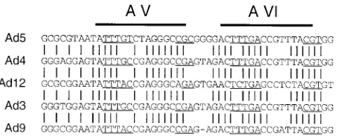
AII, as well as AV and AVI, are the functionally dominant repeats, and they are also separated from each other by 21 bp. Factors bound to these repeats would be located on the same side of the DNA double helix to potentially interact with each other and/or with factors bound to the leftmost 53 nt of the adenovirus genome to allow for DNA encapsidation. In studies to address the importance of 21-bp spacing between AI and AII, as well as AV and AVI (Fig. 2 and 3), we identified a CG dinucleotide, located in the identical position relative to AI, AII, AV, and AVI, that is 11 bp downstream of the middle thymidine residue within the thymidine triplet (Fig. 4). Our mutational studies suggest that the 11-bp spacing between the TTTG and the downstream CG is important but that the 21-bp spacing between different packaging elements is not required, at least not with respect to AI and AII (Fig. 2B). An alignment of the four functionally dominant packaging repeats together with our mutational studies defines a more extended consensus motif, 59-TTTGN8CG-39 (Fig. 4). Interestingly, these se-quences also are conserved between different adenovirus sub-groups in the regions corresponding to AV and AVI (Fig. 8). In vivo, the presence of the CG dinucleotide was found to be critical downstream of AI, AII, and AV but not downstream of AVI (Fig. 2 and 3). It is possible that functional redundancy applies not only to the AT-rich part of the new consensus motif but also to the CG sequence.
A single trans-acting packaging factor could interact with both conserved parts of the consensus motif, and it is notewor-thy that the 11-bp spacing between the TTTG and CG se-quences would present both halves of the consensus sequence on the same face of the DNA helix. Alternatively, the bipartite nature of the consensus motif could imply that distinct factors bind to the two conserved regions. Since different individual packaging repeats or combinations of repeats do not appear to be functionally equivalent, different packaging factors may in-teract with different individual A repeats. If one or more host cell factors are involved in adenovirus packaging, this may reflect the ability to recruit different binding proteins depend-ing on the cell type infected and the predominance of the respective cellular factors in that cell type. Consequently, func-tional redundancy of the packaging elements could constitute one of many strategies that the virus has developed to allow for its well-documented ability to infect a wide variety of cell types.
ACKNOWLEDGMENTS
We thank our colleagues for many helpful discussions and Tina Philipsberg for excellent technical help.
[image:9.612.319.559.69.167.2]This research was supported by funding from the Cystic Fibrosis Foundation (grant CFF Z161) and Public Health Service grant CA28146 from the National Cancer Institute to P.H.
FIG. 8. Alignment of putative packaging repeats in different adenovirus sub-groups. The nucleotide sequences corresponding to AV and AVI are shown for Ad5 (subgroup C), Ad4 (subgroup E), Ad12 (subgroup A), Ad3 (subgroup B), and Ad9 (subgroup D). The positions of AV and AVI in Ad5 are shown by lines above the sequences. Nucleotides identical between all subgroups are indicated by vertical lines.
VOL. 71, 1997 ADENOVIRUS PACKAGING 3383
on November 9, 2019 by guest
http://jvi.asm.org/
REFERENCES
1. Bjornsti, M. A., B. E. Reilly, and D. L. Anderson. 1983. Morphogenesis of bacteriophagef29 of Bacillus subtilis: oriented and quantized in vitro pack-aging of DNA protein gp3. J. Virol. 45:383–396.
2. Bolwig, G. M., J. T. Bruder, and P. Hearing. 1992. Different binding site requirements for binding and activation for the bipartite enhancer factor EF-1A. Nucleic Acids Res. 20:6555–6564.
3. Brough, D. E., A. Lizonova, C. Hsu, V. A. Kulesa, and I. Kovesdi. 1996. A gene transfer vector-cell line system for complete functional complementa-tion of adenovirus early regions E1 and E4. J. Virol. 70:6497–6501. 4. Bruder, J. T., and P. Hearing. 1989. Nuclear factor EF-1A binds to the
adenovirus E1A core enhancer element and to other transcriptional control regions. Mol. Cell. Biol. 9:5143–5153.
5. Daniell, E. 1976. Genome structure of incomplete particles of adenovirus. J. Virol. 19:685–708.
6. D’Halluin, J.-C., M. Milleville, P. A. Boulanger, and G. R. Martin. 1978. Temperature-sensitive mutant of adenovirus type 2 blocked in virion assem-bly: accumulation of light intermediate particles. J. Virol. 26:344–356. 7. D’Halluin, J.-C., M. Milleville, G. R. Martin, and P. Boulanger. 1980.
Mor-phogenesis of human adenovirus type 2 studied with fiber- and penton base-defective temperature-sensitive mutants. J. Virol. 33:88–99. 8. D’Halluin, J. C., G. R. Martin, G. Torpier, and P. A. Boulanger. 1978.
Adenovirus type 2 assembly analyzed by reversible cross-linking of labile intermediates. J. Virol. 26:357–363.
9. Earnshaw, W. C., and S. R. Casjens. 1980. DNA packaging by the double-stranded DNA bacteriophages. Cell 21:319–331.
10. Edvardsson, B., E. Everitt, E. Joernvall, L. Prage, and L. Philipson. 1976. Intermediates in adenovirus assembly. J. Virol. 19:533–547.
11. Edvardsson, B., S. Ustacelebi, J. Williams, and L. Philipson. 1978. Assembly intermediates among adenovirus type 5 temperature-sensitive mutants. J. Vi-rol. 25:641–651.
12. Engelhardt, J. F., X. Ye, B. Doranz, and J. M. Wilson. 1994. Ablation of E2A in recombinant adenoviruses improves transgene persistence and decreases inflammatory response in mouse liver. Proc. Natl. Acad. Sci. USA 91:6196– 6200.
13. Feinberg, A. P., and B. Vogelstein. 1983. A technique for radiolabeling DNA restriction endonuclease fragments to high specific activity. Anal. Biochem.
132:6–13.
14. Fredman, J. N., S. C. Pettit, M. S. Horwitz, and J. A. Engler. 1991. Linker insertion mutations in the adenovirus preterminal protein that affect DNA replication activity in vivo and in vitro. J. Virol. 65:4591–4597.
15. Freimuth, P. L., and H. S. Ginsberg. 1986. Codon insertion mutants of the adenovirus terminal protein. Proc. Natl. Acad. Sci. USA 83:7816–7820. 16. Graeble, M., and P. Hearing. 1990. Adenovirus type 5 packaging domain is
composed of a repeated element that is functionally redundant. J. Virol.
64:2047–2056.
17. Graeble, M., and P. Hearing. 1992. cis and trans requirements for the selec-tive packaging of adenovirus type 5 DNA. J. Virol. 66:723–731.
18. Graham, F. L., J. Smiley, W. C. Russell, and R. Nairu. 1977. Characteristics of a human cell line transformed by DNA from human adenovirus type 5. J. Gen. Virol. 36:59–72.
19. Hammarskjoeld, M.-L., and G. Winberg. 1980. Encapsidation of adenovirus 16 DNA is directed by a small DNA sequence at the left end of the genome. Cell 20:787–795.
20. Hearing, P., R. J. Samulski, W. L. Wishart, and T. Shenk. 1987. Identifica-tion of a repeated sequence element required for efficient encapsidaIdentifica-tion of the adenovirus type 5 chromosome. J. Virol. 61:2555–2558.
21. Hearing, P., and T. Shenk. 1983. The adenovirus type 5 E1A transcriptional control region contains a duplicated enhancer element. Cell 33:695–703. 22. Hearing, P., and T. Shenk. 1986. The adenovirus type 5 E1A enhancer
contains two functionally distinct domains: one is specific for E1A and the other modulates all early units in cis. Cell 45:229–236.
23. Hirt, B. 1967. Selective extraction of polyoma DNA from infected mouse cell cultures. J. Mol. Biol. 26:365–369.
24. Jones, N., and T. Shenk. 1979. Isolation of adenovirus type 5 host range deletion mutants defective for transformation of rat embryo cells. Cell 17: 683–689.
25. Kosturko, L. D., S. V. Sharnick, and C. Tibbetts. 1982. Polar encapsidation of adenovirus DNA: cloning and DNA sequence of the left end of adeno-virus type 3. J. Virol. 43:1132–1137.
26. Kunkel, T. A. 1985. Rapid and efficient site-specific mutagenesis without phenotypic selection. Proc. Natl. Acad. Sci. USA 82:488–492.
27. Maniatis, T., E. F. Fritsch, and J. Sambrook. 1982. Molecular cloning: a laboratory manual. Cold Spring Harbor Laboratory, Cold Spring Harbor, N.Y.
28. Pettit, S. C., M. S. Horwitz, and J. A. Engler. 1989. Mutations of the precursor to the preterminal protein of adenovirus serotypes 2 and 5. J. Virol. 63:5244–5250.
29. Robbinson, C. C., and C. Tibbetts. 1984. Polar encapsidation of adenovirus DNA: evolutionary variants reveal dispensable sequences near the left ends of Ad3 genomes. Virology 137:276–286.
30. Stow, N. D. 1981. Cloning a DNA fragment from the left-hand terminus of the adenovirus type 2 genome and its use in site-directed mutagenesis. J. Virol. 37:171–180.
31. Tibbetts, C. 1977. Viral DNA sequences from incomplete particles of human adenovirus type 7. Cell 12:243–249.
32. Yang, Y., F. A. Nunes, K. Berencsi, E. E. Furth, E. Gonczol, and J. M.
Wilson.1994. Cellular immunity to viral antigens limit E1-deleted adenovi-ruses for gene therapy. Proc. Natl. Acad. Sci. USA 91:4407–4411. 33. Yang, Y., F. A. Nunes, K. Berencsi, E. Gonczol, J. F. Engelhardt, and J. M.
Wilson.1994. Inactivation of E2A in recombinant adenoviruses improves the prospect for gene therapy in cystic fibrosis. Nat. Genet. 7:362–369.