Copyright © 1998, American Society for Microbiology. All Rights Reserved.
A Minimal Avian Retroviral Packaging Sequence Has a
Complex Structure
JENNIFER D. BANKS,1,2ASHLY YEO,1KRISTEN GREEN,1FRANZMARIE CEPEDA,1
ANDMAXINE L. LINIAL1,2*
Division of Basic Sciences, Fred Hutchinson Cancer Research Center, Seattle, Washington 98109,1
and Department of Microbiology, University of Washington, Seattle, Washington 981952
Received 15 October 1997/Accepted 26 March 1998
We have defined a 160-nucleotide region, Mc, from the 5*leader region of the Rous sarcoma virus genome that is sufficient to direct the packaging of a heterologous RNA. Mccontains the putative O3 stem structure that has previously been shown, and that has been confirmed in this study, to be important for the efficient packaging of avian leukosis-sarcoma virus RNA. Analyses of several O3 stem mutants revealed that other regions within Mccan interfere with the proper folding of altered sequences which are predicted to form a wild-type O3 stem.
The packaging signal, orc, is the cis-acting signal in retro-viral RNA necessary for encapsidation of the RNA into the virion. In an early attempt to identify the avian leukosis-sar-coma virus (ALSV) packaging signal, deletions were con-structed in the 59-untranslated region of a cloned avian sar-coma provirus. Several mutants which had packaging defects were identified. These mutants contained a deletion in an overlapping region of 31 bp located between the primer bind-ing site (PBS) and the Gag initiation codon (8). Subsequently, a 150-bp deletion that encompasses this region was engineered in an avian leukosis virus provirus and stably expressed in a quail cell line to produce the Q2bn-4D line (13). While parti-cles produced from these cells are unable to package their own genomes, they are able to package viral RNAs lacking viral structural genes, indicating that they retain trans-acting pack-aging functions. Using the Q2bn packpack-aging cell line, we previ-ously identified a 270-nucleotide (nt) packaging signal from the 59 end of the Rous sarcoma virus (RSV) genome. This se-quence, termed Ac, is sufficient for the packaging of a heter-ologous RNA (1). Acis located just downstream of the primer binding site and continues to the 59major splice site, located in the 59 end of gag coding sequences. Ac contains the last of three upstream open reading frames (uORF-3) located in the leader regions of ALSVs, which may play a role in packaging (3, 4, 12). Additionally, Accontains two 8-nt inverted repeats that are also conserved among several ALSVs and that are predicted to form the stem of the O3 stem-loop (5).
In this report we define Mc, a 160-nt region from within Ac
sufficient for packaging in our heterologous system. Mcretains uORF-3 and the O3 stem inverted repeats from Ac. We also describe the results of our initial experiments to determine the secondary structures within Mc necessary for packaging. In agreement with studies by Knight et al. (9), we find that the structure of the O3 stem, but not the sequence composing the stem, is necessary for packaging.
In order to further define the regions necessary for packag-ing, we tested sequences from within Acfor their abilities to confer packaging in our heterologous system. Acdeletion
mu-tants were placed 39of the coding sequence for the neomycin phosphotransferase gene (neo) in a derivative of pCMVneo (2, 10). This plasmid, pASY185, has an MluI linker inserted at the unique SmaI site (Fig. 1). The constructs were then transfected into the Q2bn packaging cell line (13), and mass cultures of drug G418-resistant cells were obtained (1). In initial experi-ments, Western blot analyses were performed on viral super-natants concentrated by high-speed centrifugation through a 20% sucrose cushion and quantitated, as previously described (1), in order to determine the relative amounts of viral parti-cles released by the cells. In later experiments, the more quan-titative radioimmunoprecipitation assays (RIPAs) were per-formed. For these analyses, cells were plated at a density of 107
cells per 100-mm-diameter plate at least 18 h before metabolic labeling. The cells were then labeled with [35S]methionine for
5 h, and the label was chased overnight by the addition of complete medium. Supernatants were collected, and labeled viral particles were concentrated by high-speed centrifugation through a 20% sucrose cushion. One-half of the collected particles were set aside for RNA analysis; the remaining half
[image:1.612.310.548.520.685.2]* Corresponding author. Mailing address: Division of Basic Sci-ences, Fred Hutchinson Cancer Research Center, Seattle, WA 98109-1024. Phone: (206) 667-4442. Fax: (206) 667-5939. E-mail: mlinial @fred.fhcrc.org.
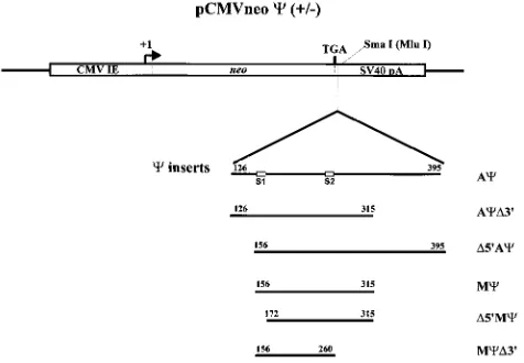
FIG. 1. Constructs used to assay regions of the genome necessary for c function. Portions of the RSV Prague C genome were cloned into pASY185, a pCMVneo derivative. Numbering is from the first nucleotide of the 59R region of the RSV Prague C genome (GenBank accession no., J02342). The S1 and S2 inverted repeats are boxed.
6190
on November 9, 2019 by guest
http://jvi.asm.org/
were precipitated in the presence of a mixture consisting of antibody buffer, 5ml ofa-PrB (11), and protein A-Sepharose beads. The antigen-antibody complexes were washed as previ-ously described (14). Bound proteins were eluted and sepa-rated on a sodium dodecyl sulfate–12.5% polyacrylamide gel.
Following electrophoresis, the gel was dried and, after an over-night exposure, scanned by a Molecular Dynamics Phosphor-Imager. Radioactive bands corresponding to the CA protein were quantitated, in machine units, with ImageQuant software. To determine the relative amounts of neo-specific RNA con-tained in the viruses, RNase protection assays were performed on concentrated virus with the Direct Protect kit (Ambion Inc.). RNAs were probed with antisense neo RNA either 170 or 250 nt in length. Protected RNA bands were scanned and quantitated as described above. Packaging efficiency was de-termined by calculating the ratio of the amount of neo RNA in the particles to the amount of CA protein, as measured by
FIG. 2. (A) RNase protection analysis to determine the amount of neo RNA in cells. Total cell RNAs were extracted from G418-resistant mass cultures of Q2bn-4D cells transfected with plasmids, as indicated above each lane, and annealed to the antisense neo probe. The sample in lane 7 had no cell RNA added. The sample in lane 8 was not treated with RNase. The arrows show the expected locations of the free probe and the neo-protected band, seen as a doublet. (B) RNase protection analysis to determine the amount of neo RNA packaged in virions released from the cells described in the legend for panel A. Virus was collected, and RNAs were extracted and annealed to the antisense neo probe. The sample in lane 1 had no RNase added. The sample in lane 8 had no viral RNA added.
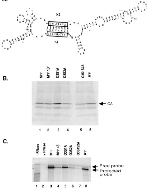
[image:2.612.53.290.70.185.2]FIG. 3. (A) The RSV genome showing the location and sequence of the minimal packaging region, Mc. The S1 and S2 inverted repeats composing the O3 stem, as well as the downstream PS3 region, are underlined in gray. The SacI restriction site and uORF-3 are underlined in black. R, repeated sequence; U5, unique 59 sequences; gag, gene encoding viral structural proteins; MA, matrix protein; CA, capsid protein; NC, nucleocapsid protein; pol, gene encoding reverse transcriptase and integrase; env, gene encoding glycoproteins; U3, unique 39sequences; 59ss, 59splice donor site; 39ss, 39splice acceptor site. (B) Predicted secondary structure of Mc. The M-Fold RNA folding program based on the computer algorithm of Zuker et al. (6, 7, 15) was used. The folding energy obtained was259.0 kcal. The S1, S2, and PS3 regions are outlined.
TABLE 1. Lengths and packaging efficienciesaof viral sequences from the 59region of RSV
Clone Length (nt) Packaging efficiency
Ac 270 111
AcD39 190 111
D59Ac 240 111
Mc 160 111
D59Mc 144 1
McD39 105 11
aPackaging efficiency was calculated as the ratio of the relative amount of neo-specific RNA packaged in particles, as measured by RNase protection
anal-ysis, to the relative number of particles, as measured by Western analysis. Pack-aging efficiencies were normalized to that of Mc. For more detail see the text. Each plus sign represents approximately 1 order of magnitude.
on November 9, 2019 by guest
http://jvi.asm.org/
[image:2.612.309.549.90.166.2] [image:2.612.120.479.383.673.2]Western blotting or RIPA. The ratios obtained for the deletion mutants were then normalized to that of the pCMVneo-Mc
construct.
The deletions were obtained by using Acas the template in the Erase-a-base system (Promega, Inc.). The sizes and loca-tions of the deleloca-tions obtained are shown in Fig. 1. AcD39
contains a deletion of 80 nt from the 39 end of Ac. D59Ac
contains a deletion of 30 nt from the 59end of Ac. By Western
and RNase protection analyses (data not shown) we found that the packaging efficiencies of AcD39andD59Acwere similar to that of Ac. These results are summarized in Table 1. Next, the packaging efficiency of the 160-base Mc, which contains the deletions found in both AcD39and D59Ac, was determined. Again, packaging levels were similar to that of Ac(Table 1). We next deleted either end of Mcto determine the effect on packaging.D59Mccontains a deletion of 16 nt from the 59end of Mc, whereas McD39contains a deletion of 55 nt from the 39
end of Mc(Fig. 1). These Mcdeletions were placed 39of neo in the sense and antisense orientations. Similar numbers of viral particles were released from the cells transfected with the mutants, as measured by Western blot analysis (data not shown). RNase protection analysis of cell RNA indicated that similar levels of neo RNA are present in the cells (Fig. 2A; data not shown for McD39constructs). RNase protection analysis of RNA from viral particles released from the cells showed sim-ilar levels of packaging of neo RNA containing Acand Mcin the sense orientation (Fig. 2B, lanes 3 and 5). In contrast, the level of packaging ofD59McRNA was reduced over 18-fold compared to that of Mc(Fig. 2B, lanes 5 and 7). The efficiency of McD39was reduced over sixfold compared to that of Mc
(Fig. 5C, lanes 3 and 4). These results are summarized in Table
FIG. 4. (A) The sequences of O3S1B and O3S2B are shown above and below, respectively, the wt O3 stem. Nucleotides differing from those of the wt sequence are highlighted. (B) RNase protection analysis to determine the amount of neo RNA packaged in virions released from G418-resistant mass cultures of Q2bn-4D cells transfected with plasmids, as indicated above each lane, and annealed to the antisense neo probe. The expected locations of free probe and the protected neo band are indicated by arrows. No RNase was added to the sample in lane 10.
[image:3.612.309.545.77.378.2]FIG. 5. (A) The sequences of O3S1A and O3S2A are shown above and below, respectively, the wt O3 stem. Nucleotides differing from those of the wt sequence are highlighted. (B) RIPA to determine the amount of viral particles released from G418-resistant mass cultures of Q2bn-4D cells transfected with plasmids, as indicated above each lane, and precipitated with thea-PrB antibody. The expected size of the capsid (CA) band is indicated. (C) RNase protection analysis of the RNA from virus released from cells described for panel B. The expected sizes of the free probe and the protected neo probe are indicated by arrows. The sample in lane 1 had no RNase added. The sample in lane 2 had no viral RNA added.
TABLE 2. Sequences and packaging efficiencies of O3 stem mutants
Clone Sequence (59to 39)
aof:
Packaging efficiencyb
S1 S2 PS3
wt Mc CUGCCCUC GAGGGCAG GAGCCCAG 1.00
O3S1B CUGAAAUC GAGGGCAG GAGCCCAG 0.06 O3S2B CUGCCCUC GAUUUCAG GAGCCCAG 0.03 O3S1S2B CUGAAAUC GAUUUCAG GAGCCCAG 1.10 O3S1A CUCGGCUC GAGGGCAG GAGCCCAG 0.18 O3S2A CUGCCCUC GAGCCGAG GAGCCCAG 0.09 O3S1S2A CUCGGCUC GAGCCGAG GAGCCCAG 0.02 O3S1S2A1PS3 CUCGGCUC GAGCCGAG CAGAUCUC 0.29
wt Mc1PS3 CUGCCCUC GAGGGCAG CAGAUCUC 1.09
aSequence differing from that of the wt is shown in boldface.
bPackaging efficiency was calculated as the ratio of the relative amount of neo-specific RNA packaged in particles, as measured by RNase protection anal-ysis, to the relative number of particles, as measured by RIPA. Packaging effi-ciencies were normalized to that of wt Mc. For more details, see the text.
on November 9, 2019 by guest
http://jvi.asm.org/
[image:3.612.49.290.88.203.2] [image:3.612.53.289.375.659.2]1. The RNAs containingcin the antisense orientation were packaged at very low levels (Fig. 2B, lanes 2, 4, and 6).
Mcis the shortest sequence which can confer a high level of packaging of a heterologous RNA that we have identified. The primary sequence of Mc is given in Fig. 3A. Mc does not contain any sequences from the Gag open reading frame. Nor are sequences from the primer binding site and 59major splice site included in the 160-nt region. Mcdoes contain the 31-nt sequence shown by Katz et al. to be involved in packaging (8). Additionally, the entire uORF-3 is located within Mc (Fig. 3A). The Mcsequence was placed in the M-Fold RNA folding program to determine the predicted secondary structure (6, 7, 15). The most stable structure is shown in Fig. 3B. The O3 stem-loop structure, which is conserved in the computer-gen-erated folding models of the leader region of several ALSV strains (5) and which has been shown to play a role in pack-aging (9), is also largely conserved in our model of Mc. The inverted repeats composing the stem, which we have named S1 and S2, are underlined in Fig. 3B.
The importance of the O3 stem in packaging is supported by ourD59Mcmutant. As described above, the deletion of just 16 nt, which includes the S1 region, from the 59end of Mcresults in an 18-fold reduction in packaging over wild-type (wt) Mc. To more directly study the role of the O3 stem in packaging, we made a series of mutations in S1 and S2 which disrupt the correct formation of the stem. These point mutations were made by standard PCR techniques. We constructed the same S1 and S2 mutations made by Knight et al. (9) for use in our heterologous system. In mutant O3S1B, three contiguous nu-cleotides from S1, CCC, have been changed to AAA. In mu-tant O3S2B, three contiguous nucleotides from S2, GGG, have been changed to UUU. Finally, O3S1S2B contains both mu-tations, restoring the complementarity between S1 and S2. These sequences are summarized in Table 2 and in Fig. 4A. The mutated packaging signals were inserted in pASY185 in both the sense and antisense orientations. To measure the number of particles released from cell lines transfected with the mutants, RIPAs were performed. These studies indicated that similar numbers of particles were released from cells. RNase protection analysis of RNA from particles released from the cells showed that the packaging of O3S1B and O3S2B sense RNAs is reduced 16- and 30-fold, respectively, over that of wt McRNA (Fig. 4B, lanes 1, 3, and 5). In particles released from the compensatory mutant, O3S1S2B, packaging cells, the amount of neo-containing RNA is similar to that of wt Mc
(Fig. 4B, lanes 1 and 7). These packaging efficiencies are sum-marized in Table 2. The antisense mutants had greatly reduced packaging levels compared to wt Mc(Fig. 4B, lanes 1, 2, 4, and 6). From these data, we concluded that the O3 stem structure plays a role in packaging, while the primary sequence of the stem is not important. The regions of the Gag polyprotein that bind to the packaging signal during encapsidation may directly interact with the O3 stem, or O3 may stabilize a region directly involved in packaging. Regions downstream of S2 may also be involved in packaging, as our McD39mutant, in which all Mc
sequences downstream of the SacI site were deleted, had a sixfold reduction in packaging compared to wt Mc.
To confirm and extend these results, we made another set of mutations which altered the O3 stem sequence at different nu-cleotides. In mutant O3S1A, three contiguous nucleotides from the S1 stem, GCC, have been changed to CGG. In mu-tant O3S2A, 3 nt from the S2 stem, GGC, have been changed to CCG. O3S1S2A contains both mutations, restoring the comple-mentarity between S1 and S2. These sequences are summa-rized in Table 2 and Fig. 5A. The numbers of particles released by the cell lines were similar as measured by RIPA (Fig. 5B). As in the case of the O3S1B and O3S2B mutants, the levels of O3S1A and O3S2A RNAs in the particles, as measured by RNase protection analysis, were reduced compared to that of wt Mc(Fig. 5C, lanes 3, 5, and 6). However, unlike the aging of the O3S1S2B compensatory mutant RNA, the pack-aging of O3S1S2A RNA was greatly reduced compared to that of wt Mc(Fig. 5C, lanes 3 and 7). The packaging efficiencies are summarized in Table 2. This was a surprising result, as the mutations were expected to restore the complementarity of the inverted repeat, presumably restoring the secondary structure of the O3 stem.
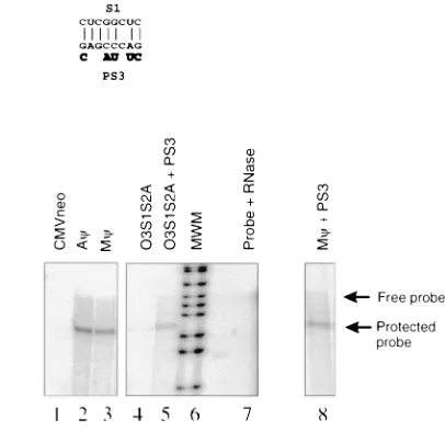
An examination of the sequences surrounding the S1 and S2 inverted repeats revealed a region, PS3 (Fig. 3), 36 nt down-stream of S2, that is complementary to S1 in 7 of 8 nt in the O3S1S2A mutant (Fig. 6A and B). To test whether PS3 was preventing the proper folding of the mutated O3 stem by interacting with S1 in the O3S1S2A mutant, we made an ad-ditional mutant, O3S1S2A1PS3, that contains the O3S1S2A mutations as well as a mutation in PS3 that destroys comple-mentarity to the S1 mutation. These sequences are summa-rized in Table 2 and Fig. 6A and B. By measuring the number of particles released from these cells (data not shown) and measuring the amount of neo RNA in these particles (Fig. 6C, lanes 3, 4, and 5) we found that the packaging efficiency of
FIG. 6. (A) The sequence of O3S1S2A1PS3. The sequences that differ from that of the wt O3 stem are highlighted. The PS3 region is outlined, and the sequences mutated in O3S1S2A1PS3 are indicated. (B) The predicted base pairing between S1 and PS3 in the O3S1S2A mutant. The nucleotide changes in the O3S1S2A1PS3 mutant are shown in boldface below the PS3 sequence. (C) RNase protection analysis of the RNA from virus released from G418-resistant mass cultures of Q2bn-4D cells transfected with plasmids, as indicated above each lane, and annealed to the neo probe. The expected sizes of the free probe and the protected neo probe are indicated by arrows. The sample in lane 7 had no viral RNA added.
on November 9, 2019 by guest
http://jvi.asm.org/
[image:4.612.329.532.70.267.2]O3S1S2A1PS3 is increased over 10-fold compared to that of O3S1S2A. The PS3 region itself does not appear to play a role in the folding of wt Mc. When the PS3 mutation was placed in the context of wt Mc, it did not affect packaging (Fig. 6C, lanes 3 and 8). These packaging efficiencies are summarized in Table 2. These experiments highlight the complexity of RNA struc-tural analysis. Mutation in one region can have an effect on the folding of regions which appear noncontiguous in computer-generated RNA folding models.
Despite the identification of Mc, there exists a paradox in our understanding of RSV packaging. In mammalian retrovi-ruses and the avian spleen necrosis virus,cis located down-stream of the 59 major splice site. As a result, the packaging signal is not found on subgenomic species. In contrast, Mcis located upstream of the 59 major splice site and therefore is found on both full-length and env and src subgenomic species. Yet the full-length message is preferentially packaged (1). One explanation is that there are regions in the intron that augment packaging in the full-length RNA. Similarly, there may be sequences in env that in some way inhibit packaging when inserted next to Mcin the spliced RNAs. This model, in which additional sequences play a role in packaging, is supported by our observation that the rate of packaging of Ac and Mc
heterologous RNAs is down 10-fold compared to the rate of packaging of the full-length RSV genome (data not shown). This possibility points out a limitation of our heterologous assay. In wt RSV infection, the subgenomic RNAs may com-pete with genomic RNA for binding to the Gag motifs that specifically recognize the packaging region. Since this compe-tition does not exist in our assay, we may be overestimating the precise efficiency of Mc. Thus, while we have identified se-quences sufficient for selective encapsidation of our heterolo-gous RNA, there may be additional sequences that further select the encapsidation of the full-length viral genome. We are currently performing experiments to investigate this possi-bility.
With this short packaging signal identified, we are presently performing a more extensive investigation of the secondary structures involved in packaging. Another important aspect of RSV packaging that has yet to be elucidated is the role, if any, of uORF-3. This open reading frame has been shown to be efficiently translated (3, 4, 12), and when the translation of uORF3 is altered, the packaging efficiency is reduced (3). Donze et al. (3) have suggested that uORF-3 contains an RNA secondary structure that inhibits the packaging of the RNA.
During translation of the open reading frame, this inhibitory structure is removed and packaging can proceed. The pre-dicted fold of Mc does show stem-loop structures in these sequences. We are currently testing the effect of mutating the AUG of uORF-3 on packaging in our heterologous system.
J.D.B. and A.Y. contributed equally to this work.
This work was supported by a grant from the National Cancer Institute (CA 18282) to M.L.L., A.Y. was supported by a Leukemia Society of America postdoctoral fellowship, and J.D.B. was supported by a National Science Foundation graduate fellowship.
We thank Julie Overbaugh for critical review of the manuscript.
REFERENCES
1. Aronoff, R., A. M. Hajjar, and M. L. Linial. 1993. Avian retroviral RNA encapsidation: reexamination of functional 59RNA sequences and the role of nucleocapsid Cys-His motifs. J. Virol. 67:178–188.
2. Aronoff, R., and M. L. Linial. 1991. Specificity of retroviral RNA packaging. J. Virol. 65:71–80.
3. Donze, O., P. Damay, and P. F. Spahr. 1995. The first and third uORFs in RSV leader RNA are efficiently translated: implications for translational regulation and viral RNA packaging. Nucleic Acids Res. 23:861–868. 4. Donze, O., and P. F. Spahr. 1992. Role of the open reading frames of Rous
sarcoma virus leader RNA in translation and genome packaging. EMBO J.
11:3747–3757.
5. Hackett, P. B., M. W. Dalton, D. P. Johnson, and R. B. Petersen. 1992. Phylogenetic and physical analysis of the 59leader RNA sequences of avian retroviruses. Nucleic Acids Res. 19:6929–6934.
6. Jaeger, J. A., D. H. Turner, and M. Zuker. 1989. Improved predictions of secondary structures for RNA. Proc. Natl. Acad. Sci. USA 86:7706–7710. 7. Jaeger, J. A., D. H. Turner, and M. Zuker. 1990. Predicting optimal and
suboptimal secondary structure for RNA. Methods Enzymol. 183:281–306. 8. Katz, R. A., R. W. Terry, and A. M. Skalka. 1986. A conserved cis-acting
sequence in the 59 leader of avian sarcoma virus RNA is required for packaging. J. Virol. 59:163–167.
9. Knight, J. B., Z. H. Si, and C. M. Stoltzfus. 1994. A base-paired structure in the avian sarcoma virus 59leader is required for efficient encapsidation of RNA. J. Virol. 68:4493–4502.
10. Linial, M. 1987. Creation of a processed pseudogene by retroviral infection. Cell 49:93–102.
11. Linial, M., J. Fenno, W. B. Burnette, and L. Rohrschneider. 1980. Synthesis and processing of viral glycoproteins in two nonconditional mutants of Rous sarcoma virus. J. Virol. 36:280–290.
12. Moustakas, A., T. S. Sonstegard, and P. B. Hackett. 1993. Alterations of the three short open reading frames in the Rous sarcoma virus leader RNA modulate viral replication and gene expression. J. Virol. 67:4337–4349. 13. Stoker, A. W., and M. J. Bissell. 1988. Development of avian sarcoma and
leukosis virus-based vector-packaging cell lines. J. Virol. 62:1008–1015. 14. Yu, S. F., and M. L. Linial. 1993. Analysis of the role of the bel and bet open
reading frames of human foamy virus by using a new quantitative assay. J. Virol. 67:6618–6624.
15. Zuker, M. 1989. On finding all suboptimal foldings of an RNA molecule. Science 244:48–52. (Review.)