Copyright © 1998, American Society for Microbiology. All Rights Reserved.
Encapsidation of the Flavivirus Kunjin Replicon RNA by Using
a Complementation System Providing Kunjin Virus Structural
Proteins in trans
ALEXANDER A. KHROMYKH,* ANDREI N. VARNAVSKI,ANDEDWIN G. WESTAWAY
Sir Albert Sakzewski Virus Research Centre, Royal Children’s Hospital, Brisbane, Queensland 4029, Australia
Received 15 October 1997/Accepted 23 March 1998
Kunjin virus (KUN) replicon RNA was encapsidated by a procedure involving two consecutive electropora-tions of BHK-21 cells, first with KUN replicon RNA C20DXrep (with prME and most of C deleted) and about 24 h later with a recombinant Semliki Forest virus (SFV) replicon RNA(s) expressing KUN structural proteins. The presence of KUN replicon RNA in encapsidated particles was demonstrated by its amplification and expression in newly infected BHK-21 cells, detected by Northern blotting with a KUN-specific probe and by immunofluorescence analysis with anti-NS3 antibodies. No infectious particles were produced when C20DXrep RNA and recombinant SFV RNAs were electroporated simultaneously. When the second electroporation was performed with a single SFV replicon RNA expressing the KUN contiguous prME genes and the KUN C gene together but under control of two separate 26S subgenomic promoters (SFV-prME-C107), a 10-fold-higher titer of infectious particles was achieved than when two different SFV replicon RNAs expressing the KUN C gene (SFV-C107) and prME genes (SFV-prME) separately were used. No SFV replicon RNAs expressing KUN structural proteins were encapsidated in secreted particles. Infectious particles pelleted by ultracentrifugation of the culture fluid from cells sequentially transfected with C20DXrep and SFV-prME-C107 RNAs were neutralized by preincubation with monoclonal antibodies to KUN E protein. Radioimmunoprecipitation analysis with anti-E antibodies of the culture fluid of the doubly transfected cells showed the presence of C, prM/M, and E proteins in the immunoprecipitated particles. Reverse transcription-PCR analysis showed that the immunoprecipitated particles also contained KUN-specific RNA. The encapsidated replicon particles sedimented more slowly than KUN virions in a 5 to 25% sucrose density gradient and were uniformly spherical, with an;35-nm diameter, compared with;50 nm for KUN virions. The results of this study demonstrate for the first time packaging of flavivirus RNA in trans, and they exclude a role in packaging for virtually all of the structural region. Possible applications of the developed packaging system include the definition of the packaging signal(s) in flavivirus RNA as well as the amino acid motif(s) in the structural proteins involved in RNA encapsidation, virion assembly, and secretion. Furthermore, it could facilitate the development of a noninfectious vaccine delivery system based on encapsidation of a noncytopathic flavivirus replicon expressing heterologous genes.
Kunjin virus (KUN) is an endemic Australian flavivirus that occasionally causes febrile illness or mild encephalitis in hu-mans. The KUN genome consists of positive-stranded RNA of 11,022 nucleotides (13) containing one long open reading frame coding for three structural (C, prM, and E) and seven nonstructural (NS) proteins in the order C-prM-E-NS1-NS2A-NS2B-NS3-NS4A-NS4B-NS5 (8). Recently we described the preparation of the first flavivirus replicon (self-replicating) RNAs with deletions in the structural region and deletions and insertions in the 39 untranslated region (UTR) (15). Using KUN replicon RNA with an insertion in the 39 UTR of the neomycin gene under control of the internal ribosomal entry site of encephalomyelocarditis virus RNA, we were able to establish BHK cell lines persistently expressing this replicon RNA (15). Importantly, replication of KUN replicon RNAs either after transfection into BHK cells or in a cell line persis-tently expressing the KUN replicon did not produce any pathic effect; this distinguishes KUN replicons from the cyto-pathic replicons of other positive-stranded RNA viruses such as Semliki Forest virus (SFV), Sindbis virus (SIN), and
polio-virus, which are widely used as vectors for expression of het-erologous genes in vitro (for references, see reference 15) and in vivo (10, 12, 42). The success of these expression systems was based mainly on the ability to produce high-titer stocks of pseudoinfectious particles containing replicon RNA packaged by structural proteins. In SFV and SIN alphavirus expression systems, this was achieved by cotransfection of replicon RNA with a defective helper RNA(s) expressing structural genes but lacking the packaging signal (6, 22). The replicon RNA expres-sion provides the enzymes for RNA replication and transcrip-tion of both RNAs, whereas the helper alphavirus RNA sup-ports the production of structural proteins for packaging of replicon RNA via expression of its subgenomic region.
Flaviviruses and picornaviruses differ from the alphaviruses SFV and SIN by their genome structure (structural genes sit-uated at the 59 end of the genome) and by the absence of synthesis of subgenomic RNA (31, 35, 38). There are no data to date on packaging of flavivirus RNA. Packaging of poliovi-rus replicon RNA was successfully achieved either by superin-fection of replicon-transfected cells with a helper poliovirus (25, 26) or by transfection of replicon RNA into cells previ-ously infected with recombinant vaccinia virus expressing structural proteins (4, 27). Although infectious particles con-taining encapsidated poliovirus replicon RNA were produced under these conditions, additional purification of particles was
* Corresponding author. Mailing address: Sir Albert Sakzewski Vi-rus Research Centre, Royal Children’s Hospital, Herston Rd., Bris-bane, QLD 4029, Australia. Phone: (617) 1568. Fax: (617) 3253-1401. E-mail: [email protected].
5967
on November 9, 2019 by guest
http://jvi.asm.org/
required in order to separate them from recombinant vaccinia virus.
The goal of these studies was to develop a KUN replicon packaging system for possible future use in defining the flavi-virus RNA packaging signal(s) and in expression of heterolo-gous genes, taking advantage of the noncytopathic nature and persistence of replication of the KUN replicon. Successful packaging of KUN replicon RNA was achieved when an SFV replicon RNA(s) expressing KUN structural proteins was elec-troporated at least 12 h after electroporation of KUN replicon RNA. A single recombinant SFV RNA expressing the KUN prME and KUN C proteins together but under control of two separate 26S promoters was more efficient in packaging exper-iments than were two SFV RNAs expressing KUN prME and KUN C separately. We demonstrated that the resulting se-creted infectious particles were uniformly spherical with an
;35-nm diameter and contained replicating KUN replicon RNA encapsidated by KUN structural proteins C, prM/M, and E. These findings represent the first demonstration of the packaging of flavivirus RNA in trans and may prove to be useful for identification of the packaging signal(s) and for development of a vaccine delivery system based on expression from a noncytopathic flavivirus replicon.
MATERIALS AND METHODS
Cells.BHK21 cells were grown in Dulbecco’s modification of minimal essen-tial medium (Gibco BRL) supplemented with 10% fetal bovine serum at 37°C in a CO2incubator.
Construction of the plasmids. (i) C20DXrep.The KUN replicon cDNA con-struct C20DXrep was concon-structed from the previously described C20rep (with the structural gene sequence except for the first 20 codons deleted) (15) by replacing an SphI2467-XhoI11021fragment representing the sequence coding for
the entire nonstructural region and the 39UTR with the corresponding fragment from a stable full-length KUN cDNA clone, FLSDX. Details of clone FLSDX will be described elsewhere; its RNA transcripts have a specific infectivity of about 104PFU/mg, which is about 100,000-fold more infectious than those from
our previously described KUN full-length cDNA clone pAKUN (13).
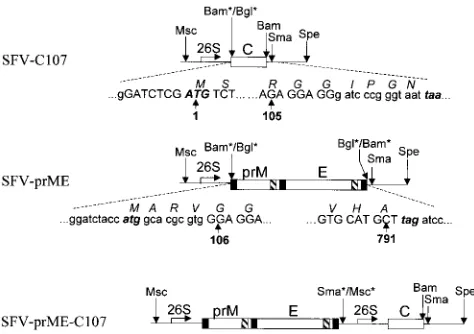
(ii) SFV-C107.An SFV replicon construct expressing the KUN core (C) gene was obtained by cloning of the BglII-BamHI fragment, representing the sequence commencing with the last 7 nucleotides of the KUN 59UTR followed by the sequence coding for the first 107 of the 123 amino acids of the KUN C protein, from the plasmid pCINeoC107 (14) into the BamHI site of the SFV replicon expression vector pSFV1 (Gibco BRL) (Fig. 1). This sequence approximates the size of the mature form of C (comprising the first 105 amino acids) after the carboxy-terminal hydrophobic sequence has been cleaved by the viral protease (33). The resulting translation product of the cloned fragment is likely to contain an extra six amino acids relative to mature C, comprising two native KUN amino acids (GG) downstream of cleavage site plus four amino acids (IPGN) derived from the SFV vector (Fig. 1).
(iii) SFV-prME.The KUN prME sequence was PCR amplified from another highly efficient full-length KUN cDNA clone, FLBSDX, modified from FLSDX (see “C20DXrep” above), by using appropriate primers with incorporated initi-ation and termininiti-ation codons and flanked by BglII sites. To minimize the number of possible mismatches which could occur during PCR amplification, we used high-fidelity Pfu DNA polymerase (Stratagene) in our PCRs. The amplified fragment was digested with BglII and cloned into the BamHI site of the SFV replicon expression vector pSFV1 to obtain the SFV-prME construct (Fig. 1).
(iv) SFV-prME-C107.An SFV replicon construct expressing both the KUN prME and KUN C genes was obtained by cloning the MscI-SpeI fragment from the SFV-C107 plasmid containing the SFV 26S subgenomic promoter, KUN C sequence, and SFV 39UTR into the SFV-prME vector digested with SmaI and
SpeI (Fig. 1). Thus, the final double subgenomic promoter construct
SFV-prME-C107 should produce SFV replicon RNA which upon transfection into BHK cells will direct synthesis of two subgenomic RNAs expressing the KUN prME and KUN C genes.
RNA transcription and transfection.KUN replicon RNA transcripts were prepared from C20DXrep plasmid DNA linearized with XhoI and from SFV plasmids linearized with SpeI by using SP6 RNA polymerase essentially as de-scribed previously (13, 15). Electroporation of RNAs into BHK21 cells was performed as described previously (15). Briefly, 10 to 20mg of in vitro-tran-scribed RNAs was electroporated into 23106BHK21 cells in 400ml in a 0.2-cm
cuvette (Bio-Rad) by using the Bio-Rad Gene Pulser apparatus.
IF analysis.Replication of KUN replicon RNA C20DXrep after initial elec-troporation and after infection of BHK cells in packaging experiments was monitored by immunofluorescence (IF) analysis with antibodies to KUN NS3
protein as described previously (15). Expression of KUN E protein after elec-troporation with SFV-prME and SFV-prME-C107 RNAs was detected by IF with a 1/10 dilution of a pool of mouse monoclonal antibodies to KUN E protein, designated 3.91D, 10A1, and 3.67G (1), which were generously provided by Roy Hall, University of Queensland, Brisbane, Australia. Expression and nuclear localization of KUN C protein after electroporation with C107 and SFV-prME-C107 RNAs were monitored by IF analysis with rabbit polyclonal anti-bodies to KUN C protein as described previously (37). Cells were fixed in acetone to detect cytoplasmic IF and in formaldehyde and methanol for nuclear IF (37).
Metabolic labeling and RIP analysis.Metabolic labeling with [35
S]methionine-cysteine of electroporated BHK cells was performed essentially as described in the SFV Gene Expression System Manual (21) with some minor modifications. Briefly, cells at 18 h after the electroporation with SFV RNAs (with or without prior electroporation with KUN replicon RNA), were pulse-labeled with [35S]methionine-cysteine either for 4 h or for 1-2 h followed by different periods
of incubation (chase) in medium with an excess of unlabeled methionine-cys-teine. Cell culture fluid was collected for analysis of secreted proteins by elec-trophoresis and radioimmunoprecipitation (RIP). Labeled cells were lysed in buffer containing 1% Nonidet P-40, 50 mM Tris-HCl (pH 7.6), 150 mM NaCl, and 2 mM EDTA, the nuclei were removed by low-speed centrifugation, and the lysate supernatant was used for parallel analysis with the culture fluid.
For RIP analysis, labeled cell culture fluids were first filtered through a 0.45-mm-pore-size filter (Sartorius AG, Gottingen, Germany) and digested with RNase A (20mg per ml) for 30 min at 37°C to ensure the removal of membrane particulate material and naked RNA. Filtered and RNase-treated culture fluids, or untreated cell lysates, were then mixed with 1/20 volume of the pooled anti-E monoclonal antibodies (see above) or with rabbit anti-C antibodies and incu-bated overnight at 4°C with constant rotation in microcentrifuge tubes. Protein A-Sepharose beads were then added to a final concentration of about 1%, and incubation was continued for another 1 h at 4°C. After three washes with RIP assay buffer (50 mM Tris-HCl [pH 7.6], 150 mM NaCl, 1% Nonidet P-40, 0.5% deoxycholic acid sodium salt, 0.1% sodium dodecyl sulfate [SDS]) and one wash with phosphate-buffered saline (PBS), the beads were resuspended in the SDS-gel sample buffer, boiled for 5 min, and subjected to electrophoresis in an SDS-polyacrylamide gel. After electrophoresis, the gels were dried and exposed to X-ray film.
[image:2.612.309.546.70.237.2]Northern blot hybridization.Five micrograms of total RNA, isolated by using Trizol reagent (Gibco BRL) from BHK21 cells infected with culture fluid col-lected from cells doubly transfected with C20DXrep RNA and SFV RNAs expressing KUN structural proteins, was electrophoresed for Northern blotting as described previously (15). The hybridization probes were32P-labeled cDNA
FIG. 1. Schematic representation of the recombinant SFV constructs. The solid line in all constructs represents the segment of the SFV replicon genome flanking the multiple cloning site; open boxes show the inserted KUN structural genes for C, prM, and E as indicated; 26S shows the position of the subgenomic SFV promoter; and the filled and hatched boxes in the KUN prM and E genes represent hydrophobic signal and anchor sequences, respectively (8). Uppercase letters in the nucleotide sequences show authentic KUN nucleotides; lowercase letters show nucleotides derived from the pSFV1 vector or encoded in the primers used for PCR amplification of KUN genes. Boldface and italicized letters show initiation (ATG) and termination (taa and tag) codons. Italicized letters above codons in the nucleotide sequence represent corresponding amino acids. Numbers with arrows represent amino acid positions in the KUN polypro-tein (8). Msc, Sma, Spe, Bam, and Bgl represent specific restriction sites. Aster-isks indicate that these restriction sites were destroyed during the cloning pro-cedure (see Materials and Methods).
on November 9, 2019 by guest
http://jvi.asm.org/
fragments representing the 39-terminal 761 nucleotides of the KUN genome, including the 39UTR (see Fig. 5B and 6), and 446 nucleotides of the SFV NSP2 region (see Fig. 5C).
Preparation of encapsidated particles and determination of their titer.For all infections with encapsidated particles, cell culture fluid was membrane filtered and treated with RNase A as described above. To prepare partially purified particles, treated culture fluids from transfected cells were clarified by centrifu-gation at 16,0003g in a microcentrifuge for 15 min at 4°C, and the particles were
pelleted from the resulting supernatant fluid by ultracentrifugation at 40,000 rpm for 2 h at 4°C in the AH650 rotor of a Sorvall OTD55B centrifuge. The pellets were resuspended in 50ml of PBS supplemented with RNase A (20mg per ml), left to dissolve overnight at 4°C, and then used for infection of BHK21 cells, for reverse transcription-PCR (RT-PCR) analysis, or for sedimentation analysis. To determine the titer of encapsidated particles, BHK21 cells on 1.3-cm2coverslips
were infected with 100 or 200ml of serial 10-fold dilutions of cell culture fluid or of resuspended pelleted material for 1.5 h at 37°C. The fluid was then replaced with 1 ml of Dulbecco’s minimal essential medium supplemented with 2% fetal bovine serum; cells were incubated for 24 h at 37°C in a CO2incubator and then
subjected to IF analysis with anti-NS3 antibodies as described above. The infec-tious titer of packaged particles was calculated by using the following formula: titer (infectious units [IU]) per 23106initially transfected cells5N3(SW/
SIA)310n3(V/VI), where N is the average number of anti-NS3-positive cells in the image area, calculated from five image areas in different parts of the coverslip; SW is the surface of the well in a 24-well plate (200 mm2); SIA is the
surface of the image area (1.25 mm2, using defined magnification parameters,
calculated according to the manual for the Wild MPS46/52 photoautomat [Wild Leitz, Heerburg, Germany]); V is the total volume of the culture fluid (usually 3 to 5 ml per 60-mm-diameter dish) collected from the population of 23106
initially electroporated BHK21 cells; VI is the volume used for infection of the coverslips (usually 100 to 200ml); and 10nis the dilution factor.
RESULTS
The SFV replicon vector was chosen for the expression of KUN structural proteins for packaging because of the high level of cytoplasmic expression of heterologous proteins, rapid construction of recombinant RNA molecules, and absence of infectious SFV viral contaminants in the culture fluid of trans-fected cells (22). It was therefore essential initially to insert KUN structural genes into the SFV replicon and demonstrate
a suitable level of their expression. Our original intention was to express the entire open reading frame coding for the KUN structural proteins C, prM, and E in the SFV replicon expres-sion vector pSFV1. Unfortunately, numerous attempts to clone a BglII-BglII PCR fragment representing the KUN C-prM-E region into the pSFV1 vector resulted in recovery of clones containing the KUN cDNA fragment inserted only in the ori-entation reverse to that required for expression directed by the SFV 26S subgenomic promoter (results not shown). To over-come this problem we decided to prepare two separate SFV constructs expressing the KUN C gene and KUN prME genes, respectively (Fig. 1).
Expression of the KUN C gene by the recombinant
SFV-C107 replicon. Electroporation of SFV-C107 RNA into
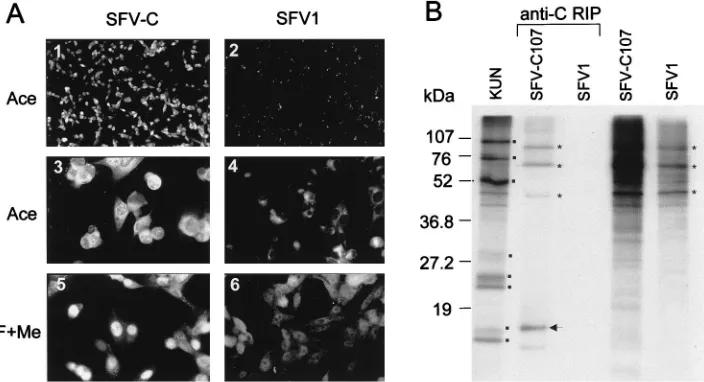
[image:3.612.123.475.71.262.2]BHK21 cells resulted in expression of KUN C protein in al-most 100% of cells as judged by IF with antibodies to KUN C protein (Fig. 2A, panel 1). KUN C protein expressed in SFV-C107 RNA-transfected cells was localized in the cytoplasm (Fig. 2A, panel 3 [acetone fixation]) and also in the nuclei (Fig. 2A, panel 5 [formaldehyde-methanol fixation]). These results are in complete agreement with our recently reported results on the nuclear localization of KUN C protein in KUN-infected cells and in a stable cell line expressing KUN C protein from pCINeoC107 plasmid DNA (37). Difficulties were experienced in identification of KUN C protein in radiolabeled lysates of SFV-C107-transfected cells (Fig. 2B), probably due to the presence of only five methionine and no cysteine residues in the whole C amino acid sequence. We therefore performed immunoprecipitation of the radiolabeled lysates with anti-C antibodies. A labeled band migrating slightly slower than KUN C protein was apparent in the lysates of SFV-C107- but not in those of SFV1-transfected cells (compare SFV-C and SFV1 in Fig. 2B). This slower migration of the C protein in SFV-C107-transfected cells may be due to the presence of an extra six
FIG. 2. Expression of KUN C protein by the recombinant SFV-C107 replicon. (A) IF analysis of BHK21 cells at 18 h after transfection with SFV-C107 RNA (panels 1, 3, and 5) by using KUN anti-C antibodies or of cells transfected with the control SFV1 RNA prepared from pSFV1 vector (panels 2, 4, and 6). Cells in panels 1 and 2 were photographed at a lower magnification than those in panels 3 to 6. Ace, acetone fixation; F1Me, formaldehyde-methanol fixation (for details of fixation, see reference 37). (B) RIP analysis with antibodies to C protein (anti-C) of SFV-C107- and SFV1-transfected BHK21 cells. Cells in 60-mm-diameter culture dishes at 18 h after transfection were labeled with 50mCi of [35S]methionine-cysteine per ml for 4 h. Labeled cell lysates for RIP were prepared as described in Materials and Methods,
and samples were electrophoresed in an SDS–15% polyacrylamide gel. Sample volumes were 1ml of 500ml of lysate for SFV-C107, 0.5ml of 300ml of lysate for SFV1, and 10ml of 30ml total recovered after RIP of 160ml of both SFV-C107 and SFV1 (anti-C) cell lysates. Dots indicate the locations of KUN proteins NS5, NS3, E, NS4B, prM, NS2A, C, and NS4A/NS2B (from top to bottom) in the radiolabeled KUN-infected cell lysate. An arrow shows the position of KUN C protein. Asterisks show the locations in gels of strongly labeled SFV and cell proteins. Numbers represent sizes of low-range prestained Bio-Rad protein standards. This figure and Fig. 3 to 8 were prepared by scanning all the original data (slides and autoradiograms, etc.) on an Arcus II scanner (Agfa) with FotoLook software (Agfa) for Macintosh at a 150-lpi resolution, followed by assembling of the montages with Microsoft PowerPoint 97 software and printing on Epson Stylus Color 800 printer at a 720- to 1,440-dpi resolution with Epson photo quality ink jet paper.
on November 9, 2019 by guest
http://jvi.asm.org/
amino acids added during plasmid construction (Fig. 1). A number of higher-molecular-weight bands were also observed in the RIP samples from SFV-C107- but not from SFV1-trans-fected cells (Fig. 2B). It is possible that this coprecipitation occurred because of some nonspecific interactions of KUN C protein with SFV or cell proteins. Nevertheless, we have dem-onstrated expression of KUN C protein from the recombinant SFV replicon, and we focused next on expressing prME genes. Expression of KUN prME genes by the recombinant SFV-prME replicon.It was shown in a number of flavivirus studies that expression of a prME cassette either from recombinant vaccinia viruses, or from a DNA expression vector, or from SIN virus replicons resulted in the secretion of subviral parti-cles containing correctly processed E and M proteins (for ref-erences, see reference 32). Importantly, expression of both full-length prM and full-length E, including all the hydropho-bic domains (signal and anchor sequences in Fig. 1), was re-quired for proper maturation and secretion of subviral parti-cles (2, 19, 28). Therefore, we included the full-length prME sequence plus the preceding signal sequence in our SFV-prME construct (see Materials and Methods) (Fig. 1). The construct was partially sequenced, and the identity to the published KUN sequence (7) of all the cleavage sites in the prM-E region responsible for proper processing of the prM, pr, M, and E proteins was confirmed (data not shown).
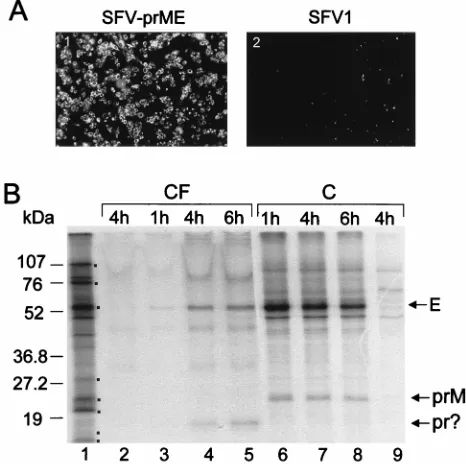
When SFV-prME RNA was electroporated into BHK21 cells, nearly 100% of cells were found to be positive in IF analysis with monoclonal antibodies to KUN E protein at 18 h after electroporation (Fig. 3A, panel 1). To confirm expression of the KUN prM and E proteins in transfected cells and to detect secretion of prME into the tissue culture fluid, we la-beled transfected cells with [35S]methionine-cysteine for 1 h,
followed by incubation for increasing chase periods. A strongly labeled band corresponding to KUN E protein was apparent in both culture fluid and cell lysates of SFV-prME-transfected cells at all times (Fig. 3B). A labeled band corresponding to KUN prM protein was detected in cell lysates, and another band corresponding in migration to the predicted molecular weight of KUN pr protein was detected in the culture fluid of transfected cells (Fig. 3B). An apparent increase in the inten-sity of labeling of the E and pr proteins in the culture fluid (Fig. 3B) and a concomitant decrease in the intensity of labeling of the E and prM proteins in the cell lysates (Fig. 3B) were observed during the chase period. Interestingly, secreted pr protein was also detected in the culture fluid of cells infected with other flaviviruses (24, 30), and protein migrating similarly was detected in the culture fluid of cells infected with recom-binant vaccinia virus expressing Japanese encephalitis virus prME genes (18). Presumably, the pr and M proteins were cleaved from their precursor prM by furin or a similar cell protease during its secretion from cells (for a review, see ref-erence 17). Because of its small size (;8 kDa), extracellular M protein probably ran off the gel during electrophoresis and therefore could not be detected. The identity of the expressed KUN E and coprecipitated prM proteins detected in the cul-ture fluid and cells (Fig. 3B) was confirmed by RIP with KUN anti-E antibodies (data not shown). These results demon-strated a correct processing of KUN prME polyprotein in cells transfected with SFV-prME RNA and the secretion of E, pr, and possibly M proteins into the culture fluid.
Expression of all three KUN structural proteins by the re-combinant SFV-prME-C107 replicon.Although we were able to package KUN replicon in cells by using transfection with two SFV RNAs expressing the prME and C genes separately (see results in the next section), the efficiency of packaging was rather low. In order to increase the efficiency of packaging and
to simplify the procedure, we prepared a single SFV replicon construct expressing the prME genes and the C gene simulta-neously. Because of the difficulties experienced with cloning of the entire C-prM-E region into the pSFV1 vector (see the first section of Results) and also to avoid possible uncertainty re-garding cleavage at the carboxy terminus of C in the absence of flavivirus protease NS2B-NS3 (3, 23, 34, 39–41), an SFV rep-licon expressing the prME and C genes under the control of two separate 26S promoters was prepared (Fig. 1, SFV-prME-C107).
IF analysis of SFV-prME-C107-electroporated BHK cells with anti-E and anti-C antibodies showed expression of both E and C proteins in nearly 100% of the cells by 18 h after transfection (results not shown). As expected, both E and C proteins were expressed in the same cell (compare dual IF labeling by anti-C and anti-E antibodies in Fig. 4A). In con-trast, only about 40% of cells were dual labeled with these antibodies when two individual SFV RNAs expressing prME and C, respectively, were electroporated simultaneously (re-sults not shown). When cells transfected with SFV-prME-C107 RNA were pulse-chased with [35S]methionine-cysteine and the
[image:4.612.310.543.71.305.2]lysates were immunoprecipitated with KUN anti-E antibodies, both E and prM proteins were coprecipitated (Fig. 4B), as was observed after transfection of SFV-prME RNA (Fig. 3B). A gradual increase of immunoprecipitated labeled E protein in culture fluids and a corresponding decrease of immunoprecipi-tated labeled E and prM proteins in the cell lysates were
FIG. 3. Expression of the KUN prME genes by the recombinant SFV-prME replicon. (A) IF analysis of SFV-prME- and SFV1-transfected BHK21 cells at 18 h after transfection by using KUN monoclonal anti-E antibodies. (B) Results of pulse-chase labeling with [35S]methionine-cysteine of SFV-prME-transfected
BHK21 cells. Culture fluid (CF) and cell lysate (C) samples were collected during chase periods (see Materials and Methods). Lane 1, radiolabeled KUN cell lysate, with dots indicating KUN proteins as in Fig. 2B. Lanes 2 and 9, samples collected after a 4-h chase period from culture fluid and cells, respec-tively, after transfection with the control SFV1 RNA. Lanes 3, 4, and 5, culture fluid samples collected at 1, 4, and 6 h of chase, respectively; lanes 6, 7, and 8, corresponding chase samples from the cells. Ten microliters of a total of 700ml of culture fluid and 5ml of a total of 300ml of cell lysate samples were used for electrophoresis. The exposure time of the dried gel for cell lysates was 1 day, and that for culture fluids was 5 days. M, the low-molecular-mass cleavage product (;8 kDa) of prM, was not resolved (see text). Numbers represent sizes of the low-range prestained Bio-Rad protein standards.
on November 9, 2019 by guest
http://jvi.asm.org/
observed during the chase period (Fig. 4B). Immunoprecipita-tion of the labeled cell lysates with anti-C antibodies confirmed expression of C protein in transfected cells and showed a decrease in the amount of precipitated C during the chase period (Fig. 4C), probably involving some degradation of C protein during a prolonged chase period as was observed pre-viously in KUN-infected cells (36). The results of RIP analysis of culture fluid, not treated with detergents, with C anti-bodies were negative (results not shown), indicating that no free C protein was secreted into culture fluid of SFV-prME-C107-transfected cells. In a later experiment (see Fig. 7C, lane 2), particles secreted from cells transfected only with SFV-prME-C107 RNA were purified, and only E and prM were
coprecipitated with anti-E antibodies; again, no secreted C was detected.
Overall, the immunofluorescence and labeling patterns in cells transfected with SFV-prME-C107 RNA were very similar to those observed in cells transfected with two different RNAs expressing prME and C proteins separately (compare the re-sults in Fig. 4 with those in Fig. 2 and 3), suggesting that proper processing and maturation of all three KUN structural proteins occurred when they were expressed from the recombinant SFV replicon.
Packaging of the KUN replicon RNA into pseudoinfectious particles by the KUN structural proteins expressed from the recombinant SFV replicons.Because with KUN replicon con-struct C20rep we were able to successfully transfect only;10% of cells (15), it was essential to use a KUN replicon with great-er transfection efficiency for attempted packaging in doubly transfected cells (i.e., cells transfected with the KUN replicon and recombinant SFV replicons expressing KUN structural proteins). Therefore, we prepared a new replicon construct, C20DXrep (see Materials and Methods), with significantly im-proved efficiency of transfection in BHK21 cells. Using an anti-NS3 antibody to detect amplification of KUN replicon RNA, we demonstrated that up to about 80% of 23106to 43
106BHK21 cells could be routinely electroporated with;10
mg of C20DXrep RNA (16). The improved replicon C20DXrep therefore was used in all packaging experiments.
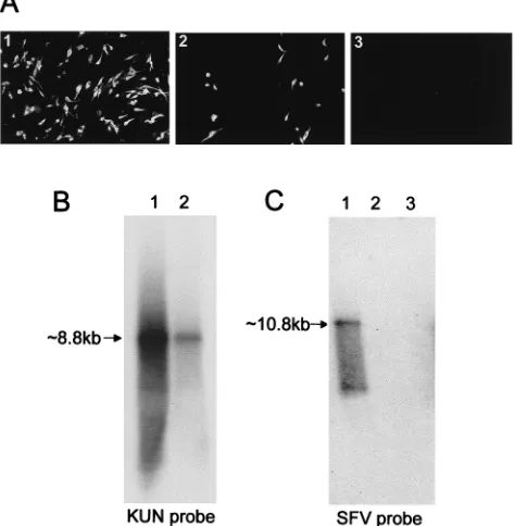
Initial cotransfection experiments showed that simultaneous transfection of C20DXrep RNA and SFV RNAs expressing KUN structural proteins did not produce detectable infectious particles (results not shown). Under these conditions, KUN replicon RNA was poorly amplified (very few cells were posi-tive by IF with anti-NS3 antibodies), whereas SFV RNAs rep-licated efficiently (there was strong IF with anti-E or anti-C antibodies in nearly 100% of cells) (results not shown). There-fore, a delay of 12 h or longer between electroporations was used in subsequent experiments in order to allow KUN repli-con RNA to accumulate before electroporation of SFV RNAs. In preliminary experiments we compared the relative efficien-cies of packaging of KUN replicon RNA with the single SFV-prME-C107 RNA or the two SFV RNAs, SFV-prME and SFV-C107, by monitoring yields by IF and Northern blot anal-yses. BHK cells were infected with the cell culture fluid col-lected at 26 h after electroporation with SFV RNAs of cells electroporated 27 h previously with C20DXrep RNA. The single SFV-prME-C107 RNA was more efficient, producing a large increase in the number of IF-positive cells and in synthe-sis of KUN-specific RNA (Fig. 5B). The titer, expressed as infectious units per milliliter (see Materials and Methods), was
;23 105, or 10-fold greater than the result when two SFV
RNAs were used. The difference in the production of infec-tious particles is probably due mainly to a lower proportion of cells simultaneously expressing both prME and C proteins after cotransfection of two SFV RNAs (40%) compared to transfection of a single SFV RNA (nearly 100%). Noticeably, the efficiency of the electroporation of SFV RNAs and subse-quent expression of KUN structural proteins from these vec-tors were not apparently affected by the previous electropora-tion and replicaelectropora-tion of C20DXrep RNA, as judged by dual IF analysis with anti-NS3 and anti-E or anti-C antibodies (data not shown). As expected, no infectious particles con-taining packaged KUN replicon RNA were detected when only SFV-prME RNA was used in packaging experiments with C20DXrep RNA (panel 3 in Fig. 5A), demonstrating that coexpression of C protein is absolutely essential for the formation of the secreted infectious particles.
[image:5.612.53.286.65.431.2]Significantly, no evidence of transmission of replicating SFV
FIG. 4. Expression of all three KUN structural proteins by the recombinant SFV-prME-C107 replicon. (A) Dual IF analysis of the same field of BHK21 cells fixed in acetone at 18 h after transfection with SFV-prME RNA by using KUN anti-C and anti-E antibodies with Texas red (TR)- and fluorescein isothiocyanate (FITC)-conjugated secondary antibodies, respectively. (B and C) Cells at 18 h after transfection with SFV-prME-C107 RNA were pulsed with [35
S]methionine-cysteine for 1 h; subsequently, 300ml (from a total of 600ml) of cell lysates (lanes C) and 1 ml (from a total of 2 ml) of culture fluids (lanes CF) collected at different chase intervals (1, 6, and 9 h) were immunoprecipitated either with KUN anti-E antibodies (B) or with KUN anti-C antibodies (C) as described in Materials and Methods. Ten microliters (from a total of 30ml) of immunopre-cipitated samples was electrophoresed in SDS–12.5% (B) and –15% (C) poly-acrylamide gels. Dots in panel B indicate KUN proteins in the labeled KUN cell lysates as in Fig. 2B. Dots in panel C represent KUN proteins prM, NS2A, C, and NS4A/NS2B (from top to bottom) in the radiolabeled KUN-infected cell lysate. Numbers represent sizes of the low-range prestained Bio-Rad protein standards.
on November 9, 2019 by guest
http://jvi.asm.org/
RNAs was obtained by IF analysis with anti-E and anti-C antibodies of cells infected with the culture fluid collected from cells transfected with any of the recombinant SFV RNAs ei-ther with or without previous transfection with C20DXrep RNA (data not shown). This assay is very sensitive, because a very low number of packaged recombinant SFV replicon RNAs able to express KUN C or E protein would have been detected by the IF analysis following the manyfold amplifica-tion of any SFV RNAs after such delivery. Similar (negative) results were also obtained by Northern blot analysis with an SFV-specific probe of RNA isolated from cells infected with the particles collected from cells sequentially transfected with KUN replicon and SFV replicon RNAs (Fig. 5C, lanes 2 and 3). The packaging observed after these sequential transfections was therefore clearly specific for KUN replicon RNA.
In order to optimize the conditions for efficient packaging of C20DXrep RNA in cotransfection experiments with SFV-prME-C107 RNA, various time points between electropora-tions (Fig. 6A) and between the second electroporation and harvesting of the infectious particles (Fig. 6B) were examined. Initially, optimization of the time between the two electropo-rations was established with a fixed time for collection of the infectious particles. Equal amounts of cells were seeded onto cell culture dishes after the first electroporation with C20DXrep RNA, and cells were subsequently electroporated
with SFV-prME-C107 RNA after a further 12, 18, 24, or 30 h of incubation. Culture fluid was then harvested from each dish at 24 h after the second electroporation, and serial dilutions were used to infect BHK21 cells for titration by IF analysis with anti-NS3 antibodies. As the period between electroporations was extended from 12 to 30 h, an increase of infectious parti-cles was observed, with the highest titer of 1.33106IU per ml
(;23 106 particles per 106 cells) corresponding to the 24-h
interval (Fig. 6A). Northern blot analysis of total RNA from infected cells with a labeled KUN-specific cDNA probe con-firmed this optimal time interval of 24 h (Fig. 6A). When;10
mg of in vitro-transcribed full-length FLSDX RNA (similar to the amount used for C20DXrep RNA [see Materials and Methods]) was electroporated into BHK cells, the titer of infectious particles accumulated in the culture fluid by 45 h, as determined by IF assay of foci in newly infected BHK cells with anti-NS3 antibodies, was ;107IU per ml (data not shown).
Thus, the yield of KUN virions after electroporation of tran-scribed full-length RNA was only ;10-fold higher than the yield of encapsidated replicon particles obtained by dual trans-fections with KUN and SFV replicon RNAs (see above).
In a separate experiment BHK cells were electroporated with SFV-prME-C107 RNA at 30 h after electroporation with C20DXrep RNA and seeded into one 60-mm-diameter culture dish. Single aliquots of the culture fluid (1 ml of a total of 3 ml) were then collected after 24, 30, and 42 h of incubation after the second electroporation, and the culture fluid was adjusted to the original volume by adding fresh medium. Collected aliquots were then used to infect BHK cells, and total cell RNA recovered from these infected cells at 24 h was then analyzed for relative amounts of amplified KUN replicon RNA by Northern blot hybridization. The gradual increase in ampli-fied KUN replicon RNA from 24 to 42 h after the second electroporation with SFV-prME-C107 RNA (Fig. 6B) was in accord with an observed increase in released infectious
[image:6.612.360.499.71.245.2]parti-FIG. 5. Packaging of KUN replicon RNA by KUN structural proteins ex-pressed from the recombinant SFV replicons. (A) IF analysis with KUN anti-NS3 antibodies of BHK21 cells infected with the undiluted culture fluid collected from BHK21 cells at 26 h after transfection with SFV-prME-C107 RNA (panel 1), SFV-prME and SFV-C RNAs (panel 2), or SFV-prME RNA (panel 3) of cells transfected 27 h previously with C20DXrep RNA. Panels 1 and 2 represent selected fields of coverslips enriched in positive cells. (B and C) Northern blot analysis of RNAs isolated from BHK21 cells infected as described for panel A, using labeled KUN-specific (B) and SFV-specific (C) cDNA probes (see Mate-rials and Methods). Lane 1 in panel B and lane 2 in panel C correspond to the cells in panel 1 of panel A. Lane 2 in panel B and lane 3 in panel C correspond to the cells in panel 2 of panel A. Lane 1 in panel C represents 5 ng of in vitro-synthesized SFV-prME-C107 RNA. The arrows in panels B and C indicate the positions of RNAs of about 8.8 kb for KUN replicon RNA and about 10.8 kb for SFV-prME-C107 RNA, determined relative to migration in the same gel of ethidium bromide-stainedlDNA digested with BstEII (New England Biolabs).
FIG. 6. Optimization of conditions for packaging of KUN replicon RNA. Northern blot analysis of BHK21 cells infected with filtered and RNase-treated culture fluid samples collected after sequential transfections with KUN replicon C20DXrep RNA and SFV-prME-C107 RNA is shown. Samples of total RNA used for Northern blotting were isolated from cells at 24 h after infection with samples of the culture fluid collected either at a fixed time (24 h) after the second transfection and with different time intervals between transfections (A) or at different times after the second transfection and a fixed time (30 h) after the first transfection (B). Titers in panel A represent the amounts of infectious units contained in the corresponding samples of culture fluids used for these infec-tions. The probe in both panels was a radiolabeled cDNA fragment representing the last 761 nucleotides of the KUN genome.
on November 9, 2019 by guest
http://jvi.asm.org/
[image:6.612.53.290.74.316.2]cles assayed by IF analysis of newly infected cells with anti-NS3 antibodies (data not shown). Further incubation of cells was abandoned in this experiment because of the severe cytopathic effects which had occurred by 42 h after electroporation of SFV-prME-C107 RNA. However, in other experiments longer incubation (68 h) of cells after the second electroporation with recombinant SFV RNA(s) resulted in reduction of the titer of accumulated infectious particles from surviving cells (data not shown).
In an attempt to improve the efficiency of the packaging system, we prepared an SFV-prME-C105 construct in which the 39 sequence of the C gene was engineered so that the resulting translation product, C, would terminate precisely at Arg105 (Fig. 1), identically to the mature form of KUN C
protein (29). When SFV-prME-C107 and SFV-prME-C105 RNAs were used in parallel in packaging experiments with C20DXrep RNA, the titers of infectious particles released in the culture fluid of transfected cells were virtually identical (;93105infectious particles per ml for both). These results
indicate either that C protein expressed from SFV-prME-C107 RNA was processed correctly (cleaved at Arg105) by KUN
NS2B-NS3 protease expressed from C20DXrep RNA or that the presence of an additional carboxy-terminal six amino acids in C107 protein (Fig. 1) did not interfere with packaging.
Characterization of the infectious particles. Having estab-lished that the infectivity of particles secreted into the culture fluid of cells transfected with C20DXrep and SFV-prME-C107 RNAs was filterable and RNase resistant, we than performed a virus neutralization test to confirm that the particles were packaged by KUN structural proteins. A 1-h incubation of this culture fluid at 37°C with a 1/10 dilution of antibodies to KUN E protein with known neutralizing activity (1) resulted in al-most complete loss of transmissible infectivity measured by IF
(panel 1 in Fig. 7A), while no loss of infectivity was observed in the control sample incubated under similar conditions in the absence of antibodies (panel 2 in Fig. 7A). Similar results were obtained when incubations with antibodies were performed at room temperature or at 4°C (data not shown). We then estab-lished the particulate nature of the particles by showing that they can be concentrated by pelleting. A clarified culture fluid of cotransfected cells was subjected to ultracentrifugation (see Materials and Methods), and resuspended pellet and superna-tant were used to infect BHK cells. Analysis by IF with anti-NS3 antibodies 24 h later showed that almost all of the infec-tious particles were present only in the pelleted material (compare panels 1 and 2 in Fig. 7B).
In order to identify the proteins and to detect the presence of KUN replicon RNA in the released infectious particles, they were immunoprecipitated with anti-E antibodies in the ab-sence of detergents from the culture fluid of cells sequentially transfected with C20DXrep and SFV-prME-C107 RNAs and radiolabeled with [35S]methionine-cysteine. Half of the
[image:7.612.142.456.70.278.2]immu-noprecipitated sample was used for separation of proteins in an SDS-polyacrylamide gel; radioautography of the gel showed the presence of the E, prM, and C proteins in the immuno-precipitates (Fig. 7C, lane 1). These same proteins were also resolved in a parallel experiment using culture fluid from KUN-infected cells (Fig. 7C, lane 3). E and prM proteins, but no C protein, were immunoprecipitated by anti-E antibodies from culture fluid of cells transfected only with SFV-prME-C107 RNA (Fig. 7C, lane 2). It was concluded that a specific interaction between KUN replicon RNA and KUN C protein was required for assembly and secretion of infectious particles. Note that secreted flaviviruses often contain significant amounts of uncleaved prM (see, for example, reference 24), as observed in Fig. 7C.
FIG. 7. Characterization of infectious particles. (A) Neutralization of infectivity of encapsidated particles, released from cells transfected sequentially with C20DXrep and SFV-prME-C107 RNAs (as in Fig. 6). Panels 1 and 2 show IF with anti-NS3 antibodies of cells infected with culture fluid collected at 24 h after transfections and incubated for 1 h at 37°C with anti-E monoclonal antibodies or culture medium, respectively. (B) IF analysis with anti-NS3 antibodies of cells infected with equal proportions of resuspended pellet (panel 1; 2ml from a 50-ml total volume) or supernatant fluid (panel 2; 200ml from a 5-ml total volume) from the culture fluid collected from cells transfected as described for panel A and subjected to ultracentrifugation (see Materials and Methods). (C) RIP analysis with anti-E antibodies of culture fluids from cells sequentially transfected with C20DXrep and SFV-prME-C107 RNAs (lane 1), transfected with SFV-prME-C107 RNA alone (lane 2), or infected with KUN virus (lane 3). Dots show bands corresponding to C and prM in lane 1 but a corresponding band for only prM in lane 2. (D) RT-PCR analysis with KUN-specific primers of RNAs extracted from the anti-E immunoprecipitates in panel C; lanes 2 to 4 are equivalent to lanes 1 to 3 in panel C. Note that immunoprecipitates were treated with RNase A (20mg per ml) for 30 min at 37°C prior to isolation of RNA in order to ensure the absence of free RNA in the RIP samples. RNA from the treated RIP samples was isolated by digestion with proteinase K (100mg per ml) in the presence of 0.5% SDS for 30 min at 37°C followed by phenol-chloroform extraction and ethanol precipitation. Lane 1,fX174 replicative-form DNA digested with HaeIII (New England Biolabs).
on November 9, 2019 by guest
http://jvi.asm.org/
on November 9, 2019 by guest
http://jvi.asm.org/
RNA extracted from the remainder of the immunoprecipi-tates pretreated with RNase A (see the previous paragraph) was reverse transcribed and PCR amplified with KUN-specific primers. As expected, a DNA fragment of the predicted size (;700 bp, NS2A region) was observed in the RT-PCR product from packaged replicon and KUN virus prepared similarly (Fig. 7D, lanes 2 and 4). No RT-PCR product was obtained from RNA extracted from the immunoprecipitate of the cul-ture fluid collected from cells transfected with SFV-prME-C107 RNA alone (Fig. 7D, lane 3) or with C20DXrep RNA alone (data not shown). These results established that replicon RNA was encapsidated in the packaged particles assayed pre-viously for infectivity and KUN proteins (Fig. 7A to C).
Further characterization of the packaged particles contain-ing replicon RNA was performed by sedimentation analysis. In parallel with KUN virions (both concentrated by ultracentrif-ugation) they were sedimented through 5 to 25% sucrose den-sity gradients. Fractions of 0.5 ml were collected, diluted, and assayed for infectivity by IF assay with anti-NS3 antibodies at 18 h for KUN virions or at 24 h for replicon particles (see the legend to Fig. 8A). The maximum infectivity for replicon par-ticles was concentrated in fractions 5 to 7, with a peak titer of
;1.33105IU/ml (fraction 6), while infectious KUN virions
were concentrated mostly in fractions 2 to 4, with a peak titer of;2.83107IU/ml (fraction 3) (Fig. 8A). These three
frac-tions from each gradient were combined, incubated with anti-E antibodies to aggregate virions and encapsidated particles, and concentrated by ultracentrifugation for electron microscopy (for experimental details, see the legend to Fig. 8B). As might be expected from the gradient sedimentation results (Fig. 8A), particles containing encapsidated replicon RNA were smaller than KUN virions, i.e.,;35 nm in diameter compared to the
;50-nm diameter of virion particles (Fig. 8B). Both replicon and virion particles appeared to be spherical and uniform in size; surface details were not resolved, probably because of attachment of some anti-E antibody molecules (Fig. 8B). Taken together, the results described in this section demon-strate that the particles produced by the dual replicon packag-ing system are in fact virus-like particles containpackag-ing KUN rep-licon RNA encapsidated by the KUN structural proteins C, prM/M, and E.
DISCUSSION
We have developed a packaging system allowing encapsida-tion of KUN replicon RNA by cotransfecencapsida-tion with SFV repli-con RNA(s) expressing KUN structural proteins. To our knowledge this is the first demonstration of packaging of a flavivirus RNA in trans, and it establishes that essential pack-aging signals do not reside in the structural region of the genome. Although KUN replicon RNA was packaged after sequential transfection with SFV RNAs C107 and SFV-prME, the yield of infectious particles was rather low (Fig. 5A
and B). A greatly improved yield was obtained when a single recombinant SFV replicon RNA, SFV-prME-C107, expressing both the KUN C and prME genes together but under control of separate 26S SFV subgenomic promoters, was prepared; this SFV RNA was used in all further encapsidation experi-ments. The less efficient production of packaged particles when two SFV RNAs (SFV-C107 and SFV-prME) were used was possibly due mainly to a significantly lower proportion of cells simultaneously expressing both the prME and C proteins (see Results). In addition, the lower yield of packaged particles could also be due to a difference in replication rates of the two SFV replicons within the same cell, especially if equimolar amounts of the KUN structural proteins were required for packaging (Fig. 5A and B).
Further optimization of conditions for encapsidation of C20DXrep RNA revealed that a time delay of about 24 h between transfections of KUN replicon RNA C20DXrep and recombinant SFV replicon RNA SFV-prME-C107 was re-quired to obtain the maximum yield of encapsidated infectious particles another 24 h later (;1.33106 particles per ml, or
;23106particles per 106transfected cells [Fig. 6A]), which
was comparable to that obtained by using SFV replicons as helpers for packaging of defective Moloney murine leukemia virus particles (;7 3 106 particles per 106 transfected cells
[20]). Interestingly, the yield of infectious SIN particles per milliliter obtained by cotransfection of two SIN RNAs (repli-con RNA and helper RNA expressing SIN structural genes) varied from 3.93106(9) to 53107to 83107(5),
represent-ing 0.5 to 5% of the yield obtained after transfection of full-length infectious SIN RNA (5). These ratios are similar to the ratios observed for the packaged KUN replicon RNA and transfected full-length FLSDX RNA (;10%) (see Results). It is important to emphasize in this context that the yields of infectious virus, viral RNA, and proteins per cell produced by flaviviruses are notoriously low compared to those of alphavi-ruses. For example, in comparative assays the rate of synthesis of KUN RNA was 10 to 100 times less than the rate of SFV RNA synthesis (7).
In the present cotransfection experiments, it is likely that a rapid switch-off of cellular and possibly KUN nonstructural protein synthesis mediated by replicating SFV RNA (22) in-hibited the initiation of KUN RNA replication, since only a minor proportion of KUN replicon-positive cells and no infec-tious particles secreted in the culture fluid were detected when C20DXrep and recombinant SFV RNAs were transfected si-multaneously (see the penultimate section of Results). In con-trast, replication of SFV RNAs was not noticeably affected by coreplicating KUN RNA (see the penultimate section of Re-sults). Previously we showed that the maximum level of syn-thesis and accumulation of KUN replicon RNA occurred at about 24 h after electroporation (15). The observed increase of the titer of infectious particles as the period between
transfec-FIG. 8. Sedimentation and electron microscopy analyses of KUN replicon and virion particles. (A) Sedimentation profiles of virions and replicon particles in parallel sucrose density gradients. Particles were collected from culture fluids of BHK cells either at 35 h after sequential transfections with C20DXrep and SFV-prME-C107 RNAs or at 24 h after infection with KUN virus and were concentrated by ultracentrifugation as described in Materials and Methods. The pelleted particles were resuspended in 300ml of PBS–0.1% bovine serum albumin overnight at 4°C and clarified by centrifugation at 16,0003g in a microcentrifuge for 10 min. The
supernatant was overlaid on top of a 12-ml 5 to 25% sucrose density gradient which was centrifuged at 38,000 rpm for 70 min at 20°C in an SW41 rotor. Fractions of 0.5 ml were collected from the bottom of the gradient and diluted 1:2 (replicon particles) or 1:100 (KUN virions) for infectivity assays by IF on coverslip cultures of BHK cells at 24 h (replicon particles) or at 18 h (KUN virions) after infection, using anti-E antibodies; titers of infectious particles were determined as described in Materials and Methods. (B) Electron micrographs of virions and encapsidated replicon particles stained with uranyl acetate. Fractions 5 to 7 of replicon particles in panel A, and fractions 2 to 4 of KUN virions, were pooled and incubated with a 1/20 dilution of anti-E antibodies for 1 h at 20°C, followed by 2 h of incubation at 4°C with constant rotation. Particles were then again concentrated by ultracentrifugation as described above, and pellets were resuspended in 175ml of PBS–0.1% bovine serum albumin overnight at 4°C. Resuspended particles were then sonicated in a Transsonic 700/h sonicating water bath (CAMLAB, Cambridge, United Kingdom) for 1 min and pelleted onto a carbon-coated Formvar grid by centrifugation in an 18° fixed-angle A-100 rotor in a Beckman Airfuge for 1 h at 80,000 rpm. Grids were stained with 4% uranyl acetate, and particles were visualized by electron microscopy. Bars, 200 nm.
on November 9, 2019 by guest
http://jvi.asm.org/
tions was increased from 12 to 24 h (Fig. 6A) indicated a direct correlation between (i) the amount of presynthesized KUN RNA and/or KUN nonstructural proteins present in cells at the time of transfection of SFV replicon RNA and (ii) the final production of infectious particles. However, we did not directly monitor the replication of KUN replicon RNA after transfec-tion of SFV replicon RNA in these experiments. Alternatively, a certain threshold of synthesized KUN RNA and proteins, or cell factors (KUN-induced membranes?), may be required for efficient replication of KUN replicon RNA in the presence of replicating SFV replicon RNA.
Comparison of the sedimentation characteristics of the en-capsidated replicon particles and KUN virions by centrifuga-tion through a 5 to 25% sucrose gradient showed that infec-tious replicon particles sedimented more slowly (Fig. 8A), suggesting that they are possibly smaller. This notion was con-firmed when the particles concentrated from the peak gradient fractions were negatively stained and visualized by electron microscopy, the diameters being;35 nm for replicon particles and ;50 nm for virions (Fig. 8B). The difference in size is probably due to the smaller size of the encapsidated replicon RNA (8,775 versus 11,022 nucleotides) and the fact that fewer protein molecules are required for encapsidation. Similarly, defective interfering particles of the alphaviruses SIN and SFV containing RNAs with deletions were also found to be smaller than virions (5, 11).
There are two major applications of the KUN replicon pack-aging system which we are exploring further. The first involves the development of a new vaccine delivery system based on the encapsidation of noncytopathic KUN replicons expressing genes of other medically important viruses, and we have a number of candidate genes for these studies. It should be noted that the use of a heterologous packaging system such as that presented here eliminates the possibility of contamination of packaged replicons with infectious virus, which appears to be a major concern with the use of homologous packaging systems such as SFV and SIN in which homologous recombi-nation between the two RNAs occurs (21, 29). A second ap-plication is based on the use of recombinant SFV replicon RNAs expressing KUN structural proteins and containing fragments of KUN replicon RNA for rapid mapping of any packaging signal(s) in the KUN RNA fragments. It is impor-tant in this regard to emphasize the observed specificity of packaging of KUN replicon RNA; no SFV replicon RNAs were transmitted by the secreted particles (Fig. 5C; see Re-sults). In view of our previous data on the strength of interac-tion between the KUN C protein and KUN UTRs (14), it was not surprising that no packaging of KUN replicon RNA to produce infectious particles occurred when C was not supplied by an SFV replicon (Fig. 5A, panel 3), indicating the essential role of C in assembly of particles.
Our results represent a significant advance in the area of flavivirus virion assembly and secretion. In summary, it is now possible to introduce mutations into the KUN replicon RNA and into genes for the structural proteins and to assess their effect on the assembly and release of virus-like particles. Thus, the dual system (KUN replicon plus SFV vector for expression of KUN structural proteins) should allow rapid mapping of the packaging signal(s) in flavivirus RNA and the amino acid mo-tif(s) in the structural proteins involved in RNA encapsidation, virion assembly, and secretion. It can also facilitate the devel-opment of a noninfectious and noncytopathic vaccine delivery system.
ACKNOWLEDGMENTS
We are grateful to Roy Hall for providing KUN anti-E monoclonal antibodies and to Alasdair McDowall and Jason Mackenzie for their help in electron microscopy of the particles.
This work was supported by grants from the National Health and Medical Research Council of Australia and from the Royal Children’s Hospital Foundation, Brisbane.
REFERENCES
1. Adams, S. C., A. K. Broom, L. M. Sammels, A. C. Hartnett, M. J. Howard,
R. J. Coelen, J. S. Mackenzie, and R. A. Hall. 1995. Glycosylation and antigenic variation among Kunjin virus isolates. Virology 206:49–56. 2. Allison, S. L., K. Stadler, C. W. Mandl, C. Kunz, and F. X. Heinz. 1995.
Synthesis and secretion of recombinant tick-borne encephalitis virus protein E in soluble and particulate form. J. Virol. 69:5816–5820.
3. Amberg, S. M., A. Nestorowicz, D. W. McCourt, and C. M. Rice. 1994. NS2B-3 proteinase-mediated processing in the yellow fever virus structural region: in vitro and in vivo studies. J. Virol. 68:3794–3802.
4. Ansardi, D. C., D. C. Porter, and C. D. Morrow. 1993. Complementation of a poliovirus defective genome by a recombinant vaccinia virus which pro-vides poliovirus P1 capsid precursor in trans. J. Virol. 67:3684–3690. 5. Barret, A. D. T., W. D. Cubitt, and N. J. Dimmock. 1984. Defective
inter-fering particles of Semliki Forest virus are smaller than particles of standard virus. J. Gen. Virol. 65:2265–2268.
6. Bredenbeek, P., I. Frolov, C. M. Rice, and S. Schlesinger. 1993. Sindbis virus expression vectors: packaging of RNA replicons by using defective helper RNAs. J. Virol. 67:6439–6446.
7. Chu, P. W., and E. G. Westaway. 1985. Replication strategy of Kunjin virus: evidence for recycling role of replicative form RNA as template in semicon-servative and asymmetric replication. Virology 140:68–79.
8. Coia, G., M. D. Parker, G. Speight, M. E. Byrne, and E. G. Westaway. 1988. Nucleotide and complete amino acid sequences of Kunjin virus: definitive gene order and characteristics of the virus-specified proteins. J. Gen. Virol.
69:1–21.
9. Dubensky, T. W., Jr., D. A. Driver, J. M. Polo, B. A. Belli, E. M. Latham,
C. E. Ibanez, S. Chada, D. Brumm, T. A. Banks, S. J. Mento, D. J. Jolly, and S. M. W. Chang.1996. Sindbis virus DNA-based expression vectors: utility for in vitro and in vivo gene transfer. J. Virol. 70:508–519.
10. Johanning, F. W., R. M. Conry, A. F. LoBuglio, M. Wright, L. A. Sumerel,
M. J. Pike, and D. T. Curiel.1995. A Sindbis virus mRNA polynucleotide vector achieves prolonged and high level heterologous gene expression in
vivo. Nucleic Acids Res. 23:1495–1501.
11. Johnson, R. E., D. R. Tovel, D. T. Brown, and P. Faulkner. 1975. Interfering passages of Sindbis virus: concomitant appearance of interference, morpho-logical variants and truncated viral RNA. J. Virol. 16:951–958.
12. Kamrud, K. I., A. M. Powers, S. Higgs, K. E. Olson, C. D. Blair, J. O.
Carlson, and B. J. Beaty.1995. The expression of chloramphenicol acetyl-transferase in mosquitoes and mosquito cells using a packaged Sindbis rep-licon system. Exp. Parasitol. 81:394–403.
13. Khromykh, A. A., and E. G. Westaway. 1994. Completion of Kunjin virus RNA sequence and recovery of an infectious RNA transcribed from stably cloned full-length cDNA. J. Virol. 68:4580–4588.
14. Khromykh, A. A., and E. G. Westaway. 1996. RNA binding properties of core protein of the flavivirus Kunjin. Arch. Virol. 141:685–699.
15. Khromykh, A. A., and E. G. Westaway. 1997. Subgenomic replicons of the flavivirus Kunjin: construction and applications. J. Virol. 71:1497–1505. 16. Khromykh, A. A., and E. G. Westaway. Unpublished data.
17. Klenk, H.-D., and W. Garten. 1994. Activation cleavage of viral spike pro-teins by host proteases, p. 241–280. In E. Wimmer (ed.), Cellular receptors for animal viruses. Cold Spring Harbor Laboratory Press, Cold Spring Har-bor, N.Y.
18. Konishi, E., S. Pincus, E. Paoletti, R. E. Shope, T. Burrage, and P. W.
Mason.1992. Mice immunized with a subviral particle containing the Japa-nese encephalitis virus prM/M and E proteins are protected from lethal JEV infection. Virology 188:714–720.
19. Konishi, E., and P. W. Mason. 1993. Proper maturation of the Japanese encephalitis virus envelope glycoprotein requires cosynthesis with the pre-membrane protein. J. Virol. 67:1672–1675.
20. Li, K-J., and H. Garoff. 1996. Production of infectious recombinant Moloney murine leukemia virus particles in BHK cells using Semliki Forest virus-derived RNA expression system. Proc. Natl. Acad. Sci. USA 93:11658– 11663.
21. Life Technologies Inc. 1994. SFV gene expression system manual. Life Tech-nologies Inc., Melbourne, Australia.
22. Liljestrom, P., and H. Garoff. 1991. A new generation of animal cell expres-sion vectors based on the Semliki Forest virus replicon. Bio/Technology
9:1356–1361.
23. Lobigs, M. 1993. Flavivirus premembrane protein cleavage and spike het-erodimer secretion require the function of the viral proteinase NS3. Proc. Natl. Acad. Sci. USA 90:6218–6222.
24. Murray, J. M., J. G. Aaskov, and P. J. Wright. 1993. Processing of the
on November 9, 2019 by guest
http://jvi.asm.org/
dengue virus type 2 proteins prM and C-prM. J. Gen. Virol. 74:175–182. 25. Percy, N., W. S. Barclay, M. Sullivan, and J. W. Almond. 1992. A poliovirus
replicon containing the chloramphenicol acetyltransferase gene can be used to study the replication and encapsidation of poliovirus RNA. J. Virol.
66:5040–5046.
26. Porter, D. C., D. C. Ansardi, W. S. Choi, and C. D. Morrow. 1993. Encap-sidation of genetically engineered poliovirus minireplicons which express human immunodeficiency virus type 1 Gag and Pol proteins upon infection. J. Virol. 67:3712–3719.
27. Porter, D. C., D. C. Ansardi, and C. D. Morrow. 1995. Encapsidation of poliovirus replicons encoding the complete human immunodeficiency virus type 1 gag gene by using a complementation system which provides the P1 capsid protein in trans. J. Virol. 69:1548–1555.
28. Pugachev, K. V., P. W. Mason, and T. K. Frey. 1995. Sindbis vectors suppress secretion of subviral particles of Japanese encephalitis virus from mamma-lian cells infected with SIN-JEV recombinants. Virology 209:155–166. 29. Raju, R., S. V. Subramaniam, and M. Hajjou. 1995. Genesis of Sindbis virus
by in vivo recombination of nonreplicative RNA precursors. J. Virol. 69: 7391–7401.
30. Randolf, V. B., G. Winkler, and V. Stollar. 1990. Acidotropic amines inhibit proteolytic processing of flavivirus prM protein. Virology 174:450–458. 31. Rice, C. M. 1996. Flaviviridae: the viruses and their replication, p. 931–960.
In B. N. Fields, D. M. Knipe, and P. M. Howley (ed.), Fields virology, 3rd ed.
Lippincott-Raven, Philadelphia, Pa.
32. Schalich, J., S. L. Allison, K. Stiasny, C. W. Mandl, C. Kunz, and F. X.
Heinz.1996. Recombinant subviral particles from tick-borne encephalitis virus are fusogenic and provide a model system for studying flavivirus
enve-lope glycoprotein functions. J. Virol. 70:4549–4557.
33. Speight, G., and E. G. Westaway. 1989. Carboxy-terminal analysis of nine proteins specified by the flavivirus Kunjin: evidence that only the intracellu-lar core protein is truncated. J. Gen. Virol. 70:2209–2214.
34. Stocks, C. E., and M. Lobigs. 1995. Posttranslational signal peptidase cleav-age at the flavivirus C-prM junction in vitro. J. Virol. 69:8125–8126. 35. Strauss, J. H., and E. G. Strauss. 1994. The alphaviruses: gene expression,
replication, and evolution. Microbiol. Rev. 58:491–562.
36. Westaway, E. G. 1973. Proteins specified by group B togaviruses in mamma-lian cells during productive infections. Virology 51:454–465.
37. Westaway, E. G., A. A. Khromykh, M. T. Kenney, J. M. Mackenzie, and
M. K. Jones.1997. Proteins C and NS4B of the flavivirus Kunjin translocate independently into the nucleus. Virology 234:31–41.
38. Wimmer, E., C. U. T. Hellen, and X. Cao. 1993. Genetics of poliovirus. Annu. Rev. Genet. 27:353–436.
39. Yamshchikov, V. F., and R. W. Compans. 1993. Regulation of the late events in the flavivirus protein processing and maturation. Virology 192:38–51. 40. Yamshchikov, V. F., and R. W. Compans. 1994. Processing of the
intracel-lular form of the West Nile virus capsid protein by the viral NS2B-NS3 protease: an in vitro study. J. Virol. 68:5765–5771.
41. Yamshchikov, V. F., D. W. Trent, and R. W. Compans. 1997. Upregulation of signalase processing and induction of prM-E secretion by the flavivirus NS2B-NS3 protease: roles of protease components. J. Virol. 71:4364–4371. 42. Zhou, X., P. Berglund, G. Rhodes, S. E. Parker, M. Jondal, and P.
Liljestrom.1994. Self-replicating Semliki Forest virus RNA as recombinant vaccine. Vaccine 12:1510–1514.