1
A DISSERTATION ON
A STUDY ON CLINICOPATHOLOGICAL
EVALUATION OF SOLITARY NODULE
THYROID
Dissertation Submitted to
THE TAMILNADU
DR.M.G.R. MEDICAL UNIVERSITY CHENNAI – 600 032
With fulfillment of the Regulations For the Award of the Degree of
M.S. GENERAL SURGERY Branch - I
DEPARTMENT OF GENERAL SURGERY KILPAUK MEDICAL COLLEGE
CHENNAI – 600 010
2
CERTIFICATE
This in to certify that the dissertation work titled “
A STUDY
ON CLINICOPATHOLOGICAL EVALUATION OF
SOLITARY NODULE THYROID
” is a bonafide researchwork of DR. R.KRISHNA KUMAR Enrolment No.
submitted in partial fulfillment of the requirements for the award of Degree
of M.S. GENERAL SURGERY in THE TAMIL NADU DR.M.G.R.
MEDICAL UNIVERSITY CHENNAI- 600 032
Signature of unit Chief
Signature of H.O.D
3
ACKNOWLEDGEMENT
I thank Dr. M. Dhanapal, M.D., D. M., Dean, Kilpauk Medical
College for permitting to use the resources and clinical material of this
hospital.
I am grateful to Dr. G. GUNASEELAN, M.S., Professor and Head
of the Department of General Surgery for his constant encouragement and
help during this study.
I profusely thank Dr. V. SUCHARITA, M.S., CHIEF S2 Unit
department of General Surgery for his guidance and encouragement.
I am indebted to the Assistant Professors in the Department of
General Surgery Govt.Kilpauk Medical College Hospital, Chennai,
Dr.A.Affee Asma,M.S, Dr.V.Vijayalakshmi M.S, Dr.Shantha
kumar,M.S, for the advise and help rendered to me.
I also thank my Post Graduate colleagues and house surgeons for all the
timely help they rendered.
Lastly, I thank MY PATIENTS not only for their
consent and co-operation towards the preparation of this study but also for
4
S.NO CONTENTS PAGE NO.
1. Introduction 1
2. Aim of Study 2
3. Review of literature 3
4. Materials and Methods used 49
5. Observation 50
6. Discussion 58
7. Conclusion 67
8. Annexure
Proforma
Abbreviations
Master Chart
5
6
INTRODUCTION
Nodular thyroid disease is a common disorder & the incidence increases with age. The prevalence of nodular goitre is 4-7%, but the autopsy studies
& USG have shown that the true incidence is to be around 50% in the adult
population. Most thyroid nodules are benign & most of them are adenomas.
Although a majority of the nodules are benign & some are shown to be
malignant. Thyroid cancers are comparatively rare disorders.
Preoperative identification & evaluation of these nodules is very
important. But this distinction often proved difficult with the reliance on the
clinical assessment & investigations of low sensitivity & specificity.
According to the clinical diagnosis and most importantly tissue diagnosis,
the treatment approach can be modified.
The FNAC is said to be the gold standard for the diagnosis of
SNG, but the FNAC has got its own pitfalls because of false negatives &
false positives reports & the same time the results have inter-observer
variation & need an experienced histopathologist. Even FNAC has got less
specificity in identifying the micro-papillary projection & can’t differentiate
between follicular adenoma & carcinoma.
The uses of USG & radioisotope studies are often additive but not
7
surgery of any type plays an important role in the treatment as well as the
diagnostic aspect but the present trend is to manage benign lesion on FNAC
with conservative line of management reserving surgery for the malignant
8
AIM OF THE
9
AIM
9 To review data regarding incidence of the solitary nodular goitre
9 To analyse & discuss the various clinical presentation
9 To find out the percentage of malignancy in solitary nodular goitre
9 To confirm the role of surgery as the diagnostic & therapeutic modality & to discuss the revised indication for surgeries
9 To stress the importance of histopathological examination as the final & confirmatory evidence of diagnosis
9 To analyse the pattern of disease affecting SNG based on HPE
10
REVIEW OF
11
SURGICAL ANATOMY
Historical Background
Goiters (from the Latin guttur, throat), defined as an enlargement of the thyroid, have been recognized since 2700 B.C. In 1619, Hieronymus
Fabricius ab Aquapendente recognized that goiters arose from the thyroid
gland. The term thyroid gland (Greek thyreoeides, shield-shaped) is, however, attributed to Thomas Wharton in his Adenographia (1656). Burnt seaweed was considered to be the most effective treatment for goiters.
The most notable thyroid surgeons were Emil Theodor Kocher
(1841–1917) Theodor Billroth (1829–1894), who performed thousands of
operations with increasingly successful results. After total thyroidectomy,
patients (children) became myxedematous with cretinous features.
Myxedema was first effectively treated in 1891 by George Murray, who
used a subcutaneous injection of an extract of sheep's thyroid; later, Edward
Fox demonstrated that oral therapy was equally effective. In 1909, Kocher
was awarded the Nobel Prize for medicine in recognition "for his works on
the physiology, pathology, and surgery of the thyroid gland."
General Topography: The thyroid gland consists typically of two lobes, a
connecting isthmus, and an ascending pyramidal lobe. One lobe, usually the
12
absent (1.7 percent). The isthmus is absent in about 10 percent of thyroid
glands, and the pyramidal lobe is absent in about 50 percent. A minute
epithelial tube or fibrous cord, the thyroglossal duct, almost always extends
between the thyroid gland and the foramen cecum of the tongue.
The thyroid gland normally extends from the level of the 5th cervical
vertebra to the body of the 1st thoracic vertebra. It may lie higher (lingual
thyroid), but rarely lower. The normal thyroid gland weighs about 30 g in
the adult —somewhat more in females than in males. Each lobe is
approximately 5 cm in length, 3 cm at its greatest width, and 2-3 cm thick.
The isthmus connecting the two lobes is about 1.3 cm in breadth. The lobes
have a broad lower portion and a relatively conical apex.
Embryogenesis
Normal Development
The thyroid gland appears by the end of the third week as an
epithelial thickening of the floor of the pharynx at the level of the first
pharyngeal pouch. This, the large median thyroid anlage, may be a
diverticulum or a solid bud. Cranial growth of the tongue, together with
elongation of the embryo, carries the origin of the thyroid gland far cranial
to the gland itself. The site of this origin is the foramen cecum of the adult
13
The thyroid gland remains connected with the foramen cecum by a
minute, solid thyroglossal duct that passes through, or anterior to, the hyoid
bone. By the fifth week of gestation, this duct usually becomes fragmented;
persistence of any portion is not unusual. In about 50 percent of the
population, the duct can be traced distally to the pyramidal lobe of the
thyroid gland. The developing gland, at first an irregular plate, develops two
lateral wings connected by the isthmus. Follicles appear during the second
month of gestation and increase through the fourth month. Colloid
formation and uptake of radioactive iodine begin at about the eleventh
week.
Epithelial structures, the paired lateral anlages, are formed from the
ventral portions of the fourth and fifth branchial pouches. This structure, the
well-known ultimobranchial body (caudal pharyngeal pouch complex),
becomes lost in the developing thyroid gland, and its cells become dispersed
as the C (calcitonin) cells among the thyroid follicles.
Present evidence suggests that the primary origin of the
calcitonin-producing cells of the thyroid gland is the neural crest of the embryo. From
the neural crest these cells migrate to the ultimobranchial body, and later
14
15
Capsule of the Thyroid Gland
The thyroid gland has a connective tissue capsule which is continuous
with the septa, and which makes up the stroma & is the true capsule of the
thyroid. External to the true capsule is a layer of fascia derived from the
pretracheal fascia. This is the false capsule, also called the perithyroid
sheath or surgical capsule. Anteriorly and laterally this fascia is well
developed; posteriorly it is thin and loose, permitting enlargement of the
thyroid gland posteriorly. There is a thickening of the fascia that fixes the
back of each lobe to the cricoid cartilage. Such thickenings are the ligaments
of Berry. The false capsule, or fascia, is not removed with the gland during
thyroidectomy.
The superior parathyroid glands normally lie between the true capsule
of the thyroid and the fascial false capsule. The inferior parathyroids may be
between the true and false capsules, within the thyroid parenchyma, or lying
on the outer surface of the fascia.
Vascular Supply
The thyroid gland competes with the adrenal glands for having the
greatest blood supply per gram of tissue.
Arteries: Two paired arteries, the superior and inferior thyroid arteries, and
16
17
Superior Thyroid Artery: The superior thyroid artery arises from the
external carotid artery just above, at, or just below the bifurcation of the
common carotid artery. It passes downward and anteriorly to reach the
superior pole of the thyroid gland. In part of its course, the artery parallels
the external branch of the superior laryngeal nerve which supplies the
cricothyroid muscle and the cricopharyngeus muscle, the lowest voluntary
part of the pharyngeal musculature. There are six branches of the superior
thyroid artery: the infrahyoid, sternocleidomastoid, superior laryngeal,
cricothyroid, inferior pharyngeal constrictor, and terminal branches of the
artery for the blood supply of the thyroid and parathyroid glands. Usually
there are two branches to the thyroid —the anterior and posterior— but
occasionally there may be a third, the so-called lateral branch.
At the superior pole, the superior thyroid artery divides into anterior
and posterior branches. The anterior branch anastomoses with the
contralateral artery. The posterior branch anastomoses with branches of the
inferior thyroid artery. From the posterior branch, a small parathyroid artery
passes to the superior parathyroid gland.
Inferior Thyroid Artery: The inferior thyroid artery usually arises from the thyrocervical trunk, but in about 15 percent of individuals it arises directly
18
divides into two or more branches as it crosses the ascending recurrent
laryngeal nerve.
The recurrent laryngeal nerve may pass anterior or posterior to the
artery, or between its branches. The lowest branch sends a twig to the
inferior parathyroid gland and supplies the lower pole of the thyroid gland.
The upper branch supplies the posterior surface of the gland, usually
anastomosing with a descending branch of the superior thyroid artery.
Thyroid Ima Artery: The thyroid ima artery is unpaired and inconstant. It arises from the brachiocephalic artery, the right common carotid artery, or
the aortic arch. It occurs in about 10 percent of individuals. Its position
anterior to the trachea makes it important in tracheostomy.
Veins
Veins of the thyroid gland form a plexus of vessels lying in the
substance and on the surface of the gland. The plexus is drained by three
pairs of veins, the superior, middle, and inferior thyroid veins.
Superior Thyroid Vein: The superior thyroid vein accompanies the superior
thyroid artery. Emerging from the superior pole of the thyroid, the vein
passes superiorly and laterally across the omohyoid muscle and the common
carotid artery to enter the internal jugular vein alone or with the common
19
20
Middle Thyroid Vein: No artery accompanies it. It crosses the common
carotid artery to open into the internal jugular vein. This vein may be absent
or, occasionally, double. The extra vein is inferior to the normal one; it has
been called the "fourth" thyroid vein. The importance of these middle
thyroid veins is in their vulnerability during thyroidectomy.
Inferior Thyroid Vein: The right vein leaves the lower border of the
thyroid gland, passes anterior to the brachiocephalic artery, and enters the
right brachiocephalic vein. The left vein crosses the trachea to enter the left
brachiocephalic vein. Rarely, the right vein crosses the trachea to enter the
left brachiocephalic vein, sometimes forming a common trunk with the left
vein. This common trunk is called the thyroid ima vein.
Lymphatic System: The thyroid gland is endowed with an extensive
network of lymphatics. Intraglandular lymphatic vessels connect both
thyroid lobes through the isthmus and also drain to perithyroidal structures
and lymph nodes. Regional lymph nodes include pretracheal, paratracheal,
perithyroidal, recurrent laryngeal nerve, superior mediastinal,
retropharyngeal, oesophageal, and upper, middle, and lower jugular chain
nodes. These lymph nodes can be classified into seven levels. The central
compartment nodes located in the area between the two carotid sheaths,
21
Nerves: The left RLN arises from the vagus nerve where it crosses the
aortic arch, loops around the ligamentum arteriosum and ascends medially
in the neck within the tracheoesophageal groove. The right RLN arises from
the vagus at its crossing with the right subclavian artery. The nerve passes
posterior to the artery before ascending in the neck, its course being more
oblique than the left RLN. Along their course in the neck, the RLNs may
branch, and pass anterior, posterior or interdigitate with branches of the
inferior thyroid artery. The right RLN may be nonrecurrent in 0.5 to 1% of
individuals and is often associated with a vascular anomaly in this situation.
Nonrecurrent left RLNs are rare, but have been reported in patients with
situs inversus and a right-sided aortic arch. The RLN may branch in its
course in the neck, and identification of a small nerve should alert the
surgeon to this possibility. Identification of the nerves or their branches
often necessitates mobilization of the most lateral and posterior extent of the
thyroid gland, the tubercle of Zuckerkandl, at the level of the cricoid
cartilage. The last segments of the nerves often course below the tubercle
and are closely approximated to the ligament of Berry. Branches of the
nerve may traverse the ligament in 25% of individuals, and are particularly
vulnerable to injury at this junction. The recurrent laryngeal nerves
terminate by entering the larynx posterior to the cricothyroid muscle.The
RLNs innervate all the intrinsic muscles of the larynx, except the
22
Injury to one RLN leads to paralysis of the ipsilateral vocal cord, which
comes to lie in the paramedian or the abducted position. The paramedian
position results in a normal, but weak voice, whereas the abducted position
leads to a hoarse voice and an ineffective cough. Bilateral RLN injury may
lead to airway obstruction, necessitating emergency tracheostomy, or loss of
voice. If both cords come to lie in an abducted position, air movement can
occur, but the patient has an ineffective cough and is at increased risk of
repeated respiratory tract infections from aspiration.
The superior laryngeal nerve:The superior laryngeal nerves also arise from the vagus nerves. After their origin at the base of the skull, these nerves
travel along the internal carotid artery and divide into two branches at the
level of the hyoid bone. The internal branch of the superior laryngeal nerve
is sensory to the supraglottic larynx. Injury to this nerve is rare in thyroid
surgery, but its occurrence may result in aspiration. The external branch of
the superior laryngeal nerve lies on the inferior pharyngeal constrictor
muscle and descends alongside the superior thyroid vessels before
innervating the cricothyroid muscle. Cernea and associates proposed a
classification system to describe the relationship of this nerve to the superior
thyroid vessels. The type 2a variant, in which the nerve crosses below the
tip of the thyroid superior pole, occurs in up to 20% of individuals and
23
vessels should not be ligated en masse, but should be individually divided,
low on the thyroid gland, and dissected lateral to the cricothyroid muscle.
This nerve also has been called the Amelita Galla Curci or "high note"
nerve after the opera singer. Injury to this nerve leads to inability to tense
the ipsilateral vocal cord and hence difficulty "hitting high notes,"
projecting the voice, and voice fatigue during prolonged speech.
Histology
The thyroid gland is surrounded by the thyroid capsule, which is a
thin layer of connective tissue. From the capsule, several septa extend
within the thyroid parenchyma, which is subdivided into several lobules.
The thyroid is divided into lobules that contain 20 to 40 follicles. There are
roughly 3 x 10⁶ follicles in the adult male thyroid gland. The follicles are
spherical and average 30 µm in diameter. Epithelial cells (cuboidal or
squamous) form the thyroid follicles; they are separated by thin connective
stroma which is rich in both lymphatic and blood vessels. Small bundles of
nerves are present. There is a colloidal gelatinous collection in the center of
the follicle. Each follicle has two types of cells: follicular and parafollicular,
or C cells. The follicular cells are responsible for the following actions:
synthesis of thyroglobulin, iodination, storage of thyroglobulin, resorption
of thyroglobulin, hydrolysis of thyroglobulin, and release of thyroid
24
PHYSIOLOGY
Iodine Metabolism
The average daily iodine requirement is 0.1 mg, which can be derived
from foods such as fish, milk, and eggs, or as additives in bread or salt. In
the stomach and jejunum, iodine is rapidly converted to iodide and absorbed
into the bloodstream, from where it is distributed uniformly throughout the
extracellular space. Iodide is actively transported into the thyroid follicular
cells by an ATP-dependent process. In fact, the thyroid is the storage site of
greater than 90% of the body's iodine content and accounts for one-third of
the plasma iodine loss. The remaining plasma iodine is cleared via renal
excretion
Thyroid Hormone Synthesis, Secretion, and Transport
The synthesis of thyroid hormone consists of several steps. The first
step, iodide trapping, involves active (ATP-dependent) transport of iodide
across the basement membrane of the thyrocyte via an intrinsic membrane
protein, the Na+/I– symporter (NIS). Thyroglobulin (Tg) is a large (660-kDa)
glycoprotein, which is present in thyroid follicles and has four tyrosyl
residues. The second step in thyroid hormone synthesis involves oxidation
of iodide to iodine and iodination of tyrosine residues on Tg, to form
25
catalyzed by thyroid peroxidase. The recently identified protein pendrin is
thought to mediate iodine efflux at the apical membrane. The third step
leads to coupling of two DIT molecules to form tetraiodothyronine or
thyroxine (T4), and one DIT molecule with one MIT molecule to form
3,5,3'-triiodothyronine (T3) or reverse 3,3',5'-triiodothyronine (rT3). When
stimulated by TSH, thyrocytes form pseudopodia that encircle portions of
cell membrane containing thyroglobulin, which, in turn, fuse with
enzyme-containing lysosomes. In the fourth step, thyroglobulin is hydrolyzed to
release free iodothyronines (T3 and T4) and mono- and diiodotyrosines. The
latter are deiodinated in the fifth step to yield iodide, which is reused in the
thyrocyte.
Comparison of T3 and T4
T3 T4
Production rate (nmol/day) 50 110
Fraction from thyroid 0.2 1.0
Relative metabolic potency 1.0 0.3
Serum concentration
Total (nmol/L) 1.8 100
Free (pmol/L) 5 20
Fraction of total hormone in free form (× 10- 2) 0.3 0.02
Distribution volume (L) 40 10
Fraction intracellular 0.64 0.15
26
KEY STEPS IN SYNTHESIS OF THYROID HORMONE
1. Active uptake of iodide (I-) in exchange for Na+.
2. Iodide may be discharged from the follicular cell by administration of
competing ions such as perchlorate, bromide or chlorate.
3. Iodide uptake, the main control point for hormone synthesis, is stimulated
by TSH.
4. Oxidation of iodide by hydrogen peroxide (H2O2) to form active iodine.
The reaction is catalyzed by thyroid peroxidase (TPO).
5. Active transport of iodine across the apical surface of the follicular cell.
6. Incorporation of active iodine into the tyrosine residues of thyroglobulin
molecules to form mono- and di-iodotyrosines (MIT and DIT).
7. Uptake of the thyroglobulin into the lumen of the follicle and lining of
iodinated tyrosine residues.
8. About 1% of stored colloid is removed each day. When the gland is very
27
28
SCHEMATIC REPRESENTATION OF THYROID HORMONE
29
PHYSIOLOGICAL EFFECTS OF THYROID HORMONES
Target Tissue
Effect Mechanism
Heart Chronotropic Increase number of -adrenergic receptors.
Inotropic Enhance responses to circulating catecholamines.
Increase proportion of -myosin heavy chain (with higher ATPase activity).
Adipose tissue
Catabolic Stimulate lipolysis.
Muscle Catabolic Increase protein breakdown.
Bone Developmental Promote normal growth and skeletal development.
Nervous system
Developmental Promote normal brain development.
Gut Metabolic Increase rate of carbohydrate absorption.
Lipoprotein Metabolic Stimulate formation of LDL receptors.
Other Calorigenic Stimulate oxygen consumption by metabolically active tissues
(exceptions: testes, uterus, lymph nodes, spleen, and anterior pituitary).
30
PITUITARY THYROID AXIS
Synthesis & liberation of thyroid hormones is controlled by TSH
from anterior pituitary. Secretion of TSH depends on the level of circulating
thyroid hormones in a classical negative feed back manner. Regulation of
TSH secretion also results from the action of thyrotropin releasing hormone
31
PATHOLOGY
A single palpable nodule in otherwise impalpable thyroid gland is
called solitary nodule thyroid. A single discrete nodule in a palpable thyroid
gland is called dominant nodule. Hence, the majority of thyroid nodules are
benign and do not require removal. Therefore, it is of utmost importance to
determine which patients with solitary thyroid nodule would benefit from
surgery.
FORMATION OF NODULES
A loss of co-ordination between iodine metabolism, epithelial
multiplication, thyroglobulin synthesis and colloid endocytosis are
important in the genesis of nodule. Iodine deficiency and ingestion of
goitrogens are the commonest cause of goitre formation. Iodine deficiency
or goitrogens or hereditary factors lead to decrease in serum thyroid
hormones with followed by increase in TSH which will produce diffuse
hyperplastic goitre. The patient will become euthyroid because of normal
thyroid hormone level, TSH level drops down and goitre disappears. If it
persists after that it is a colloid goitre with inactive follicles. Because of
32
and inactive follicles are formed. In active follicles, because of high
vascularity, haemorrhage occurs with central necrosis. Growth stimulating
antibodies are also responsible for multinodular goitre. Patient is usually
euthyroid. Firm painless nodules are palpable; hardness may be due to
calcification. Pain & sudden increase in size may be due to haemorrhage and
simulate malignancy. Many thyroid disorders, both benign & malignant may
manifest as solitary nodule.
CAUSES OF SOLITARY NODULE
A. CYST
Simple
Mixed – cystic and solid or complex
B.PALPABLE NODULE OF TRUE MULTINODULAR GOITRE
C.THYROIDITIS
Hashimoto’s thyroiditis
Sub-acute
D.INFECTION
Granulomatous disease
Abscess
E.ADENOMA
33
G. MALIGNANCY
Carcinoma – primary Differentiated
Papillay
Follicular
Medullary thyroid carcinoma
Undifferentiated
Anaplastic
- metastatic
Lymphoma
Clinically palpable nodule of a multinodular goitre is the most common
cause of solitary nodule thyroid.
ADENOMA
Virtually all adenomas of thyroid present as a small discrete
solitary nodule. They occur most commonly in young and middle aged
women. They rarely exceed 3 cm. Almost all adenomas are of follicular
variety. Rare types are papillary cystadenoma and Hurthle cell adenoma.
The differentiation of a nodule within a multinodular goitre from an
adenoma is difficult not only clinically but also anatomically.
The morphologic criteria used to identify adenoma are: complete fibrous
encapsulation, a clear distinction between the architecture inside and outside
the capsule, compression of thyroid parenchyma around the adenoma, lack
34
HISTOLOGICAL CLASSIFICATION OF ADENOMA
TYPE-I
A.EMBRYONAL ADENOMA
The follicles are premature, very cellular & arranged in the form of cords.
B. FETAL ADENOMA
Small follicles are arranged closely packed with an abundant connective
tissue stroma.
C. SIMPLE ADENOMA
Composed of closely packed follicles of normal size.
D. COLLOID ADENOMA
Contains dilated follicles filled with colloid.
E. HURTHLE CELL ADENOMA
Composed of large granular cells identical to those encountered in
various non – neoplastic thyroid lesions usually arranged in trabecular
pattern.
Type II
A. Mircofollicular
B. Macrofollicular
C. Atypical adenoma
Exhibits nuclear atypia, variability in cell morphology, including the
35
Follicular adenomas are differentiated from follicular carcinoma by
the absence of capsular or vascular invasion. Thus careful sampling of
capsule is required to exclude carcinoma.
Adenomas attain certain size and remain in that because the expansile
pressure restricts blood supply. It may suddenly enlarge and may be painful
because of haemorrhage within the nodule. Adenomas occasionally have
some dependence on TSH, so it regressed after administration of thyroid
hormones.
Adenomatous goitre
Adenomatous goitre is usually multinodular but a few may present as
solitary nodule. Most patients are euthyroid. Whether single or numerous,
adenomatous nodules have similar appearances.
Typical features include
1. Nodularity created by islands of colloid filled or hyperplastic
follicles.
2. Random irregular scarring.
3. Focal haemorrhages and haemosiderin deposition.
4. Focal calcification in areas of scarring
5. Microcyst formation.
Microscopically adenomatous nodules are composed of follicles of
36
PAPILLARY CARCINOMA
37
flattened epithelium, whereas as other are small and are more active
appearing.
CARCINOMA
Papillary Carcinoma: (70%)
Common in adults and children. Responsible for 30% of the thyroid
carcinoma occurring below 40 yrs. More common in women. It grows
slowly. Metastases to cervical lymph nodes are common. About 10-20%
may present as only cervical lymph node metastases. The primary is occult
(lateral aberrant thyroid). All the lesions below 1.0 cm are called as occult
or micro carcinoma. Blood spread is unusual. Prognosis is good. 10 years
survival rate is about 70– 80%.
Histology
Complicated branching tree like pattern of cells outlined by papilliferous
axial fibrovascular stroma. Pale, empty nuclei (Orphan Annie eyed nuclei)
and Psammoma bodies are present. Papillary carcinoma is subjected to the
influence of pituitary T.S.H.
Follicular carcinoma (25%)
It is a well differentiated carcinoma of the thyroid but more aggressive
than papillary carcinoma. More common in women. Peak incidence occurs
38
FOLLICULAR CARCINOMA
39
Two types:
i) Encapsulated - Less common
ii) Invasive mass
Haemorrhages, cystic degeneration and necrosis are common.
Microscopically picture is that of adenocarcinoma with considerable range
in size and differentiation of glands. Blood spread occurs in 70% cases.
Commonest sites are lungs, bones, brain etc. Regional lymph nodes are
involved in only 5% of cases.
Medullary carcinoma
Derived from parafollicular cells (C cells). It is an APUDOMA. 80%
occur sporadically usually in adults. 10-20% occurs in children and
teenagers with associated syndromes.
MEN IIa : MTC, Phaechromocytomas, parathyroid tumours.
MEN IIb : MTC, Phaechromocytomas + Mucosal neuroma,
Marfanoid features, ganglioneuromatosis.
90% of the patients secrete calcitonin. Less frequently histamine,
prostaglandins, ACTH and serotonin are secreted. It may present as a single
nodule or multiple nodules. Sporadic forms occur in 5th – 6th decades.
Often present in advanced forms. Familial type presents in second decade.
40
present in up to 30% of patients. Metastasis is usually to regional nodes
(50%), lung, liver and bone. Medullary carcinoma is not TSH dependent. It
does not take up radio iodine. Diagnosis of medullary carcinoma can be
made by stimulating calcitonin secretion by Pentagastrin and calcium
infusions.
Anaplastic carcinoma
Usually occurs in 7th and 8th decades of life, it is a rapidly growing,
locally infiltrative tumour with very poor prognosis. It spreads by
lymphatics and by blood stream. Two histological types are small cell
carcinoma and giant cell carcinoma. I year survival is about 20%. Other
tumours like lymphoma, sarcoma and secondaries also occur in thyroid.
Secondary tumours usually arise from kidney, breast, colon, melanomas.
Thyroiditis
(i) Hashimoto’s Thyroditis
It is an autoimmune thyroiditis. It is the commonest cause for
goitrous hypothyroidism in places where iodine intake is adequate. It is a
major cause for nonendemic goitre in children. The goitre is due to thyroid
growth immunoglobulins like auto antibodies to thyrotrophin receptors,
follicular microsomes, and thyroglobulin. Thyroid parenchyma is replaced
by fibrous tissue because of the infiltration by lymphoid cells. So
41
may develop thyrotoxicosis called ‘Hashitoxicosis’. More common in
women at menopausal age. Usually both the lobes are involved.
Nevertheless one lobe is larger than the other. It is lobulated and rubbery in
consistency. It may be associated with pernicious anaemia, vitiligo, and
Rheumatoid arthritis etc.
Histology
Excessive replacement of parenchyma by lymphocytes, plasma cells,
macrophages, lymphoid germinal centers. Follicular cells are transformed
into eosinophilic granular cystoplasmic cells called Hurthle cells (or
oncocytes or Askanazy cells). Diagnosis rests on measurement of serum
auto antibodies by Radio Immuno Assay. It is positive in over 85% of
cases. Lymphoma may develop in Hashimoto’s thyroiditis.
(ii) Subacute Thyroditis (De Quervain’s Thyroiditis)
Causative agent is a virus, probably mumps virus. Patient has flu like
illness followed by pain and rapid onset of swelling of thyroid. Swelling
may be diffuse or asymmetrical, and tender on palpation. During active
phase patient may develop hyperthyroidism, then due to extensive
42
Histology:
Aggregation of macrophages admixed with multi nucleated giant
cells. It is a self limiting condition. Acute bacterial thyroiditis is rare.
Commonest organisms are staphylococcus.
(III) Riedel’s Thyroiditis (Ligneous Thyroiditis).
Aetiology is unknown. There is extensive fibrosing reaction that
destroys more or less all the thyroid gland. The fibrous tissue may extend
beyond the capsule and involve other structures in the neck. More common
in females. It is characterized by painless enlargement of thyroid, woody
hard in consistency, asymmetrical, pressure symptoms may be present
especially tracheal compression. It may be associated with retroperitoneal
fibrosis. Occasionally it may be associated with sclerosing cholangitis.
About 25-50% of the patients are hypothyroid.
Toxic Adenoma:
This type produces hormones in sufficient quantity to give rise to
hyperthyroidism. Thyroxine output is not controlled by TSH or LATS. It is
an autonomous tumour. Patient has increased metabolic rate, loss of weight,
intolerance to heat, tremors, and fibrillations. Usually there is no
43
CLINICAL PRESENTATION
Patients with solitary nodule in thyroid may present for:
• Toxic symptoms (autonomous, hyper functioning nodule).
• Pressure symptoms – dyspnoea, dysphagia, hoarseness of voice
and rarely superior venacaval obstruction.
• Metastatic disease in the neck lymph nodes
• Distant metastasis.
• Cosmetic reasons.
CLINICAL RISK FACTORS
Age
The risk of cancer in a solitary thyroid nodule is more than 10% in
adults. This risk is more in teenagers and after 60 years of age (30% -
40%).
Sex
Nodules in men are more likely to harbour malignancy than in
women.
Growth Patterns
A nodule that has appeared recently or one that has undergone
44
45
However sudden painful enlargement of a nodule is usually due to
haemorrhage with in the nodule.
IRRADIATION
The finding of solitary thyroid nodule in an individual with a history
of external irradiation therapy over face, neck and chest should be regarded
with a high degree of suspicion for the presence of malignancy.
FAMILY HISTORY
The presence of a solitary nodule in a patient with a family history of
thyroid malignancy increases the risk for malignancy in the nodule.
PHYSICAL CHARACTERISTICS OF THYROID NODULE
1. Single versus multiple:
Malignancy occurs in 10-20% of single thyroid nodules but in only 3-
5% multinodular goitre.
2. Consistency and fixation:
Hard consistency and the clinical impression of fixation implying
invasion of adjacent structures also suggest malignancy. Indeed the
majority of cancers occurring in single nodules are mobile and
46
Solitary nodule thyroid
Follicular adenoma
47
3. Recurrent nerve paralysis.
In the absence of previous neck surgery, it is virtually pathognomonic
of malignancy.
4. Obstructive signs:
Clinical evidence of obstruction of the airway or of the great veins of
neck and mediastinum by a solitary thyroid nodule is rare, but when
present, should raise the suspicion of malignancy.
5. Lymph nodes
Associated palpable cervical lymph adenopathy points strongly to
cancer
6. Distant metastasis.
CLINICOPATHOLOGICAL STAGING OF THYROID CARCINOMA; (DEGROOT)
I. A. Unilateral confined to thyroid
B. Bilateral or multifocal
II. A. Unilateral Significant cervical nodes
B. Bilateral cervical or mediastinal lymph nodes
III. Local invasion with or without positive nodes
IV. Distant metastasis
48
49
Definitions of low risk and high risk for differentiated carcinoma as per
lahey clinic.
* AGES Scale : Age, Grade of Tumour, Extent of disease,
Size of tumor
* MACIS Scale : Metastases: Age, extent of Clearance,
Invasion,
Size of tumour.
* AMES Scale : Age, Metastases, Extent of invasion, Size of
tumour.
* Men of 40 years and younger, women of 50 years and younger
without distant metastases.
* All older patients with intra thyroid papillary carcinoma of follicular
carcinoma with minor capsular involvement, in association with
tumors less than 5 cm in diameter and no distant metastases.
High risk group:
• All patients with distant metastases
• All older patients with extra thyroid papillary carcinoma or
follicular carcinoma with major capsular involvement and tumors
50
51
Primary tumor
TX: Primary tumor cannot be assessed
T0: No evidence of primary tumor
T1: The tumor is 2 cm or less in diameter
T2: Tumor is between 2 cm and 4 cm across
T3: Tumor is larger than 4 cm limited to thyroid or with
extrathyroidal extension
T4a: Tumor of any size and has grown extensively beyond the thyroid
capsule to invade soft tissues, larynx,trachea, esophagus RLN
T4b: Tumor invades prevertebral fascia,encases carotid artery
/mediastinal vessels
For anaplastic thyroid cancers:
T4a: Tumor is still within the thyroid and may be resectable
52
Regional lymph Nodes
NX: Regional (nearby) lymph nodes cannot be assessed
N0: No regional lymph nodes spread
N1: Spread to regional nodes present
N1a: Metastases to level VI (cervical)
N1b: Metastases to unilateral bilateral or contralateral neck or the upper
mediastinal nodes
Distant Metastases:
MX: Presence of distant metastasis (spread) cannot be assessed
M0: No distant metastasis
53
STAGE GROUPING
Papillary or Follicular Tumors
Stage TNM
Younger than age 45 Years
I Any T, Any N, M0
II Any T, Any N, M1
Age 45 Years and older
I T1, N0, M0
II T2, N0, M0
III T3, N0, M0; T1-3, N1a, M0
IVA T4a, N0-1a, M0; T1-4a, N1b, M0
IVB T4b, Any N, M0
IVC Any T, any N, M1
Medullary Thyroid Cancer
Stage TNM
I T1, N0, M0
II T2-3, N0, M0
III T1-3, N1a, M0
IVA T4a, N0-1a, M0; T1-4a, N1b, M0
IVB T4b, any N, M0
IVC Any T, Any N, M1
Anaplastic Cancer
Stage TNM
IVA T4a, Any N, M0
IVB T4b, Any N, M0
54
Evaluation of Patients with Thyroid Disease
Tests of Thyroid Function
Serum TSH: The tests for serum TSH (normal 0.5 to 5 µU/mL) are based on the principle that monoclonal TSH antibodies are bound to a solid matrix
and bind serum TSH. The amount of serum TSH is proportional to the
amount of bound secondary antibody (immunometric assay). Older
radioimmunoassays for TSH were able to detect elevated TSH levels in
hypothyroidism, but were not sensitive enough to detect suppressed levels
of TSH characteristic of hyperthyroidism. Newer, second-generation,
"sensitive" TSH assays can measure levels less than 0.1 µU/mL and
third-generation or "supersensitive or ultrasensitive" assays can detect TSH levels
as low as 0.01 µU/mL. Serum TSH levels reflect the ability of the anterior
pituitary to detect free T4 levels. There is an inverse relationship between
the free T4 level and the logarithm of the TSH concentration—small
changes in free T4 lead to a large shift in TSH levels. Thus, the ultrasensitive
TSH assay has become the most sensitive and specific test for the diagnosis
of hyper- and hypothyroidism and for optimizing T4 replacement and
suppressive therapy.
Total T4 and Total T3 : Total T4 (reference range: 55 to 150 nmol/L) and T3
(reference range: 1.5 to 3.5 nmol/L) levels are measured by
55
hormones. Total T4 levels reflect the output from the thyroid gland, whereas
T3 levels in the non-stimulated thyroid gland are more indicative of
peripheral thyroid hormone metabolism and are, therefore, not generally
suitable as a general screening test. Total T4 levels are increased not only in
hyperthyroid patients, but also in those patients with elevated thyroglobulin
levels secondary to pregnancy, estrogen/progesterone use, or congenital
diseases. Similarly, total T4 levels decrease in hypothyroidism and in
patients with decreased thyroglobulin levels caused by anabolic steroid use
and by protein-losing disorders such as nephrotic syndrome. Individuals
with these latter disorders may be euthyroid if their free T4 levels are
normal. Measurement of total T3 levels is important in clinically
hyperthyroid patients with normal T4 levels, who may have T3
thyrotoxicosis. As discussed previously, total T3 levels are often increased in
early hypothyroidism.
Free T4 and Free T3: These radioimmunoassay-based tests are a sensitive
and accurate measurement of biologically active thyroid hormone. Free T4
(reference range: 12 to 28 pmol/L) estimates are not performed as a routine
screening tool in thyroid disease. Use of this test is confined to cases of
early hyperthyroidism in which total T4 levels may be normal but free T4
levels are raised. In patients with end-organ resistance to T4 (Refetoff
56
T3 (reference range: 3 to 9 pmol/L) is most useful in confirming the
diagnosis of early hyperthyroidism, in which levels of free T4 and free T3
rise before total T4 and T3. Free T4 levels may also be measured indirectly
using the T3 resin-uptake test. If free T4 levels are increased, fewer
hormone-binding sites are available for binding radiolabeled T3 that has
been added to the patient's serum. Therefore, more T3 binds with an
ion-exchange resin and the T3 resin uptake is increased.
Thyrotropin-Releasing Hormone : This test is useful to evaluate pituitary
TSH secretory function and is performed by administering 500 µg of TRH
intravenously and measuring TSH levels after 30 and 60 minutes. In a
normal individual, TSH levels should increase at least 6 µIU/mL from the
baseline.
Thyroid Antibodies: Thyroid antibodies include antithyroglobulin
(anti-Tg), antimicrosomal or antithyroid peroxidase (anti-TPO) and
thyroid-stimulating immunoglobulin (TSI). Anti-Tg and anti-TPO antibody levels
do not determine thyroid function; instead, they indicate the underlying
disorder, usually an autoimmune thyroiditis. Approximately 80% of patients
with Hashimoto's thyroiditis have elevated thyroid antibody levels, but
levels may also be increased in patients with Graves' disease, multinodular
57
Serum Thyroglobulin: Thyroglobulin is increased dramatically in
destructive processes of the thyroid gland, such as thyroiditis or overactive
states such as Graves' disease and toxic multinodular goiter. Used in
monitoring patients with differentiated thyroid cancer for recurrence,
particularly after total thyroidectomy and radioactive iodine ablation.
Thyroid Imaging
Radionuclide Imaging
Both iodine-123 (123I) and iodine-131 (131I) are used to image the
thyroid gland. The former emits low-dose radiation, has a half-life of 12 to
14 hours, and is used to image lingual thyroids or goitres. In contrast, 131I
use leads to higher-dose radiation exposure and has a half-life of 8 to 10
days. Therefore, this isotope is used to screen and treat patients with
differentiated thyroid cancers for metastatic disease. The images provide
information about the size shape of the gland, distribution of functional
activity. Areas that trap less radioactivity than the surrounding gland are
termed "cold" , whereas areas that demonstrate increased activity are termed
"hot." The risk of malignancy is higher in "cold" lesions (15 to 20%) than in
"hot" or "warm" lesions (<5%). Technetium-99m (99mTc) pertechnetate is
taken up by the thyroid gland and also used for thyroid evaluation. This
isotope is taken up by the mitochondria, but is not organified. It also has the
58
is particularly sensitive for nodal metastases. More recently, 18
F-fluorodeoxyglucose positron emission tomography (FDG PET) has been
used to screen for metastases in patients with thyroid cancer, in whom other
imaging studies are negative. However, this technique is expensive and not
widely available.
Ultrasound
Excellent, noninvasive and portable imaging method and it has the
added advantage of no radiation exposure. It is helpful in the evaluation of
thyroid nodules, distinguishing solid from cystic ones, and providing
information about size and multicentricity. Assess cervical
lymphadenopathy, guide fine-needle aspiration (FNA) biopsy. However, it
cannot be used to image thyroid tissue outside the neck (e.g., to assess the
extent of a substernal goiter).
CT/MRI Scan
Provide excellent imaging of the thyroid gland and adjacent nodes,
and are particularly useful in evaluating the extent of large, fixed or
substernal goiters and their relationship to the airway and vascular
structures. Noncontrast CT scans should be obtained in patients who are
59
FNAC
The investigation of choice for discrete thyroid swellings. Excellent
patient compliance, simple, quick to perform in OP department. USG
guided aspiration achieves more accurate sampling and reduces
unsatisfactory aspirates. There are few false positive with respect to
malignancy but definite false negative rate with respect to both benign and
malignant nodules.
Its use in the recent years has resulted in a significant
decrease in the number of thyroid surgeries being performed, while
increasing the yield of malignant lesions of patients who have undergone
surgery.
CLASSIFICATION OF FNAC REPORTS:
THY1 NON DIAGNOSTIC
THY2 NON NEOPLASTIC
THY3 FOLLICULAR
THY4 SUSPICIOUS OF MALIGNANCY
60
Flow diagram for the evaluation of thyroid nodule based on the results of fine needle aspiration biopsy
1
Consider touch preparation.2 Consider total thyroidectomy for large,
nodular, and/or bilateral lesion, as well as in patients with a history of
radiation exposure in childhood.
61
MANAGEMENT
Management opted for solitary thyroid nodules are
1. Surgical
2. Non-Surgical
SURGICAL
Advised after cytological impression
Indications:
1. All proven malignant nodules
2. All cytologically diagnosed follicular neoplasm
3. All lesions exhibiting an atypical pattern but non-diagnostic
cellular pattern on cytology.
4. All papillary adenomas
5. Cystic lesion which recurred following aspiration; > 4 cm in size;
complex in nature.
6. High suspicion of malignancy on clinical grounds even if cytology
suggests benign disease and presence of high – risk factors.
7. Hyper functioning nodule resulting hyperthyroidism.
8. Obstructive symptoms, actual or potential Patient anxiety
9. Patient anxiety
62
The standard surgical procedure in a benign single nodule should be
Hemithyroidectomy. The line of resection should be extended to the
junction of the isthmus & the contralateral lobe.
Patients with toxic nodule may be treated by surgery. Ideal procedure
is Hemithyroidectomy. Prior to surgery patient should be brought to
euthyroid state with propranolol and carbimazole. Surgical treatment is
safe, certain and without morbidity. Patients with toxic nodule over the age
of 45 years can be treated with radioiodine.
In patients with differentiated thyroid carcinoma, there are two
schools of thought,
1. HEMI THYROIDECTOMY
2. TOTAL OR NEAR TOTAL THYROIDECTOMY
The Treatment objectives in differentiated thyroid cancer are
1. Eradicate primary disease
2. Reduce the incidence of local / distant recurrence
3. Facilitate the treatment of metastasis.
4. Cure the maximum number of patients.
63
In case of micro-papillary carcinoma, and minimally invasive follicular
carcinoma and the rare encapsulated papillary carcinoma, hemi
thyroidectomy is the treatment. In all other cases of differentiated thyroid
cancers total thyroidectomy is the procedure of choice.
There are several arguments for treatment of differentiated thyroid
cancer by total / near total thyroidectomy.
i) Multifocal disease
ii) Decreased incidence of local recurrence
iii) Reduced risk of anaplasia in any residual tissue.
iv) Facilitation of diagnosing unsuspected metastatic disease by
radioactive iodine scanning or treatment with I131.
v) Greater sensitivity of blood thyroglobulin levels to predict
persistent / recurrent disease.
If total thyroidectomy is contemplated, it is not better than near total
thyroidectomy in which 1 - gms of normal thyroid tissue is preserved on the
contralateral side to protect blood supply to one or more parathyroid glands.
If conservative surgery has been done TSH is suppressed by
levothyroixine 0.2 – 0.3 mg/day. But TSH suppression is of doubtful value
64
In preparing patients for isotope scanning, T4 is stopped 8 weeks
before and T3 is used for the first 6 weeks and stopped only 2 weeks before
so that the patient will not develop thyroid insufficiency. A low iodine diet
is also recommended during those 2 weeks.
If lymph nodes are affected by secondary deposits in cases of
papillary carcinoma of MTC and if a nodule of MTC is > 2 cm in size,
modified neck dissection is performed on the ipsilateral side. Routine
central neck node dissection is done for Hurthle cell carcinoma and MTC
and in cases of involved central nodes bilateral neck dissection should be
done.
If distant metastases are found, they are treated with large doses of
131
l. The alternative is TSH suppression with T3.
In patients with medullary carcinoma the treatment of choice is total
thyroidectomy with central neck node dissection and T3 replacement
therapy. Close relatives are screened by estimating serum calcitonin in both
basal and after pentagastrin or calcium. If rise in serum calcitonin is
observed, prophylactic thyroidectomy is done. Phaeochromocytoma should
e excluded before surgery, Screening of family members with RET
65
In case of anaplastic carcinoma, total thyroidectomy with modified
neck dissection is treatment of choice but in almost all patients it is not
respectable. Isthmusectomy is done to decompress trachea and to obtain
tissue for histology. External irradiation should be given in all cases for
palliation of pain and dyspnoea. Chemotherapy with Adriamycin or
Adriamycin, chlorambucil and vincristine combination area advised.
In case of lymphoma, radical surgery is unnecessary. Once the
diagnosis is established by biopsy, Isthmusectomy is done for tracheal
decompression. Lymphoma responds well to radio therapy.
THYROIDECTOMY PROCEDURE
For Hemi thyroidectomy
1. Patient in supine position, neck extended with the help of a
sandbag placed between the shoulders and rotation of the head is
avoided by keeping the head on a ring.
2. A transverse collar incision is made about two finger breadth
above the clavicle.
3. Elevation of upper & lower flaps in the plane between platsyma
and deep cervical fascia.
4. Vertically incise the deep cervical fascia in the midline.
66
6. Ligate and divide middle thyroid vein.
7. Ligate and divide superior thyroid pedicle
8. Inferior thyroid artery to be ligated in continuity away from the
gland.
9. The thyroid isthmus is clamped at the junction with contralateral
lobe and divided.
For total thyroidectomy:
The same technique to be followed on contralateral side also. The
parathyroids are carefully separated and left insitu.
NON SURGICAL MANAGEMENT
Thyroid surgery, even in experienced hands, is associated with definite
morbidity and should not be undertaken lightly. When the question of
malignancy with a solitary thyroid nodule has been eliminated by FNAC
and in the absence of obstructive symptom, it is reasonable to offer the
patient a conservative line of management. Review the patient after 6
months, carry out a full cervical examination and repeat the FNAC.
Provided there is no clinical suspicion of cancer and the cytology again is
unequivocally benign, the individual is seen on a annual basis for re
examination and further FNAC. Suppressive dose of exogenous T4 to
67
MATERIALS
and
68
MATERIALS AND METHODS
This is a prospective study of randomly selected patients with clinically
palpable, solitary nodule thyroid diagnosed and treated at Kilpauk Medical
College Hospital. Total duration of study was two years, from September
2006 to September 2008.
Each patient’s symptoms and signs were entered in a proforma (given
at the end of the dissertation) with clinical examination in relation to the
thyroid swelling and lymph node involvement and a routine systemic and
general examination was done.
All the patients were subjected to basic investigations like complete
hemogram. Blood sugar, urea, serum cholesterol, urine analysis, chest
radiogram and neck radiogram. Tissue diagnosis was obtained by fine
needle aspiration cytology in all these patients.
Thyroid profile was done in selected patients with features of hyper or
hypofunctioning of the gland. Radioiodine study was not done since the
facility was not available at our hospital. Ultrasound of neck and Computed
Tomogram scan of, chest and skull were done in selected patients. All
operated specimens were subjected to histopathological examination.
Preoperative and postoperative complications were analysed. Most cases
were regularly followed up throughout the study period. All the
69
70
OBSERVATION
PREVALENCE OF SOLITARY NODULE THYROID
TABLE : 1
NO OF THYROID CASES ADMITTED
SOLITARY THYROID
NODULE PERCENTAGE
390 80 20.51
Solitary nodule thyroid represents thyroid pathology in about 20.51%
of case. Out of 80 patients 70 were females and 10 were males. This gives a
Male: female ratio=1:6.solitary nodule thyroid is 6 times more common in
women.
TABLE : 2
SEX DISTRIBUTION
This distribution is in accordance with most of the reported series in
our country and else where. Considering the total number of admission of
any thyroid swelling the female incidence is more partly because of
increased prevalence and partly because of increased cosmetic awareness
among young females.
SEX MALE FEMALE
Number of
patients 10 70
71
TABLE:3
AGE DISTRIBUTION
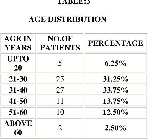
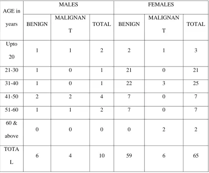
In this study the youngest patient was 16 years old and the oldest was
65 years old.80% of solitary nodules occurred during the age between 21-50
years. The highest incidence of 33.75 % was recorded during the 4th decade
of life which is comparable with study done by B. Srinivas Pai, Vijai N.
Anand, K. Rajgopal Shenoy
TABLE-4
SIDE OF INVOLVEMENT
AGE IN YEARS NO.OF
PATIENTS PERCENTAGE UPTO
20 5 6.25%
21-30 25 31.25%
31-40 27 33.75%
41-50 11 13.75%
51-60 10 12.50%
ABOVE
60 2 2.50%
SIDE RIGHT LEFT
No of
patients 54 26
[image:71.612.195.437.578.678.2]72
TABLE-5
CLINICAL PRESENTATION
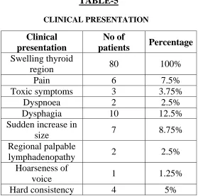
All the patients had only single palpable nodule. Toxic symptoms
were present in 3 patients. Clinical evidence of obstruction to airway or of
the great veins of neck by a solitary nodule thyroid is rare. Seven patients
complained of sudden increase in size, while hoarseness of voice was
present, only in 1 case. In this study, 2 patients had regional
lymphadenopathy; which on FNAC proved to be secondary deposits from
papillary carcinoma. 10 patients had difficulty in swallowing and 6 patients,
pain over the swelling. 2 patients had difficulty in breathing, which was
mostly uncharacteristic; neither exertional nor positional. Clinical
presentation
No of
patients Percentage Swelling thyroid
region 80 100%
Pain 6 7.5%
Toxic symptoms 3 3.75%
Dyspnoea 2 2.5%
Dysphagia 10 12.5%
Sudden increase in
size 7 8.75%
Regional palpable
lymphadenopathy 2 2.5%
Hoarseness of
voice 1 1.25%
[image:72.612.177.456.75.352.2]73 TABLE-6
HORMONAL STATUS (CLINICAL)
HORMONAL
STATUS
TOTAL NO.
OF
PATIENTS
PERCENTAGE
Euthyroid 77 96.25
Hyperthyroid 3 3.75
Hypothyroid - -
DIAGNOSTIC WORKUP:
In this study the following investigations were done in all patients
which included urine analysis, blood sugar, urea, and serum creatinine, plain
radiograph of neck, IDL scopy, chest radiograph and fine needle aspiration
cytology. USG and CT scan were done in few cases. Serum T3, T4 and TSH
estimation were done. CT scan done in a patient with extensive neck
secondaries. Radionucleotide scanning was not done for any patients due to
the non-availability of the facility in our hospital. FNAC is the investigation
of choice for solitary thyroid nodule. In this study FNAC was a very
74
TABLE-7
FNAC REPORT
SL .NO FNAC
REPORT
NO OF PATIENTS
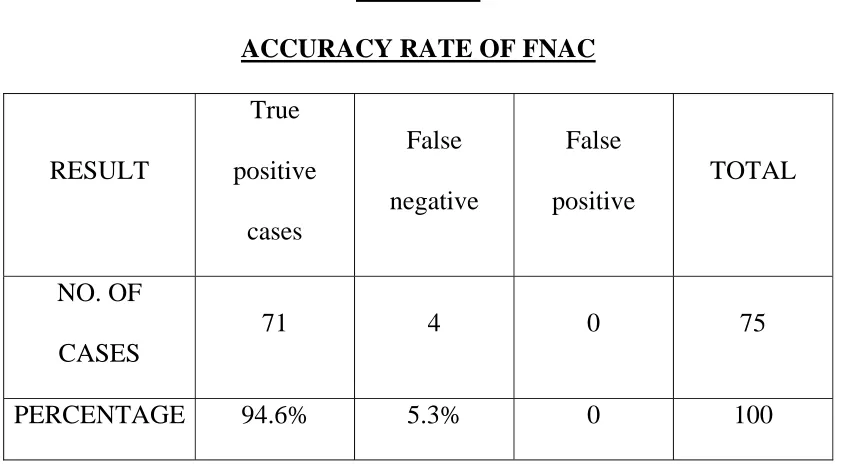
1 FOLLICULAR
NEOPLASM 40
2 NODULAR
GOITRE 31
3 PAPILLARY
CARCINOMA 6
4 HASHIMOTO’S
THYROIDITIS 3
5 THYROID
CYST -
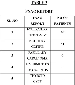
Amongst the FNAC reports, follicular neoplasm was the most
common to be reported with the inability to identify vascular/capsular
invasion. Adenomatous goitre, presenting as a solitary nodule was next
commonest eventuality. 6 Cases were reported as papillary carcinomas with
75
TABLE-8
MANAGEMENT DONE
SL.NO MANAGEMENT NO.OF PATIENTS
1 Hemithyroidectomy 64
2 Sub-total thyroidectomy 5
3 Near total-thyroidectomy 1
4 Total thyroidectomy with block
dissection 2
5 Total thyroidectomy without block
dissection 3
6 Conservative follow up 3
7 Not willing for surgery 2
TOTAL 80
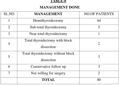
Out of 80 patients, 75 were submitted to surgery with below
mentioned indications.2 patients were given conservative line of
management because of IHD with very low ejection fraction. One another
patient with Hashimoto’s thyroiditis also was put on conservative treatment.
2 patients were not willing for surgery. These patients were advised regular
follow up, on a half yearly basis for re-examinations and repeat FNAC.
Repeat FNAC was done in 2 of these patients and were reported benign.
Provided there is no clinical suspicion of cancer and the cytology is again
unequivocally benign, the individuals are seen on an annual basis for
re-examination and further FNAC (RUSSEL).
76
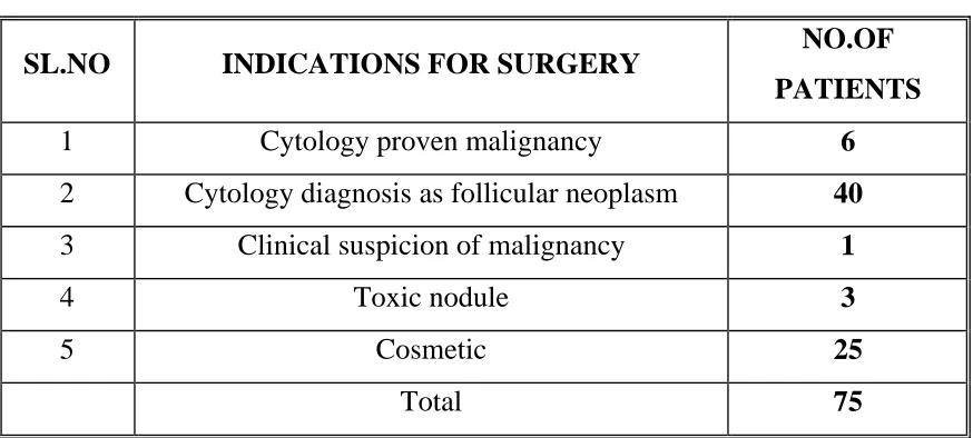
TABLE-9
INDICATIONS FOR SURGERY
SL.NO INDICATIONS FOR SURGERY NO.OF
PATIENTS
1 Cytology proven malignancy 6
2 Cytology diagnosis as follicular neoplasm 40
3 Clinical suspicion of malignancy 1
4 Toxic nodule 3
5 Cosmetic 25
Total 75
This study includes three cases of solitary thyroid nodule with
hyperthyroidism with benign cytology report. For these patients
Hemithyroidectomy was done after preparation with anti-thyroid drugs, on
the assumption that the solitary nodule is the overactive tissue, since
radioisotope scan was not available to study the functional status of the
nodule and these patients had no recurrence thyrotoxicosis.
Total thyroidectomy was done for 5 patients of papillary
carcinoma with functional neck node dissection in 2 patients. Near-total
thyroidectomy was done for a patient with papillary carcinoma leaving
behind a strip of tissue on the side other than the nodule.
Sub-total thyroidectomy was done for a 5 patients who had
77
standard surgical procedure for solitary nodule thyroid was done in 64
patients i.e., about 84% of patients who underwent surgery. All resected
specimens were submitted to histopathological examination.
TABLE-10
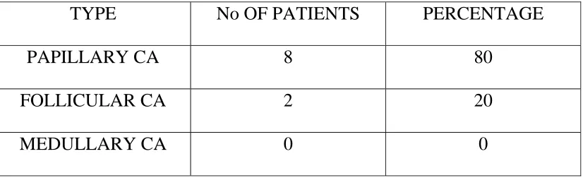
HISTOPATHOLOGICAL REPORT
HPE REPORT NO. OF PATIENTS PERCENTAGE
CARCINOMA:
Papillary carcinoma and
follicular variant of
papillary carcinoma Follicular carcinoma 10 8 2 13.33%
Follicular adenoma 36 48%
Nodular goitre 27 36%
Hashimoto’s thyroiditis 2 2.67%
Total 75
Histology proven malignancy in this series of study of solitary thyroid
nodule is 13.33% of which 80% (8 cases) were of papillary carcinoma and
its variant and the rest 20% (2 cases) were follicular carcinoma types. Of
these, six cases were diagnosed preoperatively and were offered the
confirmative treatment the rest four cases were diagnosed only on HPE
reporting & then underwent completion total thyroidectomy without neck
dissection. All these patients are under regular follow up with suppressive
78
79
DISCUSSION
The estimated life time risk of developing a nodule in the thyroid was
projected to be between 5-10%, according to a prospective study conducted
in Framingham, Massachusetts, in 1950s & 1960s. According to the study
conducted by Prof .R.L. Gupta University College of medical science, Delhi
right lobe involvement is more 51.4% compared to left lobe involvement of
38.8% which is comparable with our study with right lobe involving 68%
and left lobe 32%.
In a study conducted in MKG medical college Berhrampur Orissa
(1990-1998) under Prof. M.C.Dandapat, Dr. L.M.Mukarjee concluded that
the maximum number of cases recorded during the 3rd and 4th decade of life
with a female & male ratio of 5:1 which is comparable with our study
which has a ratio of 6:1. Nagori et al reported the maximum incidence of
SNG of 29% during the 4th decade. The commonest site of SNG was right
lobe. The percentage of malignancy was 11.1%. In a study Dr.H.Koticha &
R.M.S Kamdar Mumbai concluded that, estimation of thyroid hormone
levels has made no significant impact in deciding about surgery. FNAC is
safe, simple diagnostic but surgery is mandatory.
After an analysis of 100 cases of thyroid nodules, Prof. R.C.
Suryaprakash concluded that among the thyroid disorder adenoma thyroid