DISSERTATION ON
RADIOLOGICAL EVALUATION OF SPINAL
DYSRAPHISM USING CT AND MRI
Submitted in partial fulfillment of
requirements for
MD DEGREE BRANCH VIII RADIODIAGNOSIS
OF
THE TAMIL NADU Dr.M.G.R. MEDICAL UNIVERSITY
CHENNAI
MADRAS MEDICAL COLLEGE & RESEARCH INSTITUTE
CHENNAI - 600 003.
CERTIFICATE
This is to certify this dissertation titled “RADIOLOGICAL EVALUATION OF SPINAL DYSRAPHISM USING CT AND MRI” submitted by Dr. S. KANAKA
RAMESWARA KUMARAN, appearing for M.D. Branch VIII- RADIODIAGNOSIS degree examination in March 2007 is a bonafide record of work done by him under my direct guidance and supervision in partial fulfillment of regulations of the
Tamilnadu Dr.M.G.R.Medical University, Chennai. I forward this to the Tamilnadu Dr.
M.G.R Medical University Chennai, Tamil Nadu, India.
Signature of the Guide Signature of the Director
Prof. P. KUPPUSWAMY MD, DMRD, Prof. T.S. SWAMINATHAN MD, DMRD
Prof. of Radiology, Director,
Barnard institute of Radiology, Barnard Institute of Radiology, Madras Medical College, Madras Medical College, Govt. General Hospital, Govt. General Hospital, Chennai - 600 003. Chennai - 600 003.
Dr. KALAVATHY PONNIRAIVAN, M.D.,
Dean,
DECLARATION
I solemnly declare that this dissertation titled “RADIOLOGICAL EVALUATION OF SPINAL DYSRAPHISM USING CT AND MRI” has been done by me under the guidance and supervision of Prof.P.KUPPUSWAMY M.D,D.M.R.D, BARNARD INSTITUTE OF RADIOLOGY, MADRAS MEDICAL COLLEGE, CHENNAI. It is submitted towards partial fulfillment of requirements for the award of M.D.DEGREE (BRANCH VIII) RADIODIAGNOSIS to be held in March 2007 under
the TAMIL NADU Dr.M.G.R.MEDICAL UNIVERSITY, CHENNAI, INDIA. This has not been submitted previously by me for award of any Diploma or Degree in any other University.
Place : Date :
Dr. S. KANAKA RAMESWARA KUMARAN
MD Radiodiagnosis, (Postgraduate),
Barnard Institute Of Radiology, Madras Medical College,
ACKNOWLEDGEMENT
I express my sincere gratitude to Prof.Dr.KALAVATHY PONNIRAIVAN, M.
D., Dean, Madras Medical College for giving me permission to conduct the study in
this institution.
With extreme gratefulness, I express my indebtness to Prof.T.S.SWAMINATHAN,
M.D., D.M.R.D, F.I.C.R, Director, Barnard Institute of Radiology, Madras Medical
College, for having encouraged me to take up this study . But for his guiding spirit, perseverance and wisdom, this study would not have been possible.
I express my sincere thanks and gratitude to Prof.V.CHANDRASEKAR, M.D,
D.M.R.D, Head of the Department, Barnard Institute of Radiology, Madras Medical
College for his immense kindness, constant support and consistent encouragement in conducting this study.
I wish to thank my guide Prof.P.KUPPUSWAMY, M.D,D.M.R.D.,
Prof.N.KULASEKARAN, M.D., D.M.R.D., Prof.A. P. ANNADURAI, M.D., D.M. R.D,
Prof. K. VANITHA, M.D., D.M.R.D, D.R.M. for their support and encouragement
I am greatly indebted to my Assistant Professors Dr.UMAPATHY, D.M.R.D.,
Dr. S.SUNDARESWARAN, D.M.R.D., Dr. NESAM MANIVANNAN D.M.R.D., Dr. R.
RAVI, M.D.R.D., Dr. BABU PETER M.D.R.D., Dr. RAMESH M.D.R.D. and Dr. C.
AMARNATH, M.D.R.D. for their untiring help.
I wish to thank my fellow postgraduate colleagues for their help and cooperation during the study.
I thank all the staff of BARNARD INSTITUTE OF RADIOLOGY for
helping me conduct this study.
I thank my parents and wife for their support and encouragement. Finally I
CONTENTS
SL.NO. TOPIC Page No
1. INTRODUCTION 1
2. AIM OF STUDY 5
3. ANATOMY AND EMBRYOLOGY 6
4. REVIEW OF LITERATURE 18
5. MATERIALS AND METHODS 39
6. RESULTS AND ANALYSIS 47
7. DISCUSSION 66
8. SUMMARY 71
9. CONCLUSION 73
10. ANNEXURE
11. BIBLIOGRAPHY
12. PROFORMA
13. RESPRESENTATIVE CASES
14. MASTER CHART
INTRODUCTION
Spinal dysraphism is the most common Neural tube defect in developing countries
like India. The incidence varies from 0.5 to 11 per 1000 live births in different parts of
our country, largely affecting the lower socioeconomic strata of the population.
The etiology of Neural tube defects is Multifactorial. The interaction of
diverse factors related to Genetics, Nutrition and Environment play an important
role in the etiopathogenesis. Some of the environmental factors that may contribute
to Open Neural Tube Defects are uncontrolled maternal diabetes, and certain
prescription medications. It has been proved that deficiency of essential vitamins
especially Folic acid during pregnancy results in higher incidence of Neural tube
defects which led to prophylactic supplementation of folic acid in the antenatal
period.
Spinal dysraphism is a broad term encompassing a heterogeneous group of
congenital spinal anomalies, which results from defective closure of the neural tube
during early fetal life. Spinal dysraphism can be classified as Spina Bifida Aperta and
Spina Bifida Occulta.
Spina bifida aperta is most common type of spinal dysraphism representing a
serious congenital anomaly with severe Neurologic, Musculoskeletal, Genitourinary,
and Bowel anomalies. It encompasses three forms namely Myelomeningocele,
Myelocele and rarely Meningocele. Females show a higher incidence than Males
and most of them present at birth and are immediately taken for surgical repair and
hence are rarely imaged in unoperated cases.
Spina bifida occulta is characterized by minor Neurological manifestations and
Dermal dimple, Hemangioma, Cutis aplasia, Dermal sinus, or Hairy patch,
Rudimentary tail (caudal appendage).
Segmentation anomalies of spine are a common feature of spinal dysraphism and
along with muscle imbalances due to motor deficits result in Spinal curvature
anomalies like Scoliosis, Kyphosis and Lordosis. Scoliosis is the most common type
of spinal curvature anomaly.
Associated anomalies include Chiari malformations, Hydromyelia and
Hydrocephalus.
Imaging plays a pivotal role in the diagnosis and management of spinal dysraphism.
The various imaging modalities available are
• PLAIN RADIOGRAPH
• ULTRASONOGRAM
• CT
• MRI
PLAIN RADIOGRAPH
Plain radiograph is the base line Investigation for diagnosing various vertebral
anomalies like Spina bifida, Segmentation defects,spinal curvature anomalies, bony
septum of Diastematomyelia , Spinal canal widening and Lumbosacral soft tissue
swelling. How ever further characterization of the lesions require CT and MRI
Imaging.
ULTRASONOGRAM
Neurosonogram of the brain plays an important role in the diagnosis of associated
anomalies of spinal dysraphism like hydrocephalus and chiari malformations.
COMPUTED TOMOGRAPHY
Computed tomography with Multiplanar reformation is an excellent modality for
identification and characterization of vertebral segmentation defects, spinal curvature
anomalies and bony septum in Diastematomyelia. Various forms of spinal
dysraphism are diagnosed by CT based on the attenuation characteristics of their
contents. Thus Meningocele shows fluid atteunation, Myelomeningoceles shows fluid
and soft tissue attenuation and spinal lipomas show fat attenuation.CT is superceded
by MRI in the detection of soft tissue and associated anomalies of spinal dysraphism
like Hydromyelia, Hydrocephalus and Chiari malformations.
MAGNETIC RESONANCE IMAGING
MRI is the imaging modality of choice for characterizing the soft tissue spinal
anomalies of Spinal dysraphism especially spinal cord. Meningomyelocele,
Myelocele and Meningocele are evaluated according to the signal characteristics of
their contents. Spinal Lipomas are best characterized using Fat Suppression
Sequences. Both CT and MRI evaluates dorsal dermal sinus effectively. However
MRI supercedes CT in further characterization of the tract. MRI best depicts fibrous
septum in Diastematomyelia while bony septum is best demonstrated by CT. Further
characterization into Split cord malformation I and II, location, extent of the
hemicords, site of rejoining and associated anomalies are best demonstrated by
MRI.
MRI best characterizes tethering of cord, spinal curvature anomalies, Chiari
Thus CT and MRI are complementary to each other in diagnosis and
characterization of spinal dysraphism. Imaging also plays an important role in the
postoperative follow-ups.
AIM OF THE STUDY
To assess the role of Helical CT and MRI in
• The identification of various forms of Spinal dysraphism.
• Characterization of the lesions and associated anomalies.
NORMAL ANATOMY
LUMBOSACRAL SPINE
LS spine is made up of
¾ Bony components
Anterior elements and Posterior elements
¾ Ligaments
Anterior Longitudinal Ligament, Posterior Longitudinal Ligament, Ligamentum
flavum etc
¾ Soft tissue components
Epidural fat and veins
¾ Neural tissues and Meninges
Spinal Cord, Conus Medullaris, Nerve Roots and meninges.
BONY COMPONENTS
ANTERIOR ELEMENTS
Vertebral bodies
There are 5 lumbar and 5 sacral segments with square shape. Superior and inferior
endplates are covered by hyaline cartilage. The peripheral cortical bone is dense
while the Inner medullary is trabeculated and contains marrow tissue.
Age Marrow type T1W T2W T1 Contrast
<7yrs Red marrow Isointense Isointense Enhances
Intervertebral discs
Intervertebral discs are bean shaped with a concave posterior margin except in S1.
Nucleus pulposus shows high signal on T2 while annulus fibrosis shows low signal
on T1 and T2 weighted sequences.
POSTERIOR ELEMENTS
Pedicles
Pedicles project posterolaterally connecting the body to the neural arch forming the
spinal canal.
Articular pillars
• Pars interarticularis, superior and inferior articular facets.
Facet joints
• Diarthrodial synovial lined joints.
• Upper lumbar spine is oriented in parasagittal plane.
• Lower lumbar spine shows oblique orientation and appears
mushroom shaped on axial imaging.
Laminae and Spinous process
Laminae extend posteriorly from articular pillars to join and form the spinous process
which projects inferiorly and posteriorly.
LIGAMENTS
Anterior longitudinal ligament
• MRI-ALL appears as a thin low signal structure in both T1 and T2
weighted sequences in direct contact with the ventral surface of
vertebral bodies.
Posterior longitudinal Ligament
• PLL extends from C1 to S1.
• MRI shows PLL as a low signal structure in both T1 and T2
weighted sequences that is moulded to the posterior disc surface
but spans the posterior surface of the body like a bow string with
interposed epidural fat and veins.
Ligamentum Flavum
• Ligamentum flavum extends from the anterior aspect of the lower
margin of one lamina to the posterior surface of the lamina below.
Axial imaging shows V shaped structure.
SOFT TISSUE COMPONENTS
Epidural Fat and Veins
Epidural fat surrounds the thecal sac and root sleeves and appears high signal on T1
weighted sequences. Epidural venous plexus lie between Posterior longitudinal
Ligament and posterior surface of vertebral body. Basivertebral veins drain into the
plexus and appear as low signal voids in MRI that shows enhancement on contrast
NEURAL TISSUES
Spinal Cord
Spinal cord extends from Cervicomedullary junction to L1-2. Conus medullaris is the
diamond shaped terminal part of cord at L1-2. Cauda equina consists of nerve roots
from conus appearing like horsetail. Filum terminale is the pial extension of
meninges which shows crescent shaped appearance (axial imaging) surrounded by
the lower Sacral roots dorsally and Lumbar roots anterolaterally.
Lumbar Nerve Roots and Neural Foramina
• Nerve roots exit the spinal canal at 45o and carry a sleeve of dura
called Axillae.
• Motor nerve roots lie ventral to sensory nerve roots.
Neural Foramina and Dimensions
Boundaries
• Anterior-Vertebral body superiorly, disc and, Posterior Longitudinal
Ligament inferiorly.
• Posterior-Ligamentum flavum and articular facet.
• Superior and Inferior-Pedicles
Superior part of neural foramen is widest through, which the nerve root exits.
Sacral Plexus
THORACIC SPINE
ANTERIOR ELEMENTS
• Shape-Cone Or Triangular. Slightly wedged shaped from front to back.
• Intervertebral Discs-Height less than cervical or lumbar discs but
annulus is thicker.
POSTERIOR ELEMENTS
Pedicles and Laminae
Pedicles project posteriorly from the superior aspect of body. Laminae are broad and short and overlap like the tiles on a roof. Spinous process is long and gracile project posteriorly and inferiorly.
Articullar Pillars and Joints
Facet joint lie in the coronal plane. Transverse process project laterally. T1-T10 transverse process articulates with first 10 ribs (Costotransverse joints).
LIGAMENTS
Anterior Longitudinal Ligament and Posterior Longitudinal ligaments are thicker.
NERVES AND NEURAL FORAMINA
Spinal cord gives rise to a small ventral and large dorsal nerve root that exits through the neural foramina.
CERVICAL SPINE
clivus. Dens articulate with the anterior arch of C1. C3-C7 Vertebrae are functionally and anatomically similar.
ANTERIOR ELEMENTS
Vertebral and Uncovertebral joints
Cervical vertebrae are box shaped. There is a Increase in size from C3-7. Superior uncinate process articulates with the vertebral bodies above to form the
uncovertebral joints.
Intervertebral discs
Bean shaped with concave posterior margin. Nucleous pulposus appears high signal on T2 weighted sequence and Annulus fibrosis appears low signal on T1 and T2 weighted sequences.
POSTERIOR ELEMENTS
Pedicles
Short and project posteriorly and laterally.
Articular facets
Laminae and spinous process
Laminae are thin plates that fuse in the mid line to form the spinous process, which is often bifid. C7 has the longest spinous process.
NEURAL FORAMINA AND NERVES
Boundaries
• Anterior –Vertebral body.
• Posterior-Articular pillars and Ligamentum Flavum.
Nerves
The nerves exits at 45o and lies in the Inferior half of the neural foramina with superior half occupied by fat and epidural veins. The Dorsal roots lie behind and above the ventral roots.
LIGAMENTS
The various ligaments include Anterior longitudinal ligament, Posterior longitudinal ligament and Ligamentum flavum. The union of Superior and Inferior cruciate ligaments behind the dens forms Transverse Ligament.
Epidural fat and Veins
Fat is sparse. Epidural veins are larger.
SPINAL CORD
Shape on axial imaging
Cervical cord appears elliptical while the thoracic cord appears round. Conus
medullaris is the diamond shaped expansion of cord which terminates at L1-2. Filum terminale shows crescent shaped appearance.
Fissures
Ventral median fissure, Dorsal median sulcus, Dorsal lateral sulci, Dorsal intermediate sulci.
Central grey matter-H shaped with Anterior and Posterior Horns.
MENINGES
Dura and subdural space
Spinal dura is continuous with the cranial dura and extends upto S2 where it blends
with filum terminale and encloses the subdural space, which is usually small.
Arachnoid and subarachnoid space
Arachnoid is loosely attached to the dura and encloses the subarachnoid space.
Subarachnoid space is widest at crainovertebral junction.
AP dimensions of spinal canal
• C3-C7-10-15mm.
• D1-D12-12-13mm.
GENERAL EMBRYOLOGY
Ovum+Sperm
↓
Fertilized Ovum
↓
2cel → 3cell → 4Cell stages
↓
16cell stage (Morula)
↓
Blastocyst
↓
Bilaminar Embryonic Disc with amnionic cavity and yolk sac surrounded by Trophoblast
PROCHORDAL PLATE is a Circular area in endoderm due to change of cubical
cells of endoderm to columnar cells. It determines the central axis and the head end of the embryo.
PRIMITIVE STREAK is a Proliferation of the ectoderm along the central axis in the
tail end of embryo. These cells spread from the primitive streak between the
ectoderm and endoderm to form the intraembryonic mesoderm. The mesoderm fails to separate the ectoderm and endoderm in the region of prochordal plate and cloacal membrane.
FORMATION OF NOTOCHORD
NEURAL TUBE FORMATION
The ectoderm destined to develop into neural tube lies dorsal to the notochord called neural plate. Dorsal groove formed called neural groove becomes deeper until the two edges of neural plate come close to each other and fuse thus converting the neural groove into neural tube. The enlarged cranial part forms the brain while the caudal part forms the spinal cord.
DEVELOPMENT OF SPINAL CORD
Caudal part of neural tube gives rise to the spinal cord. It has 3 layers namely ependymal layer, mantle layer and marginal layer. Mantle layer differentiates into ventral basal lamina and dorsal alar lamina which gives rise to the anterior horn, posterior horn and roots respectively. The marginal layer gives rise to the ascending and descending tracts. The ependymal layer lines the central canal of the spinal cord and extends the entire length of vertebral column. Later the vertebral column grows faster than the spinal cord resulting in recession of spinal cord causing the spinal roots to course obliquely to reach their respective neural foramina.
FORMATION OF DISTAL CORD
The distal spinal cord forms by the processes of canalization and retrogressive differentiation. After neurulation is complete on Day 26-27 the caudal end of the neural tube and the caudal end of the notochord blend into a large aggregate of undifferentiated cells designated the caudal cell mass. The caudal cell mass extends into the tail fold, adjacent to the distal end of the developing hindgut and the
mesonephros. This juxtaposition of developing genitourinary, Notochordal, and Neural structures within the tail fold appears to account for the common concurrence of distal vertebral, Neural, Anorectal, Renal, and Genital anomalies.
focal widening of this canal at the distal conus or proximal filum is designated the terminal ventricle
The most cephalic portion of this distal cord forms the lower half of conus medullaris. Major portion of this distal cord undergoes involution to a glio-ependymal strand called the filum terminale by a process designated retrogressive differentiation.
The primitive filum extends caudally from the apex of the primitive conus to the 29th-30th vertebra. After involution of the distal cord to the filum terminale, the newly formed conus medullaris lies opposite the lower coccygeal segments. Thereafter, the spinal cord and filum do not shorten further. Rather, they elongate and thicken by interstitial growth. The caudal cell mass formed by notochord, mesoderm, and neural tissue simply segments into somites to form the sacral, coccygeal, and tail vertebrae. Retrogressive differentiation then leads to reduction of most of these segments with loss of the tail. Thereafter the vertebral column elongates with growth and grows faster than the cord. All further "ascent" of the cord results from disproportionately greater longitudinal growth of the vertebrae, not involution; the bones simply grow faster and descend away from the cord.
FORMATION OF MENINGES
The ventral part of the neural plate induces the surrounding mesenchyme to form the meninges.
FORMATION OF VERTEBRAL COLUMN
The mesoderm on either sides of the notochord is called paraxial mesoderm. It is segmented to form somites.
Somite has 3 parts
• Myotome-Paraspinal muscles
• Dermatome-Skin and subcutaneous tissue.
REVIEW OF LITERATURE
Congenital malformations of the Spine are grouped into three broad categories1, 7
SPINA BIFIDA APERTA
Lesions in which the neural tissue is exposed to view in the midline of the back are designated spina bifida aperta. The most common forms are the Myelocele and Myelomeningocele; Meningoceles without skin cover are less common.
OCCULT SPINAL DYSRAPHISM
Lesions in which the neural tissue lies deep to an intact skin cover are designated occult spinal dysraphism5, 19. and includes Dorsal dermal sinus, Spinal lipoma, Tight Filum Terminale syndrome, Neurenteric cyst, and Diastematomyelia. Midline
cutaneous stigmata such as skin dimples, hemangiomas, and hypertrichosis frequently overlie the zone of abnormal nervous tissue.
CAUDAL SPINAL ANOMALIES
Malformations of the distal end of the spine, spinal cord, and meninges are associated with disorders of the hindgut, kidneys, urinary bladder, and genitalia. These include Sacral agenesis, Terminal myelocystocele, and Anterior sacral meningocele ect.
INCIDENCE
GENDER DISTRIBUTION
The prevalence rate of Myelomeningocele3 Spinal lipomas and Diastematomyelia is slightly higher in girls (Ratio of 1.2:1). Meningocele, Dorsal dermal sinus, Thick filum terminale syndrome have no gender predilection.
AGE OF PRESENTATION
Open Spinal dysraphism22, 24 presents at birth, but spina bifida occulta may not be obvious until later in childhood or early adult life32.
SPINA BIFIDA APERTA
Definition
Form of spinal dysraphism in which the neural tissue and/or meninges are exposed to the environment, because the skin, fascia, muscle, and bone are deficient in the midline of the back. Myelocele and Myelomeningocele7 are the two commonest forms of spina bifida aperta.
Incidence
Myelocele and Myelomeningocele occur in 1-2 persons per 1, 000 live births.
Gender Distribution
Myelomeningocele afflicts females more commonly than males.
Distribution in spinal cord
Thoracic 2%, Thoracolumbar 32%, Lumbar 22%, and Lumbosacral 44%1
Clinical features
• Sensorimotor deficits of the lower extremities
Embryology
If the neural folds fail to flex and to fuse into a tube, they persist instead as a flat plate of neural tissue. This flat plate of unneurulated neural tissue is designated the neural placode. Because the neural tube does not close, the superficial ectoderm cannot disjoin from the neural ectoderm and remains in lateral position. Therefore the skin that develops from the ectoderm also lies lateral in position, leaving a midline defect. Mesenchyme then cannot migrate behind the neural tube, so the bony, cartilaginous, muscular, and ligamentous elements are also deficient in the midline. Instead, the bones, cartilage, muscle, and ligament develop in an abnormal position ventral-lateral to the neural tissue, and appear bifid and "everted. " The unfused neural plate is thus exposed to view in the midline of the back at the site of the midline deficiency of skin, bone, cartilage, muscle and ligament.
Local Examination
The neural tissue appears as a raw, reddish, vascular oval plate.
Relation of Meninges to neural Tissue
The pia-arachnoid membrane covers the ventral surface of the neural plate.
Relation of dorsal and ventral nerve roots to neural tissue
The two ventral motor nerve roots arise from the ventral surface of the neural plate just to each side of the midline ventral sulcus. The paired dorsal sensory roots arise lateral to the ventral roots.
Role of imaging
Associated anomalies
• Spinal Curvature- Scoliosis, Lordosis, Kyphosis.
• (Epi) dermal inclusion cysts
• Lipomas -6%
• Hydromyelia-30% to 50%
• Diastematomyelia-31%to46%with Myelomeningocele(complex spina bifida)21.
• Chiari II Malformation- Nearly always associated.
OCCULT SPINAL DYSRAPHISM
MENINGOCELE
Definition-Dorsal protrusion of meninges and CSF without neural elements with skin covering.
Etiology-Acquired more common than congenital.
Incidence-1per 10,000 live births.
Gender-Equal.
Location- 80% situated in lumbosacral spine
Clinical Features
• Lumbosacral mass.
• Neurological deficits uncommon.
Imaging
Plain Radiograph –Soft tissue swelling and Spina bifida.
(CT/MR)- Dorsal protrusion of meninges and CSF without neural elements.
Definition
Mid line epithelial lined tract extending from skin to a variable extent into the meninges and neural tissue of spinal cord10.
Incidence: Uncommon
Gender: No gender predilection
Embryology
If the superficial ectoderm fails to separate from the neural ectoderm at one point, a focal segmental adhesion is formed. Different rates of growth between neural and spinal tissue lead to "ascent" of the cord, the local adhesion is drawn out into an elongated epithelial-lined tube that still connects the spinal cord with the skin of the dorsal surface called Dorsal dermal sinus.
Location
Lumbosacral-57% and Occipital-24% (Wright's series10).
Clinical features
Symptoms of infection of sinus tract or mass effect of associated epidermoid/dermoid.
Local Examination
The sinus frequently appears as a pinpoint hole or a small atrophic zone in the skin typically midline, with hemangiomas and tuft of hair.
Imaging
• CT-Relatively hyperdense tract that traverses the skin, subcutaneous
• MRI- best delineates the extent and direction of tract Associated
anomalies
• Epidermoids and Dermoids (60%)
• Tethering of cord (80%)
• Lipoma (15 to 20%)
Differential Diagnosis
• Pilonidal Sinus lie in low location, near to anus, and extend
inferiorly or horizontally toward or to the dorsal surface of the coccyx. It does not enter the spinal canal. (Haworth JC etal70)
• Sacral dimples may occur together with spinal dysraphism.
DERMOID AND EPIDERMOID TUMORS
Definition-Epidermoid cystsare lined by a membrane composed only of superficial
(epidermal) elements of the skin25. Dermoid cysts are unilocular or multilocular. These tumors may arise as congenital rests or as iatrogenic implantation of viable skin elements during surgery or during spinal taps. Approximately 25% of
(epi)dermoid tumors form in association with dermal sinuses28.
Distribution-Spinal Dermoid and Epidermoid tumors constitute about 15% of all CNS (Epi)dermoids. Dermoid and Epidermoid tumors occur equally. In Lunardi's series71, Epidermoids were most frequently in Lumbar region; Dermoids were most frequently dorsal (25%) or dorsolumbar (75%). 38% were intramedullary and 63% intradural extramedullary.
Crossectional imaging-Epidermoid tumors are usually isointense with CSF or just
SPINAL LIPOMA
Definition
Distinct collections of fat and connective tissue that are partially encapsulated and have a definite connection with the spinal cord or leptomeninges
Incidence
Spinal lipomas are the most common type of occult spinal dysraphism and account for 35% of skin-covered Lumbosacral masses.
Gender
More common in females, with a sex ratio of about 1. 5:1 to 2:1.
Principal categories
Lipomyelomeningocele (84%), Intradural lipoma (4%), Filar lipoma (12%).
Clinical features
The chief complaints are usually a mass on the back (59%), urinary incontinence (23%), or weak/deformed lower extremity, perhaps with trophic ulceration (11%). Cutaneous stigmata include subcutaneous mass, Skin dimple, Hemangioma, hairy-nevus, denuded skin patch, Scar-like patch.
Embryology of lipomyelomeningocele and Intrdural lipomas
The mesenchyme surrounding the ventral surface of the closing plate, i.e, the future exterior of the neural tube is induced by the ventral surface to form meninges. No meninges would form in the midline dorsally since there is no ventral surface to induce them there. This would leave a midline dorsal defect in the meninges. Similarly, improper neurulation would prevent proper development of the neural arches, fascia, and muscle, creating a posterior spina bifida. The fat induced by the dorsal surface of the neural plate could then extend directly posteriorly, through the gap in the meninges and through the spina bifida into the subcutaneous tissue of the back. The fat would be anatomically extradural. The junction of fat and meninges would necessarily lie at the neural ridge that divides the dorsal and ventral surfaces of the neural folds.
3 Groups: Lipomas with intact dura, Deficient dura, and Filar lipomas.
Spinal Lipomas with Intact Dura (Intradural lipomas)
The intradural lipomas20 appear to arise in the dorsal midline of a cleft spinal cord and then bulge outward to form subpial masses of fat. They affect the cervical and thoracic spinal cords predominantly. In this group the spinal canal is usually nearly normal with narrow spina bifida or focal segmentation anomalies. The canal itself may be expanded by the mass. The dura is thinned, perhaps translucent, but remains intact and is displaced peripherally by the combined mass of cord and lipoma. The lipoma typically lies dorsal or dorsolateral to the cord; frequently causes cord rotation and frequently cause high-grade stenosis or block.
Imaging
• CT-Fat atteunation intradural mass
• MRI-High signal intradural mass in dorsal position in T1Weighted
Spinal Lipomas with Deficient Dura (lipomyelocele and
lipomyelomeningocele)
The most common forms of spinal lipoma (84%) are associated with definite defects in the dura through which the lipoma may extend from the spinal cord to the
subcutaneous tissue. The subcutaneous component of the mass typically forms a lump in the low back. The subjacent spinal canal shows a wide spina bifida.
Segmentation anomalies are present in 50%. The spinal cord beneath the Lipoma is cleft dorsally and very closely resembles the neural plate of a Myelomeningocele. The dura is deficient in the dorsal midline, deep to the lipoma. . Lipomyelocele and lipomyelomeningocele are exactly analogous to the Myelocele and the
Myelomeningocele, with the fat inserting into the dorsal surface of the neural plate and closure of the skin over the fat.
Imaging
• CT-Fat atteunation mass attached to neural placode
• MRI-High signal mass attached to the cord with tethering in
T1weighted images.
Filar Lipomas
Imaging –High signal mass attached to filum terminale in T1weighted images.
Associated Anomalies
DERANGED CANALIZATION AND RETROGRESSIVE DIFFERENTIATION
TIGHT FILUM TERMINALE SYNDROME
Definition
Criteria
• The filum must measure > 2 mm in diameter
• No other cause for tethering present. The tip of the conus medullaris lies
below L2 in 86%.
Embryology
Failure of complete involution of the distal cord (Raghaven N etal6)
Age; 3-35 yrs.
Gender; No predilection.
Clinical features
Sesorimotor Deficits and Bowel And Bladder Disturbances.
Imaging
• Plain radiograph and CT-Dysraphic spine, vertebral segmentation
anomalies.
• Myelography (Conventional and CT) –Shows low lying conus and thick
filum. Exiting nerve roots have a lateral or uphill course.
• MRI- Low lying conus and thick filum with associated lipoma.
Associated anomalies
Filar fibrolipomas 29%, Kyphoscoliosis 15% to 25%.
SACROCOCCYGEAL TERATOMAS
Etiology
Sacrococcygeal teratomas most probably arise from totipotential cells derived from Hensen's node.
Incidence
1 per 35,000-40,000 births.
Gender
Females predominate (80%). Hereditary forms have been reported.
Presentation
Patients may present asymptomatically because of an external mass or they may manifest hydrops, high output cardiac failure, respiratory failure, and renal
insufficiency related to the bulk and vascularity of the mass
Altman Classification
Type I tumors (47%) have predominantly external component with minimal
presacral component.
Type II tumors (35%) are evident externally but have significant intrapelvic
extension.
Type III tumors (9%) can be detected externally but lie predominantly within the
pelvis and abdomen.
Type IV tumors (10%) are entirely presacral.
Crossectional Imaging-Most sacrococcygeal tumors are mixed solid and cystic
TERMINAL VENTRICLE
The terminal ventricle is the normal slight expansion of the central canal of the cord within the distal conus and/or proximal filum. It appears to represent the point of union between the portion of the central canal made by neurulation and the portion made by canalization of the caudal cell mass. MRI studies often demonstrate a tiny drop of CSF at the site of the terminal ventricle. Slight expansion of this in patients without other pathology is presently regarded as normal.
TERMINAL SYRINGOHYDROMYELIA
Large cystic expansion of the distal one-third of the cord is found alone or in association with diverse forms of occult spinal dysraphism. Terminal
hydrosyringomyelia57 may be seen by MRI in up to 30% of patients with occult spinal dysraphism. It is found most frequently in patients with concurrent anorectal
anomalies (67%), Meningocele (54%), Diastematomyelia (38%), Tight filum terminale syndrome (33%), Lipomyelomeningocele (19%), and Dermal sinus tract (17%)
CAUDAL SPINAL ANOMALIES WITH ANORECTAL AND UROGENITAL
MALFORMATIONS
Embryogenesis
TERMINAL MYELOCYSTOCELE (SYRINGOCELE)
Terminal myelocystocele is a cystic dilatation of the distal spinal cord. Terminal myelocystoceles constitute 1% to 5% of skin-covered lumbosacral masses. They are typically associated with the OEIS and other severe anomalies of the hindgut and genitourinary systems. It may also be related to the teratogen retinoic acid.
SYNDROME OF CAUDAL REGRESSION
The syndrome of caudal regression12 designates a constellation of anomalies of the hind end of the trunk, including partial agenesis of the thoracolumbosacral spine, imperforate anus, malformed genitalia, bilateral renal dysplasia or aplasia,
pulmonary hypoplasia, and, in the most severe deformities, extreme external rotation and fusion of the lower extremities (Sirenomelia).
Incidence
Sacral Agenesis14 occurs in approximately 1 per 7, 500 births.
Gender
Males and females are affected equally. Nearly all cases are sporadic.
Diabetes Association
There is a definite but incomplete association with Diabetes mellitus.
Clinical Features
Sensory/motor deficits and bowel/bladder disturbances.
Imaging
• PLAIN RADIOGRAPH-shows variable absence of distal spine.
ANTERIOR SACRAL MENINGOCELES
Definition
Anterior sacral meningoceles are diverticulae of the thecal sac that protrude anteriorly into the extra peritoneal presacral space15.
Inheritance
Most anterior sacral meningoceles18, 50 occur sporadically. They may be seen in conditions with dural ectasia such as Neurofibromatosis and Marfan's syndrome, or may be associated with the Currarino triad of anorectal malformations, sacral defects, and presacral masses.
Incidence
Clinically, anterior sacral meningoceles account for 3.7% of retrorectal tumors48.
Gender
In adults, females appear to be more affected (approximately 10:6). The lesion is equally frequent in boys and girls below age 15,
Clinical Features
Local pressure on pelvic organs causes unremitting constipation, urinary frequency and incontinence, dysmenorrhea, dyspareunia, and back pain. Pressure on the nerve roots causes sciatica, diminished rectal and detrusor tone, numbness and paresthesias in the lower sacral region.
Imaging
unilateral sacral agenesis that becomes remodeled around the hernia ostium with time. In approximately 20% the sacral defect is midline. The sacral dural sac is often widened and patulous. The stalk is typically narrow. The meningocele sac may be unilocular or multilocular.
LATERAL LUMBAR AND THORACIC MENINGOCELES
These lesions are characterized by CSF-filled protrusions of dura and arachnoid through one or several enlarged neural foramina into the paraspinal extrapleural and retroperitoneal tissue. They may be unilateral or bilateral, and are commonly
associated with scoliosis. Lateral meningoceles 47are most common in patients with mesenchymal disorders such as Neurofibromatosis (85%), Marfan's and Ehler-Danlos syndromes.
DERANGEMENTS IN THE NOTOCHORD
(SPLIT NOTOCHORD SYNDROME)
Persistence of a midline adhesion between ectoderm and endoderm could cause derangement in the migration of notochordal cells with consequent deflection or splitting of the notochord. This appears to be the genesis of Dorsal enteric fistula, Neurenteric cyst, and Diastematomyelia.
DORSAL ENTERIC FISTULA
Persistence of a patent notochord canal (canal of Kovalevsky) would create a patent fistula from the mesenteric surface of gut through the mesentery and prevertebral tissue, through the vertebral bodies, spinal canal, and spinal cord, and through bifid laminae to an ostium in the midline skin of the back. Such a complete
NEURENTERIC CYST
Definition
Neurenteric cysts30, 45 are enteric-lined cysts that present within the spinal canal and exhibit a definite connection with the spinal cord and/or vertebrae. They may
communicate with an extra spinal component of cyst in the mesentery or
mediastinum around a hemi vertebra or through a butterfly vertebra and/or they may attach by a fibrous stalk to the vertebra, mesentery, or gut.
Etiology
Failure of foregut notochord separation during embryogenesis.
Incidence It is exceedingly rare.
Age and Gender
1st or 2nd decade. Slight male Predominance.
Distribution
Thoracic spine-42%. Cervical spine-32%. Posterior fossa-13%. CVJ-10%. Lumbar-rare.
Imaging
• Myelography –Intrdural extramedullary mass anterior to spinal
cord.
• CT-CSF density mass protruding from the spinal canal into the
sacrum.
• MRI – T1-iso to slightly hyperintense to CSF.
Associated Anomalies-Vertebral anomalies-43%
Differential Diagnosis
Arachnoid cyst, Epidermoid cyst and Inflammatory cysts.
DIASTEMATOMYELIA
Definition
Diastematomyelia signifies a sagittal clefting of the spinal cord, conus medullaris, and/or filum terminale into two, not necessarily symmetrical, hemi cords.
Embryology
Adhesions between the embryonic ectoderm and endoderm40
↓
Accessory neuroenteric canal
↓
Endomesenchymal tract condenses around the accessory neuroenteric canal
↓
Split spinal cord
Incidence
Uncommon
Gender
Female predominance
Age
Clinical Features
In children, the common presenting complaints39 are (i) Musculoskeletal deformities (98%) and Pes cavus, (ii) Neurologic deficits (84%) and Incontinence of bladder and bowel, and (iii) Scoliosis (79%) and in adults, Sensorimotor changes (69%) and pain (58%) . Cutaneous stigmata like hair patches, Nevi, Lipomas are common.
Location
Lumbar or lumbosacral spine 45%, Thoracic spine 31%, Thoracolumbar spine 12%. Cervical spine 7% and the sacrum in 1%. "Double" Diastematomyelia occurs in < 1%. In Diastematomyelia, 16, 17 the conus medullaris is usually low in position. The two Hemi cords nearly always (91%) reunite distally into a reformed cord below the cleft. Hemi cords are asymmetrical in 30%. The filum terminale is thickened and tether the reunited cord.
SPLIT CORD MALFORMATION 5
• Type 1-Two hemi cords separated by osteocartilagenous septum
and contained in separate dural sacs.
• Type11-Two hemi cords separated by fibrous septum and contained
common dural sac.
Imaging
• PLAIN FILM and NECT shows the bony septum and associated
vertebral anomalies.
• MYELOGRAPHY-Depicts the hemi cords.
• MRI- Depicts the hemi cords, nature of septum especially fibrous
Associated Anomalies
Segmentation anomalies (85%), Scoliosis and kyphosis(50% - 60%), Klippel-feil syndrome(2%-7%), Sprengel's deformity (7%), Chiari I (3%) Hydrosyringomyelia (50%), Dermal sinus (3%), Lipomyelomeningocele (3%) Teratomas(3%)and Horseshoe kidneys.
SEGMENTATION ANOMALIES OF VERTEBRAL BODIES
Embryology
During the 9-10th week of gestation two ossification centres develop for the ventral and dorsal half of the vertebral body.
Anomalies
½ Asomia-Complete absence of vertebral body.
½ Hemivertebra
• Unilateral wedge vertebra (right or left)-Scoliosis at birth.
• Dorsal hemi vertebra-Rapidly progressive scoliosis.
• Ventral hemivertebra(very rare).
½ Coronal cleft
Failure of fusion of anterior and posterior ossification centres
½ Butterfly vertebra
Failure of fusion of lateral halves due to persistence of notochordal tissue.
MATERIAL AND METHODS
• The study comprises of 70 patients including 33 males and 37
females, age ranging from birth to 30 years. The study was conducted for a period of 20 months from January 2005 to August 2006 in Barnard Institute of Radiology, Madras Medical College, Chennai.
• The patients were referred from Institute of Child Health, Egmore
and Institute of Neurology, Government General Hospital, Chennai to Barnard Institute of Radiology for radiological evaluation.
• Clinically the most common cause for referral was swelling in the
back predominantly Lumbosacral region. The other symptoms were sensory/motor deficit, bladder/ bowel disturbances, spinal curvature deformities, cutaneous features like dermal dimple, hypertrichosis, silky hair, dermal sinus and capillary hemangioma ect.
CRITERIA
Inclusion Criteria
• All cases of open spinal dysraphism.
• Cases presenting with lumbosacral swelling.
• Cases presenting with cutaneous stigmata like Dermal dimple, tuft
of hair, Nevi, dermal sinus ect.
• Cases showing vertebral anomalies in Plain radiograph.
• Cases presenting with congenital scoliosis/ kyphoscoliosis/
lordosis ect.
• Cases presenting with bladder/bowel incontinence since
• Cases presenting with motor or sensory deficit since childhood.
Exclusion Criteria
• Treated cases.
• Spinal tumors.
CT TECHNIQUE
All the examinations were performed on a Toshiba Asteion Spiral CT scanner. PARAMETERS
LUMBAR SPINE
• Axial contiguous images from L1 TO S1.
• Slice thickness=4mm.
• Intersection gap=3-4mm.
• Window settings=wide and narrow.
• Sagittal and coronal reconstruction using multiplanar reconstruction
technique.
DORSAL SPINE
• Axial contiguous images D1 TO D12.
• Slice thickness=4mm.
• Intersection gap=4mm.
• Window settings=wide and narrow.
• Sagittal and coronal reconstruction using multiplanar reconstruction
technique.
CERVICAL SPINE
• Axial contiguous images CVJ TO D1.
• Slice thickness=2-3mm.
• Intersection gap=2mm.
• Sagittal and coronal reconstruction using multiplanar reconstruction
technique.
MRI TECHNIQUE
MRI was performed using 1.5 Tesla super conducting SIEMENS MAGNETOM , symphony.
PARAMETERS
LUMBAR SPINE
1. Sagittal fast spin echo, T1-weighted image from conus to S1.
• Field Of View=28cm.
• Slice thickness=4-5mm.
• Intersection gap=1mm.
• Matrix=256x512.
2. Sagittal fast spin echo T2-weighted image.
Field Of View=28cm.
Slice thickness=4-5mm.
Intersection gap=1mm.
Matrix=320x512.
3. Axial fast spin echo T2-weighted image.
• FOV=20cm.
• Slice thickness=4-5mm.
• Intersection gap=1mm.
• Matrix=256X256.
DORSAL SPINE
1. Sagittal T1-weighted sequence including the entire Dorsal spine.
• FOV=40cm.
• Slice thickness=3-4mm.
• Intersection gap=1mm.
• Matrix=512x512.
2. Sagittal fast spin echo T2-weighted sequence including the entire dorsal spine
• FOV=40cm.
• Slice thickness=3-4mm
• Intersection gap=1mm.
• Matrix=512x512.
3. Axial T2-weighted sequence including the entire Dorsal spine.
• FOV=40cm.
• Slice thickness=3-4mm.
• Intersection gap=1mm.
• Matrix=512x512.
CERVICAL SPINE
1. Sagittal fast spin echo, T1-weighted image including from the cerebellar tonsils to D1.
• FOV=26-28cm.
• Slice thickness=3-4mm.
• Intersection gap=1mm.
• Matrix=256x512.
2. Sagittal fast spin echo T2-weighted sequence including from the cerebellar tonsils to D1.
• FOV=40cm.
• Slice thickness=3-4mm.
• Intersection gap=1mm.
• Matrix=384x512.
3.Axial 2D GRE covering C1-C7.
• FOV=20cm.
• Slice thickness=3mm.
• Intersection gap=1mm.
• Matrix=384x512.
For interpretation the following Characteristics of Spinal dysraphism were studied and analyzed in these patients.
1. TYPES
Open Spinal Dysraphism
• Myelomeningocele.
• Myelocele.
• Meningocele.
Occult Spinal Dysraphism
• Spinal lipomas.
• Diastematomyelia.
• Dorsal dermal sinus.
• Tight filum terminale syndrome.
• Anterior sacral meningocele.
• Sacral agenesis.
2. CT CHARACTERISTICS
Lesion Attenuation
• Fluid-Menigocele .
• Soft tissue with fluid-Menigomyelocele.
• Soft tissue-Myelocele.
• Fat with soft tissue-Lipomyelocele.
• Fat with soft tissue and fluid-Lipomyelomeningocele.
• Fat-Dural lipomas, Filar lipomas.
Spinal Location
• Lumbosacral.
• Cervical.
Vertebral Anomalies
• Spina bifida.
• Butterfly vertebra.
• Hemi vertebra.
• Block vertebra.
• Others.
Spinal Curvature anomalies
• Scoliosis
• Kyphosis
• Lordosis
Septum in Diastematomyelia
• Bony
• Fibrous
3. MRI CHARACTERISTICS 2
Signal Intensities of lesion in T1-W, T2-W and FLAIR SEQUENCES
• CSF Intensity –Meningocele.
• CSF Intensity+Neural tissue-Myelomeningocele.
• Neural tissue-Myelocele.
• Fat intensity+neural tissue-Lipomyelocele.
• Fat intensity+CSF intensity + neural
tissue-Lipomyelomeningocele.
• Fat intensity-Intradural lipomas, Filar lipomas
Septum In Diastemetamyelia
• Fibrous
Tethering of cord
Vertebral anomalies
Spinal distribution
Spinal curvature anomalies
Chiari association
Hydromyelia
Hydrocephalus
The above mentioned characteristics of spinal dysraphism were analysed using CT and MRI to arrive at radiological diagnosis.
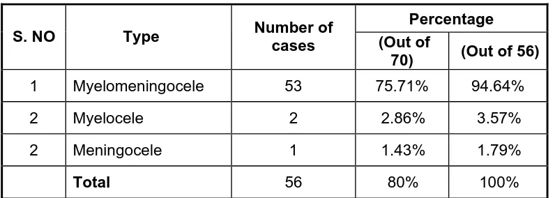
[image:48.612.109.507.468.610.2]RESULTS AND ANALYSIS
TABLE 1
OPEN SPINAL DYSRAPHISM
Percentage
S. NO Type Number of
cases (Out of
70) (Out of 56)
1 Myelomeningocele 53 75.71% 94.64%
2 Myelocele 2 2.86% 3.57%
2 Meningocele 1 1.43% 1.79%
Total 56 80% 100%
Myelomeningocele accounting for 94.6% of the total 56 cases followed by Myelocele and Meningocele.
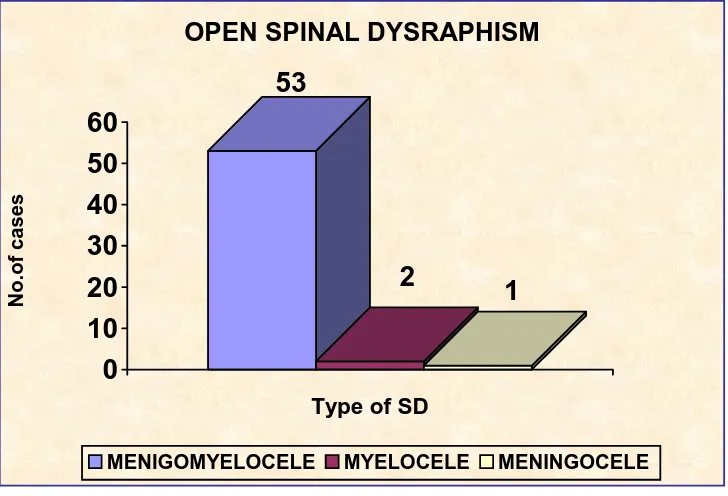
53
2
1
0
10
20
30
40
50
60
No.of cases
Type of SD
OPEN SPINAL DYSRAPHISM
[image:49.612.125.489.132.380.2]MENIGOMYELOCELE MYELOCELE MENINGOCELE
TABLE 2
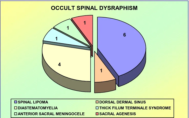
OCCULT SPINAL DYSRAPHISM
Percentage S.No Type Number
of cases (Out of 70) (Out of 14)
1 Spinal lipomas 6 8.57% 42.86%
2 Diastematomyelia 4 5.71% 28.57%
3 Dorsal dermal sinus 1 1.43% 7.14%
4 Tight filum terminale 1 1.43% 7.14%
5 Anterior sacral meningocele
1 1.43% 7.14%
6 Sacral agenesis 1 1.43% 7.14%
Total 14 20% 100%
common type of occult spinal dysraphism is Spinal lipoma accounting for 42.86% of the total 14 cases.
OCCULT SPINAL DYSRAPHISM
1 4
1 1
1 6
SPINAL LIPOMA DORSAL DERMAL SINUS
[image:50.612.122.491.131.362.2]DIASTEMATOMYELIA THICK FILUM TERMINALE SYNDROME ANTERIOR SACRAL MENINGOCELE SACRAL AGENESIS
TABLE 3
GENDER DISTIBUTION IN OPEN SPINAL DYSRAPHISM
S.No Open spinal dysraphism
Number of cases
Male % Female % Total
1. Meningomyelocele 53 22 41.51 31 58.49 100%
2. Myelocele 2 1 50 1 50 100%
3. Meningocele 1 0 0 1 100 100%
Total 56 23 41.07 33 58.93 100%
22
1
31
1 1
0 5 10 15 20 25 30 35
No.of cases
MALE FEMALE
GENDER
GENDER DISTRIBUTION IN OPEN SPINAL DYSRAPHISM
TABLE 4
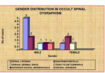
GENDER DISTRIBUTION IN OCCULT SPINAL DYSRAPHISM
Type Number Male % Female % Total
Spinal lipoma 6 5 83.33 1 16.67 100
Diastematomyelia 4 2 50 2 50 100
Dorsal dermal sinus 1 0 0 1 100 100
Tight filum terminale 1 1 100 0 0 100
Anterior sacral meningocele
1 1 100 0 0 100
Sacral agenesis 1 1 100 0 0 100
Total 14 10 71.43 4 28.57 100
M:F=2.5:1
5
2
1 1 1 1
2 1 0 0.5 1 1.5 2 2.5 3 3.5 4 4.5 5 No.of cases MALE FEMALE Gender
GENDER DISTRIBUTION IN OCCULT SPINAL DYSRAPHISM
TABLE 5
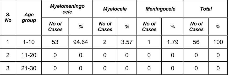
AGE GROUP DISTRIBUTION IN OPEN SPINAL DYSRAPHISM
Myelomeningo
cele Myelocele Meningocele Total
S. No
Age group
No of
Cases %
No of
Cases %
No of
Cases %
No of
Cases %
1 1-10 53 94.64 2 3.57 1 1.79 56 100
2 11-20 0 0 0 0 0 0 0 0
3 21-30 0 0 0 0 0 0 0 0
All cases of open spinal dysraphism occurred in 1 – 10 age group.
Mean age of presentation is 1.23yrs
TABLE 6
AGE GROUP DISTRIBUTION IN OCCULT SPINAL DYSRAPHISM
Spinal lipomas Diastema-tomyelia Dorsal dermal sinus Tight filum ternminale Anterior sacral meningo cele Sacral agenesis Age group
No % No % No % No % No % No %
1-10 4 66.67 4 100 1 100 1 100 0 0 0 0
11-20 2 33.33 0 0 0 0 0 0 1 100 1 100
21-30 0 0 0 0 0 0 0 0 0 0 0 0
Total 6 100 4 100 1 100 1 100 1 100 1 100
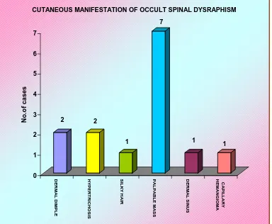
[image:53.612.89.505.481.654.2]TABLE . 7
CUTANEOUS MANIFESTATIONS OF OCCULT SPINAL DYSRAPHISM
Cutaneous Signs Dermal dimple Hyper-trichosis Silky hair Palpable mass Dermal sinus Capillary hemangioma Total
No.of cases 2 2 1 7 1 1 14
% 14.29 14.29 7.14 50 7.14 7.14 100
The most common cutaneous manifestation is palpable mass in the back
2 2 1 7 1 1 0 1 2 3 4 5 6 7 No.of cases
DERMAL DIMPLE HYPERTRICHOSIS SILKY HAIR PALPABLE M
A
SS
DERMAL SINUS
CAPILLARY
HEMANGIOMA
TABLE 8
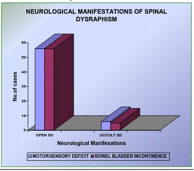
NEUROLOGICAL MANIFESTATIONS OF SPINAL DYSRAPHISM
Type Motor and
Sensory deficit
Bowel and Bladder incontinence
Open spinal dysraphism 56 56
Occult spinal dysraphism 6 5
Neurological manifestations occurred in all cases of open SD while in occult SD 11 of the 14 cases showed neurological manifestations.
0 10 20 30 40 50 60
No.of cases
OPEN SD OCCULT SD
Neurological Manifesations
NEUROLOGICAL MANIFESTATIONS OF SPINAL
DYSRAPHISM
TABLE 9
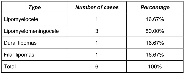
SPINAL LIPOMAS
Type Number of cases Percentage
Lipomyelocele 1 16.67%
Lipomyelomeningocele 3 50.00%
Dural lipomas 1 16.67%
Filar lipomas 1 16.67%
Total 6 100%
Lipomyelomeningocele is the most common type of spinal lipoma accounting for 50% of the total cases.
SPINAL LIPOMAS
3
1 1
1
TABLE 10
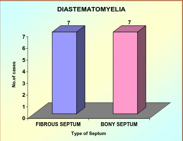
DIASTEMATOMYELIA
Type Fibrous
septum
Bony
septum Total %
Diastematomyelia in occult SD 2 2 4 28.57
Diastematomyelia in open SD 5 5 10 71.43
Total 7 7 14 100
Bony and fibrous septum occurred equally in both open and occult SD.
7 7
0 1 2 3 4 5 6 7
No.of cases
FIBROUS SEPTUM BONY SEPTUM
Type of Septum
TABLE 11
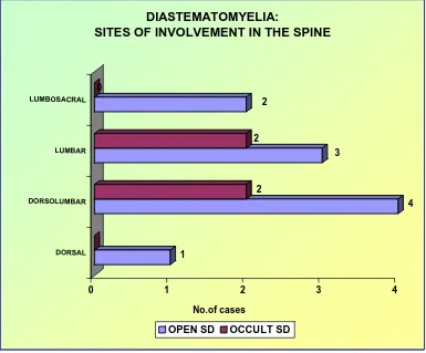
DIASTEMATOMYELIA: SITES OF INVOLVEMENT IN THE SPINE
Type Cervical Dorsal Dorsolumbar Lumbar Lumbosacral Total %
Open SD 0 1 4 3 2 10 71.43
Occult SD 0 0 2 2 0 4 28.57
Total 0 1 6 5 2 14 100
In open SD the most common site of occurrence of Diastematomyelia are DL and L regions. In occult SD Diastematomyelia occurred equally in both DL and L regions.
1
4 2
3 2
2 0
0 1 2 3 4
No.of cases DORSAL
DORSOLUMBAR LUMBAR LUMBOSACRAL
DIASTEMATOMYELIA:
SITES OF INVOLVEMENT IN THE SPINE
TABLE 12
TETHERING OF CORD
Type Tethering No tethering Total
Spinal lipomas 4 2 6
Diastematomyelia 1 3 4
Open SD 4 52 56
Dorsal dermal sinus 0 1 1
Tight filum terminale 1 0 1
Anterior sacral meningocele
0 1 1
Sacral agenesis 0 1 1
TOTAL 10 60 70
% 14.29 85.71 100
Tethering of cord occurred in 14.29% of the total cases.
TETHERING OF CORD IN SPINAL DYSRAPHISM
60
10
TABLE 13
VERTEBRAL ANOMALIES
Hemivertebra Butterfly vertebra
Block vertebra
Spina bifida Others
Open SD 21 23 10 56 2
Spinal Lipomas 2 3 1 6 0
Dorsal dermal sinus 0 1 0 1 0
Diastematomyelia 2 0 1 3 0
Tight Filum Terminale 0 1 0 0 0
Anterior Sacral Meningocele 1 0 0 1 0
Sacral Agenesis 0 1 0 1 0
TOTAL 26 29 12 68 2
% 37.14 41.43 17.14 97.14 2.86
Spinar biffila is the most common vertebral anomaly occurring in 97.14% of total cases.
VERTEBRAL ANOMALIES IN SPINAL DYSRAPHISM
68 26
12 2
29
TABLE 14
SPINA BIFIDA DISTRIBUTION IN SPINE
Distribution in spine
Cervical Dorsal Lumbar Lumbosacral Total
Types
Spina bifida Cases
No % No % No % No % No %
Open SD
56 4 7.14 12 21.43 18 32.14 22 39.29 56 100
Occult SD
12 1 8.33 2 16.67 4 33.33 5 41.67 12 100
Total 68 5 7.35 14 20.59 22 32.35 27 39.71 68 100
Spina bifida occurs in 97.14% of the total cases. Lumbosacral spine is most common site of occurrence in both open and occult spinal dysraphism.
5 14 22 27 0 5 10 15 20 25 30 No.of cases
Distribution in spine
SPINA BIFIDA DISTRIBUTION IN SPINE
TABLE 15
DISTRIBUTION OF SPINAL DYSRAPHISM IN SPINE
Distribution in spine Total
Types No
Cervical Dorsal Lumbar Lumbosacral No. %
Open SD 56 4 7.14% 12 21.43% 18 32.14% 22 39.29% 56 100
Occult SD
14 1 7.14% 2 14.29% 4 28.57% 7 50% 14 100
Total 70 5 7.14% 14 20% 22 31.43% 29 41.43% 70 100
The most common site of occurrence of spinal dysraphism is LS spine.
5
14
22
29
0 5 10 15 20 25 30
No.of cases
Distribution in spine
DISTRIBUTION IN SPINE
TABLE 16
SPINAL CURVATURE
Spinal
curvature Scoliosis Kyphosis Lordosis
Region Cervical Dorsal Lumbar Dorsal Lumbar Lumbar Total
OPEN SD 1 6 5 4 2 4 22
OCCULTSD 1 5 4 5 3 3 21
TOTAL 2 11 9 9 5 7 43
% 2.86 15.71 12.86 12.86 7.14 10 61.43
The most common spinal curvature anomaly is scoliosis.
22
14
7
0 5 10 15 20 25
No.of cases
Type of spinal curvature
SPINAL CURVATURE IN SPINAL DYSRAPHISM
SCOLIOSIS KYPHOSIS LORDOSIS
TABLE 17
HYDROMYELIA ASSOCIATION
Hydromyelia Type
Present Absent Total
Open SD 15 41 56
Occult SD 7 7 14
Total 22 48 70
% 31.43 68.57 100
Hydromyelia occurred in 31.43%. It was found to be more common in open SD.
HYDROMYELIA IN SPINAL DYSRAPHISM
31%
69%
TABLE 18
HYDROCEPHALUS IN SPINAL DYSRAPHISM
Hydrocephalus Type
Present Absent Total
OPEN SD 25 31 56
OCCULT SD 5 9 14
TOTAL 30 40 70
% 42.86 57.14 100
Hydrocephalus is present in 42.86% of the total cases.
Hydrocephalus is more common in Open spinal dysraphism.
HYDROCEPHALUS
25 5
TABLE 19
CHIARI ASSOCIATION
Type Chiari II Chiari I %
OPEN SD 51 0 91.07
OCCULT SD 0 2 14.29
Chiari II malformation is associated with open spinal dysraphism in 91.07% of cases. Chiari I malformation is associated with occult spinal dysraphism in 14.29% of cases.
2
51
0 10 20 30 40 50 60
No.of cases
Chiari 1 Chiari 2
Chiari Type CHIARI ASSOCIATION
TABLE 20
COMPARISON OF CT AND MRI IN SPINAL DYSRAPHISM
S. No Characteristics CT MRI
1 OPEN SPINAL DYSRAPHISM
Meningomyelocele ++ ++++
Myelocele ++ ++++
Meningocele ++ ++++
2 OCCULT SPINAL DYSRAPHISM
Spinal lipomas ++ ++++
Diastematomyelia ++ ++++
Dorsal dermal sinus ++ ++++
Tight filum terminale + ++++
Anterior sacral meningocele ++ ++++
Sacral agenesis ++ ++++
3 VERTEBRAL ANOMALIES ++++ ++
4 DISTRIBUTION IN SPINE +++ +++
5 SPINAL CURVATURE +++ +++
6 TETHERING + ++++
7 CHIARI ASSOCIATION ++ ++++
8 HYDROMYELIA ++ ++++
9 HYDROCEPHALUS ++ ++++
KEYS
+ POOR
++ GOOD
+++ EQUAL
DISCUSSION
A total of 70 cases of spinal dysraphism were analyzed using Helical CT and MRI.
INCIDENCE
56 Patients were of open spinal dysraphism type and 14 patients were of occult spinal dysraphism accounting for 80% and 20% respectively. This is comparable with study conducted by Kumar R singh et al showing an incidence of 76.77% and 23.23% for open and occult spinal dysraphism respectively69.
GENDER
In open spinal dysraphism there were 23 males and 33 females accounting for 41.01% and 58.93% respectively thus showing female predominance (M:F 1:1.43) comparable with the study by Steinbok P et al54
In occult spinal dysraphism males constituted 10 cases and females 4 cases accounting for 71.43 % and 28.53% respectively showing marked male
predominance. (M: F 2.5:1). This comparable with Tripathy P et al showing male female ratio of 2.2:122.
AGE OF PRESENTATION
All cases of open Spinal dysraphism occurred in the first two years of life with no cases beyond that age group (Mean age of presentation is 1.23yrs). Occult Spinal dysraphism patients presented at later age group in the first, second and third decade with most of the cases occurring in the first decade.