A STUDY OF THE EFFICACY OF
TRAMADOL AS AN ADJUVANT TO
BUPIVACAINE IN
BRACHIAL PLEXUS BLOCK
Dissertation submitted for the degree of
DOCTOR OF MEDICINE
Branch – X (ANAESTHESIOLOGY)
APRIL
–
2013
THETAMILNADUDR. M.G.R.MEDICALUNIVERSITY,
CERTIFICATE
This is to certify that this dissertation entitled “A STUDY OF THE
EFFICACY OF TRAMADOL AS AN ADJUVANT TO BUPIVACAINE
IN BRACHIAL PLEXUS BLOCK” is a bonafide record of the work done by
Dr. HARIBASKAR R under my supervision and guidance in the Department
of Anaesthesiology at Thanjavur Medical College, Thanjavur during the period
of his post graduate study from April 2010 to March 2013 for the partial
fulfillment of M.D. (Branch X - Anaesthesiology) degree.
Professor and Head of Department,
Department of Anaesthesiology,
Thanjavur Medical College and Hospital, Thanjavur.
The Dean,
DECLARATION
I, solemnly declare that the dissertation titled “A STUDY OF THE
EFFICACY OF TRAMADOL AS AN ADJUVANT TO BUPIVACAINE
IN BRACHIAL PLEXUS BLOCK” is a bonafide work done by me at
Thanjavur Medical College Hospital, Thanjavur, during 2010 – 2013.
The dissertation is submitted to “The Tamilnadu Dr. M.G.R. Medical
University, Chennai”, Tamilnadu as a partial fulfillment for the requirement of
M.D Degree examinations– Branch -X (Anaesthesiology) to be held in April
2013.This has not been submitted previously by me for the award of any degree
or diploma from any other university.
ACKNOWLEDGEMENT
First and foremost I would like to express my deepest gratitude to my
Parents, Wife and Children’s who prepared me for life, whose love and
blessings made me the person I am today.
I am extremely thankful to Prof. Dr. C. Gunasekaran M.D., DCH,
Dean i/c, Thanjavur Medical College and Hospital, for his kind permission to
carry out this study.
I am immensely grateful to Prof. Dr.R.Muthukumaran M.D., D.A,
Professor and Head of the Department of Anaesthesiology, for his concern and
support in conducting the study.
I sincerely extend my thanks to Prof. Dr. R.Thenmozhi M.D., D.A., for
her expert guidance and teaching through every step.
Iam thankful to Prof. Dr. A.L.Meenachisundaram M.D., D.A.,
Department of Anaesthesiology, for his valuable suggestions and support in
I am greatly indebted to my guide Dr.C.KUMARAN M.D, Assistant
Professor Department of Anaesthesiology, for his inspiration, guidance and
comments at all stages of this study.
I am thankful to all Assistant Professors of the department of
Anaesthesiology and the statistician, for their guidance and help.
I am thankful to all my Colleagues for the help rendered in carrying out
this dissertation.
I would like to express my thanks to staff members and postgraduates of
the Department of Orthopedics and Department of Plastic surgery, Thanjavur
Medical College and Hospital, Thanjavur for giving me the opportunities to do
this work on their patients.
Finally, I would like to extend my sincere gratitude to all my patients in
Thanjavur Medical College
THANJAVUR, TAMILNADU,INDIA-613004
(Affiliated to the T.N Dr. MGR Meical University,Chennai)
ETHICAL COMMITTEE
CERTIFICATE
Name of the candidate : R. HARIBASKAR
Course : M.D. ANAESTHESIOLOGY
Period of Study : 2010-2013
College : THANJAVUR MEDICAL COLLEGE
Dissertation Topic : A STUDY OF THE EFFICACY OF TRAMADOL
AS AN ADJUVANT TO BUPIVACAINE IN BRACHIAL PLEXUS BLOCK
The Ethical Committee Thanjavur Medical College has
decided to inform that your Dissertation Topic is accepted and you
are permitted to proceed with the above study.
2
CONTENTS
S.NO TOPIC PAGE NO.
1. INTRODUCTION 1
2. AIM OF THE STUDY 4
3. HISTORY 5
4. ANATOMICAL CONSIDERATIONS 10
5. PHYSIOLOGICAL CONSIDERATIONS 22
6. BASICS OF NERVE LOCATOR 31
7. PHARMACOLOGY OF BUPIVACAINE 37
8. PHARMACOLOGY OF TRAMADOL 45
9. REVIEW OF LITERATURE 52
10. MATERIALS AND METHODS 62
11. OBSERVATIONS AND RESULT 70
12. DISCUSSION 84
13. SUMMARY 92
14. CONCLUSION 94
15. BIBLIOGRAPHY, PROFORMA &
ABSTRACT
Background and objectives: Supraclavicular plexus block provides good alternative
to General anaesthesia for upper limb surgeries with good postoperative analgesia.
Various drugs have tried as adjuncts to local anaesthetics for brachial plexus block to
enhance the quality and duration of analgesia. The present study was undertaken to
assess the effect of Tramadol added to brachial plexus block by supraclavicular
approach for onset and duration of block and postoperative analgesia.
Methods: A prospective, randomized, double blinded study was conducted on 60 ASA
I or II adult patients undergoing upper limb surgeries under supraclavicular brachial
plexus block. Patients were randomly divided into two groups. Patients in Group B (n
= 30) were administered 38mL of 0.25% Bupivacaine + 2ml Normal saline and Group
BT (n = 30) were given 38mL of 0.25% Bupivacaine + 2ml Tramadol (2mg/kg). The
onset time and duration of sensory and motor blockade were recorded. Haemodynamic
variables (i.e., heart rate, systolic and diastolic blood pressure, oxygen saturation), and
rescue analgesic requirements were recorded for 24 hrs postoperatively.
Results: The onset of sensory and motor block was significantly faster in Group BT
compared to Group B (P < 0.05). Rescue analgesic requirements were significantly
less in Group BT compared to Group B (P < 0.05). Haemodynamic variables did not
differ between groups in the post-operative period.
Conclusion: Thus Tramadol (2mg/kg) in combination with 38mL of Bupivacaine
(0.25%) was found to be good agent for hastening the onset of sensory and motor
block and improved postoperative analgesia when used in brachial plexus block
without producing any adverse events.
3
1.INTRODUCTION
“Man uses his arms and hand constantly… as a result he
exposes his arms and hands to injury constantly… Man also eats
constantly… Man’s stomach is never really empty… The
combination of man’s prehensibility and his unflagging appetite
keeps a steady flow of patients with injured upper extremities and
full stomachs streaming into hospital emergency rooms. This is why the brachial plexus is so frequently the anaesthesiologist’s favorite
group of nerves” - Classical Anaesthesia Files, David little, 19631 .
Brachial plexus block is an alternative technique to general
anaesthesia for upper limb surgeries. They produce complete
muscular relaxation, maintaining stable intraoperative hemodynamic
condition and sympathetic block which reduces postoperative pain.
Brachial plexus block is used today to provide anaesthesia for
upper limb surgeries. There are four usual sites of approach.
1. Interscalene approach
2. Supraclavicular approach a. Classic approach b. Plumb –bob technique
4 3. Axillary approach
4. Infraclavicular approach
Among the four approaches, Supraclavicular brachial plexus
block is a very popular mode of anaesthesia for various upper limb
surgeries. This approach is attractive due to its effectiveness in terms
of cost and performance, margin of safety, along with good
postoperative analgesia. It also has the reputation of providing most
complete and reliable anaesthesia for upper limb surgeries. The
plexus is blocked at the level of trunk where it is most compact i.e. at
the middle of brachial plexus, resulting in homogenous spread of
anaesthetic throughout the plexus with a faster onset and complete
block.
Bupivacaine is one of the commonly used local anaesthetics as
it has a longer duration of action varying from 3 to 8 hours.
However, it has limiting factors like delayed onset, patchy or
incomplete analgesia. To minimize these drawbacks many drugs like
neostigmine, opioids, hyaluronidase, midazolam, clonidineetc., have
been added to local anaesthetics to improve the quality and duration
5 A variety of opioids have been studied for brachial plexus
blockade including tramadolκ. Tramadol is a synthetic
4-phenyl-piperidine analog of codeine has a unique mode of action. First, it stimulates the μ receptor and to lesser extent δ and κ-opioids
receptors. Then by nonopioid mechanism it also activates spinal
inhibition of pain by decreasing the reuptake of norepinephrine and
serotonin from the nerve endings and potentiates the effect of local
anaesthetics when mixed together in peripheral regional nerve block.
It has less respiratory depressant effect due to weak µ receptor
affinity.
The present study is being undertaken to evaluate the onset
time, duration and postoperative analgesic efficacy of bupivacaine
6
1.
AIM OF THE STUDY
To evaluate the effects of adding tramadol (2mg/kg) as an
adjuvant to bupivacaine (0.25%) in brachial plexus block by
supraclavicular approach with regard to the following parameters:
Onset time and duration of sensory blockade
Onset time and duration of motor blockade
Duration of analgesia
Untoward side effects
Hemodynamic variables
7
2.
HISTORY
HISTORICAL REVIEW
“History, although sometimes made up of the few acts of the
great, is more often shaped by the many acts of the small”
- Mark Yost.
1901- Harry Cushing’s first used the term Regional
anaesthesia to describe pain relief by nerve block. The term regional
analgesia denotes the interruption of pain impulses by physiological
blockade at a certain point along their pathway of transmission in the
peripheral nerves.
HISTORY OF BRACHIAL PLEXUS BLOCK
1. 1885 - William Stewart Halsted performed the first brachial
plexus block.
2. 1886 - Carl Koller demonstrated the anaesthetic properties of
cocaine on the eye of patient.
3. 1897 - George Crile used a similar technique in which the
plexus was exposed under local anaesthesia in a 12 year old
8
EVOLUTION OF SUPRACLAVICULAR BRACHIAL
PLEXUS BLOCK2
1. 1911-1912 - Kulenkampff described the first percutaneous
supraclavicular approach. He pointed out that above the
clavicle the plexus lies under the skin as it passes over the first
rib and accessible to a percutaneous technique.
2. 1922- Labat G advocated an injection at three separate points
which failed to elicit parasthesia by Kulenkampff’s method.
First injection, beneath the deep fascia in the direction of the
first rib, second towards the chassaignac’s tubercle and third
towards the lateral margin of the first rib behind the clavicle (5
ml with each injection).
3. 1926 - Livingston carried out Kulenkampff’s technique
without the production of parasthesia as soon as the deep
cervical fascia had been penetrated. He wrote that the plexus
and the artery are separated from the surrounding structures by
a fascial investment.
4. 1940 - Patrick chooses to lay down a “wall of anaesthetic”
through which the plexus must pass in its course over the first
rib, where 60-70 ml of solution was being injected during 5-6
9 supraclavicular block, subsequently referred to by many as the “classical supraclavicular technique”.
5. 1942 - Knight modified Patrick’s technique by making the
three injections through three separate needle insertions,
parallel to one another. For the first time he directed the needle
insertion caudally.
6. 1944- Murphey used a single injection technique and used
lateral border of anterior scalene muscle as the landmark and
direction of needle insertion caudal as with Knight’s
technique, not medial or dorsal, as with most other techniques.
7. 1949 - Bonica and Moore utilized Kulenkampff’s and Patrick’s technique and developed a technique where it begins
with utilizing the classical landmarks and direction of needle
insertion and demands a definite parasthesia prior to first injection. Then continued as Patrick’s technique by laying
down a wall of anaesthetic solution by “Walking the rib” and
makes multiple injections during each withdrawal of the
needle. This was used over the subsequent twenty years.
8. 1958 - Lookman fully realized the potential of the fascial
sheath, who like Livingston realised on the fascial investment
10 plexus lies in a closed compartment. He said this space lies
between the anterior and middle scalene muscles and is
pyramidal in shape, with its apex pointing upwards and
medially towards the extent of the fourth cervical vertebra. He did not verify the needle’s proper placement within space
before the injection. He admitted the tendency for the point of
the needle to pass too posteriorly and hence to come to be
within the substance of (or even behind) the middle scalene
muscle.
9. 1964 - Winnie after numerous anatomical dissections showed
that the relation of the plexus and the subclavian artery to the
midpoint of the first rib is not constant. He showed that there
is a constant relationship between the anterior and middle
scalene muscles, the plexus and the first rib. The plexus
between the scalene muscles always insert on the first rib. He
inserted needle between the two muscles in the direction of the
space between them. Once a parasthesia is obtained, a single
injection is made into the space.
10.Fortin and Tremblay advocated the use of a short needle which
was long enough to reach the plexus but too short to reach the
11
History of Local Anaesthesia - Bupivacaine
1. 1956 – Bupivacaine was synthesized by Ekenstam.
2. 1963 – Bupivacaine was introduced into clinical practice by
Telivuo.
History of Tramadol
1. 1970 - Tramadol was introduced by Grunenthal in German
market.
History of Peripheral Nerve Stimulator
1. 1912 Perthes and 1955 Pearson - demonstrated peripheral
nerve could be identified by electro stimulation.
2. 1962 Greenblatt and Denson -introduced the nerve stimulator
12
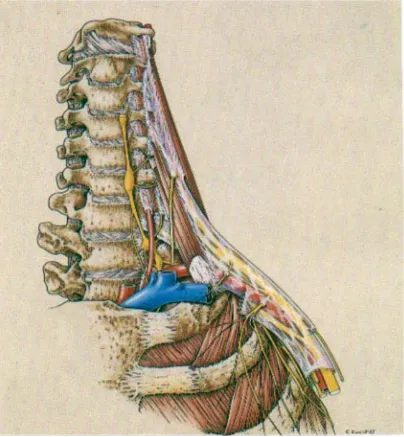
4. ANATOMICAL CONSIDERATIONS
3THE BRACHIAL PLEXUS:
Knowledge of the formation of brachial plexus and of its
distribution is absolutely essential for the use of brachial plexus
anaesthesia for the upper limb surgeries. Close familiarity with the
vascular, muscular and fascial relationships of the plexus throughout
its formation and distribution is also essential to the mastery of the
various techniques of brachial plexus blockade.
In its course from intervertebral foramina to the upper arm, the
fibres form roots, trunks, divisions, cords and terminal nerves passes
through a complex process of combining, dividing, recombining and
finally redividing.
FORMATION OF PLEXUS
Roots
The plexus is formed by the anterior primary rami of the
cervical nerves 5th to 8th, together with the bulk of the thoracic nerve
1st (C8 and T1). In addition there is frequently a contribution above
from C4 to the 5 th
13
FIGURE 1
THE RELATIONS OF THE BRACHIAL PLEXUS
14 thoracic nerve. Occasionally the plexus is mainly derived from C4 -8
(Pre –fixed plexus) or from C6 – T2 (post – fixed plexus).
Trunks and Divisions
The five roots of the plexus emerge from the intervertebral
foramina. They lie in the gutter between the anterior and posterior
tubercles of the corresponding transverse process. All five roots then
become sandwiched between scalenus anterior and medius. Here the
roots of C5 and C6 unite to form the upper trunk. The root of C7
continues as the middle trunk and those of C8 and T1 form the lower
trunk. Each trunk then divides behind the clavicle, into anterior and
posterior divisions, which unite to form the cords in the axilla.
Cords:
The six divisions unite up into three cords lateral, medial and
posterior into the axilla.
These cords are composed as follows:
The lateral cord is formed by the union of the anterior
divisions of the upper and middle trunks. The anterior division of the
lower trunk forms the medial cord. All the three posterior divisions
15
FIGURE 2
16
DISTRIBUTION OF BRACHIAL PLEXUS
These are divided with relation to clavicle - the supraclavicular
branches that arise above the clavicle and the infraclavicular
branches that arise below it.
The composition of the brachial plexus can be summarized as
follows:
SUPRACLAVICULAR BRANCHES
1. Five roots – the anterior primary rami of C5 – 8 and T1
2. Three trunks.
Upper trunk, C5 and C6
Middle trunk, C7 alone and
Lower trunk, C8 and T1
3. Six divisions – each trunk divides into an anterior and
posterior division.
INFRACLAVICULAR BRANCHES
4. Three cords
17
FIGURE 3
A. CUTANEOUS DISTRIBUTION OF THE CERVICAL ROOTS.
18
Branches are given off from
1. Roots 2. Trunks and 3. Cords
Branches from the Roots
1. Nerve to the serratus anterior (C5, C6 and C7)
2. Muscular branches to
i. Longus cervices (C5- C8)
ii. Three scalene (C5 – C8)
iii. Rhomboids (C5)
3. A twig of phrenic nerve (C5)
Branches from the trunks
1. Suprascapular nerve (C5, C6)
2. Nerve to subclavius (C5, C6)
BRANCHES FROM THE CORDS
LATERAL CORD
Lateral pectoral nerve (C5, C6, C7)
Lateral head of median nerve (C5, C6, C7)
19
FIGURE 4
20
MEDIAL CORD
Medial pectoral nerve (C8, T1)
Medial head of median nerve (C8, T1)
Medial cutaneous nerve of arm (C8, T1)
Medial Cutaneous nerve of forearm (C8, T1)
Ulnar nerve (C7, C8, T1)
POSTERIOR CORD
Upper subscapular nerve (C5, C6)
Lower subscapular nerve (C5, C6)
Nerve to latissimus dorsi (C6, C7, C8)
Axillary nerve (C5, C6)
Radial nerve (C5, C6, C7, C8, T1)
SYMPATHETIC CONTRIBUTION TO BRACHIAL PLEXUS:
The segmental preganglionic sympathetic contributions are
variable, but generally extend more caudal. The highest contribution
is usually from T2 with T1 contributing only rarely, while lowest
may be as far as T8, T9 or even T10.The post ganglionic
contributions are from grey rami communicantes from the
21
RELATIONS OF BRACHIAL PLEXUS:
In its passage from the cervical transverse processes to the first
rib, the plexus is "sandwiched" between the anterior and middle
scalene muscles and invested in the fascia of those two muscles.
The 'interfascial compartment', along with subclavian artery
which crosses the first rib immediately infront of the trunks. Artery
is close to the scalenus anterior and the plexus is close to the
scalenus medius. Subclavian vein is separated from the artery by the
scalenus anterior. The fascia covering the muscles is derived from
the perivertebral fascia, which splits to invest these muscles and
rejoins again at their lateral margins to form an enclosed space, the
interscalene space. As the plexus cross the first rib, the three trunks
are arranged one on top of the other vertically. Not infrequently, the
inferior trunk gets trapped behind and even beneath the subclavian
artery above the rib, during embryologic development.
This may be the reason why local anaesthetics injected via the
interscalene techniques sometimes fail to provide anaesthesia in the
distribution of the ulnar nerve, which may be buried deep within
inferior trunk behind or beneath the subclavian artery. After crossing
22 subclavian artery becomes the axillary artery. Above the clavicle, the
axillary artery lies central to the three cords, in the axilla the lateral
and posterior cords are lateral to the first part of the axillary artery,
the medial cord being behind it. Around the second part of the artery,
they are related according to their names. In the lower axilla, cords
divide into nerves for the upper limb.
In passing over the first rib under the clavicle, the subclavian
vein also becomes the axillary vein and its relationship with the
neurovascular bundle changes. Above the first rib the subclavian
vein does not lie within the neurovascular bundle, it is separated by
the insertion of scalenus anterior. As it passes over the first rib,
becoming the axillary vein it joins the neurovascular bundle so that
parts of the plexus are sandwiched between artery and vein. As all
the three enter the axilla, they invaginate the perivertebral fascia at
the lateral margins of the anterior and medial scalene muscles,
carrying this fascial investment of the neurovascular bundle into the
axilla as the axillary fascia, an extension of the perivertebral or
scalene fascia forming the axillary perivascular space, a tubular
extension of the interscalene space. In its course through the axilla
and upper arm the fascia of the surrounding muscles contribute to the
23
FIGURE 5
24 click' to the anaesthetic while entering the sheath. It is
important to note that major terminal nerves leave the sheath high in
the axilla under the cover of pectoralis minor muscle.
The musculocutaneous nerve enters the substance of
coracobrachialis and continues down within this muscle. The axillary
nerve also leaves the sheath immediately after arising from the
posterior cord. The interocostobrachial nerve travels parallel to but
outside the axillary sheath and medial cutaneous nerve of the arm
runs similarly but occasionally it may remain within the sheath.
THE BRACHIAL PLEXUS SHEATH
The connective tissue of the prevertebral fascia and the
anterior and middle scalenus envelops the brachial plexus as well as
the subclavian and axillary artery in a neurovascular "sheath".
Volume of the sheath: 42ml.
Shape of the sheath: Cylindrical to conical - Wide proximally
and narrow distally.
Length: 8-10cms.
The tissue is densely organized as it leaves the deep cervical
25 sheath blends with the fascia of the biceps and brachialis muscle
distally.
Anaesthetic implications
Because of these connective tissue septae, anaesthesia might
be complete and rapid in onset in some nerves, but delayed and
incomplete or completely absent in others. The incidence of partial
block is an exception rather than the rule, so septa apparently are of
little clinical significance as the local anaesthetic can percolate
through them.
TECHNIQUE OF BRACHIAL PLEXUS BLOCK
Surgical anaesthesia of the upper extremity and shoulder can
be obtained following neural blockade of the brachial plexus at
several sites. The various approaches that can be used for this
blockade are as follows.
1. Interscalene approach
2. Supraclavicular approach
a. Classic approach b. Plumb –bob technique
c. Subclavian perivascular technique 3. Axillary approach
26
TECHNIQUE OF BLOCKADE - SUPRACLAVICULAR SUBCLAVIAN PERIVASCULAR APPROACH TO
BRACHIAL PLEXUS
Anatomical Land marks:
The three trunks are clustered vertically over the first rib
cephaloposterior to the subclavian artery. Neurovascular bundle lies
inferior to the clavicle at above its mid point.
The essential landmarks to be identified are
1. Cricoid cartilage
2. Interscalene groove
3. Clavicle midpoint
4. Subclavian artery
PROCEDURE:
Position: Supine position with the head turned to the opposite side to
be blocked. The arm is pushed down to depress the clavicle.
The posterior border of sternocleidomastoid is felt, by asking
the patient to raise the head while keeping the head turned to
opposite side. The interscalene groove should be located behind the
midpoint of the posterior border of the muscle. The anterior and
middle scalene muscles can be made prominent by asking the patient
27 clavicle the pulsation of the subclavian artery can be felt in the
interscalene groove while standing on the side of the patient. On the
right side interscalene groove is palpated with the left index finger
and the needle is inserted with the right hand. Subclavian artery is
guarded with thumb. After aseptic measures and intradermal weal
raised with local infiltrations of 1 ml of 2% Lignocaine intradermally
in the interscalene groove 1 to 1.5 cm above the clavicle, a 22G, 50
mm short bevelled unipolar insulated needle connected to a nerve
locator is directed posteriorly and caudally towards the ipsilateral
nipple and slightly medially. End point in a nerve stimulator is a
motor response in ulnar distribution side of hand with an output less
than or equal to 0.5mA. To avoid intra vascular injection aspiration
done every 3-5 ml of the drug injected.within one ml of injection
muscular twitch disappear. The solution should flow without
resistance. High resistance or pain on injection may indicate
intraneural injection and the needle must be repositioned.
Volume of local anaesthetic (either 1% lignocaine or 0.25%
Bupivacaine) that can be used is 25-40 ml depending on the weight
of the patients. When large volumes are used the sheath may be felt
28 subcutaneous swelling of an extra fascial injection. To encourage the
spread proximally, digital pressure distal to the needle point may be
used and digital pressure proximal to needle insertion point may help
to encourage distal spread.
COMPLICATIONS
a) Related to procedure
Vessel puncture - Haematoma formation
Pneumothorax
Neuropraxia
b) Related to Local anaesthetic
Intravascular injection
Circumoral numbness
Convulsions
29
5. PHYSIOLOGICAL CONSIDERATIONS4
International association for the study of pain has defined pain as “An unpleasant sensory and emotional experience associated with
actual or potential tissue damage, or described in terms of such
damage”.
Pain perception requires a noxious stimulus which is transformed
from its native form by the activated nociceptors into electrical
signals which are then transmitted along the corresponding
nociceptive fibres. These fibres in turn synapse onto second order
neurons in the spinal cord. These interneurons are located in the
dorsal horn. It is at these interneurons where the initial modulation of
nociceptive input occurs. From the spinal cord nociceptive input is
transmitted to the brain stem, thalamus and cortex.
Peripheral neuroanatomy of nociception
C and A fibres are the main peripheral nociceptors. The skin,
joints and periosteum are richly innervated with C and A nociceptors
30 A fibres are responsible for the sensation of first pain, the initial
sharp pain experienced following an injury. C fibres are
unmyelinated and are responsible for second pain, the slowly
building throbbing burning pain experienced following an injury.
Classification of sensory fibres
Sensory receptors
Speed of
transmission Sensory function Myelination
C fibres 0.5 – 2 m/sec1
Noxious chemical,
mechanical, thermal
activation (slow burning second pain)
Unmyelinated
Aα fibres 70 – 120m/sec
noxious chemical thermal, mechanical stimuli (sharp fast, first pain)
slightly myelinated
Aβ fibres 30 – 70 m/sec
nonpainful, light touch,
pressure, vibration,
proprioception
heavily myelinated
Aγ fibres 30 – 70 m/sec proprioception, motor to
muscle spindle myelinated
Aδ fibres 12 – 30 m/sec pain, cold, touch myelinated
B fibres 3- 15 m/sec pre ganglionic autonomic
(sympathetic) myelinated
Peripheral neurochemistry and neurotransmitters
Commonly released inflammatory mediators implicated in
pain and hyperalgesia include bradykinins, potassium, substance-P,
31 neurotransmitters either activate or sensitize the peripheral
nociceptors to pain.
Peripheral neurochemistry of Algogenic agents:
Algogenic agent Action on nociceptors
Bradykinin activates
Substance P sensitizes
Potassium activates
Hydrogen activates
Arachidonic acid sensitizes
Cytokines sensitizes
Serotonin sensitizes
Noradrenaline high concentration activates and sensitizes after injury.
Physiology of nerve conduction
Neurons are the basic building blocks of the nervous system
that responds to various stimuli. Integration and transmission of
nerve impulses are specialized functions of neurons.
All peripheral nerves are elongated axons of neurons situated
centrally. A typical peripheral nerve consists of bundles of motor,
sensory and other fibres enclosed in the outermost covering called
32 neurilemma or the axonal membrane. Depending upon the presence
or absence of myelin sheath, it can be a myelinated nerve fibre or
unmyelinated fibre.
The axonal membrane itself is made up of a bimolecular lipid
palisade, interspersed with large protein molecules. The membrane
lipids are largely phospholipids composed of a polar head group and
a non polar hydrocarbon tail.
The primary function of the cell membrane is to separate the
extracellular from the intracellular environment. The major
difference between these two environments is the ionic
concentration. This disequilibrium provides the means for impulse
conduction.
The most important ions in this respect are sodium and
potassium. A membrane bound protein sodium potassium ATPase
maintains normal resting equilibrium potential between -50mv to -
90mv by pumping potassium ions into the cell and sodium ions out
of the cell. A positive ion gradient from inside the membrane to
33 During nerve conduction the following changes occur in the
cell membrane.
In the resting phase:
There is a potential difference across the membrane the inside
is negative, due to a higher concentration of sodium ions outside than
inside the cell.
Na+ moves in and K+ moves out of cells but because of more
K+ channels opened at rest, K+ permeability is greater than Na+
permeability. Therefore K+ channels maintain the resting membrane
potential.
Depolarization phase:
During excitation, Na+ channels in the cell membrane open
briefly allowing sodium ions to flow into the cell, thereby
depolarizing the membrane.
Repolarization Phase:
During this phase, opening of voltage gated K+ channel
occurs, results in passing of potassium ions out of the cell to restore
34
Restoration Phase:
During this phase, sodium ions return to the outside and
potassium ions re - enter the cell.
Distribution of ion channels in Myelinated Neurons:
Voltage gated Na+ channels are highly concentrated in the
nodes of Ranvier and the initial segment in myelinated neurons.
The initial segment and in sensory neurons, the first node of
Ranvier are the sites where impulses are normally generated and the
other nodes of Ranvier are the sites to which the impulses jump
during saltatory conduction.
The sodium channel is believed to be an integral membrane
spanning protein. Depolarization of the cell induces a configurational
change on the sodium channel which causes it to open and allow ion
passage. In many myelinated neurons, the Na+ channels are flanked
by K+ channels that are involved in repolarization.
Action of Local Anaesthetics on Nerve Fibres5:
The primary action of local anaesthetics on the nerves is
electrical stabilization. The large transient increase in permeability to
35 prevented. Thus the resting membrane potential is maintained and
depolarization in response to stimulation is inhibited.
Local anaesthetics block sodium conductance by:
1. binding of local anaesthetics to sites on voltage gated Na+
channels prevents opening of the channels by inhibiting the
conformational changes that underlie channel activation.
2. local anaesthetics produce nonspecific membrane expansion.
There is an unfolding of membrane protein together with a
disordering of the lipid component of the cell membrane with
consequent obstruction of the sodium channel.
PAIN PATHWAY
SPINAL CORD
The gray matter of the spinal cord is divided into ten lamina
with lamina I – IV representing the dorsal horn. The dorsal horn is capped by the Lissauer’s tract which consists of branches of
cutaneous A and C – fibres and few visceral afferents.
Nociceptive fibres terminate in the superficial layers of lamina
I & II while the non-painful myelinated fibres terminate in the deeper
layers of lamina III, IV. Lamina II has the highest concentration of
36 nociception may occur at this level through the use of opioids
(systemic and neuraxial).
Ascending sensory pathways
Peripheral sensory neurons synapse onto the secondary
interneurons of the dorsal horn. The axons of the non nociceptive
secondary neurons travel isobilateral in the dorsal columns of the
spinal cord as fasciculus cuneatus (upper body through T6) and
fasciculus gracilis (lower body below T6) and synapse in the
thalamus.
The axons of the nociceptive secondary neurons after
synapsing travel contra laterally in the anterolateral aspects of the
spinal cord as the neospinothalamic and paleospinothalamic tract.
Neospinothalamic tract carries fine discrimination of pain e.g.
location, intensity, and first pain.
Paleospinothalamic tract responds to noxious stimuli. The
paleo spinothalamic tract synapses in the thalamus, hypothalamus
and limbic system and plays a role in emotional aspects of pain via
limbic system. The thalamus has multiple connections to limbic
37
Descending inhibitory pathways
The descending controls of pain project specifically onto
laminas I, II, V of the dorsal horn from mesencephalon, raphenuclei
and reticular tract. The mesencephalon is rich in opioid receptors.
This area sends excitatory transmissions to the rostroventral medulla
which sends noradrenaline and serotonin inhibitory tracts via the
dorsolateral funiculus to lamina I, II, V of spinal cord.
The noradrenaline and serotonin fibres mediate transmission
between the primary afferents and the secondary neurons of the
dorsal horn. Increased activity of these fibres leads to increased
inhibition of pain transmission.
Location of opioid receptors (central)
Opioid receptors are found in the various regions in CNS
namely, cerebral cortex, limbic cortex (anterior and posterior
amygdale, hippocampus) hypothalamus, medial thalamus, mid brain,
periaqueductal gray matter, extrapyramidal areas, substantia
gelatinosa and sympathetic preganglionic neurons.
38
6. BASICS OF NERVE LOCATOR
6, 7Perivascular technique and elicitation of paraethesia had been
the classical methods for locating nerves in peripheral nerve blocks.
Peripheral nerve locator technology is a newer one, utilizing
objective end points for effective nerve localization.
Peripheral nerve locator is used to elicit Evoked Motor
Response (EMR). They are used to assess functioning of
Neuromuscular (NM) junction. The other name for Peripheral nerve
locator (PNL) is Peripheral nerve stimulator (PNS) .When the high
intensity current is used to assess the NM junction function through
cutaneous electrodes it is called as PNS. When low intensity current
is used to locate the nerve it is called peripheral nerve locator.
Physiological basis of PNL Technology
The ability of a nerve locator to evoke a motor response
depend on i) intensity of current
ii) duration of current
iii) polarity of stimulating current used
39 Assuming a square pulse of the current is used to stimulate the
nerve the total charge applied is the product of intensity of current
and duration of the pulse.
Rheobase and Chronaxie:
Rheobase is the minimal current required to stimulate the
nerve with a long pulse width.
Chronaxie is the duration of the stimulus required to stimulate
at twice the rheobase.
I = Ir (1+c/t)
Where I - current required, C - chronaxie,
Ir - rheobase, t - duration of stimulus.
Chronaxie is useful when comparing different nerves or nerve
fibre types. The larger fibres are more readily stimulated than the
smaller fibres. It is possible to stimulate the larger Aα motor fibre without stimulating the smaller Aδ or C -fibres responsible for pain.
Chronaxies of Peripheral Nerves
* A-α 50 -100 μ seconds
* A-δ 170 μ seconds
40
Principles of peripheral nerve stimulation:
i) Preferential cathodal stimulation:
Significantly less current is needed to obtain a response to
nerve stimulation when cathode is adjacent to the nerve, rather than
the anode.
ii) Variation of stimulus intensity with varying needle nerve distance.
Stimulation intensity will be variable as determined by
Coulomb’s law.The relationship between the stimulus intensity and
the distance from the nerve is governed by Coulomb’s law
I = K (Q/r2)
Where I is the current required to stimulate the nerve, K is a
constant, Q is the minimal current needed for stimulation, and r is the
distance from the stimulus to the nerve.. A very high stimulus current
is required to stimulate the nerve when the needle tip is far away
from the nerve.
Components of peripheral nerve locators
Oscillator
Display
Constant current generator
41
Characteristics of an ideal PNL
1) Constant current output:-
The constant current designs of the locator allows for an
automatic compensation for changes in tissue or connection
impedance during nerve stimulation assuring accurate delivery of the
specified current.
2) Options for different pulse width:-
Shorter pulse width corresponds to the chronaxie of motor
fibres in a mixed peripheral nerve. Wider pulse width (>100μ sec) is
useful for stimulating a sensory nerve or a nerve with compromised
conduction i.e. Diabetic neuropathy.
3) A wide range of current output - 0.01 to 5.0 mA:-
A higher current output is needed for patients with neuropathy
and sensory nerve stimulation.
4) Digital display of the delivered current.
5) Variable current output dial.
6) Clearly identifiable polarity
7) Disconnect indicator - shows circuit connection status
8) Battery indicator
42 If the stimulating frequency is higher, it allows faster
manipulation of the needle.
Clinical points to be noted while using PNL:
An effective use of PNL technology mandates knowledge of
anatomy with respect to
a) Optimal needle insertion site to achieve needle tip – target nerve
contact.
b) Muscle innervations scheme of the targeted nerve to identify
desired evoked motor response (EMR).
c) Ability to differentiate desired EMR from the alternate EMR
elicited by the stimulation of adjacent muscle and collateral nerves.
d) The relationships of adjacent neuromuscular structures generating
those alternate EMR to the targeted nerve.
e) The highest rate of success is attained when a brisk EMR occur
between 0.2-0.4mA.
An EMR at currents higher than 0.5mA may result in failed
block because the needle tip is too far from the nerve. A brisk EMR
at stimulating current lower than 0.1mA may risk nerve damage
43
Peripheral Nerve Locator Settings:
1) Mixed nerve (most PNB)
Current 1 mA
Current duration (Pulse with) 0.1ms Frequency 1-2 HZ
2) Sensory nerve (e.g. lateral cutaneous and saphenous nerve) Current 2-5 mA
Current duration 1ms Frequency 1 Hz
3) Diabetic neuropathy (PNB)
Current 2 mA
Current duration 0.3 ms Frequency 1-2 Hz
APPROPRIATE EVOKED MOTOR RESPONSE FOR EACH
PNB
PNB Technique Optimal EMR
Interscalene
Flexor: Deltoid, Biceps, Pectoralis major
Extensor: Triceps, Brachioradialis, Wrist extensors (EMR of > 2 muscles)
Deep Cervical
plexus Rhomboids, Shoulder girdle
Infraclavicular
Muscles of wrist and hand
Radial – extension of wrist/ fingers. Median – flexion of wrist / fingers.
Ulnar – adduction of thumb / 4th and 5th finger flexion.
Femoral Quadriceps – patellar tap
44
7. PHARMACOLOGY OF BUPIVACAINE
8, 9, 10BUPIVACAINE
Bupivacaine is a local anesthetic agent with long duration of
action
Pharmacology
Bupivacaine hydrochloride is 2 piperidine carboxamide, 1
butyl N-2, 6 dimethyl phenyl, monohydrochloride, and monohydrate.
Bupivacaine molecule is a tertiary amine separated from an
aromatic ring system that is a benzene ring by an chain. The tertiary
intermediate amine is a base that is a proton acceptor. The chain
contains an amide linkage (-NHCO-) hence it is classified as an
amino amide compound. This amide linkage contributes to the
anesthetic potency.
The aromatic ring system gives a lipophilic character to its
portion of molecule whereas the tertiary amine end is relatively
45
STRUCTURE
Structure - Activity relationship:
Bupivacaine being more lipophilic (because of butyl group) it
is very potent and produces longer lasting blocks.
PHARMACODYNAMICS
Mechanism of action:
The uptake of the drug by the tissues is largely due to
lipophilic absorption. This shifts effective pKa downward and
thereby favors the neutral base form.
Bupivacaine blocks impulses by reducing the currents through
voltage-activated Na+ channels. The inhibition is not specific;
however, K+ currents are also reduced. Binding of bupivacaine to
sites on voltage gated Na+, channels prevents opening of the
46 It is similar to that of any other local anaesthetics. The primary
action of local anaesthetics is on the cell membrane of the axon, on
which it produces electrical stabilization. The large transient increase
in permeability to sodium ions necessary for propagation of the
impulse is prevented. Thus the resting membrane potential is
maintained and depolarization in response to stimulation is inhibited.
The mechanism by which local anaesthetics block sodium
conductance is as follows:
a) Local anaesthetics in the cationic form act on the receptors
within the sodium channels, on the cell membrane and blocks it. The
local anaesthetic can reach the sodium channel either via the
lipophilic pathway directly across the lipid membrane, or via the
axoplasmic opening. This mechanism accounts for 90% of the nerve
blocking effects of amide local anaesthetics.
b) The second non specific mechanism of action is by
membrane expansion.
PHARMACOKINETICS:
Pka 8.1
Bound in plasma 95%
47
Volume of distribution 0.4 – 0.9 litres / kg
Half life 1.2 – 2.4 hours
Peak time 0.17 – 0.5 hours
Peak concentration 0.8 micrograms/ml
Toxic plasma concentration >1.5 micrograms/ml
Clearance – 0.47 litres /min
Metabolism – Liver by dealkylation to
Pipecolyloxilidine
Excretion – 5% by the kidney as unchanged drug
[image:53.595.129.531.470.757.2]and the rest as metabolites.
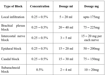
TABLE: 4. Dosage and concentration of Bupivacaine in various
block
Type of Block Concentration Dosage ml Dosage mg
Local infiltration 0.25 – 0.5% 5 – 20 ml upto 175mg
Brachial plexus
block 0.25 – 0.5% 20 – 40 ml 75 – 225mg
Intercostal nerve
block 0.25 – 0.5% 3 – 5 ml
15 – 20 mg per each nerve
Epidural block 0.25 – 0.5% 15 – 20 ml 50 – 200mg
Caudal block 0.25 – 0.5% 15 – 30 ml 75 – 150mg
Subarachnoid
48
DRUG DOSAGE11, 12
Bupivacaine upto 3mg/kg.
Addition of epinephrine to bupivacaine has no effect on its
duration of action but it delays absorption of local anaesthetic due to
vasoconstriction from the site of administration.
Toxicity of Bupivacaine
It is relatively free of side effects if administered in an
appropriate dosage. It is more cardiotoxic than Lignocaine and this is
made worse by hypoxia, hypercapnia, acidosis and pregnancy.
1. Central nervous system toxicity
CNS is more susceptible to bupivacaine. The initial symptoms
involve feeling of light headedness and dizziness followed by visual
and auditory disturbance. Disorientation and occasional feeling of
drowsiness may occur. Objective signs are usually excitatory in
nature which includes shivering, muscular twitching and tremors;
initially involving muscles of the face (perioral numbness) and part
of extremities. At still higher doses cardiovascular or respiratory
49 Bupivacaine, since the elevation of PaCO2 enhances cerebral blood
flow, so that more anesthetic is delivered rapidly to the brain.
2. Cardiovascular system toxicity
Bupivacaine depresses rapid phases of depolarization (Vmax)
in purkinge fibres and ventricular musculature to a greater extent
than lignocaine. It also decreases the rate of recovery from a
dependent block than that of lignocaine. This leads to incomplete
restoration of Vmax between action potential at high rates, in
contrast to complete recovery by lignocaine. This explains why
lignocaine has antiarrhythmic property while bupivacaine has
arrhythmogenic potential. High level of bupivacaine prolongs
conduction time through various parts of heart and extremely high
concentration will depress spontaneous pacemaker activity, resulting
in bradycardia and arrest. Cardiac resuscitation is more difficult
following bupivacaine induced cardiovascular collapse and hypoxia
along with acidosis which markedly potentiates cardiac toxicity.
Bretylium raise the ventricular tachycardia threshold that was
50 The cardiovascular collapse / central nervous system ratio for
bupivacaine is 3.7 0.5.
3. Respiratory system:
Respiratory depression may be caused if excessive plasma
level is reached which in turn results in depression of medullary
respiratory center.
4. Autonomic nervous system:
Myelinated preganglionic beta fibers have a faster conduction
time and are more sensitive to the action of local anesthetic including
bupivacaine. Involvement of preganglionic sympathetic fibers is the
cause of widespread vasodilatation and consequent hypotension that
occurs in epidural and paravertebral block. When used for
conduction blockade all local anesthetic agents particularly
bupivacaine produces higher incidence of sensory blockade than
motor fibers.
Treatment of adverse reaction:
Treatment is mainly symptomatic i.e. maintaining circulation
with IV fluids and vasopressors if required to restore the
51 controlled ventilation. Convulsions may be controlled with diazepam
(0.1- 0.2 mg/kg) or thiopentone (2-3 mg/kg) or a muscle relaxant and
controlled ventilation with oxygen. Corticosteroids, if allergic
reactions are suspected. Treatment of ventricular fibrillation and
tachycardia by amiodarone (5mg/kg IV), bretylium (5mg/kg) or
defibrillation (2-6 joule/kg).
Role of additives:
i. Adrenaline: Onset time reduced and duration prolonged.
ii. Sodium bicarbonate: Onset time reduced and duration
variable.
iii. Clonidine: Onset time reduced and duration prolonged.
iv. Hyaluronidase: Onset time reduced and duration variable.
v. Opioids: Onset time reduced and duration prolonged. Reports
controversial.
52
8. PHARMACOLOGY OF TRAMADOL
13, 14, 15, 16Tramadol is an analgesic with unique dual mechanism. It is a
synthetic 4 – phenyl piperidine codeine analogue. Tramadol is a
racemic mixture of 2 enantiomers.
Structure of Tramadol
• Chemical name: Tramadol belongs to the aminocyclohexanol
group.
Mechanism of action
Tramadol has both opioid and non-opioid actions
1. Tramadol has a low affinity for opioid receptors. It acts as a selective μ-receptor agonist, but also binds weakly to kappa and delta
receptors.
2. Non-opioid mechanism is by monoaminergic pathway. It inhibits
noradrenaline and 5-hydroxy tryptamine (serotonin) neuronal
53 tramadol i.e., tramadol (+) and tramadol (-) have complementary and
synergistic anti-nociceptive interaction. Tramadol (+) has greater affinity for μ receptors and inhibits serotonin reuptake. Tramadol (-)
inhibits norepinephrine reuptake. The synergistic effect of both these
enantiomers may be responsible for its low potential for the
development of tolerance, dependence, and abuse and production of
analgesia with the absence of ventilatory depression. Interestingly,
the racemate may produce less sedation and gut inhibition than either
enantiomer alone.
Pharmacodynamics
Effects on respiration
In clinically recommended doses, tramadol is unlikely to
produce relevant respiratory depression.
Effects on cardiovascular system
Tramadol increases transiently heart rate, both systolic and
diastolic blood pressure; it increases peripheral vascular resistance,
decreases pulmonary arterial resistance, and exerts a negative
54
Tolerance and dependence
Tolerance is minimal. It has low physical and psychological
dependence.
Other effects
Tramadol in clinical dosage has no effect on plasma histamine
levels and it does not cause any systemic anaphylactoid reactions.
Pharmacokinetics
Bioavailability
Oral 68 %( 70 – 75%)
Intramuscular 100%
Elimination T1/2 6hours (4.5 – 7.5%)
Metabolite 7.5hours
Percentage of drug bound in plasma 20%
Clearance 8ml/min/kg (6 – 12ml)
Volume of distribution 2.7litres/kg (2.3 – 3.9)
Onset of analgesia 1 hour
Peak time 2.3 ± 1.4 hours
55
Metabolism – Liver by O- demethylation to O-
desmethyl tramadol which shows 200 times higher
affinity for µ receptors.
Elimination – 15 – 30% as unchanged drug by kidney.
Therapeutic efficacy
On intravenous administration, tramadol is equivalent to
pethidine, l/5th as potent as nalbuphine, 1/10th as potent as
morphine.
Dosage
Tramadol can be given in doses of 50-100 mg upto 4 times a
day. Total daily dose should not exceed 400mg for adults. In children
> l year of age the dosage is 1 – 2 mg/kg.
Routes of administration
Oral, parenteral, epidural, rectal, caudal.
Adverse effects: Mild, transient and rare
1. CNS: Nonspecific CNS irritation, dizziness, sedation, euphoria,
dysphoria.
2. GIT: Nausea, vomiting, constipation, GI irritation
56 4. CVS: Orthostatic hypotension, tachycardia
5. Others: Motor weakness, urinary retention
Respiratory depression with tramadol is less than that with
morphine. Respiratory depression is unusual in recommended doses
and was not found in neonates whose mother had been given
tramadol. The advantage of tramadol over other opioids with respect
to less respiratory depression is limited by lack of efficacy of
tramadol in severe pain.
Drug interactions
Tricyclic antidepressants, selective serotonin reuptake
inhibitors (SSRI), neuroleptics: tramadol when given to patients on
these drugs decreases the seizure threshold. Concomitant
administration of tramadol and SSRI causes serotonin syndrome.
Concomitant administration of tramadol with monoamino
oxidase inhibitors causes hypertensive reactions. Quinidine inhibits
tramadol metabolism. Hence serum tramadol concentration
increases, when these 2 drugs are used. Carbamazepine enhances
tramadol metabolism, hence tramadol halflife is decreased as much
as 50%. When these two drugs are used concomitantly, tramadol
57
Overdosage:
Symptoms are similar to other opioids. Miosis, vomiting,
coma,
respiratory depression, respiratory arrest and cardiovascular collapse.
Opioid antagonist naloxone will reverse coma and respiratory
depression.
Advantages:
1. Can be given through different routes - oral, parenteral etc.
2. Less respiratory depression
3. Less dependence, abuse, tolerance
4. Less secretion in the milk of lactating mother.
5. Freely available. No narcotic prescription restriction.
6. Comparatively cheap.
Indications:
1. Tramadol is indicated for moderate to severe pain in adults.
Tramadol 50-150 mg IV was equivalent to morphine 5-15 mg but a
preservative free preparation had 1/13 of the potency of morphine
58 Despite being relatively less potent than pure opioids,
tramadol has achieved efficacy when used to treat moderate pain
after surgery. Because of its efficacy as an analgesic, tramadol is
considered to be effective in step two of the world health
organization guideline for treatment of patients with cancer pain.
2. In peripheral nerve blocks along with local anaesthetics to prolong
the duration of anaesthesia as well as analgesia.
Contraindications:
As tramadol enhances monoaminergic transmission, the drug
is contraindicated in patient receiving mono-amino oxidase inhibitors
and caution advised in patients with epilepsy. Not recommended in
59
9. REVIEW OF LITERATURE
1. Suman Chattopadhyay et al 200717 conducted a prospective
double blind study in supraclavicular brachial plexus block to
evaluate the effect of weak opioid tramadol with nonopioid
mechanisms of action, improves post operative analgesia when used
as an additive along with bupivacaine in 70 patients who underwent
surgery of various upper limb surgeries. These patients are randomly
allocated into 2 equal groups so that 35 patients received inj.
bupivacaine (0.25%) - 38 ml + 2 ml normal saline (Group C) and the
remaining 35 received inj. bupivacaine (0.25%) - 38 ml +tramadol
100 mg (2 ml) (Group T) [total volume in both group 40 ml].The
onset, quality and duration of the block and duration of analgesia
was assessed as well as the possible side effects and also the
incidence of various complications following the procedure. Earlier
the onset of motor (6.1 ± 1.2 vs 8.6 ± 1.4 mins) and sensory
blockade(11.2 ± 2.1 vs 18.4 ± 2.5mins) and the duration of pain
relief (410.1 ± 95.1 min vs 194.8 + 60.4 min) produced with addition
of tramadol was longer and superior in comparison to control group
60 rate, blood pressure and spo2. No side effect was noted in any of the
patients.Tramadol is a useful adjuvant for brachial plexus block.
2. Renu Wakhlo et al 200918 conducted a study on 60 patients to
compare the adjuncts- tramadol and butorphanol to lignocaine with
adrenaline for onset and duration of block and post operative
analgesia for upper limb surgeries following supraclavicular brachial
plexus block. All patients received total volume of 30 cc of
anaesthetic. Patients were randomly divided into three equal groups
so that 20 patients received only lignocaine with adrenaline
(1:200,000 ) 20 cc and 10 cc saline (Group I), next 20 patients
received lignocaine with adrenaline + tramadol 100 mg (2 cc) +8cc
saline (Group II) and remaining 20 received lignocaine + adrenaline
+ butorphanol 1 mg. (1 cc) + saline 9 cc (Group III). The onset of
sensory and motor block, duration of block and post operative
analgesia was compared. Statistical analysis was done by ANOVA
test and intergroup comparison done by Bonferroni's t test. It was
found that Group II patients had earlier onset and prolonged duration
of sensory and motor block while Group III patients had prolonged
duration of postoperative analgesia lasting upto mean of 12 hours.
61 and prolonging the sensory and motor block while butorphanol is
suitable agent for prolonged postoperative analgesia.
3. S.Antonucci et al 200119 compared the adjuvant effects of
Clonidine (1.5 mcg/kg), Sufentanil (20 mcg) and Tramadol (100mg)
with 0.75% Ropivacaine in brachial plexus axillary blockade among
80 patients of each group20 with control group as normal saline and
concluded that the use of tramadol 100mg as adjuvant provides a
significant reduction of onset time and provides a prolonged
anaesthesia and analgesia with a quality of block similar to that of
Clonidine and Sufentanil and incidence of side effects like sedation,
bradycardia and hypotension and itch are lower. They concluded that
tramadol 100mg may be a useful alternative as adjuvant in periphery
block with same effects of other drugs commonly used and a lower
incidence of side effects.
4.Sukran Geze et al 201220 compared tramadol 100mg and fentanyl
50mcg by adding to 40ml of 0.25% levobupivacaine plus 40mg
lignocaine mixture for axillary plexus block by randomized double
blind study and concluded that tramadol hydrochloride or fentanyl
when added to local anaesthetic mixtures as an adjuvant agent
62 orthopaedic upper limb surgeries. Furthermore tramadol improves
the block quality more than fentanyl.
5. Shrestha BR et al 200721 evaluated the postoperative analgesia
of tramadol 2mg/kg and dexamethasone 8mg as admixture to
bupivacaine by conducting a prospective, randomized, double blind
study in 60 patients. Patients were randomly allocated in to two
groups of 30 each. The duration of postoperative analgesia was
recorded in both groups using pain VAS score which was determined
by maximum VAS score of 8-10 and when patient demands for
additional analgesics. Dexamethasone significantly prolonged the
postoperative analgesic duration (mean 1028 minutes) which was
significant than tramadol (453.17 minutes) (P <0.05).This helps to
minimize the cost and provides patient comfort.
6. Sebastien Robaux et al 200422 designed a prospective,
randomized, controlled and double-blind clinical trial to assess and
evaluate the time of onset and quality of postoperative analgesia, and
occurrence of adverse effects of tramadol in one-hundred patients
scheduled for carpal tunnel release surgery. All patients received
1.5% mepivacaine 40ml.The patients were randomly divided into
63 (Group P), 22 patients received tramadol 40 mg (Group T40), other
20 patients received tramadol 100 mg (Group T100) and remaining
20 received tramadol 200 mg (Group T200).Onset and duration of
sensory and motor blocks were not different among groups. The
number of patients requesting analgesia in the postoperative period
was significantly less in the 3 tramadol groups compared with the
placebo group (P=0.02); this was also noted with the placebo and
T40 groups compared with the T200 group. No statistical
significance was demonstrated between the placebo and the T40
group or the T100 group and the T200 group. Furthermore, there was
a significant trend effect among groups applying the
Cochran-Armitage tendency test (P=0.003), suggesting a dose-dependent
decrease for additional postoperative analgesia requirements when
tramadol was added. Side effects did not differ among groups,
although they were more frequently recorded in the T groups. Study
suggests that tramadol added to 1.5% mepivacaine for brachial
plexus block enhances in a dose-dependent manner the duration of
analgesia with acceptable side effects. However, the safety of
tramadol has to be investigated before allowing its use in clinical
64 7. Olfa Kaabachi et al 200923 conducted a prospective randomized
study to evaluate the effect of varying doses of tramadol as an
adjuvant to axillary block with lidocaine 1.5% (epinephrine 1/2,
00,000). Three groups were allotted randomly, control group
received 4ml saline, TL group received100mg tramadol and 2ml
saline and TH group received 200mg tramadol along with lidocaine.
The results showed that the addition of 200mg tramadol prolonged
the analgesic duration with a delayed onset time.
8. Stephan Kapral et al 199924 studied in patients randomly
assigned to receive either 40ml of 1% mepivacaine with 2ml of
isotonic sodium chloride solution (Group A), 40 ml of 1%
mepivacaine with 100mg tramadol or 40ml of 1% mepivacaine with
2ml of isotonic sodium chloride and 100mg tramadol
intramuscularly and evaluated that when tramadol 100mg added to
1% mepivacaine prolongs the duration of blockade without any
adverse effects in brachial plexus axillary block. Tramadol may be a
useful alternative to epinephrine and clonidine as an adjuvant to local
65 9. F. Alemanno et al 201225 studied in 120 patients allocated in 3
groups. Group P received 0.4ml/kg of 0.5% levobupivacaine plus
isotonic sodium chloride and isotonic sodium chloride
intramuscularly. Group TPN ( perineural tramadol) received 0.4ml/kg
of 0.5% levobupivacaine plus 1.5mg/kg tramadol perineurally and
isotoni