FUNCTIONAL OUTCOME ANALYSIS OF PARALLEL PLATE
TECHNIQUE FOR DISTAL HUMERUS FRACTURES
Dissertation submitted in
Partial fulfilment of the requirement for
M.S. DEGREE-BRANCH II
ORTHOPAEDIC SURGERY
MADRAS MEDICAL COLLEGE AND
RAJIV GANDHI GOVT. GENERAL HOSPITAL
THE TAMILNADU DR.M.G.R.MEDICAL UNIVERSITY
CHENNAI-TAMILNADU
CERTIFICATE
This is to certify that this dissertation titled “Functional Outcome Analysis of
Parallel-Plate technique for distal humerus fractures” is a bonafide record of
work done by DR.DINESH.L, during the period of his Post graduate study from
May 2010 to April 2013 under guidance and supervision in the Institute of
ORTHOPAEDICS AND TRAUMATOLOGY, Madras Medical College and Rajiv
Gandhi Government General Hospital, Chennai-600003, in partial fulfilment of the
requirement for M.S.ORTHOPAEDIC SURGERY degree Examination of The
Tamilnadu Dr. M.G.R. Medical University to be held in April 2013.
Dr. KANAGASABAI.M.D. PROF.M.R.RAJASEKAR. M.S.ORTHO., D.Ortho. Dean Director
Madras Medical College & Rajiv Institute of Orthopaedics and traumatology Gandhi Government General Hospital, Madras Medical College & Rajiv Gandhi Chennai -600 003. Government General Hospital
DECLARATION
I declare that the dissertation entitled “
FUNCTIONAL OUTCOME
ANALYSIS OF PARALLEL PLATE TECHNIQUE FOR DISTAL
HUMERUS FRACTURES
” submitted by me for the degree of M.S is the record work carried out by me during the period of May 2010 to November 2012under the guidance of Professor Dr.M.R.RAJASEKAR M.S.ORTHO., D.Ortho., Professor & Head of the Department of Orthopaedics, Institute of Orthopaedics and traumatology, Madras Medical College, Chennai. This dissertation is submitted to the Tamilnadu Dr.M.G.R. Medical University, Chennai, in partial fulfilment of the University regulations for the award of degree of M.S.ORTHOPAEDICS (BRANCH-II)) examination to be held in April 2013.
Place: Chennai Signature of the Candidate
Date: (Dr.L.DINESH)
Signature of the Guide
Prof.Dr.M.R.RAJASEKAR M.S.ORTHO., D.Ortho.,. Director
ACKNOWLEDGEMENT
Any prospective study in a place as big as this Institution requires the support and
guidance of a lot of people. It would only be appropriate if all the hours they have put in
are properly remembered and acknowledged. My sincere thanks to
Dr.V.KANAGASABAI, M.D , Dean, Madras Medical College, for permitting the
utilization of the resources and clinical material in this hospital.
I will forever be indebted to our Director Prof M.R.RAJASEKAR M.S.Ortho;
D.Ortho Director, Institute of Orthopaedics and Traumatology Madras Medical
College & Government General Hospital. He has been my guide for this study and
always been a constant source of inspiration with his advice and guiding me through the
finer aspects of this study. Without him this study would not have been possible.
I sincerely thank Prof.N. DEEN MOHAMED ISMAIL M.S.Ortho., D.Ortho.,
for his support during this study. Without his guidance and constant encouragement , this
study would not have been possible.
I sincerely thank Prof.V.SINGARAVADIVELU M.S.Ortho., D.Ortho., for his
support, guidance and encouragement during the study.
I am grateful to PROF. A.PANDIASELVAN., M.S.Ortho., D.Ortho., for his
valuable guidance.
I am thankful to PROF. ANBALAGAN M.S. Ortho., D.Ortho for his support in
I am grateful to Dr.R.SUBBIAH. M.S.Ortho. D.Ortho, Reader in Spine Surgery,
for his support in this study.
I am profoundly thankful to Dr.NALLI R UVARAJ,M.S. Ortho., D.Ortho.,
DNB Ortho., Reader in Spine Surgery, for all his valuable inputs to this study.
My sincere thanks to Prof. R.H. GOVARDHAN. M.S,Ortho., D.Ortho., former
director, Prof. S.SUBBAIAH., M.S,Ortho., D.Ortho., and Prof.V.THULASIRAMAN
M.S,Ortho., D.Ortho., retired professors, Institute Of Orthopaedics and Traumatology, for
their valuable advice and guidance.
My special thanks to ,Dr.A.SHANMUGASUNDARAM M.S.Ortho, Dr.MOHAMMED
SAMEER., M.S.Ortho. DNB ortho for his constant encouragement and valuable
guidance throughout the study.
I, sincerely thank Dr.K.P.MANIMARAN, Dr.S.KARUNAKARAN,
Dr.VELMURUGAN, Dr N. MUTHALAGAN, Dr.NALLI. R.GOPINATH,
Dr.PRABHAKARAN, Dr. SENTHIL SAILESH, Dr.P.KINGSLY, DR.KANNAN,
DR.PALANI ,DR.HEMANTH KUMAR, DR.KALIRAJ , DR.MUTHUKUMAR
who have each been great mentors in their own way.
I thank all anaesthesiologists and staff members of the theatre for their endurance during
this study.
I am grateful to all my post graduate colleagues for helping in this study. Last but not
least, my sincere thanks to all our patients, without whom this study would not have been
CONTENTS
S.NO TITLE PAGE NO
1 INTRODUCTION 1
2 AIM AND OBJECTIVE 4
3 HISTORY & REVIEW OF LITERATURE 5
4 ANATOMY & BIOMECHANICS 15
5 CLASSIFICATION 30
6 SURGICAL APPROACHES 34
7 TREATMENT PROTOCOL 37
8 MAYO ELBOW PERFORMANCE SCORE(MEPS) 44
9 MATERIALS AND METHODS 45
10 OBSERVATION AND RESULTS 53
11 DISCUSSION 57
12 CONCLUSION 66
13 CASE ILLUSTRATIONS 67
PROFORMA
BIBLIOGRAPHY
ABBREVIATIONS
CASE PROFORMA
Functional Outcome Analysis of parallel plating in
distal humeral fractures
Case no:……… Unit:………
Name:…….……… Age/Sex:…..… /………
I.P No: …….………Occupation:………..
Address:………
………..Phone:………
Date of injury : ………./………/……….
Date of admission : ………./………/……….
Date of definitive surgery : ………./………/……….
Date of discharge : ………./………/……….
Mechanism of injury:
Road traffic accident
Accidental fall
Fall from height
Assault with weapon Others………
General condition:
Conscious
Drowsy
Unconscious Haemodynamic status:
Moderately stable (Systolic BP 70 to 90 mmHg, PR 90 to 110/min)
Unstable (Systolic BP<70 mmHg, PR>110/min) Side involved: (Right/Left)
Type of injury: (a) Closed (b) Open
Grade I
Grade II
Grade III A
Grade III B
Grade III C Type of the fracture:( Xray findings)
Type A: Extra-articular
A1: simple # of metaphysic
A2: metaphyseal wedge #
A3: complex metaphyseal# Type B: Partial-articular
B1: lateral condyle sagittal
B2: medial condyle sagittal
B3: frontal # of the capitellum or trochlea Type C: Complete articular
C1: simple # of both the articular surface and the metaphysic
C2: simple # of articular surface, multifragmentery at metaphysic
C3: multifragmentary # of articular surface Associated other long bone injuries: (Yes/No)
Associated head injury: (Yes/No)
Treatment history:
Date of surgery:
Implants used :
Blood transfusion: (yes/no)
Other fracture fixation:
Intra operative complication:
Immediate post operative complication:
Late post operative complication:
1
st Fo
llo w u p Da te : Complaints Wound x-ray MEPS score ROM Advice Asst. Sign 2
st Fo
ROM
Advice
ABBREVIATIONS
M : Male
F : Female
R : Right
L : Left
RTA : Road Traffic Accident
MVA : Motor Vehicle Accident
OTA : Orthopaedic Trauma Association
LCP : Locking Compression Plate
MCL : Medial Collateral Ligament
LCL : Lateral Collateral Ligament
3D : Three Dimensional
CT : Computed Tomogram
1
INTRODUCTION
Fractures of the distal humerus accounts for 2-6% of all fractures and 1/3 of
all humeral fractures. Intraarticular distal humerus fractures are very rare
accounting for 0.5% of all fractures1. In this modern society with a growing
elderly population and a extremely active young population, the incidence of distal
humeral fractures is increasing and having a bimodal distribution .In young adults,
most distal humerus fractures occur from high-energy trauma like sideswipe
injuries, motor vehicle accidents(MVA) .In elderly persons with more osteoporotic
bone, these injuries occur from simple falls2.
In this era of modern orthopaedics, despite various advances ,distal humeral
fractures remain one of the most challenging injuries to treat. Composite problems
in distal humerus fracture management include frequent articular involvement,
metaphyseal communition, bone loss and osteopenia. The forementioned issues
along with the complex three dimensional geometry pose great difficulties in
internal fixation. Poor outcomes like contracture, secondary to prolonged
immobilization thought to be necessary to protect the fixation, nonunion, high
failure rate are noted with old internal fixation techniques. Attempt to achieve
painless, stable yet mobile elbow requires a systematic approach in for open
2
The treatment of these fractures is still debated, and an ongoing quest for the
ideal solution still remains. The chances of functional impairment and deformity
are very high following conservative treatment of distal fractures of the
humerus3,4,5. In the elbow, the principles of good anatomical alignment, absolute
stabilization and early mobilization is of prime importance than in any other joint.
Majority of current recommendations in the management of distal humeral
fractures include open reduction and internal fixation (ORIF) with plates and
screws. ORIF of the fracture allows the surgeon to restore anatomical alignment of
the fracture fragments and permit early range of motion (ROM) exercises which
may aid in the return of a functional ROM of the elbow postoperatively. Various
forms of internal fixation have been evolved over time in an attempt to best restore
anatomical alignment of the distal humerus. The anatomical location to place the
plates on the distal humerus has recently been debated throughout the literature
with the majority of authors currently recommending at least two plates be utilized
to provide adequate stability and allow for adequate restoration of anatomy.
The guidelines proposed by the AO/ASIF group for fixation of distal
humeral fractures has been a gold standard till now with 2 plates placed at a 90°
angle to one another (orthogonal/perpendicular/90°/90° plating).Using these
fixation techniques, different authors have reported unsatisfactory results in 20% to
3
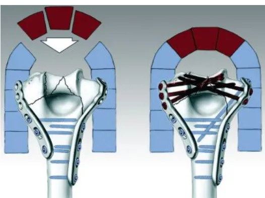
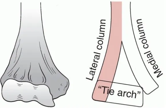
As a result of ongoing search for a more secure technique, later evolved the
concept of parallel plating (180°), which involves placing one plate along the
medial column of the distal humerus and the other plate along the lateral column,
with the screws in the distal fragment interdigitating with each other restoring the
‘tie-beam arch’ of the distal humerus. Several biomechanical studies have proven the superiority of parallel over traditional plating methods, yet there are only fewer
clinical studies to analyse the functional outcome of parallel plating in distal
4
AIM AND OBJECTIVE
To analyse the functional outcome of patients treated with parallel plate
technique in distal humeral fractures in Institute of Orthopaedics and
Traumatology, Rajiv Gandhi Government General Hospital, Madras Medical
5
HISTORY AND REVIEW OF LITERATURE
Distal humeral fractures represents a constellation of complex articular
fracture, resulting from severe trauma to elbow, which are difficult to treat. The
complex three dimensional structure of distal humerus poses a challenging task for
reconstruction if fractured. The diversity of views on the subject is an indication of
poor quality of results.
Among patients, who sustain a fracture in the distal humerus, there is a
bimodal distribution, with respect to age and gender, with peaks of incidence in
males aged 12 to 19 years and females aged 80 years and over. The proportion of
elderly patients who sustain these injuries is increasing, and this trend will
continue. With this change in population, come fresh challenges for reconstruction,
including poor bone quality, fracture comminution, and reduced capacity for
rehabilitation.
Injury to distal humerus occurs from a spectrum of low velocity to high
velocity injuries. Low velocity injuries, are simple domestic falls in middle-aged
and elderly females, in which the elbow is either struck directly or axially loaded,
6
injuries, are more common cause of high velocity injury, in younger males. These
patients, often have open fractures and other injuries, (17% other orthopaedic
injuries and 5%multisystem injuries)25 . These, young population when injured,
adds to the socio-economical burden of the community.
In 1811, Desault was the first one to come to a conclusion that, these
fractures are the most difficult of all fractures, with treatment options, ranging
from essentially no treatment to replacement of joint. In early 20th century, many
authors like Hitzrot (1932), Eastwood (1937), Evans (1953) Watson jones (1956),
Deplama (1959) and Brown & Morgan(1971) were in favour of conservative
approach. But, as the results of conservative approach were, incongruous joint,
non-union, malunion, and stiff elbow, most condemned conservative management
in all type of fractures, and advised surgical management. The goals of treatment
are a stable, painless and functionally useful elbow, and this can be achieved by
proper anatomical restoration of articulating surface by open reduction, and stable
internal fixation followed by early rehabilitation.
It was Van Gordner (1940) and Cassebaum (1952), who first approached
these fractures, by posterior means. They emphasized the advantages of posterior
7
fragments. 2. It allows more freedom in the use of implants. 3. It involves
dissection of soft parts that contain no major neurovascular structures, the ulnar
nerve have been identified and retracted previously. 4. It is the only approach that
can give clear view of the joint surface. 5. With this, not only the posterior surface,
but also the borders of distal humerus can be utilized for fixation purposes 6. Less
number of cutaneous nerves, when compared to medial and lateral approaches48.
The trans-olecranon osteotomy approach, which is considered to be the gold
standard, for management of distal humeral fractures was, first employed by
Cassebaum in 1952 and achieved good results. Other approaches which are proved
useful, include the paratricipital (Alonso-Llames) 27, 28, triceps-reflecting
(Bryan-Morrey)29, triceps-reflecting anconeus pedicle (TRAP)30,triceps- splitting31.32 .
On the basis of the available evidence, a Grade-C recommendation can be
made for the use of the paratricipital approach for extra-articular or simple
intra-articular fractures. There is fair evidence to suggest that, the use of a triceps-
splitting approach leads to functional outcomes, equivalent to those provided by an
olecranon osteotomy, while potentially avoiding the complications associated with
8
Chen G in 2011 came to a conclusion after analysis of 67 patients, that ORIF
via the triceps-sparing approach confers inferior functional outcomes for
intercondylar distal humerus fractures in patients over the age of 60 years, for
whom the olecranon osteotomy approach may be a better choice. However, for
patients less than 60 years of age, especially those less than 40 years of age, either
approach confers satisfactory outcomes34.
In 1953, Mervin Evans treated distal humeral fractures by reduction and
fixation of the articular surfaces, followed by attaching it to the humeral shaft.
Restoration of articular surface is of prime importance, and any residual
displacement between the fixed articular fragments and the shaft, will not have
great deleterious effects on the ultimate function.
Rehabilitation of the injured elbow, following surgery is equally important,
as elbow is prone for stiffness when immobilized for long time35. For early
rehabilitation, the fractures should be fixed with a stable construct. The stable
fixation is achieved by internally fixing the reconstructed articular block, with the
shaft by plating on both pillars12. Without this dual plate arrangement, stability
of fixation can be inadequate, and this has been proven by many
9
and over ridge, on medial side (perpendicular plating) or over ridges on both sides
(parallel plating).
In the last quarters of the century, improved outcomes of surgery for distal
humeral fractures were reported, AO-ASIF group set out their principles of
anatomical articular reduction and rigid internal fixation, through their
perpendicular plating techniques. In 1990, Helfet , Hotchkiss13 did biomechanical
analysis of the perpendicular plating technique and added creditability to this
technique. A number of subsequent clinical studies, revealed nearly 75–85% good
to excellent results with 90–90 plating.
In 2006, Doornberg et al 47 concluded from a long term follow-up study of
19 years, results of open reduction and internal fixation of 19 Type C fractures of
the distal part of the humerus are similar to those reported in the short term. This
10
In 2001, O’Driscoll et al12
defined the principles of fixation of these
fractures using parallel plating technique and defined two goals that should be
met: First, fixation within the distal fragment must be maximized, and second, all
fixation in distal fragments should contribute to stability between the distal
fragments and the shaft. In addition, he defined eight technical principles by
11
12
All these principles can be achieved by using parallel plate orientation 12,
while the principle of locking of screws by interdigitation in the distal fragment is
limited in orthogonal plate orientation. Linking the plates together through the
bone with screws, thereby creating the architectural equivalent of an arch, offers
the greatest biomechanical stability for comminuted distal humeral fractures .
The arch is formed by inter-digitation of locking screws passing through the
distal fragments from both plates in the sagittal plane. Small osteochondral
fragments can be fixed with countersunk screws, headless screws or absorbable
screws. Before the invent of principle based parallel plating, perpendicular plating
[image:25.612.178.441.256.453.2]proposed by AO-ASIF was followed universally.
13
After parallel plating concept was introduced, numerous biomechanical
studies were conducted between parallel and perpendicular plating for validation of
the superior one39, 40. Of these mechanical studies, two studies by Schemitsch et al
(1994) and Self et al (1995) , Arnader (2008) showed parallel plate fixation to be
substantially more stable than 90-90 plate fixation21,39, and two demonstrated no
difference23,40. Zalavras et al37 (2011) concluded that higher degree of stiffness and
higher degree of resistance in torque, cyclical varus loading axial and sagittal
loading to failure was exhibited by parallel plating compared to orthogonal plate
constructs.
Many studies have documented 20% to 25% of unsatisfactory outcomes
after the usual orthogonal plating6,7,8,18,19,20. Henley et al6 reported failure in 5 of 33
patients in his series, 5 of 88 fractures in his series by Letsch et al.8, 3 of 57
reported on by Holdsworth and Mossad19, 9 failures in 72 cases in the series of
Wildburger et al.38, and 16 of 96 reported on by Sodergard et al.35. The cause of
failure being, less number of screws in distal lateral column, leading to loss of
screw purchase, with resultant instability at both columns, causing non union at
supra-condylar level6,13,17,20,21 .There were no failures of fixation in series of O
Driscoll’s parallel plating12,22
. The perpendicular technique requires less soft tissue
dissection, technically easy and the reports of non-union, in this technique are
14
than perpendicular as per cadaveric bone studies, clinical comparison of these two
plates in large groups is not available till date.
The Various plates that are available for fixation are Locking compression
plates(LCP), 3.5 mm reconstruction plates (simple and locking), one third tubular
plates, lambda plates and precontoured distal humeral plates (parallel and
perpendicular). Deshmukh and Deivendran et al43 in 2010 showed less implant
failure with distal humeral locking plates . The pre-contoured geometry allows
easier reduction and saves operating time in fixation of these complex fractures
44
.A study by Corradi A et al 42 in the same year compared the effectiveness
between distal humeral locking compression plates and conventional
reconstruction plates showed no significant differences between the two fixation
methods based on clinical outcome, complications and function of the affected
limb . The principle of each long screw engaging a fragment on the opposite
column fixed by a plate of the ipsilateral column creates a locked arch even
15
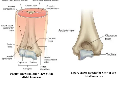
ANATOMY OF DISTAL HUMERUS
The distal humerus is defined as the square of the epicentre between the
epicondyles as described by Mueller.
The distal humerus consists of two condyles, forming the articular surface of
the trochlea and capitellum. Proximal to the trochlea, the prominent medial
epicondyle serves as a source of attachment of the ulnar collateral ligament and the
flexor-pronator group of muscles. Laterally, the lateral epicondyle is located just
above the capitellum and is much less prominent than the medial epicondyle. The
lateral collateral ligament and the supinator-extensor muscle group originate from
the flat, irregular surface of the lateral epicondyle. The posteroinferior aspect
[image:28.612.237.365.120.298.2]serves as a partial origin of the anconeus muscle.
16
Just above the articular surface of the capitellum, the radial fossa
accommodates the radial head during flexion. The coronoid inserts into a large
coronoid fossa superior to the trochlea. Posteriorly, the olecranon fossa serves a
similar purpose, receiving the tip of the olecranon during extension.A thin
membrane of bone separates the olecranon and coronoid fossae in about 90 percent
of individuals, although there is some race and sex variation with this anatomical
feature. The coronoid and olecranon fossae are bordered by the strong lateral
[image:29.612.65.309.99.394.2]supracondylar column and a smaller medial supracondylar column. The difference
[image:29.612.61.537.109.461.2]Figure shows anterior view of the distal humerus
17
in size of these two structures is important because the smaller medial column may
be vulnerable to fracture during insertion of some designs of humeral components
at the time of elbow prosthetic replacement surgery. The posterior aspect of the
lateral supracondylar column is flat, whereas the anterior surface is slightly
curved. This allows ease of application of contoured plates to the posterior aspect
of the lateral column and forms the basis of routine orthogonal plating. The
prominent lateral supracondylar ridge separates the two surfaces into the
so-called safe interval between the brachioradialis and extensor carpi radialis longus
anteriorly and the triceps posteriorly. This serves as an important landmark for
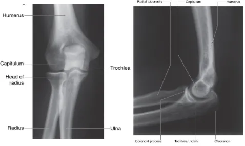
many lateral surgical approaches. The radiologic appearance of the various bony
[image:30.612.61.539.430.715.2]landmarks is shown in the pictures below.
18
Proximal to the medial epicondyle, about 5 to 7 cm along the medial
intermuscular septum, a supracondylar process is observed in 1 to 3 percent of
individuals. A fibrous band termed the ligament of Struthers may originate from
this process and attach to the medial epicondyle. When present, this spur serves as
an anomalous insertion of the coracobrachialis muscle and an origin of the
pronator teres muscle. Various pathologic processes have been associated with the
supracondylar process, including fracture and median and ulnar nerve entrapment.
NERVES IN RELATION TO DISTAL HUMERUS:
Surgical Anatomy of the Ulnar nerve:
In the midportion of the arm the ulnar nerve lies anterior to the medial head
of the triceps and posterior to the medial intermuscular septum. In 70% of
19
arcade is located approximately 8 cm proximal to the medial epicondyle and is
composed of the deep fascia of the arm, superficial fibers of the triceps, and the
internal brachial ligament arising from the coracobrachialis tendon. The nerve then
passes into a fibroosseous groove that is bordered anteriorly by the medial
epicondyle, posterior and laterally by the olecranon and ulnar humeral ligament,
and medially by a fibroaponeurotic band. In this region, numerous branches of the
superior and inferior collateral and posterior ulnar recurrent arteries, as well as
several veins, accompany the nerve. Also at this level, a small articular branch
leaves the ulnar nerve to innervate the joint capsule.
After exiting the fibroosseous groove, the ulnar nerve travels between the
humeral and ulnar heads of the flexor carpi ulnaris covered by a fibrous called
Osborne’s ligament or arcuate ligament. It is often very thick and is a common
cause of ulnar nerve compression. While lying within the muscle of the flexor
carpi ulnaris, the ulnar nerve gives off motor branches to this wrist flexor.
Traveling distally, the nerve pierces the flexor pronator fascia and then lies
between the flexor digitorum superficialis (FDS) and the flexor digitorum
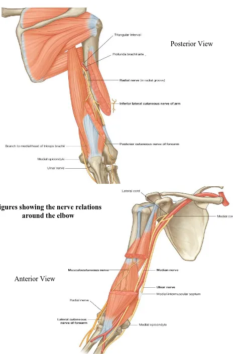
20 Figures showing the nerve relations
around the elbow
Posterior View
21
The radial nerve winds around from the medial to the lateral side of the
humerus in a groove with the profunda brachii artery, between the medial and
lateral heads of the Triceps brachii. It pierces the lateral intermuscular septum
approximately 10 cm proximal to the lateral epicondyle and enters the anterior
compartment. It later passes between the Brachialis and Brachioradialis to the front
of the lateral epicondyle, where it divides into a superficial and a deep branch.
The median nerve descends through the arm, it lies at first lateral to the
brachial artery; about the level of the insertion of the Coracobrachialis it crosses
the artery, usually in front of, but occasionally behind it, and lies on its medial side
at the bend of the elbow, where it is situated behind the lacertus fibrosus (bicipital
fascia), and is separated from the elbow-joint by the Brachialis
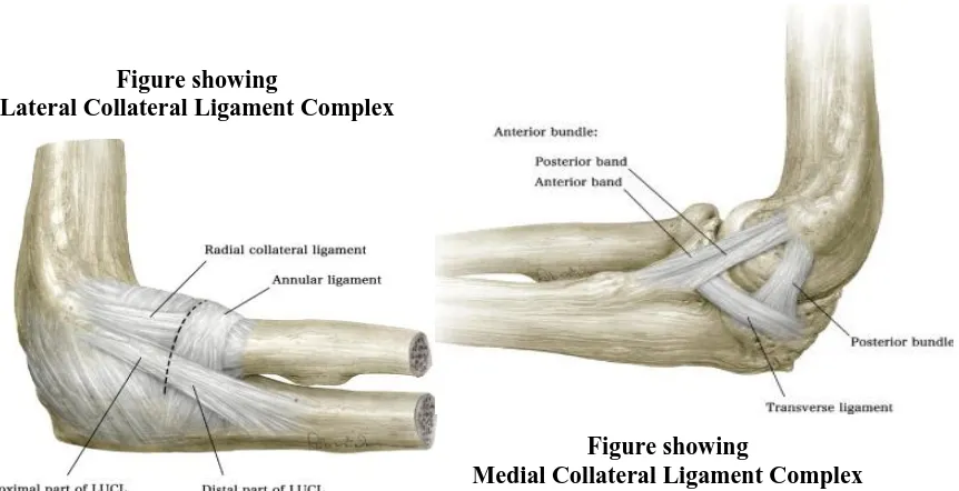
LIGAMENTS AROUND THE ELBOW
The lateral collateral ligament (LCL) complex
It consists of the radial collateral ligament, the lateral ulnar collateral
ligament and the annular ligament. The annular ligament attaches to the anterior
and posterior margins of the lesser sigmoid notch, whereas the radial collateral
ligament originates from an isometric point on the lateral epicondyle and fans out
22
from the isometric point on the lateral epicondyle and attaches to the crista
supinatoris of the proximal ulna. The LCL complex functions as an important
restraint to varus and posterolateral rotatory instability. The LCL complex is
vulnerable to injury during application of a direct lateral plate; therefore, exposure
of the lateral aspect of the distal lateral column should not extend past the equator
of the capitellum.
The medial collateral ligament (MCL) consists of an anterior bundle,
posterior bundle and transverse ligament. The anterior bundle is of prime
importance in elbow stability. It originates from the anteroinferior aspect of the
medial epicondyle, inferior to the axis of rotation, and inserts on the sublime
tubercle of the coronoid. The MCL functions as an important restraint to valgus
and posteromedial rotatory instability.It is susceptible to injury at its origin during
placement of a medial plate that curves around the medial epicondyle to lie on the
[image:35.612.95.526.522.743.2]ulnar aspect of the trochlea.
Figure showing
Medial Collateral Ligament Complex Figure showing
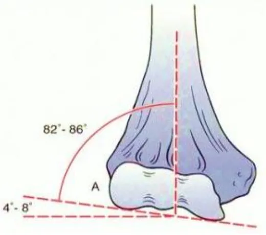
23 SURGICAL ANATOMY
The elbow is anatomically a trocho-ginglymoid joint, meaning that it has trochoid
(rotatory) motion through the radiocapitellar and proximal radioulnar joints and
ginglymoid (hinge-like) motion through the ulnohumeral joint.
The olecranon of the ulna articulates around the trochlea of humerus. The trochlea
normally is tilted in 5 degree of valgus in males and 8 degrees of valgus in females,
thus creating the carrying angle of the elbow53. The line drawn tangential to the
articular surface on the AP view of the distal humerus makes an angle of between
4 and 8 degrees of valgus to the shaft axis. . In the male, the mean carrying angle is
[image:36.612.149.409.437.667.2]11 to14 degrees, and in the female, it is 13 to 16 degrees.
Figure showing
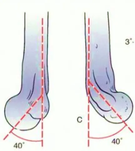
24
The trochlea is externally rotated 3-8 degrees from a line connecting the medial
and lateral epicondyles, resulting in external rotation of the arm when the elbow is
flexed 90 degrees.
The articular segment juts forward from the line of the shaft at 40 degrees and
functions architecturally at the arch at the point of maximum column divergence
distally. It is to noted that the medial epicondyle is on the projected axis of the
[image:37.612.179.411.167.288.2] [image:37.612.192.408.438.679.2]shaft, whereas the lateral epicondyle is projected slightly forward from the axis .
Figure showing Externally rotated trochlea
25
The trochlea must be restored to its normal position, acting as a tie beam between
medial and lateral columns of the distal humerus and thus acts as a keystone of the
arch.This forms the triangle of the distal humerus, which is crucial for stable
elbow motion. Both columns must be securely attached to the trochlea. So every
attempts to restore the proper valgus and external rotation of the trochlea to allow
for stability, full motion and a normal carrying angle.
The medial column diverges from the humeral shaft at approximately 45 degrees,
continues and ends in the medial epicondyle. As nothing articulates with the
anteriomedial epicondyle, the entire surface is available for internal fixation
hardware. Care should be taken to protect and transfer the ulnar nerve anteriorly.
The lateral column diverges from the humeral shaft at approximately 20 degrees
and is largely cortical bone with a broad flat posterior surface, making it ideal for
[image:38.612.159.450.263.453.2]plate placement.
26
The coronoid is important to elbow stability and should be reduced and fixated if
displaced.
The recessed and thinned bone just cephalad to the waist of the trochlea anteriorly
is the coronoid fossa and its counterpart posteriorly is the Olecranon fossa. The
thin wafer of bone that separates the depth of these fossae may be partially
deficient in a small percentage of the population. These fossae are designed for the
receipt of the radial head and the coronoid and olecranon processes with full
flexion and extension respectively (These are important points to bear in mind in
the seating of screws on the distal lateral or medial columns for the address of
distal humeral fractures). Safe screw placement assures no violation of these
fossae. Impingement by a misdirected implant blocks terminal joint motion. If the
medial and lateral columns can be securely fixated to the trochlea, early motion
should be tolerated.54
At the posterior capitellum, cancellous screws must be used to avoid interrupting
the anterior capitellar cartilage.
A second range of motion occurs with the elbow joint in supination and the
forearm in pronation; this ROM is allowed by the radial head articulation with the
27 BIOMECHANICS
The ulnohumeral articulation is the cornerstone of osseous Stability and mobility in
the flexion - Extension plane – especially the coronoid process.
The coronoid process resists posterior subluxation in extension beyond 30o or
greater, depending on the other injuries55. The medial facet of the coronoid is
especially crucial to stability in varus stress. At the extremes of ulno-humeral
motion, the coronoid or olecranon processes may ‘lock’ into their corresponding
fossae, adding additional stability from muscular contraction and with little input
from the ligaments.
However, most activities in most patients rely on a combination of ligamentous
integrity and bony integrity of the articulation.
The anterior band of the medial collateral ligament secures the medial side of the
joint, running from an area just medial and distal to the medial epicondyle and to
the sublime tubercle, slightly distal and medial to the coronoid itself. The
brachialis muscle inserts more distally on the anterior surface of the proximal ulna.
Fracture near the base of the coronoid may compromise these important
28
The radial head also contributes to elbow stability by widening the base of support
of the forearm, tensioning the posterolateral ligament and acting as an anterior
buttress.
Fracture of the coronoid process, radial head, medial epicondyle, os olecranon may
be associated with elbow dislocation, making treatment more complex.
Soft tissue structures about the elbow are responsible for as much as 40% of the
resistance to valgus stress and 50% of that to varus stress in the extended position.
The anterior bundle of the medial collateral ligament may provide one-third to one
half of the elbow’s resistance to valgus stress depending on the amount of elbow
flexion.
A large fracture of the coronoid process, a fracture of the medial epicondyle, and
rupture of the medial collateral ligament may completely disrupt the medial
components of the elbow.
The lateral collateral ligament complex inserts onto the annular ligament. Injury to
this ligament is responsible for posterolateral rotatory instability that may lead to
29
The muscles surround the elbow, besides the biceps / brachialis and triceps,
theoretically stabilize the elbow as well. However, it is difficult to quantify the
importance of the supinator tendon, ECU and the extensor origin.
Except for anecdotal recommendations, repair of these muscles after acute injury
has never been documented to be crucial in preventing redislocation, despite
30
CLASSIFICATION OF DISTAL HUMERUS FRACTURES
ANATOMICAL CLASSIFICATION:
Supracondylar fractures, transcondylar fractures, intercondylar fractures, fractures
of the condyles (lateral and medial), fractures of the articular surfaces(capitellum
and trochlea), and fractures of the epicondyles.
31
32
THE MEHNE AND MATTA CLASSIFICATION 46
It is based on, jupiter’s model of distal humerus, which is composed of
two divergent columns, that support an intercalary articular segment.
1. Intra articular
a. Single column: high medial, high lateral, low medial, low lateral and
divergent single column fracture
b. Bicolumn: high T, low T, Y, H, medial lambda, lateral lambda fractures
c. Articular surface: capitellum, trochlea or both
2. Extra-articular intra capsular fractures
high flexion, low flexion, high extension and low extension, trans column
fractures, high abduction and high adduction fractures.
3. Extra- capsular fractures
33
34
SURGICAL APPROACHES
1. TRICEPS- SPLITTING APPROACH (CAMPBELL) 31.32:
Distal part of the triceps is split through the
aponeurosis
Distally extend the split on to the olecranon
Proximally extend till the radial nerve is
identified
The approach provides only a limited exposure
to the articular surface
2. TRICEPS-REFLECTING APPROACH (BRYAN- MOOREY) 29 :
The entire triceps muscle is elevated
subperiosteally from the posterior distal
humerus
The triceps can be removed with some part of
ulna to facilitate bone to bone attachment
entire triceps is reflected upwards and laterally
35 3. TRAP APPROACH (0’DRISCOLL) 30 :
It combines the reflection of triceps along with the laterally placed anconeus
muscle as a single unit.
The anconeus is first exposed distally and later reflected as a whole proximally
The medial exposure is similar to the triceps reflecting approach
4. PARA- TRICIPITAL APPROACH
(ALONSO- LLAMES) 27, 28:
triceps muscle is elevated subperiosteally from
posterior distal humerus.
36
triceps muscle.
This approach is can be used for type A and type C1 fractures with expertise.
Can produce excellent outcomes as extensor mechanism is not disturbed.
5. OLECRANON- OSTEOTOMY APPROACH 7, 12,49:
This approach can give a excellent exposure of the articular surface
Ideal for type C fractures
‘V’ shaped chevron osteotomy is preferred for good
union and stable fixation12.
It has inherent rotational stability as well as
translational stability when compared to the
37
TREATMENT PROTOCOL
CLINICAL HISTORY AND EXAMINATION:
A detailed history regarding name, age, sex, date of injury, mechanism of
injury, residential address, occupational status and associated injuries were
recorded. Patients general condition, vitals were noted. Patients affected limb were
x rayed in both true antero-posterior and true lateral views in slight traction after
removing slab if applied previously.
LABORATORY WORK UP:
The patients were submitted to a battery of routine investigations required
for pre-anesthetic checkup. Associated medical comorbidities were dealt with if
present. 3D reconstruction CT of elbow joint were taken for evaluating the number
of fragments, degree of comminution and displacement in Intraarticular fractures
41
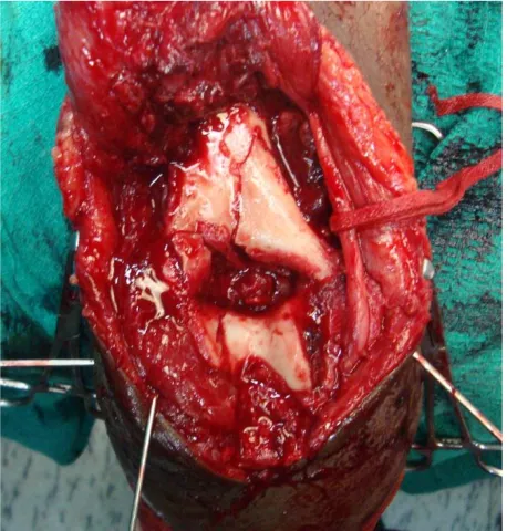
38 SURGICAL TECHNIQUE:
The patient, were given a general anesthesia or regional anesthesia and were
positioned in the lateral position, with the involved limb supported over bolsters in
OT table as depicted in the picture below.
A midline posterior skin incision made48, deep fascia incised and before
proceeding further, the ulnar nerve is identified, dissected out and retracted gently
with an umbilical cotton tape. Triceps muscle identified and released on either side
from the intermuscular septum. Fracture site exposed further with chevron V
shaped olecranon osteotomy 7,12,49 incompletely with saw and completed with an
osteotome in complex articular fractures. as it provides adequate exposure of the
aricular fragments16.In other types we utilized any of the described approaches like
39 TECHNIQUE OF PARALLEL PLATING 12:
We attempted to achieve the eight technical principles derived from the two
major goals of (1) maximizing fixation in the distal fragments and (2) ensuring that
all fixation in the distal segment contributes to stability at the supracondylar level.
Once the fracture is exposed the following steps are carried out.
Step 1: Articular reduction. Articular fragments were aligned in anatomy and
were fixed provisionally with K wires placed subchondrally in a way not
40
Step 2: Plate placement and provisional fixation. Slightly undercontoured
3.5mm plates were placed in medial and lateral ridges in a way that both end at
different levels at the shaft region and atleast 3 screws were placed in shaft. A (first
proximal) screw were placed in one of the proximal hole of each plate but not fully
tightened, leaving some freedom for the plate to move proximally later during
compression. K wires were used in distal fragments for provisionally fixation.
Step 3:Articular fixation. Long medial and lateral distal screws fixing maximum
fragments were applied
Step 4: Supra condylar compression
a. the proximal screw on one side was backed out and a large bone clamp was
[image:53.612.189.418.265.505.2]applied distally on that side and proximally on the opposite cortex to eccentrically
41
load the supracondylar region. A second proximal screw was inserted through the
plate in compression mode, and then the backed out screw is retightened.
b. This step repeated for other column also.
c.Diaphyseal screws was to be applied to achieve residual compression through
undercontoured plates.
Step 5: Final fixation:
Provisional K wires in the distal fragment were removed and replaced with screws
After fixing the fracture segments, TBW of osteotomized olecranon was
carried out either with two K wires or a 6.5mm Cancellous screw. Meticulous
[image:54.612.166.411.318.551.2]repair of soft tissues was done in layers with a suction drain.
42
43 POST OP PROTOCOL:
Patients were placed in a well-padded plaster extension splint applied anteriorly
and the limb elevated for first 3 days.
Active finger movements started from day 1.
Intravenous antibiotics were given for 3 days; Oral antibiotics were given for 3
days.
Drain removal at 48 hours ; Suture removal done on 12th day
Indomethacin prophylaxis for heterotopic ossification was given for the first
postoperative month (75 mg/day)
Elbow range of motion was started between days 3 and 7 postoperatively, as
tolerated by the patient.
Generally, active-assisted and active range of motion were encouraged (flexion,
pronation, and supination) of elbow.
Passive supported(gravity assisted) extension was reserved for patients that
underwent an extensor mechanism disrupting approach.
at 6 months patients were allowed to their routine full activities
Follow up at 2nd, 4th , 6th, 8th week . At each follow up, patients were evaluated
clinically and radiologically for union, and the outcomes were measured in terms
44
MAYO ELBOW PERFORMANCE SCORE (MEPS)
OUTCOME RATING BASED ON MEPS:
greater than 90 excellent
Score 75 to 89 good
Score 60 to 74 fair
45
MATERIALS AND METHODS
STUDY DESIGN:
A prospective study was done to evaluate the functional outcome of parallel
plating technique in treatment of distal humeral fractures and to analyse the results.
STUDY GROUP:
The study group consists of 24 Patients with distal humeral fractures, who
underwent osteosynthesis with parallel plating technique between May 2010 and
Oct 2012 at the institute of orthopaedics and traumatology, Madras medical college
and Rajiv Gandhi Government General Hospital, Chennai. The study was done
with clearance from Hospital ethical committee. Those who fulfilled the inclusion
criteria given below were invited to participate in the study. Informed consent was
obtained from all the patients willing to take part in the study.
a. INCLUSION CRITERIA:
1. Intra articular fractures of distal humerus
2. Age >18 years
3. AO Types A2,A3 and C (supracondylar and bicondylar)
4. Closed ,Grade I and grade II compound injuries
46
7
6
4 4
3 0 1 2 3 4 5 6 7 8
20-30 yrs 31-40 yrs 41-50 yrs 51-60 yrs >60 yrs
Age incidence
b. EXCLUSION CRITERIA:
1. With vascular injuries
2. Grade III compound Open fractures
3. severe unreconstructable intra-articular communited fractures in elderly
4. uncooperative patients for the rehabilitation and follow up
5. Patients who were not medically fit for surgery
6. not willing to participate
On admission, careful history was elicited from the patients or attendants to
reveal the mechanism of injury and associated injuries.A detailed clinical
examination and radiological assessment was done to assess the fracture pattern,
deformity, neurovascular status associated injuries and for vital signs. Then the
injured limb was immobilized in a above elbow plaster slab until surgery.
47
Age No of Patients Percentage
20 to 30 Years 7 29.2%
31 to 40 Years 6 25%
41 to 50 Years 4 16.7%
51to 60 years 4 16.7%
>60 years 3 12.5%
The Mean age of the patients was 39 years ranging from 20 to 65 years.
SEX DISTRIBUTION:
Males dominated our study .Male: Female ratio was 2:1 (16:8)
16 8
Sex Distribution
Males
48 MODE OF INJURY:
Majority of the patients suffered Motor vehicle Accidents(MVA) . The
second most common mode of injury was simple accidental falls.Other mode of
injuries were fall from heights(FFH) , assault. 71% of the fractures were closed
injuries while the Open fractures (Grade I and II Gustilo Anderson types)
constituted remaining 29% of the cases.
Mode of injury No. of Patients Percentage
Closed Open Total
MVA 10 5 15 62.5%
Simple Fall 5 - 5 20.8%
FFH 1 2 3 12.5%
Assault 1 - 1 4.2%
0 2 4 6 8 10 12 14 16
MVA
Fall
FFH
Assault
10
5
1 1
5
2
Open
49 GENDER Vs MODE OF INJURY:
Males constituted two-thirds of our study.Young males predominantly sustained
injury by Road traffic Accidents(RTA) whereas older females predominantly
sustained accidental fall . Male:Female= 2:1
Mode of injury Male Female
MVA 12 3
Simple Fall - 5
FFH 3 -
Assault 1 -
TOTAL 16 8
50
14
10
Side of Injury
Right
Left
SIDE OF INJURY:
14 patients(58.3%) had fracture of right distal humerus and 10 (41.7%) patients
had fracture of left side.
FRACTURE DISTRIBUTION:
Intraarticular fractures constituted majority of our cases accounting for about
75%.Extraarticular Metaphyseal fractures constituted the remaining 25%.
Fracture type (AO-OTA)
No. of
Patients Percentage
C3 3 12.5% Complete Articular
75%
C2 8 33.3%
C1 7 29.2%
A3 4 16.7% Extraarticular
25%
51 ASSOCIATED INJURIES
In our study we noted the following associated injuries.
Associated injuries No. of Patients
Fracture of Distal radius 2
Fracture shaft of contralateral humerus 1
Fracture of pubic rami 2
Intertrochanteric Fracture of Femur 1
Fracture shaft of Femur 1
Fracture Metatarsals 1
Radial Nerve palsy 1
Ulnar Nerve palsy 1
Head injury 1
Type A2 - 2
TypeA3-4
Type C1-7 Type C2-8
Type C3 - 3
Type A2 - 2
Type A3 - 4
Type C1 - 7
Type C2 - 8
52 SURGICAL APPROACHES :
We used chevron osteotomy of the olecranon for fracture fixation in 14 of
our cases(58.33%) as intraarticular fractures dominated our study.Other
approaches used were paratricipital approach in 3 cases(12.5%),triceps splitting
approach in 4 cases(16.66%) and TRAP approach in 3 cases(12.5%).
Procedure No. of Patients
Olecranon osteotomy 14
Paratricipital 3
Triceps splitting 4
TRAP 3
0 2 4 6 8 10 12 14
Olecranon osteotomy Paratricipital Triceps splitting TRAP
14 3
4 3
53
OBSERVATION AND RESULTS
The following observations were made in our study.
1) The Mean age of the patients was 39 years ranging from 20 to 65 years . Nearly
30% patients belong to 3rd decade followed by 4th decade (25%). 71% of the
patients belong to less than 50 years.
2) Males dominated our study group with a ratio of 2: 1
3) Right limb injuries were more common.
4) Motor vehicle accidents and accidental simple falls were the common mechanisms
of injury.
5) Motor Vehicle accidents was a major form of injury in younger males whereas
simple falls from standing height had been the most common mode of violence in
elderly females.
6) Completely articular fractures constituted 75% while extraarticular transcondylar
fractures constituted only 25% of our cases.
7) Of the complete articular types, the order of most common types were
54
8) Seven patients had associated skeletal injuries. One patient had radial nerve
injury,one patient ulnar nerve injury, and one patient had a head injury.
9) Most of the patients were operated by Chevron osteotomy approach (14 Patients).
Four patients were operated by Triceps splitting approach. TRAP approach and
paratricipital approach were used each in three patients.
10) In our study, the average surgical time delay was 6 days ranging from 5 to 17 days.
11) The average surgical time was 110 minutes ranging from 60 minutes to 3 hours.
12) Complications encountered in our study were paraesthesia along ulnar nerve
distribution, ulnar nerve and radial nerve neuropraxia, infection, stiffness, delayed
union, heterotopic ossification, non-union at osteotomy site and hard ware
prominence.
13) Three patients had infection. One patient was treated conservatively with
antibiotics. One patient who had a wound gapping on the 5 day over the olecranon
healed by secondary intention and Split skin grafting. One patient who had a initial
open injury developed a deep seated infected which warranted wound debridement
and finally closed with a abdominal flap cover. He had a delayed union, stiffness
55
14) 3 patients reported numbness and paraesthesia along ulnar border of little finger
which was treated conservatively. One patient developed ulnar and radial nerve
neuropraxia. Radial neuropraxia showed recovery but anterior transposition was
done after 12 weeks for the ulnar nerve which recovered partially after 8 months.
15) Stiffness was noted in 5 patients. Heterotopic ossification with reduced elbow
ROM was observed in 2 patients.One patient who developed deep seated infection
required flap cover, also developed stiffness. Another patient stiffness occurred
due to pain , post fixation and other had a soft tissue contracture.
0 1 2 3 4 5 3 2 5
1 1
4 4
Complications
Infection
Myositis
stiffness due to any cause
Non union at osteotomy
Hardware prominence
pain
56
16) One patient who had a nonunion at the osteotomy site was done a revision
osteosynthesis with tension band wiring.
17) No patient died during treatment or follow up.
18) Twenty four patients of distal humeral fractures were treated surgically with
parallel plating and analyzed with average follow up of 13months (6 months – 2 ½
years).
19) In our study, solid radiologic union was achieved primarily in all patients. The
average time to union was about 14 weeks. Hardware failure or Nonunion did not
occur in any patient.
20) The mean flexion-extension arc was 107°. The mean MEPS score was 82 in our
study. The results were excellent for 8 elbows, good for 11, fair for 3, and poor for
57
DISCUSSION
Functional elbow is very essential for an individual for social and economic
thriving. Fractures of the distal humerus may directly affect the functional
movement of elbow especially intercondylar (intra-articular) fracture. The
relationship of the radiohumeral joint and ulnohumeral joints must be perfect for a
good functional outcome.
The majority of distal humerus fractures presenting to our centre were
resulting from road traffic accidents (62.5%) compared to study by Sanchez-Sotelo
et al 50 where the major mechanism of injury was accidental fall from standing height(56%). This is probably reflective of the fact that several trauma cases are
being referred to our centre which is the tertiary referral centre for trauma care of
this region.
The high male:female ratio seen in our centre (2:1) as compared to 1:1
recorded by Sanchez-Sotelo et al 50 is the resultant of the high number of trauma
cases treated in our centre and the fact that males are more prone for road traffic
accidents compared to females because in our society females travel less.
Fracture configuration according to the OTA type had a significant bearing
58
outcome than group A patients. This has again stressed the importance and
prognostic significance of the OTA classification. Study by Sanchez-Sotelo et al 50
revealed that the commonest fracture type was OTA class A and C which our
study concurs It is also important to stress on the fact that incidence of type C
fractures is more than the type A fractures suggesting that the incidence of high
velocity injuries is on the rise.
The restoration of elbow function is dependent on three salient features:
exposure, fixation and the post-operative rehabilitation, with later two are of
primary consideration. Adequate exposure is necessary for visualization fixation of
the fracture fragments. The optimal exposure is provided by the posterior approach
with osteotomy of the olecranon.
Olecranon osteotomy was done in 14 of our cases. Eight of them were fixed
with modified TBW with K wires and three of our cases were fixed with
cancellous screws with TBW. This allowed us complete examination of the
articular surfaces of trochlea, capitellum, olecranon and radial head. It also gives
access to the medial and lateral supracondylar ridges. Full evaluation of the
fragments of the fracture and reduction can then be performed. Although
non-union of the osteotomy may be regarded as a potential complication of this
59
for immediate use of the elbow through a secure range of motion. Only one case in
our 14 osteotomized elbows showed a non-union which united with revision
osteosynthesis with modified TBW.
24 cases in our study were operated with parallel plating which provided
absolute stability for early mobilisation. The lateral plate placement directly on the
lateral column allows for lengthy screw placement which is limited in traditional
orthogonal plating due the fear anterior capitellar breach in the same. Since we use
the 3.5mm reconstruction plates, it allows for easy contourability for both column
fixation. The previous concept of using the more malleable 1/3 tubular plate for
the medial column which requires heavy contouring is now in question and several
authors recommend at least a stronger 3.5mm plates or Precontoured plates for
both columns to achieve a more stable rigid construct to allow for early
mobilization. . In our study we have not met any implant failures or non-union at
the fracture site which is on par with the fact that parallel plating offers a
inherently stable construct in a given clinical situation and in concurrence with
studies done on parallel plating by Sanchez-Sotelo et al 50 and Atalar et al 51.
Complications :
Elbow stiffness (secondary to infection(1) ,heterotopic ossification(2) and
60
complication encountered in 5 patients. We had infection requiring further
operative intervention in 2 patients. Both of them required surgical debridement
once and repeated dressings before the wound was closed by skin grafting and flap
cover one in each case. Another patient who developed a superficial infection was
treated conservatively with antibiotics .Hardware prominence was a major
complaint in one patient. All but four elbows were completely painless at the final
follow up. All fractures united within the study period .There were no cases of
non-union at the fracture site except for a non-union at the osteotomy site due to
distraction which united with revision osteosynthesis with modified tension band
wiring.
Sanchez-Sotelo et al 50 describes complication rates of 43% which included wound-healing complications (6%), deep infection (3%), nonunion (3%),
heterotopic ossification (16%),Osteonecrosis 1 (3%),Posttraumatic arthritis 2 (6%)
Permanent ulnar neuropathy(6%). Gofton et al reported a complication rate of
48%, which included heterotopic ossification(17%), olecranon nonunion(9%), and
infection (9%). Atalar et al 51 showed a complication rate of 48% in their study
group of 21 patients.The other previously referenced studies reported complication
rates of 11% to 29% 21,24. In the recently published retrospective series of Athwal et
al.52 assessing the Mayo Elbow parallel plate technique, they noted a complication
61
common complication noted was postoperative nerve injuries ( 16%), wound
complications(12%) including two wound dehiscences requiring surgical
debridement. One olecranon nonunion was noted which was treated
non-operatively. Our study showed a similar complication rate of 41 % which is
concurrent with the international literature which included infection (12.5%),
heterotopic ossification (8.3%) ,Nonunion at osteotomy (4%),permanent ulnar
neuropathy(4%),stiffness with pain excluding myositis and infection(8.3%),hard
ware prominence(4%).
0 10 20 30 40 50 60
Sanchez-Sotelo et al Gofton et al &Atalar et al
Athwal et al. Our study
43% 48%
53% 41%
62
Elbow stiffness due to all causes was the commonest complication(21%) in
our study group .Poor compliance to physiotherapy and mobilisation was a major
determinant in elbow stiffness. Though the construct might favour early
mobilisation ,the motivation and compliance for physiotherapy plays a major role
in instituting earlier range of motion exercises after surgery and to get a better
functional outcome.
Iatrogenic nerve complications were noted in 4 patients(16.6%) in our study.
Post-operative ulnar nerve paraesthesia was observed in 3 patients. These
paraesthesia were transient and all of them recovered without any particular
treatment within 2 months post op. Medial plates or ulnar nerve handling may be a
reason for this. One patient had both sensorimotor involvement of the ulnar nerve
and radial nerve with neuropraxia postoperatively. Initially the patient was treated
conservatively when he showed recovery of only the radial nerve symptoms.
Anterior transposition of the ulnar nerve was done at the end of three months. He
showed a partial recovery of the ulnar nerve function at the last follow up. Ulnar
neuropathy can occur during the initial injury or iatrogenically during surgical
fixation. The rate of ulnar neuropathy following ORIF of distal humerus fractures
has been reported as being between zero and 12% in the previously described
63
failed elbow reconstruction; they found mostly good to excellent recovery from
ulnar neuropathy when they performed neurolysis and transposition of the nerve.
The mean age of our study group was 39 years as compared to 58 years in
the study by Sanchez-Sotelo et al 50 and 47 years in study by Atalar et al 51.This
shows a rising incidence of these injuries among younger population due to the
higher incidence of road traffic accidents in developing countries like India.
Younger patients, often males had these high velocity injuries like motor vehicle
accidents and fall from height in working place associated with soft tissue injury.
The incidence of open fractures in our study group was 29%.One of our patients
who at the presentation had a grade II compound injury was treated with initial
wound debridement and parallel plating with primary skin grafting .But he
developed wound infection which after settled after serial debridement and
ultimately ended in a flap cover. He had a delayed union and post traumatic
stiffness and had a poor outcome.
Bony union took an average of 13.4 weeks in our study which is comparable
to 12 weeks obtained by Sanchez-Sotelo et al50. All patients had bony union at end
of the study period, except for the one patient with deep infection had a delayed