Thesis Submitted to
The Tamilnadu Dr. M.G.R Medical University, Chennai In partial fulfillment of the requirements
for the award of the Degree of MASTER OF PHARMACY
IN
PHARMACOLOGY Submitted by
Ms. ANSA P.U, B.Pharm. Reg. No: 26116393 Under the guidance of
Institutional Guide Industrial Guide
Mrs. C. Maheswari, M.Pharm, (Ph.D)., Dr. Achuthan C.Raghavamenon Lecturer, Department of Pharmacology, Assistant Professor,
RVS College of Pharmaceutical Sciences, Amala Cancer Research Centre,
Sulur, Coimbatore. Amala Nagar, Thrissur.
DEPARTMENT OF PHARMACOLOGY,
RVS COLLEGE OF PHARMACEUTICAL SCIENCES, SULUR, COIMBATORE - 641402.
COMPONENTS FROM UVARIA NARUM (DUNAL) WALL SEED"
DEGREE DISSERTATION WORK SUBMITTED TO THE TAMILNADU Dr.M.G.R MEDICAL UNIVERSITY, CHENNAI IN PARTIAL FULFILLMENT
FOR THE AWARD OF
MASTER OF PHARMACY
(PHARMACOLOGY)
Submitted by
Ms. ANSA P.U Reg. No: 26116393
UNDER THE GUIDANCE OF
Institutional Guide Industrial Guide
Mrs. C. Maheswari, M.Pharm, (Ph.D)., Dr. Achuthan C.Raghavamenon Lecturer, Department of Pharmacology, Assistant Professor,
RVS College of Pharmaceutical Sciences, Amala Cancer Research Centre,
(Affiliated To The Tamilnadu Dr.MGR Medical University, Chennai) 242-B, Trichy road, Sulur, Coimbatore-641402.
Phone :( 0422), 2687241, 2687480, 2687603 Ext 302 Fax: +91-422-2687604
CERTIFICATE
This is to certify that this project work entitled “PARTIAL PURIFICATION OF
ANTI-TUMOR AND ANTIOXIDANT COMPONENTS FROM UVARIA NARUM (DUNAL) WALL SEED" submitted in partial fulfilment of the requirements for the award of Degree of Master of Pharmacy in Pharmacology to The Tamilnadu Dr.
M.G.R Medical University, Chennai is a bonafied work carried out by Ms. ANSA P.U (Reg. No: 26116393) at Amala Cancer Research Centre, Thrissur, Kerala, and completed the work at Department of Pharmacology, R.V.S College of
Pharmaceutical Sciences, Sulur, Coimbatore-641402 under the supervision and guidance of Mrs. C. Maheswari, M.Pharm, (Ph.D).,(Institutional Guide) and Dr.
Achuthan C. Raghavamenon (Industrial Guide) during the academic year 2012-2013.
Dr. R. Venkatanarayanan, M.Pharm., Ph.D Principal,
Place: Coimbatore RVS College of Pharmaceutical Sciences,
Date: Sulur, Coimbatore-641402.
(Affiliated To The Tamilnadu Dr.MGR Medical University, Chennai) 242-B, Trichy road, Sulur, Coimbatore-641402.
Phone :( 0422), 2687241, 2687480, 2687603 Ext 302 Fax: +91-422-2687604
CERTIFICATE
This is to certify that this project work entitled “PARTIAL PURIFICATION OF
ANTI-TUMOR AND ANTIOXIDANT COMPONENTS FROM UVARIA NARUM (DUNAL) WALL SEED" submitted in partial fulfilment of the requirements for the award of Degree of Master of Pharmacy in Pharmacology to The Tamilnadu Dr.
M.G.R Medical University, Chennai is a bonafied work of Ms. ANSA P.U (Reg. No: 26116393) carried out at Amala Cancer Research Centre, Thrissur, Kerala, and completed the work at Department of Pharmacology, R.V.S College of
Pharmaceutical Sciences, Sulur, Coimbatore-641402 under the supervision and guidance of Mrs. C. Maheswari, M.Pharm, (Ph.D)., (Institutional Guide) and Dr.
Achuthan C. Raghavamenon (Industrial Guide) during the academic year 2012-2013 to my fullest satisfaction. This work is original and contributory. .
Dr. D. Benito Johnson, M.Pharm., Ph.D Professor and Head,
Department of Pharmacology,
Place: Coimbatore RVS College of Pharmaceutical Sciences,
(Affiliated To The Tamilnadu Dr.MGR Medical University, Chennai) 242-B, Trichy road, Sulur, Coimbatore-641402.
Phone :( 0422), 2687241, 2687480, 2687603 Ext 302 Fax: +91-422-2687604
CERTIFICATE
This is to certify that this project work entitled “PARTIAL PURIFICATION OF
ANTI-TUMOR AND ANTIOXIDANT COMPONENTS FROM UVARIA NARUM (DUNAL) WALL SEED" submitted in partial fulfilment of the requirements for the award of Degree of Master of Pharmacy in Pharmacology to The Tamilnadu Dr.
M.G.R Medical University, Chennai is a bonafied work of Ms. ANSA P.U (Reg. No: 26116393) carried out at Amala Cancer Research Centre, Thrissur, Kerala, and completed the work at Department of Pharmacology, R.V.S College of
Pharmaceutical Sciences, Sulur, Coimbatore-641402 under my guidance and supervision and Dr. Achuthan C. Raghavamenon (Industrial Guide) during the
academic year 2012-2013 to my fullest satisfaction. This work is original and
contributory. .
Mrs. C. Maheswari, M.Pharm., (Ph.D) Lecturer, Department of Pharmacology,
Place: Coimbatore RVS College of Pharmaceutical Sciences,
Date: Sulur, Coimbatore- 641402.
DECLARATION
I ANSA P.U, hereby declare that the dissertation work entitled "Partial
purification of anti-tumor and antioxidant components from Uvaria narum (Dunal) Wall
seed" submitted by me, in partial fulfillment of the requirements for the degree of MASTER OF
PHARMACY in PHARMACOLOGY to The Tamilnadu Dr.M.G.R Medical University,
Chennai is the result of my original and independent research work carried out under the
guidance and supervision of Dr. ACHUTHAN C.RAGHAVAMENON, Assistant Professor of
Amala Cancer Research Centre, Thrissur and under the internal supervision of Mrs. C.
MAHESWARI, M.Pharm, (Ph.D)., during the academic year 2012-2013. This has not formed
the basis for the award of any Degree/ Diploma/ Fellowship or similar title to any candidate of
any university.
ANSA P.U
Reg.No.26116393
Dept of Pharmacology,
Place: Coimbatore RVS College of Pharmaceutical Sciences,
This is to certify that the dissertation work entitled "PARTIAL PURIFICATION OF ANTI-TUMOR AND ANTIOXIDANT COMPONENTS FROM UVARIA NARUM (DUNAL) WALL SEED" submitted by Ms. ANSA P.U (Reg.No:26116393) to THE TAMILNADU DR.M.G.R. MEDICAL UNIVERSITY, CHENNAI in partial fulfillment for the Degree of MASTER OF PHARMACY in PHARMACOLOGY is a bonafied work carried out during the academic year 2012-2013 by the candidate at the Department of Pharmacology, R.V.S College of
Pharmaceutical Sciences, Sulur, Coimbatore and was evaluated by us.
Examination centre:
Date:
Internal Examiner External Examiner
Apart from my effort, the success of the dissertation mainly depends on the encouragement and
guidance of many others, I take this opportunity to express my thanks and gratitude to the people
who have been instrumental behind the successful completion of this task.
First and foremost, I wish to express my thanks to GOD ALMIGHTY for his love and unseen
guidance throughout my work.
I would like to express my gratitude to Dr. R. Venkatanarayanan, Principal, R.V.S College of
Pharmaceutical Sciences, providing me all the facilities for the postgraduate studies and
dissertation work.
I express my heart-felt gratitude to my institutional guide Dr. D. Benito Johnson, Professor and
Head, Department of Pharmacology, R.V.S College of Pharmaceutical Sciences for providing
necessary facilities, encouragement, constructive suggestions and constant inspiration
throughout the entire course of study.
I extend deep sense of gratitude to my beloved teacher, Mrs. C. Maheswari, Lecturer,
Department of Pharmacology for her support and encouragements.
I express thanks from my heart to my Industrial Guide, Dr. Achuthan C. Raghavamenon, Amala
Cancer Research Centre, Thrissur for his valuable guidance and critical suggestions throughout
the work and preparation of the project.
I sincerely thank Dr. Ramadasan Kuttan, Research Director, Amala Cancer Research Centre
for allowing me to do my project on this institution and providing necessary facilities during the
course of the work.
I take this opportunity to express my gratitude to, Mr. R. Suresh (Ph.D), Professor in
department of pharmacology, for his valuable guidance and support for my work on this topic.
I specially thank Mrs. Smitha Rajesh, Research fellow, Amala Cancer Research Centre for her
Amala Cancer Research Centre, Thrissur for their help and support during my project.
I would like to express thanks to my ever loving friends, Johncy, Jithu, Anu Alex, Prasobh
(R.V.S College of Pharmaceutical Sciences) and Annu Sony, Suraja, Sandhya, Rahul, Rohini,
Noby (Project students, Amala Cancer Research Centre) for their support as this project would
not be completed without their word of encouragements.
Above all, I would like to express my profund gratitude to my beloved PARENTS, HUSBAND
(Mr.Amarraj Vazhoor) and SISTER who rendered me priceless support during the whole
tenure of life to achieve more.
INDEX
SL.NO: CONTENTS PAGE NO:
1 INTRODUCTION 1
2 LITERATURE REVIEW 24
3 OBJECTIVES 35
4 PLAN OF THE WORK 36
5 PLANT PROFILE 37
6 MATERIALS AND METHODS 41
7 RESULTS 58
8 DISCUSSION 85
9 CONCLUSION 89
10 BIBLIOGRAPHY 90
11 ANNEXURE 99
SL.NO: TABLES PAGE NO:
1 Clinically important Tumor markers 13
2 Biologically significant free radicals 17
3 Plant-derived anticancer agents 36
4
Yield and characteristics of Uvaria narum seed oil extract. 58
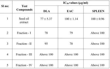
5 IC50 values of U. narum seed oil extract and its separated fractions 59
6 Phytochemical analysis of U. narum seed oil extract 64
7 Rfvalues of fractions obtained in TLC analysis of U. narum
seed oil extract
65
8 Phytochemical analysis of the fractionated seed oil on TLC by various spray
reagents
67
9 Characteristics of the four fractions obtained from U. narum seed oil extract by
TLC separation
68
10 Characteristics of the various compounds obtained by HPTLC of
U. narum seed oil extract
71
13 IC50 values for U. narum seed oil extract, fraction IV and standard,
vitamin C in various in vitro anti oxidant assay systems
74
14 Ferric reducing power of U. narum seed oil extract and its fraction- IV 77
15 Effect of fraction-I of U. narum seed oil extract on tumor volume of mice 80
16 Effect of fraction-I of U. narum seed oil extract on body weight of tumor
bearing mice
81
17 Effect of fraction-I of U.narum seed oil extract on total WBC count of tumor
bearing mice
82
18 Effect of Fraction-I of U.narum seed oil extract on haemoglobin count of tumor
bearing mice
83
19 Inhibition of DLA induced solid tumor in mice by fraction-I of U. narum
seed oil extract
SL.NO: FIGURES PAGE NO:
1 Stages of development of cancer 7
2 Complications produced by free radicals in the body 15
3 Formation of free radicals and its neutralisation by antioxidants 16
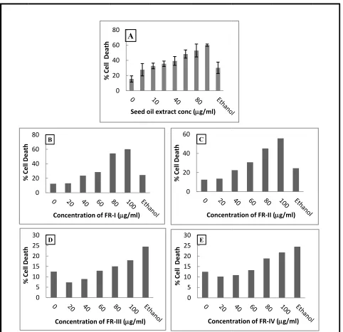
4 In vitro cytotoxicity of U.narum seed oil extract and its separated fractions on
DLA cell lines
60
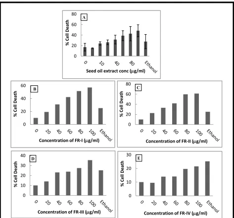
5 In vitro cytotoxicity of U.narum seed oil extract and its separated fractions on
EAC cell lines
61
6 In vitro cytotoxicity of U.narum seed oil extract and its separated fractions on
normal spleen cells
62
7 TLC analysis of the U.narum seed oil extract under white light, UV 254 nm
and Iodine vapour
65
8
Phytochemical detection of U.narum seed oil extract on TLC plates spraying
with (A)-Dragondorff's reagent,(B)-AS reagent,(C)-Ferric ferrocyanide
reagent, (D)- 20% aq.sulphuric acid reagent and (E)-Methanol-potassium
hydroxide reagent
66
9 HPTLC analysis of the U.narum seed oil extract, fraction -I and IV under
(A)254 nm , (B) 366 nm and(C) white remission
69
10 3D image of HPTLC chromatogram of U.narum seed oil extract, fraction-I
and IV
HPTLC analysis
12 Integrated graph of showing peaks of fraction-I in HPTLC analysis 71
13 Integrated graph of showing peaks of fraction-IV in HPTLC analysis 72
14 Effects of U.narum seed oil extract, fraction-IV and the vitamin C on
superoxide radical scavenging activity
75
15 Effects of U.narum seed oil extract, fraction-IV and vitamin C on
AAPH-induced lysis of human erythrocyte
75
16 DPPH radical reducing activity of U.narum seed oil extract, fraction-IV and
vitamin C.
76
17 Hydroxyl radical scavenging activity of U.narum seed oil extract, its
fraction-IV and the standard, vitamin C
76
18 Ferric-reducing antioxidant power of ascorbic acid 77
19 Inhibition of DLA induced solid tumor by fraction-I of U. narum seed oil
extract in Swiss albino mice.
% : Percentage sign
μl : micro litre
0
C : Degree celsius
ANOVA : Analysis of variation
cm3 : Cubic centimetre
Fig : Figure
Tab : Table
gm : Gram
U. narum : Uvaria narum
hr : Hour
i.p : Intraperitonially
m mole : millimoles
mg/dl : milligrams per decilitre
mg/kg b.wt : milligram per kilogram body weight
mg/kg : 1 milligram per kilogram
mg/m2 : 1 milligram per square meter
min : minute
sec : seconds
ml : Milli liter
mm : Millimeter
[image:15.612.177.427.126.707.2]nm : nanometer
OD : Optical density
T.vol : Tumor volume
Hb : Hemoglobin
ROS : Reactive oxygen species
SD : Standard deviation
WBC : White blood cells
SOD : Superoxide dismutase
DPPH : 2, 2- Diphenyl-1-picryl hydrazyl
AAPH : 2,2'-azobis-2-amidino propane hydrochloride
FRAP : Ferric reducing antioxidant power activity
INTRODUCTION
Cancer is a group of diseases which is characterized by uncontrolled growth and spread of abnormal cells. If the spread of abnormal cells is not controlled, it can result in death. Cancer is supposed to cause by both external factors (tobacco, chemicals, radiation, and infectious organisms) and internal factors (inherited mutations, immune conditions, hormones, and other mutations that occur from metabolism). These causal factors may act either together or in sequence to initiate or promote carcinogenesis. Most cancers requires multiple steps for their development which occur over many years.
According to the estimates from the International Agency for Research on Cancer (IARC) (Ferlay et al., 2008), there were 12.7 million recent cancer cases in 2008 worldwide, of which
5.6 million occured in economically developed countries and 7 million in economically developing countries.
Nowadays, there is an increasing demand for medicinal plants in chemotherapy. This is because of their ability to produce the significant therapeutic effect without causing much side effects. Hence the studies for evaluating the medicinal properties of various plants have prominent role in medicine.
TUMOR
A tumor is commonly used as a synonym for a neoplasm (Saunders., 2007). It is a solid or fluid filled (cystic) lesion that may or may not be formed by an abnormal growth of neoplastic cells that appears enlarged in size. However tumor is not synonymous with cancer. By definition cancer is malignant, while a tumor can be benign, pre-malignant or malignant, or it can represent a lesion without any cancerous potential so ever. Cancer stem
cells may play a major role in tumor growth. Scientists believe cancer might have its own stem cells that result in the regrowth of tumors.
According to Medilexicon's medical dictionary (Nordqvist et al., 2012), a Tumor is:
1. Any swelling or tumefaction.
CANCER CAUSES: THEORIES THROUGHOUT HISTORY:
From the earliest times, physicians have puzzled over the causes of cancer. However the ancient Egyptians blamed cancers on the gods.
• Humoral theory
Hippocrates believed that the body had 4 humors (body fluids) - blood, yellow bile, phlegm and black bile. When the four humors were balanced, a person was healthy. They believed that too much or too little of any of the humors caused disease.
• Lymph theory
Stahl and Hoffman theorized that cancer was composed of fermenting and degenerating lymph, varying in density, alkalinity and acidity. This theory gained rapid support. John Hunter, from the 1700s, supported that tumors grow from lymph constantly thrown out by the blood.
• Blastema theory
In 1838, Johannes Muller proposed that cancer is made up of cells and not lymph, but he believed that a cancer cell does not originate from normal cells. Also he proposed that cancer cells developed from budding elements (blastema) between normal tissues. Rudolph Virchow demonstrated that all cells including the cancer cells are derived from other cells.
• Chronic irritation theory
Virchow determined that chronic irritation was the cause for cancer, but he believed incorrectly that cancers spread like a liquid. In the 1860s, Karl Thiersch, showed that cancers metastasize through the spread of malignant cells and not through some unidentified fluid.
• Trauma theory
Despite the advances in the understanding of cancer, from the late 1800s to 1920s, trauma was thought by some to cause cancer. This belief was rejected as injury failed to cause cancer in experimental animals.
• Infectious disease theory
CAUSES OF TUMOR:
In general, tumors (Moscow et al., 2011) occur when cells divide excessively in the body.
Typically, the cell division is strictly controlled. New cells are formed to replace older ones or to perform new functions. Cells which are damaged or no longer needed would die to make room for healthy replacements. The common causes of tumor can be:
−If the balance between cell division and death is disturbed, a tumor may form.
−Problems with the body's immune system can also lead to tumors. Tobacco causes more deaths as cancer than any other environmental pollutants. Other causes include:
• Benzene, other chemicals and toxins
• Drinking too much alcohol
• Environmental toxins, such as certain poisonous mushrooms and a type of poison that can grow on peanut plants (aflatoxins)
• Excessive sunlight exposure, obesity, radiation, viruses and genetic problems
Tumors which are known to be caused by viruses are:
Cervical cancer (human papilloma virus) Hepatocellular carcinoma (hepatitis B virus)
Some tumors are more common in one gender compared to the other. Some are more common among children than elderly and viceversa. Others causes are related to diet, family history and environment.
SYMPTOMS OF TUMOR:
Symptoms depend on the type and location of tumor. For instance, lung tumors may cause shortness of breath, coughing or chest pain and tumors of the colon may cause diarrhea, weight loss, constipation, blood in the stool or iron deficiency anaemia.
TYPES OF TUMOR
Tumors are groups of abnormal cells that may form lumps or growths. Tumors grow and behave differently depending on whether they are non-cancerous (benign) or cancerous (malignant). Precancerous conditions may have the potential to develop into a cancer. Tumor is classified as:
¾ Benign Tumor ¾ Premalignant Tumor ¾ Malignant Tumor
Benign Tumor:
Benign tumors (Mazumdar et al., 2001) grow slowly, and do not spread into other tissues.
They are non-cancerous and are not usually life-threatening. They cause no harm if they are left alone. But, some benign tumours can cause problems. Most benign tumors are not harmful to human health. Some may press against nerves or blood vessels and cause pain or other adverse effects. Benign tumors of endocrine tissues may result in the excessive production of some hormones. Examples of benign tumors include:
• Adenomas- tumors that arise from glandular epithelial tissue. Examples include hepatocellular adenoma, pituitary adenoma, bile duct adenoma, adrenocortical adenoma, basal cell adenoma, follicular adenoma, chromophobe adenoma, and nipple adenoma. Although adenomas are not cancerous, they can change and become cancerous, then they are called adenocarcinomas.
• Hemangiomas - are benign tumors which consist of many blood cells. Sometimes they may be seen on the surface of the skin and are called strawberry marks. The majority of them
appear at birth and they gradually goes with time. Usually they do not require any treatment. If they affect the patient's sense organs, the doctor may recommend treatment with corticosteroids. If the patient is above 10 years of age, they are commonly removed using laser surgery.
• Lipomas -are soft-tissue tumors. Lipomas consist of adipose tissue (fat cells). Most of them are very small, painless, movable and often soft to the touch. They are more common among people above 40 years. Experts disagree on whether lipomas change and become cancerous (malignant). Examples are angiolipoleiomyoma, angio lipoma, chondroid lipoma, intradermal spindle-cell lipoma, neural fibrolipoma, pleomorphic lipomas and superficial subcutaneous lipoma (most common type which is found just below the skin's surface).
Premalignant Tumor:
A premalignant (Ambrosi et al., 2002) or precancerous tumor is one that is not yet malignant
or cancerous, but is about to become so. Examples of premalignant growths include:
• Actinic keratosis - also known as senile keratosis or solar keratosis, this is a premalignant growth consisting of crusty, scaly and thick patches of skin. Fair people are more susceptible to these types of growths, especially people who are exposed to sunlight. They are potentially premalignant because a number of them progress to squamous cell carcinoma. There is about 20% risk that untreated lesions may eventually become cancerous. However continuous sun exposure increases the risk of malignancy.
• Dysplasia of the cervix - the normal cells lining the cervix of the uterus change. The growth can be premalignant and may result in cervical cancer. Cervical dysplasia is usually diagnosed with PAP smear. According to the National Institutes of Health, USA, about 5% of PAP smears detect the presence of cervical dysplasia. They are common in women age group 25 to 35. They may be removed with cryotherapy (freezing), or conization (the cone of tissue from the cervix is removed).
• Leukoplakia - thick, white patches on the gums, bottom of the mouth, inside cheeks and on the tongue (less commonly). They cannot be scraped off easily. Experts believe tobacco smoking or chewing is the main cause. As leukoplakia is rarely dangerous, a small percentage is premalignant and can eventually become cancerous. Many mouth cancers occur in the areas next to leukoplakia.
Malignant Tumor:
Malignant tumors (Mazumdar et al., 2001) are cancerous tumors, they tends to become
progressively worse, and can result in death. Unlike benign tumors, malignant grows fast, and they spread (metastasize).Metastasis is the process by which cancer cells spread from their primary site to distant locations in the human body. For instance, a patient may have started off with melanoma (skin cancer) which metastasized in their brain. There are many types of tumors, which are made up of specific types of cancer cells:
• Carcinoma - these tumors are derived from the skin or tissues that line body organs (epithelial cells). For example, carcinomas can be of the lung, liver, stomach, pancreas, colon, prostate or breast. Many of the most common tumors are of this type, especially among the older patients.
• Sarcoma - these are tumors that start off in connective tissue, such as bones, cartilage, fat and nerves. They originate from the mesenchymal cells outside the bone marrow. Majority of them are malignant. They are so called after the cell, tissue or structure they originate from, for instance osteosarcoma, angiosarcoma, liposarcoma, chondrosarcoma and fibrosarcoma.
• Lymphoma/Leukemia - cancer arises from the blood forming (hematopoietic) cells that originate in the marrow and generally mature in the blood or lymph nodes. Leukemia accounts for 30% of childhood cancers. Leukemia is the only cancer where tumors are not formed.
• Germ cell tumor - these are tumors that arise from germ cells or pluripotent cells. They are most commonly present in the ovary or testicle. The majority of testicular tumors are of germ cells. Less commonly, germ cell tumors may also appear in the brain, abdomen and chest.
Fig.1: Stages of development of cancer
EXAMINATIONS AND TESTS FOR TUMOR:
When a tumor is found, a biopsy is performed to determine if the tumor is noncancerous (benign) or cancerous (malignant). Depending on the location of the tumor, the biopsy can be a simple procedure or a serious operation. Most patients with tumors have CT or MRI scans to determine the exact location of the tumor and how far it has spread. Recently, positron emission tomography (PET) scans have been used to find certain tumor types.
Other tests include:
• Biopsy of the tumor
• Blood tests (to look for chemicals such as tumor markers)
• Bone marrow biopsy (most often for lymphoma or leukemia)
• Chest X-ray
[image:23.612.112.550.78.340.2]TREATMENTOF TUMOR:
Treatment varies based on:
• The type of tumor
• Whether it is noncancerous or cancerous
• Its location
If the tumor is benign (meaning it has no potential to spread) and is located in a safe area where it will not cause symptoms or affect the function of the organ, sometimes no treatment is needed. Benign tumors of the brain may be removed because of their location or harmful effect on the surrounding normal brain tissue.
If a tumor is cancerous, possible treatments include:
¾ Chemotherapy ¾ Radiation ¾ Surgery
¾ A combination of these methods
If the cancer sticks to one location, the goal of treatment is usually to remove the tumor with surgery. If the tumor has spread only to local lymph nodes, sometimes these can also be removed. If the cancer cannot be removed with surgery, the options for treatment include radiation and chemotherapy or both. Some patients need a combination of radiation, surgery, and chemotherapy. Lymphoma is rarely treated with surgery. Radiation therapy and chemotherapy are most often used for treating lymphoma.
CHEMOTHERAPY
Chemotherapy (Tripathi., 2008) is an important modality in cancer treatment. Chemotherapy involves administering powerful chemical agents (drugs) to destroy cancer cells in the entire organism. It causes a greater proportion of cell death among neoplastic as opposed to normal cells. However damage to normal cells result in chemotherapy toxicities and side effects, it can be seen that those actively dividing cells are most vulnerable.
Antineoplastics are general category of drugs used in chemotherapy and they are classified as: 1. Alkylating Agents :
Chlorambucil (Leukeran) Melphalan (Alkeran)
Mechlorethamine hydrochloride (Mustargen) Ifosfamide (Ifex)
• Alkyl sulphonates - Busulphan (Myleran) • Nitrosoureas -
Carmustine (BCNU) Lomustine (CCNU) • Ethylenimines -
Thiotepa • Triazenes -
Dacarbazine (DTIC-Dome) 2. Antimetabolites
Folate antagonist -
Methotrexate (Folex, Mexate) Purine analogues -
Mercaptopurine (Purinethol) Pentostatin (Nipent)
Pyrimidine analogues - Cytarabine (Cytosar-U) Fluorouracil
3. Anthracycline Antibiotics Doxorubicin, Daunorubicin Mithramycin, Actinomycin D 4. Vinca Alkaloids
Vinblastin (Velban) Vincristine (Oncovin) 5. Epipodophyllotaxins
Etoposide, Teniposide 6. Taxanes
7. Camptothecins
Irinotecan, Topotecan 8. Enzymes
L-Asparaginase 9. Hormone derivatives
Tamoxifen (Nolvadex), Prednisolone Ethinyl estradiol
CYCLOPHOSPHAMIDE
Cyclophosphamide is a nitrogen mustard alkylating agent from oxazophorines group (McEvoy., 2007). It is a medicine used to treat severe inflammatory illnesses such as complicated systemic lupus erythematosus (SLE/lupus), polymyositis (muscle inflammation), scleroderma and vasculitis (inflamed blood vessels) like Wegener’s granulomatosis.
Brand name : Cycloblastin, Endoxan, Cytoxan, cytophosphane Molecular Formula : C7H15Cl2N2O2P
Molecular Mass : 261.1 Mechanism of Action :
Cyclophosphamide
⇓ P450 in liver Hydroxylated metabolite
⇓
Phosphoramide mustard
⇓ +DNA
Alkylated DNA Side effects:
Nausea and vomiting, alopecia, mouth ulcers and skin rash, bone marrow depression, bladder inflammation, Infections, neurotoxicity and reduce fertility.
Drug Interactions:
Cyclophosphamide (Takimoto et al., 2008) should be used carefully if taken at the same time
as:
• anti-fungal treatments such as ketoconazole, voriconazole and posiconazole • anti-HIV medicines such as efavirenz, ritonavir, nevirapine, tipranavir, atazanavir
• other antibiotics such as erythromycin, ciprofloxacin, rifampacin, clarithromycin and norfloxacin
• warfarin, anticoagulant
• Avoid eating grapefruit or drinking grapefruit juice while taking this medicine, as it can affect the level of cyclophosphamide in the body.
• If cyclophosphamide is taking, it is recommended not immunized with live vaccines such as MMR (measles, mumps and rubella), OPV (oral polio virus) or yellow fever.
Dosage:
Cyclophosphamide (Novack SN et al., 1971) can be taken by mouth as tablets or it can be
given via a vein as an infusion or injection. For long-term treatment it is normally taken in tablet form. For adults and children, usual dose is 1 to 5mg/Kg body wt/day.
Uses:
Cyclophosphamide is used in the treatment of Hodgkin & Non-Hodgkin lymphoma, Multiple myeloma, Leukaemia, cutaneous T-cell lymphoma, neuroblastoma, retinoblastoma, cancer of the ovary and breast, small cell cancer of the lung and sarcoma.
BIOPSY:
To decide whether a tumor is malignant or not, a sample is taken by a surgeon or an interventional radiologist and sent to the laboratory and examined under a microscope by a pathologist - the sample is called a biopsy (Nordqvist et al., 2012). There are three different
types of biopsies:
• Excisional biopsy - the entire lump or suspicious area is surgically removed.
• Incisional (core) biopsy - a sample is surgically removed from the tumor
• Needle aspiration biopsy - fluid or a sample of tissue is removed with a needle
PREVENTION OF TUMOR:
The risk of cancerous (malignant) tumors can be reduced by:
• Eating a healthy diet
• Limiting alcohol
• Maintaining a healthy weight
• Minimizing the exposure to radiation and toxic chemicals
• Not smoking or chewing tobacco
• Reducing sun exposure, especially if you burn easily
TUMOR MARKERS:
These are usually glycoproteins (soluble molecules) in the blood, which can be detected by monoclonal antibodies. Highly raised levels of a tumour marker (Sturgeon et al., 2009) can
provide helpful information but inappropriate use can have economic implications, cause patients additional anxiety and distress, and unnecessary investigations may be associated with side-effects and may delay correct diagnosis and treatment.
Uses
Each tumour marker has a variable profile of uses (Perkins et al., 2003)
¾ Screening -
Screening tests require high sensitivity to detect early stage disease.
¾ Disease staging - For diagnosis and prognosis.exa summary to use in appraisal
¾ Monitoring for cancer recurrence -
When monitoring these patients, tumour
marker levels should be determined only when there is a potential for meaningful
treatment
¾ Assessing response to therapy
• Tumour marker values returning to normal may indicate cure despite radiographic evidence of persistent disease. In this circumstance, the residual tumour is often non-viable.
• However an increase in tumour marker levels, coupled with lack of clinical improvement, may indicate treatment failure.
• Residual elevation after specific treatment usually indicates persistent disease.
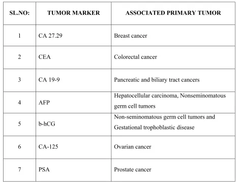
SL.NO: TUMOR MARKER ASSOCIATED PRIMARY TUMOR
1 CA 27.29 Breast cancer
2 CEA Colorectal cancer
3 CA 19-9 Pancreatic and biliary tract cancers
4 AFP
Hepatocellular carcinoma, Nonseminomatous germ cell tumors
5 b-hCG
Non-seminomatous germ cell tumors and Gestational trophoblastic disease
6 CA-125 Ovarian cancer
[image:29.612.91.557.89.447.2]7 PSA Prostate cancer
Table.1: Clinically important Tumor markers
FREE RADICALS
The free radical theory of aging states that organisms age because cells accumulate free radical (Herrling et al., 2008) damage over time. Free radicals are molecular species which contain an
unpaired electron (represented as R·). Free radicals are some of the most chemically reactive molecules that need to pair its single electron, thus it must abstract a second electron from a neighbouring molecule. This results in the formation of yet another free radical and self-propagating chain reaction ensues.Chain reactions involving free radicals (Halliwell et al.,
• Initiation reactions are those that result in a net increase in the number of free radicals. They involve in the formation of free radicals from stable species or they may involve reactions of free radicals with stable species to form more free radicals.
• Propagation reactions are those in which the total number of free radicals remains the same.
• Termination reactions are those reactions resulting in a net decrease in the number of free radicals. However, two free radicals combine to form a more stable species, for example: 2Cl·→ Cl2
Free radicals in human body can arise from fatty food, alcohol, smoking, environmental pollutants, ozone, hydrogen peroxide, ionisation, toxins, carcinogen toxins etc. The vast majority of free radicals originates within the body, an unavoidable byproduct of living system. Under normal conditions free radical intermediates are produced in living systems, the body handles free radicals formed by the breakdown of compounds through the process of metabolism. The major sources of free radicals (such as O2- and HO2·) are modest leakages from the electron transport chains in mitochondria, endoplasmic reticulum and chloroplasts.
DANGER OF FREE RADICALS:
There are four types of free radicals damages (Morrison et al., 1992):
1. Damage to fat compounds: The fatty membranes surrounding the cells being the prime target of free radicals attacks, these damaged membranes then loose its ability to transport oxygen, water or nutrients to the cells.
2. Damage to protein molecules: Free radicals also attack the nucleic acid which comprises the genetic code in every cell. The function of nucleic acids is to regulate the normal cell function, growth and repair the damaged tissues.
3. Cell damage: Damages done to the chromosomes and nucleic acids might initiate the growth of abnormal cells in the body, which was the first step in cancer development.
Fig.2: Complications produced by free radicals in the body
EFFECTS OF FREE RADICAL OXIDATION STRESS:
Production of reactive oxygen species and other free radicals(Acworth et al., 1997) and theirs
damages to various molecules and cells may result not only in the toxicity of xenobiotics but also results in ageing and various age-related diseases, including :
¾ Cardiovascular: Heart Attack, High Blood Pressure, Angina, Stroke, Atherosclerosis. ¾ Lungs : Asthma, Allergies, COPD, Cancer, Chronic Bronchitis
¾ Brain : Alzhemer's, Parkinson's, Migraine, Insomnia, Lack of Mental Clarity, Depression, Bipolar, Dementia
¾ Joints : Osteoarthritis and Rheumatoid arthritis ¾ Eyes : Cataracts, Macular & Retinal Degeneration
¾ Skin : Wrinkles, Acne, Skin Aging, Eczema, Cancer, Psoriasis
¾ Immune : Cold and Flu, Autoimmune, HIV, Herpes, Cancer, Ulcerative colitis, Chronic Viral Disease, Crohn's, Lupus, Hepatitis
SOURCES OF FREE RADICALS :
Free radicals (Dillard et al., 1978) are produced during the various metabolic activities carried
out inside our body and also get exposed to free radicals present in the environment. For example:
Exogenous sources: Sunlight, Automobile Exhaust, Cigarette Smoke, Alcohol Consumption, Emotional Stress, Pollutants, UV light, Xenobiotics, Ionising radiation and Exposure to Heavy metals like Mercury, Lead, Cadmium etc
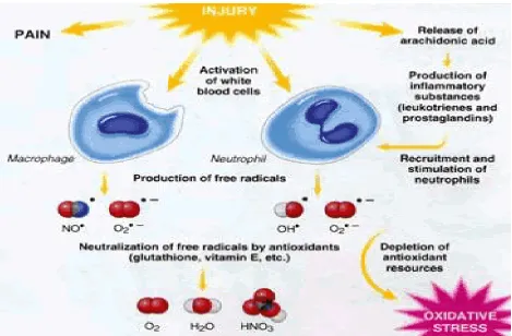
Endogenous sources: Mitochondrial leak, Respiratory burstand Enzyme reactions
[image:32.612.118.523.303.582.2]
Fig.3: Formation of free radicals and its neutralisation by antioxidants
TYPES OF FREE RADICALS IN THE BODY:
free radical that arises from the attack of an oxidizing radical on an organic molecule. Hydrogen-centered radicals result from attack of the H atom. Another form is the sulfur-centered radical produced in the oxidation of glutathione resulting in the thiyl radical. A nitrogen-centered radical also exists for example the phenyl diazine radical.
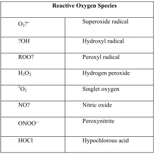
Reactive Oxygen Species
O2?⁻ Superoxide radical
?OH Hydroxyl radical
ROO? Peroxyl radical
H2O2 Hydrogen peroxide
1O2 Singlet oxygen
NO? Nitric oxide
ONOO⁻ Peroxynitrite
[image:33.612.165.467.166.469.2]HOCl Hypochlorous acid
Table.2: Biologically significant free radicals.
ANTIOXIDANTS
The body also has several natural chemical means or systems for neutralizing free radicals. These agents that counteract and minimize free radical damage, donate or provide unpaired electrons to which free radicals can attach without causing harm. Antioxidants (Singh et al.,
Alzheimer's disease and cataracts. Antioxidant substances include Beta-carotene, Lycopene, Lutein, Selenium and Vitamins A,C,E.
Antioxidants get their name because they prevent oxidation. Oxidation is a reaction in which a molecule looses its electron. The two major sources of antioxidants are:
• Those that you get from food or food supplements • Those produced within your own body.
TYPES OF ANTIOXIDANTS
¾ Antioxidant Nutrients
Antioxidants (Karlsson., 1997) from our diet appear to be of great importance in controlling damage by free radicals. Each nutrient is unique in terms of its structure and antioxidant function.
Vitamin E is actually a generic term that refers to all entities (eight found so far) that exhibit biological activity of the isomer tocopherol. Alpha-tocopherol has the highest biopotency, or strongest effect in the body. Because it is fat-soluble, it safeguard cell membranes from damage by free radicals. Alpha-tocopherol also protects the fats in low-density lipoproteins (LDLs or bad cholesterol) from oxidation.
Vitamin C, also known as ascorbic acid, is a water-soluble vitamin. As such, it scavenges free radicals that are in an aqueous environment, such as inside your cells. Vitamin C works synergistically with vitamin E to quench free radicals and regenerates the reduced (stable) form of vitamin E.
Beta-carotene, also a water-soluble vitamin, is the most widely studied of the 600 carotenoids identified to date. It is thought to be the best quencher of singlet oxygen. Beta-carotene is also especially excellent at scavenging free radicals in low oxygen concentration.
¾ Antioxidant Enzymes
The antioxidant enzymes superoxide dismutase (SOD), catalase (CAT) and glutathione peroxidase (GPx) serve as your primary line of defense in destroying free radicals.
SOD first reduces (adds an electron to) the radical superoxide (O2-) to form hydrogen peroxide (H2O2) and oxygen (O2).
2O2- + 2H ----SOD--> H2O2 + O2
Catalase and GPx then work simultaneously with the protein glutathione to reduce hydrogen peroxide and ultimately produce water (H2O).
2H2O2 ---CAT---> H2O + O2
H2O2 + 2glutathione ---GPx---> oxidized glutathione + 2H2O
(The oxidized glutathione is then reduced by another antioxidant enzyme -- glutathione reductase.)
Together, they repair oxidized DNA, destroy oxidized lipids and degrade oxidized protein. Various other enzymes act as a secondary antioxidant defense mechanism to protect you from further damage.
¾ Other Antioxidants
In addition to enzymes, minerals and vitamins, there are many other nutrients and compounds that have antioxidant properties. Among them is coenzyme Q10 (CoQ10, or ubiquinone), which is essential to energy production and can also protect the body from destructive free radicals. Also product of DNA metabolism, uric acid, has become increasingly recognized as an important antioxidant. Additionally, phytochemicals are being investigated for their antioxidant activity and health-promoting potential.
ANTIOXIDANT PROCESS
Antioxidants (Kaczmarski et al.,1999) block the process of oxidation by neutralizing free
• Chain-breaking - When a free radical releases or captures an electron, a second radical is formed. This molecule then turns around and does the same thing to a next molecule, thus generating more unstable products. This process continues until termination occurs, either the radical is neutralised by a chain-breaking antioxidants such as beta-carotene and vitamins C, E or simply it decays into a harmless product.
• Preventive - Antioxidant enzymes like catalase, superoxide-dismutase and glutathione peroxidase prevent oxidation by reducing the rate of chain initiation by scavenging initiating radicals, such antioxidants can stabilise oxidation chain from ever setting in motion. It also prevent oxidation by stabilizing transition metal radicals such as copper and iron.
The effectiveness of any given antioxidant in the body depends on type of free radical involved, its generation and the target of damage. Thus, in one particular system an antioxidant may protect against free radicals, while in other systems it could have any effect at all. In certain circumstances, an antioxidant may even act as a pro-oxidant that generates toxic oxygen species.
ROLE OF MEDICINAL PLANTS IN CHEMOTHERAPY
India is the largest producer of medicinal plants and is rightly called the Botanical garden of the World. Medical information in the old Indian literatures includes several medicinal herbs, which have been in the use for thousands of years, under the indigenous system of medicine. In India, about 45 thousand plant species have been identified, out of which about 15-20 thousand plants are having good medicinal value. However, among these plants, traditional communities use only about 7000-7500 plants for medicinal purposes. The Ayurvedic system of medicine uses about 700, Siddha 600, Unani 700 and the modern medicine uses about 30 medicinal plants for treating a variety of diseases in both man and animal. Only few medicinal plants are attracted by the scientists for investigating them as a remedy for tumour.
prevention. Consumption of huge amount of fruits and vegetables can prevent the development of cancer. Doctors recommend that people who like to reduce their risk of cancer should eat several pieces of fruits and vegetables daily. Several plant-derived products exhibit potent antitumor activity against many cancer cell lines.
A good number of medicinal plants (Khare., 2007) are found mentioned in the ancient classical Ayurvedic texts 'Charaka Samhita'. 'Susruta Samhita' and 'Astanga Hrdaya Samhita'. But many of them still remain to be properly identified. During the process of urbanization the contact with plants in their natural habitat was lost, creating confusion in the correct identity of many plants.
Anticancer properties of plant derived or natural products :
Plants have a long history of use in the treatment of cancer. Hartwell, in his review of plants used against cancer (Cragg et al., 2005) lists more than 3000 plant species that have reportedly
been used in the treatment of cancer. It is significant that over 60% of currently used anticancer agents are derived in one way or another from natural sources, including plants, marine organisms and micro-organisms.
Plant derived compounds have played an important role in the development of several clinically useful anticancer agents. Several anticancer agents including taxol, vinblastine, vincristine and topotecan are in clinical use all over the world. Vinblastine and vincristine from the Madagascar periwinkle, Catharanthus roseus (Apocynaceae), introduced a new era in
using plant material as a medication for treatment. They were the first agents in clinical use for the treatment of cancer. Vincristine, Vinblastine are used in combination with other cancer drugs,as the treatment for various kinds of cancers, including breast and lung cancers, leukemias, lymphomas, advanced testicular cancer. A number of promising agents such as combrestatin, betulinic acid and silvesterol are in clinical or preclinical development.
Medicinal plants (Kaur et al., 2011) maintain the health and vitality of individual and also cure
Since chemotherapy and radiation cause severe toxicity, herbal plants are becoming popular throughout the world nowadays, and are also used as a therapy for tumors or cancer. The antitumour (antineoplastic) activities of several medicinal plants (Sharma et al., 2009) have
been reported by various authors. Many of these plants include: Abrus precatorious , Aglaia
roxburghiana, Cassia fistula , Catharanthus roseus, Crocus sativus , Ervatamia
heyneana , Hygrophila spinosa , Hippocratea murcantha , Indigofera mysorensis , Ocimum
sanctum , Olea polygama , Plumbago rosea, Podophyllum hexandrum , Semecarpus
anacardium , Solanum dulcamara, S. indicum , S. khasianum, S. surattense , Terminalia
arjuna, Trigonella foenumgraecum , Vanda parviflora, Wedelia calendulacea , Withania
somnifera and Zingiber capitatum .
Antioxidative properties of plant derived or natural products :
Several herbs and spices (Farrukh et al., 2006) have been reported to exhibit antioxidant
activity, including rosemary, turmeric, thyme, nutmeg, white pepper, ginger chili, pepper and several other chinese medicinal plant extracts. The majority of the active antioxidant compounds are flavonoids, flavones, isoflavones, coumarins, lignans, anthocyanins, catechins, and isocatechins. In addition to the above compounds found in natural foods, α-tocopherol, β -carotene and vitamins C and E are known to possess antioxidant potential. A direct relationship between antioxidant activity and phenolic content of plant extracts has been reported. Epidemiological studies have shown that the consumption of foods and beverages rich in phenolic content can reduce the risk of heart disease.
Many Indian medicinal plants are considered as potential sources of antioxidant compounds. In some cases, their active constituents are known. Terminalia chebula, T. bellerica, T.
muelleri, and Phyllanthus emblica, all of which have antioxidant activity, showed high content
of phenolics like gallic acid. Conversely, the antioxidant activity of Withania somnifera,
Hemidesmus indicus, Ocimum sanctum, Cichorium intybus, Mangifera indica and Punica
granatum as determined by several methods have been poorly documented.
In the present study, Uvaria narum seeds are used. It is a medicinal plant, fam. annonaceae
phenols, tannins, antioxidants. The presence of these phytoconstituents in the plant has been attributed to its various medicinal properties. The whole root extract of Uvaria narum could be
medicinally employed as a possible free radical scavenger. The plant may also be considered against ageing and other disease caused by free radicals. Hence intention of the study is to evaluate the antitumor and antioxidant activities in oil extract of their seeds.
REVIEW OF LITERATURE
Cancer remains to be one of the leading causes of death in the United States and around the world. The advent of drug targeted therapies has undeniably improved cancer patients cares. However, advanced metastasized cancer remains untreatable. Hence, continued searching for a safer and more effective chemoprevention and treatment is clearly needed for the improvement of the efficiency and to lower treatment cost for cancer care. Cancer chemoprevention with natural phytochemical compounds is an emerging strategy to prevent, impede, delay, or cure cancer (Wang et al., 2012).
Chemoprevention refers to the use of agents to inhibit, reverse or retard tumor genesis. Numerous phytochemicals derived from edible plants have been reported to interfere with a specific stage of the carcinogenic process. Many mechanisms have been shown to account for the anticarcinogenic actions of dietary constituents, but the attention has recently been focused on intracellular-signaling cascades as common molecular targets for various chemo preventive phytochemicals (Surh., 2003).
Phytochemicals are compounds made by plant-based foods that consist of nutrients such as vitamins, minerals and antioxidants that impart a great effect in the body. The majority of the phytochemicals are known as antioxidants, carotenoids,beta carotene, folic acid, vitamin C, and vitamin E. Phytochemicals could provide health benefits as: (1) substrates for biochemical reactions; (2) cofactors of enzymatic reactions; (3) inhibitors of enzymatic reactions; (4) absorbents/sequestrants that bind to and eliminate undesirable constituents in the intestine; (5) ligands that agonize or antagonize cell surface or intracellular receptors; (6) scavengers of reactive or toxic chemicals; (7) compounds that enhance the absorption and or stability of essential nutrients; (8) selective growth factors for beneficial gastrointestinal bacteria; (9) fermentation substrates for beneficial oral, gastric or intestinal bacteria; and (10) selective inhibitors of deleterious intestinal bacteria which include terpenoids, phenolics, alkaloids and fiber (Dillard et al., 2000).
Camptotheca acuminata also have been extensively studied. Other important compounds
include Vincristine, Vinblastine, Colchicine, Ellipticine and Lepachol along with Flavopiridol (Mukherjee et al., 2001).
MAJOR PLANT DERIVED CHEMOTHERAPEUTIC DRUGS:
Since 1961, nine plant-derived compounds have been approved for use as anticancer drugs in the US: vinblastine (Velban), vincristine (Oncovin), etoposide (VP-16, 1 ), teniposide (VM-26, 2 ), Taxol (paclitaxel), navelbine (Vinorelbine), taxotere (Docetaxel), topotecan (Hycamtin), and irinotecan (Camptosar). The last three drugs were approved by the Food and Drug Administration in 1996, (Dholwani et al., 2008)
Sl.no: Compound Source Mechanism Cancer Use
1
Vincristine Catharanthus roseus mitotic block
Lymphoma, Leukemia, breast, lung cancers
2 Vinblastine Catharanthus roseus mitotic block Breast, lymphoma
3 Paclitaxel
Taxus brevifolia
Nutt, Taxus baccata Anti-mitotic Ovary, lung, breast, bladder
4 Docetaxel
Taxus brevifolia
Nutt, Taxus baccata Anti-mitotic Breast and lung cancer
5 Topotecan Camptotheca acuminate
DNA
topoisomerase I inhibition
Ovarian, lung and pediatric cancer
6 Irinotecan Camptotheca acuminate
DNA
topoisomerase I inhibition
Colorectal and lung cancer
7 Flavopiridol Amoora rohituka & Dysoxylum binectariferum
Inhibits cell cycle progression at G1 or G2 phase
colorectal, lung and renal cell carcinoma, non-Hodgkin’s lymphoma, leukemia, solid tumors
8 Ellipticine Ochrosia borbonica, Excavatia coccinea, Ochrosia elliptica
DNA intercalation and inhibition of topoisomerase II
Various cancer cell types
9
Etoposide and Teniposide
Podophyllum peltatum and
Podophyllum emodi not known
Lymphomas, bronchial and testicular cancers
10 Colchicine Colchicum autumnale and Gloriosa superba L.
Anti-mitotic Leukemia, solid tumors
MAJOR SETBACK OF EXISTING CHEMOTHERAPEUTIC DRUGS:
Chemotherapeutic techniques have a range of side-effects that depend on the type of medications used. Most medications affect mainly the fast-dividing cells of the body, such as blood cells and cells lining the mouth, stomach, and intestines, (Keidan et al.,1989). Common side-effects include:
• Depression of the immune system, which results in potentially fatal infections such as typhlitis.
• Fatigue. The treatment can be physically exhausting the patient, who might be already very tired from cancer-related fatigue. It may also produce mild to severe anemia.
• Tendency to bleed easily. Medications that kill rapidly dividing cells or blood cells are likely to reduce the number of platelets in the blood, which can result in bleeding.
• Gastrointestinal distress. Nausea and vomiting are the common side effects of chemotherapeutic medications. This also produces diarrhea or constipation. Malnutrition and dehydration may result when the patient did not eat or drink enough, or when the patient vomits frequently, because of the gastrointestinal damage.
• Hair loss
• Myelosuppression and Immunosuppression
• Typhlitis (life-threatening gastrointestinal complication of chemotherapy)
• Secondary neoplasm
• Infertility
• Teratogenicity
• Neurological adverse effects
Damage to some specific organs is also possible:
• Cardiotoxicity (heart damage)
• Hepatotoxicity (liver damage)
• Ototoxicity, producing vertigo
• Encephalopathy (brain dysfunction)
OTHER STUDIES OF PHYTOCHEMICALS RELATED TO CANCER DRUG DEVELOPMENT:
A clinical and preclinical study was conducted in six adult patients with non resectable liver cancer as well as in mature New Zealand white rabbits with implanted VX2 carcinoma in the liver. An oily anticancer drug injected into the hepatoproximal lumen of the ligated hepatic artery can intensify the anticancer effects of a ligation of the hepatic artery for liver cancer was concluded from that study, (Kenichiro et al., 2006). Another antitumor study with MDV3100
was conducted in patients with castration-resistant prostate cancer by noting antitumor effects at all doses, (Howard et al., 2010).
The antitumor activity of saffron (Crocus sativus) extract against intraperitoneally transplanted
sarcoma-180 (S-180), Ehrlich ascites Carcinoma (EAC) and Dalton's lymphoma ascites (DLA) tumors in mice was studied, (Nair et al.,1991). In a study Curcumin (diferuloylmethane),
polyphenol derived from the plant Curcuma longa was studied for its anticancer potential to
suppress proliferation of a wide variety of tumor cells, down-regulate transcription factors and down-regulate the expression of COX2, LOX, NOS, chemokines, TNF, cell surface adhesion molecules and cyclin D1. It also down-regulate growth factor receptors (such as EGFR and HER2); and inhibit the activity of protein tyrosine kinases, N-terminal kinase and protein serine/ threonine kinases (Bharat et al., 2003).
RELEVANT STUDIES OF ANNONACEAE FAMILY AND UVARIA SPECIES RELATED TO
CHEMOTHERAPEUTIC DRUG DEVELOPMENT:
Another work on evaluation of anti-tumor constituent from Annonaceae plant family were done by isolating 50 new Annonaceous acetogenins, 12 new styrylpyrones and 25 new polyoxygenated cyclohexenes from 5 Uvaria species. Preliminary bioassay tests showed most
of the new isolates exhibit significant anti-tumor activities (Yu., 1999).
In addition to the cytotoxic, antitumor, and antimicrobial activities of plant extracts from the Annonaceae family, other root bark extracts are reported in the literature as having medicinal purposes. One compound, 15-desacetylundulatone, a quassinoid, was found active against P-388 mouse lymphocytic leukemia cells and Colon-38 adenocarcmoma (Lumonadio et al.,
1991).
By activity-directed fractionation, goniothalamicin and annonacin, two acetogenins, was isolated from the ethanolic extracts of the stem bark of Goniothalamus giganteus Hook. of Annonaceae family and studied for cytotoxic and insecticidal activity and was found to be weakly active against 3PS murine leukemia (Alkofahi.,1988). Similarly, three bioactive annonaceous acetogenins, annomocherin, annonacin and annomontacin, had been isolated from the fractionated ethanolic extracts of the seeds of Annona cherimolia, supported by brine shrimp lethality test (BST) and their cytotoxicities against the breast carcinoma (MCF-7) and kidney carcinoma (A-498) cell lines was studied (Kim et al., 2001).
Similar anticancer studies was conducted by isolating three annonaceous acetogenins from seeds of Annona cherimolia (Woo et al., 1999); another by isolating flavone from the stems
and leaves of Dasymaschalon trichophorum Merr. of annonaceae family (Liu et al., 1992) and
yet another by isolating Annonaceous acetogenins from the ethanolic extract of the bark of Annona bullata of annonaceae family (Hui et al., 1992).
The genus Uvaria, of annonaceae have been found to contain compounds such as acetogenins
useful as pesticides, and recently as anticarcinogenic agents. The compounds taken from the roots of Uvaria chamai has also been implicated as a causative agent for liver cancer. Two
separate studies out of sub-Saharan Africa have shown that the active compound in Uvaria
chamaii, chamuvaritin, has shown that it has mutagenic effects. One of the studies also showed
disease is multi-factorial in etiology, including a combination of causative agents such as Hepatitis B Virus, and chemical carcinogens, the compounds contained in the medicinal plants were implicated as potential sources of causative agents for liver cancer (Uwaifo et al., 1984
and Uwaifo et al., 1972).
The phytochemical analysis and cytotoxic activity of leaves of Uvaria chamae against L929
transformed cells was studied and found that at a concentration corresponding to their IC50 against L929 cells, they were nontoxic against mouse thymocytes (Philipov et al., 2000).
In a Ethnobotanical survey of the plants used in the management of cancer, carried out in Ogun State, Nigeria, seventy-three different species of Angiosperms were found to be used for the management of cancer. Out of this bark of Uvaria chamae and roots of Uvaria afzelii,
annonaceae family was found to be effective as anticancer drug (Mike et al., 2010). Another
anticancer study was studied in the roots of the Uvaria acuminata by isolating Uvaricin, a
potent anticancer compound from the plant (Jolad et al., 1982).
RELEVANT STUDIES OF ANNONACEAE FAMILYAND UVARIA SPECIES RELATED TO
ANTIOXIDANT THERAPY:
The antioxidant and antibacterial activities of Uvaria narum (Dunal) Wall was studied in the
essential oil and different plant extracts (petroleum ether, ethyl acetate, chloroform and methanol) which showed that the leaf methanol extract and leaf essential oil of Uvaria narum
are good sources of natural antioxidants and antimicrobial compounds and can be incorporated into the drug formulations (Joji et al., 2012).
Other reported literature has shown that the compound, Uvaricin, taken from the purified root extracts of Uvaria acuminata, another Annonaceous plant, demonstrated an in vitro activity
against P-388 lymphocytic leukemia in mice (Jolad et al., 1982). Another extract, Uvaratin,
also from Uvaria acuminata, was also shown to have anti-tumor activity (Cole et al., 1976).
OTHER STUDIES OF PYTOCHEMICALS RELATED TO ANTIOXIDANT THERAPY:
The in vitro antioxidant activity of melatonin was studied using thiocyanate method. Also, the
and the extract showed a significant inhibitory effect on the peroxidation of rat-brain phospholipid and inhibited DNA damage by bleomycin or copper-phenanthroline systems, (Gregorio et al., 2000).
In a study it was concluded that tea possesses a strong antioxidant activity (in vitro) which was
believed to be exerted by its poly phenols moiety and also tea had a potent in vivo activity
in man. The inhibition of this effect by milk was thought to be due to the complexation of tea polyphenols by milk proteins. Thus it clarified the putative role of dietary poly- phenols in modulating oxidative stress in vivo (Serafini et al., 1996).
Similarly antioxidant activities of the methanolic extract of Cinnamomum verum bark was
studied and evaluated with reference to antioxidant compounds like butylated hydroxyl anisole, trolox and ascorbic acid (Mathew et al., 2006). A Polyherbal Formulation (PHF),
Rumalaya Forte that has a combination of medicinal herbs such as the powders of Boswellia
serrata, Alpinia galanga, Commiphora wightii and Glycyrrhiza glabra and the extracts of
Tribulus terrestris and Tinospora cordifolia was tested for its antioxidant and free radical
scavenging activity in vitro and demonstrated a significant NO free radical scavenging activity (Subash et al., 2012).
Another in vitro and in vivo antioxidant study was conducted in alcoholic and aqueous extracts
of the roots of Toddalia asiatica and it was concluded that the extracts exhibited potential
scavenging effects on hydrogen peroxide, DPPH and nitric oxide free radicals. The in vivo antioxidant activity was also investigated using Wistar albino rats (Madhavan et al., 2010).In
a study the in vitro antioxidant effect of the ethanolic extract of Mimosa pudica (Mimosaceae)
was evaluated against free radical damage by different standard methods such as DPPH, Nitric Oxide, ABTS and Hydrogen peroxide free radical model. Out of these four free radicals the extract showed potent activity on DPPH and Nitric oxide, which is compared to that of ascorbic acid and rutin taken as standards(Muthukumaran et al., 2011).
The alcoholic extract of Medicago sativa was studied for its in vitro antioxidant activity using
different models namely DPPH and ABTS radical scavenging, iron chelates activity, lipid peroxidation assay, alkaline DMSO assay and nitric oxide scavenging assay. The results were analyzed statistically by the regression method and indicate that Medicago sativa was effective
In a study the total phenolic, flavonoid contents and in vitro antioxidant activity of methanol extract of Dioscorea esculenta (Lour). Burkill was evaluated. Total phenolic content was
estimated using the Folin Ciocalteu method, flavonoid content using aluminium chloride and In vitro antioxidant activities and reducing power capacity were determined using standard methods (Manickam et al., 2012).
OTHER RELEVANT STUDIES OF ANNONACEAE FAMILYAND UVARIA SPECIES:
In a study the phytochemical analysis of the root barks essential oil of Uvaria narum Wall.
(Annonaceae) were done by GC/MS and about 52 components were detected, out of which 22 components were identified. Bornyl acetate (15.2%) and patchoulenone (8.1%), a tricyclic sesquiterpene ketone were found to be the major constituents of the oil (Abdul et al., 1992).
The antiproliferative activity of Madagascan plant Uvaria sp. against the A2780 ovarian cancer
cell line was done by isolating two new acetogenins from the plant and it was observed that the two acetogenins display weak antiproliferative activity against the A2780 ovarian cancer, the A2058 melanoma, and the H522 lung cancer cell lines (Dai et al.,2012). The antibacterial,
antifungal and anthelmintic activities for the hexane and ethyl acetate extracts of the root barks
of Uvaria narum Wall. and Uvaria hookeri King was evaluated in one of the study (Padmaja
et al., 1993). Similarly, the antibacterial and antioxidant potential of the root of Uvaria narum
(Dunal) Wall was carried out by using various extracts in another study (Padyana et al., 2011).
In fact, the literature contains numerous reports that plant extracts from the Annonaceae family have been investigated for their medicinal and toxicological effects. In one pharmacological screening, substantial antibacterial, antifungal, and anthelminthic activities were observed using extracts of the root barks of Uvaria narum wall, and Uvaria hookeri king (Padmaja et
al., 1993).
The hepatoprotective potential of leaves of Uvaria narum (Annonaceae) by noting elevation
favorably to that of streptomycin sulfate, an inhibitor of protein synthesis (Parmer et al.,
1978).
In a study, venom neutralizing properties of Uvaria chamae was evaluated by in vivo
neutralization of Naja nigricollis venom in rats. For this Uvaria chamae leaves were extracted
in methanol and the results indicated the presence of potent snake venom neutralizing capacity in the plant extract(James et al., 2013).
Other reported literature has shown that the compound, Uvaricin, taken from the purified root extracts of Uvaria acuminata, another Annonaceous plant, demonstrated an in vitro activity
against P-388 lymphocytic leukemia in mice (Jolad et al., 1982). Another extract, Uvaratin,
also from Uvaria acuminata, was also shown to have anti-tumor activity (Cole et al., 1976).
In another study, species from Uvaria originating from Tanzanian plants were tested for their
in vitro activity against the multi-drug resistant K I strain of Plasmodium falciparum, the
causative agent for the disease malaria. In this study, root extracts from the stem and root barks
of Uvaria iucida and Uvaria sp. (pande), were reported as having anti-malarial activity.
Among the compounds isolated in this study were Uvaretin, Diuveretin, and 8',9'-dihydroxy-3-faraesylindole as the most active compounds The particular Uvaria species consisted of nine
varieties from Tanzania and included: U dependens, U.faulknerae, U. kirkii, U. leptocaldon,
U. Iucida, Uvaria sp. (Pande), U. scheffleri, and U. tanzaniae (Nkunya et al., 1991).
A in vitro study was done on the acaricidal activity of three Uvaria species, namely U.
klaineana, U. mocoli and U. versicolor against Dermatophagoides pteronyssinus, the
European house dust mite (Akendengueet al.,2003). In another study the methanolic extracts
of the stem, root and leaves of Uvaria chamae were evaluated for their antibacterial activity
against Methicillin-Resistant S. aureus, S. aureus, E. coli, Klebsiella spp, Proteus spp, and P.
aeruginosa using standard agar diffusion method. Out of this methanoilc leaf extract was
found to be significant (Oluremi et al., 2010). The antiplasmodial activity of Pleiocarpa
mutica, Cleistopholis patens and Uvaria chamae were studied by screening its activity against
P. falciparum (Jonathan et al., 2001 and Nkunya et al.,1991).
Another study focussed on the antioxidant and antibacterial activities of hexane, chloroform, ethanol extracts of leaves and barks of Uvaria grandiflora Roxb (Annonaceae) and found that
extract (Noushin et al., 2011). The antifungal activity of dichloromethane, petroleum ether and
ethanolic extracts of the stem bark and leaves of Uvaria scheffleri Diels (Annonaceae) was
studied against Aspergillus niger , Aspergillus fumigatus and Penicillium species (Moshi et
al., 2004).
OBJECTIVES
¾ Preparation of oil extract