0022-538X/94/$04.00+0
Copyright ©D 1994,American SocietyforMicrobiology
The
EBNA-2
Arginine-Glycine
Domain
Is
Critical but Not Essential
for
B-Lymphocyte
Growth
Transformation;
the Rest of
Region
3
Lacks Essential
Interactive Domains
XIAO TONG, RAMANAYALAMANCHILI, SHIZUKOHARADA,ANDELLIOTTKIEFF* DepartmentofMicrobiologyand Molecular Genetics andDepartment ofMedicine,
Harvard University, Boston, Massachusetts 02115 Received 3February1994/Accepted27 June1994
Sincedeletion ofregion 3 (aminoacids [aa] 333to425) ofEpstein-Barrvirus nuclearprotein2 (EBNA-2) results in EBVrecombinants whichcannot transformprimary Blymphocytes (J. I.Cohen, F.Wang, and E. Kieff, J. Virol. 65:2545-2554, 1991),theroleof domains ofregion 3wasinvestigated.Deletion of the
Arg-Gly
repeatdomain,R-337GQSRGRGRGRGRGRGKG354,resultsinEBV recombinants that transformprimaryB lymphocytes withmodestly decreased
activity.
Thetransformed cells grow slowlyandaredifficult toexpand. EBNA-2 deletedfortheArg-Glydomain does notassociate with the nuclearchromatin fraction. The Arg-Gly repeathas anintrinsicability to bindtohistoneHi,
tootherproteins,including EBNA-1,and tonucleicacids, especially poly(G).Twoindependent deletionsof eachpartof the rest ofregion3(aa359 to383 and 385 to430) havelittle effect ontransformation, while deletion of therestofregion3 (aa 361to425) as asinglesegment substantiallyreducestransformation efficiency.EBNA-2deletedfor all ofregion3 can still transactivatethe LMP1promoterintransientexpression assays but is less active than EBNA-2intransactivatingtheBamHI-C promoter. EBNA-2 deleted for the Arg-Gly domain is better than EBNA-2 at transactivating the LMP1 promoter and is as active as EBNA-2 in transactivating the BamHI-C promoter. These data are most compatible withamodel inwhichtheArg-Gly domain of region 3 isamodulatorof EBNA-2 interactionsand activities, while the restofregion 3 isimportantinpositioningtheregion2Jkappabindingdomain relative totheregion4acidic transactivating domain. Despitethe nullphenotypeof theregion 3deletion,region3 is unlikelytomediate essentialinteractions withotherproteins.The ability of Epstein-Barr virus (EBV) to acutely and efficiently transform primary B lymphocytes into perpetually proliferating lymphoblastoid cell lines (LCLs) is linked to expression from the viral genome of six EBV nuclearproteins (EBNA-1, EBNA-2, -3A, -3B, -3C, and -LP), two latent membrane proteins (LMP1 and -2) and two small RNAs (EBERs) (31). Ofthese 10 gene products, the EBERs (45), EBNA-3B(46),and LMP2(33, 34)are notrequiredfor latent infection or cell growth transformation in vitro, although LMP2mayberequired for the maintenance of latent infection invivo(37).EBNA-1is essential forefficient persistence of the EBV genome in latentinfection (54),while EBNA-2(10, 23), EBNA-3A and -3C (47), and LMP1 (27) are essential and EBNA-LP (35)iscritical for latent infection orB-lymphocyte growth transformation.
EBNA-2 is one of the first two genes expressed in latent infection. At least one essential function of EBNA-2 is as a transactivator of viral and cell gene expression. EBNA-2 induces expressionof the LMP1 (1, 52)and LMP2 (62) genes andof cellulargenes,including CD21(11), CD23 (49-51), and
c-fgr
(29). EBNA-2-responsive elements upstream of theLMP1(17,48), LMP2A (61), BamHI-C (Cp) (44), and cellular CD23 (51) promoters have been shown to convey EBNA-2 responsiveness to heterologous promoters.
Molecular genetic and biologic analysis of the role of EBNA-2 in latent growth-transforming infection identified at
*Correspondingauthor. Mailing address: Department of
Microbi-ology and Genetics, Harvard University, 12 South Thorn Bldg., 75 Francis St., Boston, MA 02115. Phone: (617) 732-7048. Fax: (617) 278-6964. Internet address: KIEFFE@TNP01.BWH.HARVARD.
EDU.
least fourregions of EBNA-2 whichareessential for transfor-mation and for transactivation of the LMP1 promoter (9). Regiononeisdefinedbyadeletion of amino acids (aa) 19to
110,which consistmostlyofapolyproline repeat. Region2is defined bythe insertion ofa linkerencoding GRSS between W-320 and P-321. This region mediates EBNA-2 interaction with a 63-kDa cell protein, J kappa, which recognizes a sequence common to EBNA-2 response elements (22, 32). EBNA-2 aa310to 336are sufficient for Jkappainteraction, and the short sequence P-317PWWPP-322 conserved between type 1and type 2 EBNA-2probablymediates this interaction (22, 55). Region 3 is definedby adeletionof aa 333 to 425, a segment which includes an Arg-Gly repeat at aa 337to 354. Region 4 is defined byadeletion ofaa426 to472, a segment whichincludesacoreacidictransactivating domain at aa 449to 462(7, 8).The adverse effectof each of these mutationsonthe transformingandtransactivating phenotypes of EBNA-2 is not because themutantproteinsareunstable or fail totranslocate
to the nucleus (9) and is therefore most consistent with the hypothesis that eachregion includes aninteractive domain(s) involved in viral or cellular gene transactivations or other functions essential for transformation.
This report focuses on region 3 (aa 333 to 425) and the unusual R-337GQSRGRGRGRGRGRGKG-354 domain in region 3. Although this domain can serve to ensure nuclear translocation when the more usual nuclear localization se-quenceatthe end of EBNA-2 is mutated(9), that is unlikely to be the principal function of this domain. Arg-rich repeat domains in other proteins have been implicated in protein-RNA(13) orprotein-protein (59) interactions. We have now investigatedthe role of theArg-Gly domain and of the rest of region3 in EBV-mediated cellgrowth transformation, trans-6188
on November 9, 2019 by guest
http://jvi.asm.org/
activation, and interactions with cellular and viral proteins and nucleic acids.
MATERIALSAND METHODS
Celllines, culture conditions, and labeling of cell proteins. BJAB cells are derived from an EBV-negative (EBV-) B-cell lymphoma (36). BL30/P3HR-1 is an EBV- Burkitt lymphoma cell line which has been infected in vitro by EBV strain P3HR-1 (6). The P3HR-1 genome is deleted for an EBV DNA segment which encodes the last two exons of EBNA-LP and the entire EBNA-2 exon. P3HR-1 clone 16 cells were obtained from G. Miller, Yale University (41). IB4 is an EBV-trans-formed B-lymphoblastoid cell line (28). All cells were main-tained in RPMI 1640 medium supplemented with 10% fetal bovine serum. Cells (108) were grown to
106/ml
and labeled overnight in 1 mCi of [35S]methionine (New England Nuclear) per 20 ml in methionine-free RPMI medium (GIBCO BRL) supplementedwith 10% dialyzed fetal bovine serum (GIBCO BRL).Cosmids and plasmids. pSG5-EBNA 2 is an expression vector for EBNA-2 (48). pSG5-E2 plasmids, deleted for vari-ous parts of EBNA 2 region 3, were constructed by PCR-facilitated mutagenesis (24). The PCR primer sequences were asfollows:5'-GATTACATTTITGAGACA-3' (5' outside
prim-er) and
5'-ACAAGTTCTGC'T'l'AATA-3'
(3' outsideprim-er); for pSG5-E2,d337-354, 5'-CAAGGCCAGAGCAAGTC CAGGGACAAGCAA-3' (5' inside primer) and 5'-GCTCT GGCClT'GAGTC(T-3' (3' inside primer); for pSG5-E2, d359-383,
5'-AAGTCCAGGGACGTCCTCGGTCT'TCATC
AG-3' (5' inside primer) and 5'-GTCCCTGGACTTFGCCC TT-3' (3' inside primer); for pSG5-E2,d385-430, 5'-CTAAG
TCCAGTCCAGGCTCCCATTC'TTTCC-3'
(5' inside primer)and 5'-GACTGGACT1'AGTTCAGG-3' (3' inside primer). TheBstEII-to-BglII fragment of pSG5-EBNA 2 was replaced by the deletion fragments from the PCR-amplified DNA. pSG5-E2,d333-425 was made by cloning the BsmI fragment fromcosmid T1EBNA 2 d333-425 (9) in place of the wild-type (wt)BsmI fragment of pSG5-EBNA 2. pSG5-E2,d361-425 was made by using a 5' outside primer (5'-CATCACCACCACG CATGCATC-3'), a 3' outside primer that was the same as described above, a 5' inside primer (5'-GCAAGTCTAGAGA
CAAGCAATCCCATAATAGCCCAGAGGC-3'),
and a 3'inside primer
(5'-GCCTCTGGGCTATTATGGGATTGCT[
GTCTCTAGACTTGC-3').
The sites of genetic alterationswere sequenced by the dideoxy method (U.S. Biochemical Corp.). Cosmids with EBNA-2 deletions were constructed by cloning the mutated NsiI fragments of the EBNA-2 open reading frame into theHindIII-SstII fragment from the EBV DNA EcoRI A fragment. The mutated HindIII-CpoI frag-mentswere then used toreplace the wtHindIII-CpoIsequence of cosmid WEAH, a clone of the W91 EBV DNAEcoRI A fragment in MAU3 (40). Two cosmid clones of each deletion mutant wereindependentlyderived. Glutathione S-transferase (GST) fusion proteinexpression vectors were constructed by PCR amplification of the appropriate part of wt or mutated EBNA-2, using 5' and 3' primers which have BamHI and EcoRI sites in frame with the GST expression vector pGEX-2TK (26). GST-(RG)8 was made by inserting a synthetic oligonucleotide encoding eight Arg-Gly repeats into the
BamHI-to-EcoRI
site ofpGEX-2TK.Transformationassay andvirus passage. Cosmid clones of wt or specifically mutated EBV W91 EcoRI-A DNA were digestedwithEcoRItorelease the EBV DNA from the vector, and 10 ,ug of DNA was mixed with 50 ,ug ofpSVNaeIZDNA. TheDNAs were transfected into 15 million P3HR-1 clone 16
cells by electroporation at 200 V and 960
,uF
in a Bio-Rad Gene Pulser cuvette. The cells were then diluted into 15 ml of RPMI medium with 10% fetal bovine serum and incubated at37°C
for 6 days. On the 6th day, culture supernatant containing the virus was filtered through a 0.45-jim-pore-size filter and used to infect freshly isolated T-cell-depleted human periph-eral blood lymphocytes (9, 46, 47). The infected cells were plated on a 96-well plate at 5 x10⁴
cells in 150jl
of medium per well. The cells were fed once every 2 weeks with 100jl
of medium per well. LCLs were macroscopically visible at 4 to 6 weeks.Lytic EBV infection was induced by transfecting 15 million cells with 50 jig of pSVNaeIZ DNA and incubating the cells in the presence of 20 ng of phorbol 12-myristate 13-acetate (GIBCO BRL) per ml. Four days after electroporation, culture supernatant containing the virus was used to infect freshly prepared human non-T peripheral blood lymphocytes.
Nuclear fractionation. Cell nuclei were isolated and frac-tionated as described previously (39). Briefly, cells were lysed in hypotonic buffer (10 mM N-2-hydroxyethylpiperazine-N'-2-ethanesulfonic acid [HEPES], 50 mM NaCl, 150 mM sucrose,
5 mM
KCl,
1mM
MgCl2,
2 mM dithiothreitol[DTT],
1 mMphenylmethylsulfonyl fluoride [PMSF], 2 jig of aprotinin per ml), and nuclei were isolated by centrifugation at 1,000x g for 10 min. The nucleus pellet was extracted twice with nuclear buffer (10 mM HEPES, 120 mM NaCl, 0.5% Nonidet P-40 [NP-40], 1 mM PMSF 2 jig of aprotinin per ml), and the combined
supernatants
were considered the nucleoplasm frac-tion. The NP-40-extracted pellet was resuspended in nuclease digestion buffer (10 mM HEPES, 100 mM NaCl, 50 mMKCl,
5mM
MgCl2,
0.1
mM EDTA, 1 mM PMSF, and 2 jig ofaprotinin per ml), digested with DNase I for 30
min,
and then extracted with high-salt buffer (10 mM HEPES, 2 M NaCl, 0.5% NP-40, 10 mM EDTA, 1 mM PMSF, 2 jig of aprotinin per ml). The extracts were centrifuged at 15,000 x g for 10min,
and the pellet was washed once in nuclear buffer. The com-bined
supernatants
were considered the chromatin fraction. The nuclear matrix fraction was prepared by solubilizing the final pellet in sodium dodecyl sulfate (SDS) protein sample buffer.GST fusion proteins. GST fusion proteins were expressed in
Eschenchia
coli after induction with 0.1 mMisopropyl-p-D-thiogalactopyranoside (IPTG) (26). Cells were harvested 3 h after induction. After sonication and centrifugation, the ex-tracts were incubated with glutathione-agarose beads (Phar-macia) for 1 h at
4°C.
The beads were precipitated and washed with NETN (20 mM Tris [pH 8.0], 100 mM NaCl, 1 mM EDTA, 0.5% NP-40).Affinity
binding assay. Labeled cells were lysed in buffer(108
cells per 2 ml of lysis buffer) containing 150 mM NaCl, 10 mM HEPES (pH 8.0), 1% NP-40, 2 jig of aprotinin per ml, 1 mM PMSF, 1 mM DTT, and 25 mM betaine (Sigma). Insoluble material was removed by centrifugation. The lysates were precleared by incubation with glutathione-Sepharose beads loaded with GST. Aliquots of precleared lysates were incu-bated with Sepharose beads loaded with various GST fusion proteins as indicated for 1 h at
4°C.
The beads were washed in lysis buffer plus 0.1% SDS. Proteins bound to beads were eluted with SDS sample buffer and analyzed by SDS-polyacryl-amide gel electrophoresis (PAGE) using 8.5% gels.Far-Western blot analysis. GST fusion proteins were la-beled and incubated with nitrocellulose filters containing pro-teins transferred from gels after SDS-PAGE (26). Briefly, glutathione-Sepharose beads loaded with fusion proteins were resuspended in 1 x HMK buffer (20 mM Tris [pH 7.5], 100 mM NaCl, 12 mMMgCl2)containing the catalytic subunit of cyclic
on November 9, 2019 by guest
http://jvi.asm.org/
AMP-dependent protein kinase (Sigma),
[y-32P]ATP
(New EnglandNuclear),and 1mMDTT.The mixturewasincubated for 30 min at 4°C. Fusion protein was eluted with 20 mM reducedglutathione (Sigma) in 100 mM Tris(pH 8.0) and 120 mM NaCl. The 32P-labeled fusion proteins were added to hybridizationbuffer(Hyb75; 20 mM HEPES [pH 7.7],75 mMKC1,
0.1 mM EDTA, 2.5 mM MgCl2, 1 mM DTT, 0.05% NP-40) at 2.5 x 10⁵ cpm/ml. To reduce background, Hyb75was supplemented with equal volume of lysate from E. coli expressing GST.
Nucleic acidbindingassay.Single-stranded DNA (ssDNA)-double-stranded DNA
(dsDNA)-cellulose,
cellulose, andpoly(G)-, poly(A)-,
poly(U)-, and poly(C)-agarose werepur-chased from Sigma. GST fusion proteins were 32P-labeled as described above. Nucleic acidcoupled tomatrixwasincubated with labeled GST fusion protein in hybridization buffer (105 cpmper samplein
NETN)
supplementedwith 1/10volumeof lysatefrom bacteriaexpressing GST(see above).After30 min ofincubation, beads were washed with NETN and Cerenkov counted (Beckman LS 5000TD). For blocking experiments, labeled GSTfusion proteinwaspreincubatedwith blocker for 15 min before addition of the labeled protein to the nucleic acid matrix.Dimethylsulfatemethylation. Oligonucleotideswere meth-ylated byaddition offreshlydiluted 1% dimethyl sulfate to a finalconcentration of0.05%and incubationat65°Cfor15 min
(53).
Transfections and CAT assays. BJAB cells (107) were
suspended
in 0.3 ml of RPMI 1640 and placed in aBio-RadGene Pulser cuvette; 5 ,ug of
pUC-3gal,
10 ,ug ofpSG5 orpSG5
witha wtormutatedEBNA-2, and 5jig
of LMP1CAT(-234/+40)
reporterplasmid(48)
orCpCATreporterplasmid(55)
were added to each cuvette. Cells were electroporated with 220 Vat 960 ,uF.1-Galactosidase
and chloramphenicolacetyltransferase (CAT)
activitieswereassayedafter 3daysasdescribedpreviously
(48).
RESULTS
The Arg-Gly repeat is important in latent infection and growthtransformation ofprimaryBlymphocytes.Toevaluate the role of the region 3 (aa333 to425) domains in primary B-lymphocyte growth transformation, DNA fragments con-taining a wt orspecifically mutated EBNA-2 gene were
com-pared
for the ability to marker rescue transforming virusactivityfrom EBV-infected P3HR-1 cells. P3HR-1 EBV DNA iswt exceptfora deletionofaDNAsegmentwhichincludes the lasttwoexonsof EBNA-LP and the entire EBNA-2 open reading frame. Transfection of P3HR-1 cells with a wt EBV
DNA
fragment
which spans the deletion and with anexpres-sion plasmid for the EBV BZLF1 immediate-early transacti-vatoroflyticinfection results in lytic infection and recombina-tion of the transfected DNA with the replicating P3HR-1 genome. Transforming recombinants can then be identified and enumerated by virtue of their ability to infect and to growthtransformprimaryB lymphocytes.
Deletion of all of region 3 (aa 333 to 425) has been previously reported to be a null mutation in such marker rescue experiments (9). The null transforming activity was confirmed with new constructs of the marker-rescuing DNA
fragment (E2d333-425).
Two different constructs failed tomarker rescue transformation in each of three independent
experiments
inwhichasimilar fractions of the virus from cellstransfected withwtDNAgave rise to 19 to 28 transformants. Twodifferent constructsofEBNA-2deleted for DNA
encod-ing
theArg-Gly
domain(E2d337-354)
generated 0 to 7 or 0 to14transformants in fiveindependent experiments inwhichwt DNAgave rise to 19 to84 transformants. Themeanvalue for the E2d337-354 markerrescueefficiency relative to wtcontrols was 8%. In two independent experiments, two different con-structsdeleted fortherestof region3(E2d361-425)each gave rise to 3 to 8 or 4 to 5 transformants, while the wt DNA generated 61 to 63 transformants. The mean value for the E2d361-425 transforming efficiencyrelativetothewtvaluewas 8%. Surprisingly,when the restofregion3was studiedas two separatedeletions,E2d359-383 andE2d385-430, eachmutated DNAmarkerrescued transforming activityfrom P3HR-1 cells with anefficiencyfully equalto orgreaterthan thatofwtDNA. Since EBVrecombinants deleted for EBNA-2 aa 359 to 383or 385 to 430 had wt transforming activity, these sequences are unlikely to directlymediate importantinteractions of EBNA-2 with other proteins.Thus,the reduced transformingactivityof the combined deletion, E2d361-425, is most likely due to an effect on the folding or presentation of other domains of EBNA-2.
LCLs infected with and transformed byE2d337-354 recom-binants also grew differently from wt recombinant-infected LCLs, while LCLs transformed byE2d359-383 orE2d385-430 recombinants were indistinguishable from wt-infected LCLs. Wild-type, E2d359-383, or E2d385-430 recombinant-infected LCLs reached saturationin the original 96-well platesby 4to 6weeks. In contrast, E2d337-354 recombinant-infectedLCLs reached saturation at 6 to 8 weeks. LCLs infected with E2d337-354 recombinants also continued to grow more slowly than wt recombinant-infected LCLs over theensuing 4months. While more than 80% of the wt-infected LCLs could be expanded to >107 cells, only 50% of the E2d337-354 recom-binant-infected LCLs could be expanded.
Putative E2d337-354 mutant recombinant-infected LCLs were confirmed to have the E2d337-354 DNA by PCR with primers corresponding to bp 49228 to 49651 and 49651 to 49670 (3), which amplify a 388-bp fragment across the site of the deletion or a 442-bp fragment from wt EBV DNA (data notshown).E2d359-383-, and E2d385-430-infected LCLs were similarlyconfirmed to have their respective deletions by PCR using primers corresponding to bp 49443 to 49640 and 49962 to 49979, which amplify 462- and 399-bp fragments from the mutants and 537-bp fragments from wt EBV DNA (data not shown).
To further characterize the E2d337-354 recombinant phe-notype, lytic infection was induced in LCLs infected with wt or E2d337-354 EBV recombinants. Dilutions of the resultant virus from wt- and mutant recombinant-infected cells which had been induced to similar levels of permissivity for lytic infection were used to infect primary B lymphocytes. At 4 weeks,E2d337-354 recombinant virus-transformed primary B lymphocytes into macroscopically visible LCLs with 30% of the wtrecombinant activity; by 9 weeks, the number of macroscop-ically visibleLCLs generated by E2d337-354 recombinant virus was 80% of the wt level (Fig. 1). Thus, the most evident phenotype was the slower outgrowth of LCLs transformed by E2d337-354 recombinant virus. The near wt ultimate efficiency of the E2d337-354 recombinants in primary B-lymphocyte transformation in these experiments as opposed to experi-ments with virus derived, de novo, from P3HR-1 cell transfec-tions is probably due to an inhibitory effect of coinfection of the primary Blymphocytes with parental P3HR-1 virus, since a decreased multiplicity of infection of primary B lymphocytes with virus stocks from P3HR-1 cells resulted in an enhanced efficiency of transformation (data not shown).
EBNA, LMP, andBZLF1 expression in LCLs transformed by E2d337-354 or wt EBV recombinants. EBNA and LMP1
on November 9, 2019 by guest
http://jvi.asm.org/
E2d337-354c1
E2d337-354c2 E2d37-354c3
E2d337-354c4 WildType
0 20 40 60 80 100 Number ofTransformants
FIG. 1. Summaryof outgrowth of LCLs following infection with wt orE2d337-354 recombinantvirus. LCLs infected with one wt and four independent E2d337-354recombinants were rendered similarly per-missive for lytic EBV infection. Four days later, culturesupernatants containing the viruswereused toinfect primary human B lymphocytes. The number of macroscopically visible transformants was scored at 4 weeks(graybars) or at 9 weeks (black bars).
VVT E2d337-354
r X- Z m r X- Z
CD Z ) Z 0 Z C) Z
97-
66-
33-
21-I-E2
+Hl
FIG. 3. EBNA-2 andE2d337-354 differentially associate with the nuclear chromatin fraction. LCLs transformed bywt EBV recombi-nant orE2d337-354werelysedand fractionated into cytoplasm(CYT), nucleoplasm (NP), chromatin (CN), and nuclear matrix (NM). EBNA-2 was detectedbyWesternblotting withmonoclonal antibody PE2; histone Hi was detected by immune rabbit serum. Sizes are indicatedinkilodaltons.
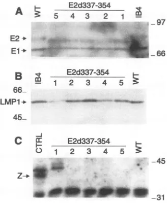
expressions were analyzed in LCLs transformed by wt or 354 EBV recombinants. LCLs infected with E2d337-354 EBVrecombinant virus consistently expressed slightly less EBNA-1 relative to EBNA-2 than was found in wt recombi-nant-infected LCLs. EBNA-2 expression was similar to that in wtrecombinant-infected LCLs,and the level of EBNA-1 was reduced lessthan twofold (Fig. 2A). The EBNA-2 size inthe E2d337-354-infected LCLs was also smaller because of the deletion.LMP1wasexpressedatsimilar levels in bothmutant andwtLCLs(Fig. 2B).
BZLF1 expressionwasevaluated as amarker of spontane-oustransition of LCLstolyticinfection. As ischaracteristic of most recently derived LCLs, BZLF1 expression was barely detectable in most of the LCLs. Among five mutant LCL
A ~~ E2d337-354
5 5 4 3 2 1
....
r...j
-97E2+ >..
El-b ^-
'r;-* .
*';S§:.' ... -'? 66
E2d337-354
Bm 1 2 3 4 5
66_
LMPt
-
45-C a E2d337-354
0 1 2 3 4 5
-45
Z--1p-M
go VW -31
FIG. 2. EBV gene expression in LCLs infected with wt and
E2d337-354 EBV recombinants. LysatesfromIB4 cells,wt
recombi-nant-infectedLCL,orfive E2d337-354-infected LCLsweresubjected toSDS-PAGE,transferredtonitrocellulose,andprobedforEBNA-1
andEBNA-2, usinganEBV-immunehumanserum(A), LMP1, using
monoclonal antibody S12 (B), BZLF1, using monoclonal antibody
BZ.1(C). Alysatefromcellstransfected with BZLF1wasusedas a
positivecontrol (CTRL).Sizesareindicated in kilodaltons.
clones tested, one expressed easily detectable BZLF1 (Fig. 2C). Thus, E2d337-354does notaffectthepermissivityofLCLs forlyticEBVinfection.
Byimmunofluorescencemicroscopy,E2d337-354inmutated recombinant-infected LCLs was slightly more aggregated in nuclearclumpsthanwas wtEBNA-2 inLCLs infected withwt
recombinants. E2d337-354-infected LCLs alsotendedtovary more in immunofluorescence intensity, with some cells ex-pressinghigherEBNA-2levels than anywtEBNA-2 recombi-nant-infected LCLs (data not shown). However, E2d337-354
was notdistinctive enoughtobereadilyidentifiableby immu-nofluorescence microscopy.
CD23expressionwasalso evaluatedbyimmunofluorescence microscopy, since EBNA 2 transactivates CD23 expression both directly and through LMP1 transactivation
(49,
50).
Wild-type and mutant recombinant-infected LCLs were
equallyCD23positive (datanotshown).
Nuclear localization of wt and E2d337-354 from LCLs. EBNA-2isnormallyfound in thenucleoplasm,chromatin,and nuclear matrix fractions
(39).
Tostudythe nucleardistribution ofE2d337-354, cell lysates from LCLs transformedby
wt orE2d337-354 EBV recombinants were fractionated and EBNA-2wasdetectedbyWestern
blotting.
Asshown inFig. 3,
wtEBNA 2wasdetected in thenucleoplasm,
chromatin,
and nuclear matrix fractions, while E2d337-354 was absent from thechromatin fraction andwas moreabundant in the nucleo-plasm and nuclear matrix fractions. The absence of E2d337-354 from the chromatin fraction was not due to inefficient extraction, since similaramountsofhistone Hiweredetected inchromatin fractionsfrombothwtand mutated LCLlysates
(Fig. 3). Therefore, the Arg-Gly domain is
likely
to mediate EBNA-2associationwithchromatin.Interaction of the
Arg-Gly
domain withcellular and viral proteins.Tostudy therole of theArg-Gly
domainin efficient transformation and chromatinassociation,
the interaction of theArg-Glyand otherregion
3 domains with cellularproteins
was investigated by
using
GST fusionproteins.
GST fusion proteinswith region2 plusregion
3(GST-E2,310-432),
withthe rest of
region
3 downstream of theArg-Gly
repeat(GST-E2,355-432),
with theArg-Gly
domain(GST-E2,335-360), orwitha
simplified
eight
Arg-Gly
repeats[GST-(RG)8]
wereincubated with35S-labeled
lysates
from IB4(EBV+)
orBJAB
(EBV-)
cells. Asimilarrepertoire
ofmultiple
proteins
from EBV- orEBV+ B
lymphocytes
bound toon November 9, 2019 by guest
http://jvi.asm.org/
[image:4.612.359.524.75.194.2] [image:4.612.66.305.76.193.2] [image:4.612.102.266.446.647.2](N4 C( 0 (N4
co) C') co c')
G
S
T
-
I'
coC'
o c n W LO
u)
C) m Ln >
GST- eoee
140-87
---
48--33
-29
--A
EBV + + +
B C CD 0CD C)D NC) + GST-' e C)
200
.I
j
4-_116 -97
--66
31
EBV +
FIG. 4. Cellular proteins bound to EBNA-2 region 3. IB4 cells (EBV+)orBJABcells(EBV-)werelabeled with[35S]Met, lysed,and precleared by incubation with glutathione-Sepharose beads loaded with GST. Aliquots ofprecleared lysatewere incubated with beads loaded with GST-E2,310-432, GST-E2,335-360, GST-(RG)8, GST-E2,355-432,ortheGSTvectorcontrol. Thecellularproteins boundto
the fusionprotein beadsweresubjectedtoSDS-PAGEandvisualized byfluorography. Sizesareindicatedinkilodaltons.
432, GST-E2,335-360, or GST-(RG)8. Noprotein specifically boundtoGST-E2,355-432comparedwith GST(Fig. 4). Since somanyuninfected cellproteinsboundto
GST-(RG)8,
subtle differences in proteins retrieved from EBV+ or EBV- cell extractscouldnotbedetected; also,thedifferences inproteins bound to larger GST fusionproteins are difficult to discern. Very few proteins were precipitated by the GST-E2,355-432 and GST control under thesame condition (Fig. 4). Thus, alarge number of cellularproteinsbind totheArg-Gly repeat domain ofregion3or to an evensimpler Arg-Glyrepeat,while therestofregion3doesnotappeartospecificallyinteract with anyB-lymphocyteprotein.
To further investigate direct interactions of the Arg-Gly domain with cell proteins, 32P-labeled GST fusion proteins
were used to probe an electrophoretic separation of cell proteins immobilized on nitrocellulose filters. The far-Western analysisrevealed that thesamecellproteinswere detectedby labeled GST-E2,310-432 and GST-E2,335-360 but not by GST-E2,310-336orother GST-fusion proteins from region 3 which lack theArg-Gly repeats(Fig.5Aand data notshown). The same proteins were detected in extracts from EBV-infected or unEBV-infected lymphocytes (Fig. 5B). Further, GST-E2,310-432 bound to itself, to E2,335-360, or to GST-E2,335-376, each of which had the Arg-Gly repeat, but did not bind to GST-E2,310-336 or to GST-E2,355-432 (Fig. 5C and data not shown). Thus, the Arg-Gly repeat can interact with itself on far-Western blots, even though it is highly positively charged.
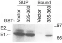
SinceEBNA-2and EBNA-1 both have Arg-Glydomains, we investigated whether the EBNA-2 Arg-Gly domain could mediate an interaction between EBNA-2 molecules or be-tweenEBNA-2and EBNA-1.EBV-infected cell proteins were incubated with GST-E2,335-360 or with GST control beads, andthe proteins which bound to the beads were analyzed with anEBV-immune human serum.About5% of the EBNA-1 in
-21
0 (D CM C %J C-4
CD C) CO CO) cO CY) CO t N
U) 0 0 0 6
[image:5.612.327.548.74.282.2][32P]GST- X - -v
FIG. 5. Far-Western analysis showing directbinding of EBNA2 region 3 to cell proteins. Total cell proteins were subjected to
SDS-PAGE andtransferredtonitrocellulose. GST fusionproteins, 32p labeledinvitroby protein kinase A,wereusedtoprobethe filter.(A) Proteins from IB4 cells (EBV+) were probed with labeled fusion protein GST-E2,335-360, GST-E2,310-336, or GST-E2,310-432. (B)
Proteins from BJAB(EBV-)orIB4(EBV+)cellswereprobedwith labeled GST-E2,310-432. (C) Purified fusion proteins GST-E2,335-376, GST-E2,335-360, GST-E2,310-336, and GST-E2,310-432 were
subjected to SDS-PAGE, transferred to nitrocellulose, and probed with labeledGST-E2,310-432. Sizesareindicatedinkilodaltons.
the lysate bound to GST-E2,335-360 beads and not to GST beads. EBNA-2 was not detected under the same conditions, althoughasimilar level of EBNA-2 boundtoGST-E2,335-360 beads would not be detectable with available sera (Fig. 6). Attempts to detect an interaction between EBNA-2 and EBNA-1 by coimmunoprecipitation from EBV-infected cell nuclear extracts with the EBNA-2-specific monoclonal anti-body PE2 have not been successful, possibly because of the limited sensitivity of EBNA-2 and EBNA-1 antisera. Never-theless, these data raise the possibility that EBNA-2 can interact with EBNA-1or itselfthrough Arg-Gly domains.
Among the cellproteins which bindtolabeled GST-E2,310-432 is aprotein similar in size tohistone Hi (Fig. 5A). Since EBNA-2 deleted for the Arg-Gly domain differs from wt EBNA-2 in association with the chromatin fraction and
SUP Bound
C o
CDco o CDc
0 0 c')
0 LO
a) g) co
GST-~> cv) C')
E2
E1/---97
66
FIG. 6. EBNA-1is able toassociate withthe Arg-Gly domain of EBNA-2. Extracts of IB4 cells were incubated with GST-E2,335-360 or with GST(Vector).Proteins boundtoGST beads and proteins left in the supernatants (SUP) after incubation were subjected to SDS-PAGE, transferred to nitrocellulose, and probed with an immune human serum.Thepositionsof EBNA-1 and EBNA-2 areindicated. Sizesareindicatedinkilodaltons.
.,t
401v4ftmwr.
.,
'-...
"D
on November 9, 2019 by guest
http://jvi.asm.org/
[image:5.612.116.239.75.276.2] [image:5.612.383.491.580.655.2]CL
B_
-66_
31...
A Bound
<: 0 o z z
0 o
oL
oL
CD-c
116_ 97-66_
Bound
<: a o z z
0 0 a a0 0
CL r q cn '
SUP SUP
< CD o Z z cd C C o Z z C
aO '5 X 0 ao O 0. a lo o
mL XL IL 1 ct) 9 rL al 0 X tn
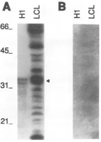
FIG. 7. HistoneHiis detectedonfar-Western blots by the labeled Arg-Gly repeat. (A) Purified histoneHi (lane H1) or proteins from IB4 cells (lane LCL) were subjected to SDS-PAGE, transferred to nitrocellulose, and probed with 32P-labeled GST-E2,335-360. The position of histone Hi is indicated by an arrowhead. In panel B, soluble histone Hi was added to thehybridization buffer. Sizes are indicated inkilodaltons.
EBNA-2 has been reported to bind to histone
Hi
(21), we furtherinvestigatedwhether the Arg-Gly domain might medi-atethatinteraction. In fact, labeled GST-E2,335-360 bound to purified histone Hi or to histoneHi
from nuclear extracts (Fig. 7A).Furthermore, soluble histoneHi
when added to the probe buffer prevented the binding of labeled GST-E2,335-360 to histone Hi and to other cellular proteins immobilized on filters (Fig. 7B). These experiments indicate that the Arg-Gly domaincanbindtohistoneHi
and thathistoneHi
canblock thebindingoftheArg-Gly domain to most other proteins even though histoneHi and the Arg-Gly repeat are both positively charged proteins.The Arg-Gly repeat can interact with nucleic acids and mediates much of the EBNA-2 interaction with nucleic acids. EBNA-2also bindstoboth ssDNA- and dsDNA-cellulose(14), but the DNA binding domain is not known. To investigate whethertheArg-Gly domain could mediate EBNA-2 interac-tion with nucleicacids,
32P-labeled
GST-E2,335-360was incu-bated with ssDNAordsDNA immobilizedoncelluloseorwith poly(A), poly(C),poly(G),orpoly(U)immobilized on agarose beads, and the bound fusion proteinwas quantitated by the amount ofradioactivityretained on the matrix. GST-E2,335-360 bound bettertossDNAthantodsDNA and bound besttopoly(G) (Table 1). GST-E2,335-360 didnotbind
significantly
tocelluloseor topoly(A)-, poly(C)-,orpoly(U)-agarose (datanotshown).
[image:6.612.122.225.72.217.2]To evaluate the role of the Arg-Gly domain in EBNA-2
TABLE 1. Binding of the Arg-gly Domaintonucleic acids
Nucleic Degree ofbinding"
acid' Expt 1 Expt 2 Expt 3 Expt 4 Expt5 Expt 6 Avg
ssDNA 26.6 126.7 122.6 91 25 32.3 70.7±44.2
dsDNA 9.2 NA NA NA 7.2 10.4 89±1.3
Poly(G) 386 534.6 259.9 692 NA NA 468± 161
aNucleic acidscoupledtomatrixwereused for incubation with32P-labeled
GSTfusionproteins.
b Definedas countsretainedby matrix afterincubation withGST-E2,335-360/
counts retained by matrix after incubationwith irrelevant protein. NA, not
available.
116_ 97-66_
E2d337-354 E2WT
FIG. 8. Binding ofwtEBNA 2orE2d337-354tonucleic acids. (A) BJAB cells were transfected with pSG5-E2,d337-354. Wild-type
EBNA-2wasexpressed from IB4 cells (E2WT). Lysateswere made
fromIB4 orfrom BJABtransfectants, and aliquots were incubated
with eachtypeof nucleic acid coupledtomatrix: ssDNA-or
dsDNA-cellulose (sDNA ordDNA), control cellulose (celo), orpoly(A) or
poly(G)-agarose (polA orpolG). Bound proteinswere subjectedto
SDS-PAGE,and EBNA-2wasdetectedbyWesternblotting.Inpanel B, 10% of the proteins from the startinglysatesorfrom the superna-tants(SUP) after incubation with nucleic acidasinpanel Awererun on agel,and EBNA-2wasidentifiedbyimmunoblotting. Positions of
EBNA-2areindicatedby arrowheads. Sizesareindicated in
kilodal-tons.
interaction with nucleic acid, extracts from B lymphocytes expressing wt EBNA-2 or E2d337-354 were passed through
poly(A)-or poly(G)-agarose orssDNA- ordsDNA-cellulose.
As is characteristic of the Arg-Gly domain alone, EBNA-2 bound to ssDNA- and dsDNA-cellulose and even more
strongly to poly(G)-agarose. In contrast, E2d337-354 bound somewhat less to ssDNA- and dsDNA-cellulose and very
poorlyto poly(G)-agarose (Fig. 8A). Very little wtEBNA-2 passed through ssDNA-or dsDNA-cellulose or
poly(G)-aga-rose, while more E2d337-354 passed through ssDNA- or
dsDNA-cellulose and almost all of the E2d337-354 passed through poly(G)-agarose (Fig. 8B). Thus, EBNA-2 binds to ssDNA, dsDNA,orpoly(G);moreover, theArg-Glyrepeatis
an important component of the ssDNA and dsDNAbinding activity and thekeycomponentofpoly(G) bindingactivity.
Poly(G),poly(dG),andpoly(I)canformverystablequartet structures (43,53),which could underlie thespecificityofthe Arg-Glyrepeatforpoly(G)asopposedtopoly(C),poly(U),or
poly(A).Toinvestigatewhether theabilityofpoly(G) toform quartet structures underlies the interaction, poly(dG)15 or
poly(I)wasincubated in 0.1or1Msalt under conditions which would favorquartet formation and tested as competitors for
poly(G) interaction with GST-E2,335-360. While soluble poly(G) in 10-foldexcesscould effectivelyblock GST-E2,335-360 binding to poly(G)-agarose, poly(dG) and poly(I) were
much less effective.Furthermore,although methylationofthe
N-7 position in G by dimethyl sulfate interrupts hydrogen bonding between neighboring guanine bases and prevents quartet formation (53), methylated poly(dG) had no less
blocking activity thanpoly(dG) (Table 2). Thus, theArg-Gly B
on November 9, 2019 by guest
http://jvi.asm.org/
[image:6.612.335.548.72.273.2] [image:6.612.64.305.619.683.2]TABLE 2. Block of bindingtopoly(G) by G-quartetstructurea Degree ofblocking"
Blocker
Expt 1 Expt2 Expt3 Expt4 Avg
None 1.0 1.0 1.0 1.0 1.0
Poly(dG) 0.88 0.26 0.35 0.62 0.53±0.24
Methylated poly(dG) 0.93 0.45 0.35 0.70 0.61 ±0.22
Poly(I) 0.21 0.19 0.80c 0.52C 0.43±0.25
Poly(G) 0.016 0.015 0.043C 0.018C 0.023±0.011
a32P-labeled GST-E2,335-360 was preincubated with a 10-fold excess of
blocker beforebeing addedtopoly(G)-agarose.Thesalt is 100 mMNaCloras
indicated.
bCalculated as countsretainedbypoly(G) with blocker/counts retained by poly(G)without blocker.
cThe salt usedis1M KCI.
repeatspecifically binds to poly(G), and the G-quartet struc-tureprobably isnotthebasisfor thespecificityforpoly(G).
Activityof EBNA-2region 3 deletionmutants on transacti-vation. Since LMP1 is essential for primary B-lymphocyte growth transformation and EBNA-2 is an important transac-tivator of the LMP1 promoter, one essential function for EBNA-2 in transformation is through LMP1 promoter trans-activation. To determine if E2d337-354 has an adverse effect onLMP1transactivationwhich wouldaccountfortheadverse effects on cell growth transformation, transactivation by E2d337-354, E2d359-383,andE2d385-430wascomparedwith transactivationbywtEBNA-2as apositivecontrol(48) orby E2d333-425 as a nullmutant control (9). The EBNA-2 open reading frames were cloned into thepSG5 expression vector and transfected into BJAB cellsalongwith areporterplasmid consisting of the CAT gene under the control of thethymidine kinase promoter and LMP1 upstream sequence (-234/ +40LMPCAT) (48).A,B-galactosidaseexpression plasmidwas included in each transfection as an internal control. Surpris-ingly,E2d337-354 induced four-tofivefold-higherCAT activ-ity than wt EBNA-2, while E2d359-383, E2d385-430, and E2d333-425werenearly asactive as wtEBNA-2(Table 3).
Toinvestigate the relativeefficiency of pSG5-E2,d337-354 in transient transactivation of the LMP1 gene from EBV
epi-someswhichareinnucleosomalstructures(15),pSG5-EBNA 2, pSG5-E2,d337-354, or pSG5-E2,d333-425 was transfected into BL30/P3HR-1 cells. Since the P3HR-1 genome lacks EBNA-2, there is little LMP1 protein in BL30/P3HR-1 cells, but expression can be induced by wtEBNA-2 (9). In
BL3O/
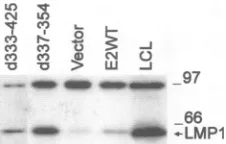
[image:7.612.381.494.72.144.2]P3HR-1 cells, the effects ofwtEBNA-2orofE2d337-354on LMP1 expression paralleled the effects on LMP1CAT plas-mids.E2d337-354inducedtwo- tofourfold-higher LMP1 levels than didwtEBNA-2 (Fig. 9 and data not shown).Although EBNA-2deleted for all of region 3 has been reported to be a null mutant in inducing LMP1 expression in BL30/P3HR-1 cells(9),theprevious result was probably due to experimental error,sincerepetition of the previous experiment showed that
TABLE 3. Summary of CAT assays
CATactivity' (mean ±SD)
Promoter
wt E2d337-354 E2d333-425 E2d359-383 E2d385-430 LMP1 4.9±2.0 27.7± 10.6 3.0±1.0 5.2±2.6 5.2± 1.4 Cp 2.9±0.6 2.6±0.5 0.8±0.3 1.6±0.3 3.2± 1.2
LO 19
CN U)
c' N
'a > w -J
mm
._ _* _ __-97 _66
dl_.-LMP1
FIG. 9. Transactivation of LMP1 from the P3HR-1 genome by EBNA-2 and region3deletions. BL30/P3HR-1cellsweretransfected
with pSG5-EBNA 2 (E2WT), the pSG5 vector control (Vector), pSG5-E2,d337-354 (d337-354), or pSG5-E2,d333-425 (d333-425). Cellswereharvested,andlysatesfromtransfectantsorfromIB4cells
(LCL)were immunoprecipitatedwith monoclonalantibodyS12. The
immunoprecipitatesweresubjectedtoSDS-PAGE.LMP1proteinwas
detectedby monoclonal antibodyS12.Sizeswereindicated in kilodal-tons.
E2d333-425wasactive intransactivation of LMP1 gene(Fig.9 and reference 6a). Thus, E2d333-425 hasa nulltransforming phenotype and a near wt LMP1 transactivating phenotype, while E2d337-354 is deficient in transformation and has greaterthanwtLMP1 transactivating activity.
EBNA-2 has also been shown to transactivate the Cp promoterthroughanupstreamelement which is similartothe LMP1 element in having one J kappa binding site (22, 32). EBNA-2, E2d337-354, E2d359-383, E2d385-430, or E2d333-425was transfected into BJAB cells with a CpCAT reporter plasmid, and CATactivitywasassayed(55). In contrast tothe results with the LMP1 promoter, E2d337-354wassimilartowt EBNA-2 in Cp transactivation. E2d333-425 and E2d359-383 appeared to be deficient in transactivation, and E2d385-430 was similar to wtEBNA-2. These data suggest that EBNA-2 interacts differentlywith the LMP1 and Cp promoters, even
thoughthey both haveasingleJkappabinding site. DISCUSSION
These experiments confirm that region 3 is critical to primaryB-lymphocyte growthtransformation and identify the Arg-Glyrepeat domainas an importantcomponent ofregion 3. Deletion of theArg-Glyrepeat domain reduces transform-ing efficiency. The transformed cells grow slowly and are
persistently difficult to maintain in culture. In contrast, inde-pendentdeletions of each part of the restofregion 3 haveno adverseeffectonprimaryB-lymphocyte growth transformation or on thegrowth of the transformed cells. The only region 3 residuesnotindependentlyinvestigatedin this study are aa 333 to 336, 355 to 358, and 384. These few residues are highly unlikely to constitute essential interactive domains, since de-letions of immediately adjacent sequences do not affect trans-formation. Thus, the data indicate that region 3 does not interact with anessential mediator of cell growth transforma-tion.
What then is the basis for the stringent requirement for region 3 in primary B-lymphocyte growth transformation? From anEBVrecombinant genetic perspective, the stringent requirement is probably the sum of several less significant effects. The Arg-Gly domain deletion results in less efficient transformation, and the transformed cells grow poorly. The restofregion 3 when independently deleted as two parts has no significant effect on cell growth transformation, but when deletedas onesegment ithas a substantial effect on transfor-mation efficiency. The Arg-Gly and other region 3 deletions probably cooperatively result in the null transforming effect characteristic of the wholeregion 3 deletion.
aCATactivityfromcotransfectionwith EBNA-2expressionvectordivided by CATactivity from cotransfection withthe pSG5 vector control. Each value was
determinedfrom at least fiveindependentexperiments.
on November 9, 2019 by guest
http://jvi.asm.org/
[image:7.612.57.299.85.165.2]From a biochemical perspective, region 3 (aa 333 to 425) joins region 2 (aa 310 to 336) to the region 4 (aa 430 to
464) acidic transactivating domain. Region 2 is a proline-rich
sequence,
L-31OHNLPSPPWWPPICDPPPQPSKIQGS-336
throughwhichEBNA-2bindstothe cell protein, J kappa (22, 55). J kappa directs EBNA-2 toaGTGGGAAsequence that
is a component of all EBNA-2 response elements. GPPWW PPXXDP is likely to be the core of the interacting domain,
sincetheunderlined residuesare thoseconserved between the
EBNA-2genesof EBVtypes1 and 2(12)and mutation of the WWtoSS orFFablates J kappainteraction (22, 55). EBNA-2
with the WW-to-SS mutation is inactive in marker-rescuing transforming activity, providingagenetic link between J kappa
interaction and cell growth transformation (55). Residues 333 to 336 are not likely to be important in J kappa interaction, since the type 2 EBV sequence is PPTN instead of QGQS. Therefore,thedeletion of these residuesin the whole region 3 deletion is not likely to be an important component of the effect of thelargerdeletion,although this alternativecannotbe completely dismissed. Rather, the impressive effects of the region 3 deletionontransformation are morelikelytobe due tothe tight approximation of the acidic andJkappabinding domainswhich may disrupt the interaction betweenEBNA-2
andJ kappa. The negative effect of deletion E2d361-425 on
transformation may have a similarbasis. The deletion of the
sequencesbetweentheacidic domain and the highly charged Arg-Gly domain mayaffect the activities of the Arg-Gly, the acidic,ortheJkappadomain.
An expectation of this simple model is that the region 3 deletionwould haveasubstantialnegative effect on
transacti-vation. Such an effectisobserved ontransient transactivation
of theCppromoter. However,incontrasttoapreviousreport (9), the region 3 deletion has little or no effect on transient
transactivation of the LMP1 promoter. The LMP1 and Cp promoters each have a single J kappa binding site (32).
Deletion ofthe site from the Cp promoter ablates transacti-vation(25),while deletion fromthe LMP1promoterdoesnot totallyablate LMP1transactivation(17). Thus, EBNA-2must interact with another protein(s) which recognizes the LMP1 promoter.
Theregion 3 deletion has the expectednegative effectonJ kappa-mediated transactivationof the Cp promoter andmay
also have a similar effect on transactivation of the LMP1
promoter. However,theputative negativeeffectonthe LMP1
promoter maybe counterbalancedbyapositiveeffectonthis promoter from theArg-Glydeletion.
TheArg-Glydomain islikelytobeanegativemodulator of
EBNA-2 interactions witha second component of the LMP1
promoter. Although deletion of this domain results in de-creased transforming efficiency, poor growth of the trans-formed cells, and decreased EBNA-2 chromatin association, EBNA-2deleted for theArg-Glydomain transactivatestheCp promoter and has abnormally high transactivatingeffects on
the LMP1 promoter. The supranormal effect on the LMP1 promoter in transient transactivation assays indicates that an
Arg-Gly interaction negatively regulates the LMP1 promoter. The absence ofasimilar effectontheCppromoter suggeststhat the effectis notmediatedthroughJkappaorthroughinteractions between the Arg-Gly and acidic transactivating domains and
favorsthe possibility that theArg-Glydomainnegativelyaffects theputative secondcomponentof the LMP1promoter.
TheArg-Glyrepeatishighly chargedand thereforelikelyto constitute a surface domain of EBNA-2. This domain can
interact with many proteins and nucleic acids. EBNA-2
ex-tracted from EBV-transformedcells ispartofalarge complex
[image:8.612.322.565.82.253.2](20), and the Arg-Gly domain may mediate some of these
TABLE 4. Summary ofproteins containing the Arg-Gly domain First amino
Proteina acid of
(reference) Arg-Gly Sequence domain
EBNA-2(3) 337 RGQS(RG)6KGKS
EBNA-1(3) 356 RGRERARGGSRERA(RG)4 SNF2 transactivator (30) 1505 (RG)9RP
DNA-methyltransferase (4) 990 (KG)6KH
Small nuclearRNP(42) 98 (RG)9 BTF3a(60) 13 RGRGRARG Papillomavirus E2 protein
BPV-4(38) 195 (RS)5
HPV-5(57) 267 (RS)2RH(RS)5KS HPV-8(18) 302 RP(RS)3RGRA
Splicing factor
U1-70K snRNP (16) 390 (RD)3RR(RD)3RERD
U2AF65 (58) 27 RSHS(RS)2RDRKR(RS)2
SF2/ASF(19) 204 (RS)8NS(RS)2
a RNP,ribonucleoprotein; BPV-4, bovine papillomavirustype4; HPV-5 and HPV-8, humanpapillomavirustypes5and 8; snRNP,small nuclear
ribonucle-oprotein.
interactions. The Arg-Gly domain may also mediate EBNA-2 association with chromatin, possibly through interaction with histoneHi.
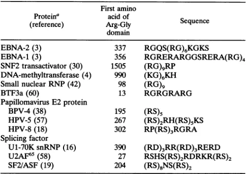
The interactions of the Arg-Gly repeats with other proteins and with nucleic acids are not likely to be simply related to positive-negative chargeeffects. HistoneHiis also a basic protein but binds to theArg-Gly domain, and the Arg-Gly repeat can bind toitself. Amongnucleicacids, onlypoly(G) has high affinity for thedomain. Argininehasalarge extendedguanidiniumside chain whichcaninteract withnucleotides(56). Similar Arg-Gly motifs are also present in other proteins (Table 4) involved in transcription regulation, such as SNF2, a yeast transactivator; BTF3a, a general transcription factor; or papillomavirus E2 protein, a regulator of viral gene expression. Notably, many splicing factors haveArg-Ser-richregions(Table 4),which medi-ateinteractions among splicing factors(2,5, 58,63).
ACKNOWLEDGMENTS
Bill Kaelin kindly provided the pGEX-2TK plasmid and helpful advice.JamesWilliamsoncontributed useful discussiononG-quartet
structure.We thankDavid-ThorleyLawsonfor monoclonalantibody S12,Alan Rickinson and Martin Roweformonoclonalantibody BZ.1, and Alan Wolffe and Stefan Dimitrov forthehistone Hi antibody. Qian Miao and LisaVaraprovided excellent technical assistance.
R.Y. is supported by Public Health Service National Research ServiceAwardAI08548-02; S.H. issupported TheNaitoFoundation. This researchwassupported by Public Health ServicegrantCA47006 from the National Cancer Institute.
REFERENCES
1. Abbot, S.D.,M.Rowe,K.Cadwallader,A.Ricksten, J.Gordon,F. Wang,L. Rymo, and A. B. Rickinson. 1990. Epstein-Barr virus nuclearantigen 2inducesexpression of thevirus-encoded latent membraneprotein. J. Virol.64:2126-2134.
2. Amrein, H., M. L. Hedley, and T. Maniatis. 1994. The role of specific protein-RNA andprotein-proteininteractions inpositive andnegativecontrol ofpre-mRNAsplicing bytransformer 2.Cell 76:735-746.
3. Baer, R, A. T.Bankier,M. D.Biggin,P. L.Deininger,P.J.Farrell, T.J.Gibson, G.Hatfull,G. S.Hudson,S. C.Satchwell,C.Seguin, P.Tufnell,andB. Barrell.1984. DNA sequence andexpressionof the B95-8Epstein-Barrvirusgenome.Nature(London)
310:207-211.
4. Bestor, T., A. Laudano, R. Mattaliano, and V. Ingram. 1988.
on November 9, 2019 by guest
http://jvi.asm.org/
Cloning and sequencing of acDNAencodingDNA methyltrans-ferase of mouse cells. The carboxyl-terminal domain of the mammalian enzymes is relatedto bacterial restriction methyltrans-ferases. J. Mol. Biol. 203:971-983.
5. Caceres, J. F., and A. R. Krainer. 1993. Functional analysis of pre-mRNA splicing factorSF2/ASF structural domains. EMBOJ. 12:4715-4726.
6. Calender, A., M. Billaud, J. P. Aubry, J. Banchereau, M. Vuil-laume, and G. M.Lenoir.1987. Epstein-Barr virus(EBV)induces expression of B-cell activation markers on invitro infection of EBV-negative B-lymphoma cells. Proc. Natl. Acad. Sci. USA 84:8060-8064.
6a.Cohen, J.Unpublished data.
7. Cohen, J.I. 1992. A region of herpes simplex virus VP16 can substitute for a transformingdomain ofEpstein-Barrvirusnuclear protein 2. Proc. Natl. Acad. Sci. USA89:8030-8034.
8. Cohen, J. I., and E. Kieff. 1991. An Epstein-Barr virus nuclear protein 2 domain essential fortransformation is adirect transcrip-tionalactivator. J. Virol. 65:5880-5885.
9. Cohen, J. I., F. Wang, and E. Kieff. 1991. Epstein-Barr virus nuclearprotein 2 mutations defineessential domains for transfor-mation andtransactivation. J. Virol. 65:2545-2554.
10. Cohen, J. I., F. Wang, J.Mannick, and E. Kieff. 1989. Epstein-Barr virus nuclear protein 2 is a key determinant of lymphocyte transformation. Proc.Natl. Acad. Sci. USA 86:9558-9562. 11. Cordier, M., A.Calender, M. Billaud, U. Zimber, G. Rousselet, 0.
Pavlish, J. Banchereau, T. Tursz, G. Bornkamm, and G. M. Lenoir. 1990. Stable transfection of Epstein-Barr virus (EBV) nuclear antigen 2 in lymphoma cells containing the EBVP3HR1 genome induces expression of B-cell activation molecules CD21 and CD23. J. Virol. 64:1002-1013.
12. Dambaugh, T., K. Hennessey, L.Chamnankit, and E.Kief. 1984. U2region ofEpstein-Barr virus DNAmay encode Epstein-Barr virus nuclear antigen 2. Proc. Natl. Acad. Sci.USA81:7632-7636. 13. Delling, U., S. Roy, S. M. Sumner, R. Barnett, L. Reid, C.A.Rosen, and N.Sonenberg. 1991. The number of positivelychargedamino acidsinthebasic domain of Tat is critical fortrans-activation and complexformation with TAR RNA. Proc. Natl.Acad. Sci. USA 88:6234-6238.
14. Dillner, J., H. Rabin, N. Letvin, W. Henle, G. Henle, and G. Klein. 1987. NuclearDNA-binding proteins determinedbythe Epstein-Barr virus-related simian lymphotropic herpesviruses H. gorilla, H. pan, H. pongo and H. papio. J. Gen. Virol. 68:1587-1596. 15. Dyson, P. J., and P. J. Farrell. 1985. Chromatin structure of
Epstein-Barr virus. J. Gen. Virol. 66:1931-1940.
16. Etzerodt, M., R. Vignali, G. Ciliberto, D. Scherly, I. W. Mattaj, and L. Philipson. 1988. Structure and expression of a Xenopus gene encoding an snRNP protein (Ul 70K). EMBO J. 7:4311-4321.
17. Fahraeus, R., A. Jansson, A.Sj0blom,T.Nilsson, G. Klein, and L. Rymo. 1993. Cell phenotype-dependent control ofEpstein-Barr virus latent membrane protein 1 gene regulatory sequences. Virology195:71-80.
18. Fuchs, P. G., T. Iftner, J. Weninger, and H. Pfister. 1986. Epidermodysplasia verruciformis-associated human
papillomavi-rus 8: genomic sequence and comparative analysis. J. Virol. 58:626-634.
19. Ge, H., P. Zuo, and J. L. Manley. 1991. Primary structure of the human splicing factor ASF reveals similarities with Drosophila regulators. Cell66:373-382.
20. Grasser, F. A., P. Haiss, S. Gottel, and L. N. Mueller. 1991. Biochemical characterization ofEpstein-Barr virus nuclear anti-gen2A. J. Virol. 65:3779-3788.
21. Grasser, F. A., C.Sauder,P.Haiss, A. Hille, S.Konig,S.
Gdttel,
E. Kremmer, H. P.Leinenbach, M. Zeppezauer, and L. N. Mueller. 1993. Immunological detection of proteins associated with the Epstein-Barrvirus nuclear antigen 2A.Virology195:550-560. 22. Grossman, S.R, E.Johannsen,X.Tong, R.Yalamanchili, and E.Kief. 1994. The Epstein-Barr virus EBNA-2 transactivator is directed toits responseelement by the JK recombination signal bindingprotein. Proc. Natl. Acad. Sci. USA91:7568-7572. 23. Hammerschmidt, W., and B. Sugden. 1989. Genetic analysis of
immortalisingfunctions ofEpstein-Barrvirus in human B
lympho-cytes. Nature(London) 340:393-397.
24. Higuchi, R. 1990. Recombinant PCR, p. 177-183. In M. A. Innis (ed.), PCR protocols: a guide to methods and applications. Academic Press, Inc., San Diego, Calif.
25. Jin, X. W., and S. H. Speck. 1992. Identification of critical cis elements involved inmediating Epstein-Barr virus nuclearantigen 2-dependent activityof an enhancerlocated upstream of the viral BamHI C promoter. J. Virol. 66:2846-2852.
26. Kaelin, W. J., D.C. Pallas, J. A.DeCaprio, F.J. Kaye,and D. M. Livingston. 1991. Identification of cellularproteins thatcan inter-act specifically with theT/ElA-binding region of the retinoblas-toma geneproduct. Cell 64:521-532.
27. Kaye,K. M., K. M. Izumi, and E. Kieff. 1993. Epstein-Barr virus latent membrane protein 1 is essential for B-lymphocyte growth transformation. Proc. Natl. Acad. Sci. USA90:9150-9154. 28. King, W., A. Thomas-Powell, N. Raab-Traub, M. Hawke, and E.
Kieff. 1980.Epstein-Barr virus RNA. V. Viral RNAin a restrin-gently infected, growth-transformed cell line. J. Virol. 36:506-518. 29. Knutson, J. C. 1990. The level of c-fgr RNA is increased by EBNA-2, an Epstein-Barr virus gene required for B-cell immor-talization. J. Virol. 64:2530-2536.
30. Laurent, B. C., M. A.Treitel, and M. Carlson. 1991.Functional interdependence of the yeast SNF2, SNF5, and SNF6 proteins in transcriptional activation. Proc. Natl. Acad. Sci. USA 88:2687-2691.
31. Liebowitz, D., and E. Kieff. 1993. Epstein-Barr virus, p. 107-172. In B. Roizman, R. J. Whitley, and C. Lopez (ed.), The human herpesviruses. Raven Press Ltd., New York.
32. Ling, P. D., D. R. Rawlins, and S. D. Hayward. 1993. The Epstein-Barr virus immortalizing protein EBNA-2 is targeted to DNA by a cellular enhancer-binding protein. Proc. Natl. Acad. Sci. USA 90:9237-9241.
33. Longnecker, R., C. L. Miller, X. Q. Miao, B. Tomkinson, and E. Kieff. 1993. The last seven transmembrane and carboxy-terminal cytoplasmic domains of Epstein-Barr virus latent membrane pro-tein 2 (LMP2) are dispensable for lymphocyte infection and growth transformation in vitro. J. Virol. 67:2006-2013.
34. Longnecker, R., C. L. Miller, B. Tomkinson, X. Q. Miao, and E. Kieff. 1993. Deletion of DNA encoding the first five transmem-brane domains of Epstein-Barr virus latent memtransmem-brane proteins 2A and 2B. J. Virol. 67:5068-5074.
35. Mannick, J. B., J.I. Cohen, M. Birkenbach, A. Marchini, and E. Kief. 1991. The Epstein-Barr virus nuclear protein encoded by the leader of the EBNA RNAs is important in B-lymphocyte trans-formation. J. Virol. 65:6826-6837.
36. Menezes, J., W. Leibold, G. Klein, and G. Clements. 1975. Establishment and characterization of an Epstein-Barr virus (EBV)-negative lymphoblastoid B cell line (BJA-B) from an exceptional, EBV-genome-negative African Burkitt's lymphoma. Biomedicine 22:276-284.
37. Miller, C. L., J. H. Lee, E. Kieff, and R. Longnecker. 1994. An integral membrane protein (LMP2) blocks reactivation of Epstein-Barr virus from latency following surface immunoglobulin crosslinking. Proc. Natl. Acad. Sci. USA 91:772-776.
38. Patel,K. R, K. T. Smith, and M. S. Campo. 1987. The nucleotide sequence and genome organization of bovine papillomavirus type 4. J. Gen.Virol. 68:2117-2128.
39. Petti, L., C. Sample, and E. Kieff. 1990. Subnuclear localization andphosphorylation of Epstein-Barr virus latent infection nuclear proteins. Virology 176:563-574.
40. Raab-Traub, N., T. Dambaugh, and E. Kieff. 1980. DNA of Epstein-Barr virus VIII: B95-8, the previous prototype, is an unusual deletion derivative. Cell 22:257-267.
41. Rabson, M., L. Gradoville, L. Heston, and G. Miller. 1982. Nonimmortalizing P3J-HR-1 Epstein-Barr Virus: a deletion mu-tantof its transforming parent, Jijoye. J. Virol.44:834-844. 42. Rokeach, L. A., J. A. Haselby, andS.0.Hoch. 1988. Molecular
cloningof a cDNA encoding the human Sm-D autoantigen. Proc. Natl.Acad. Sci. USA85:4832-4836.
43. Sen,D.,and W. Gilbert. 1990. A sodium-potassium switch in the formation offour-stranded G4-DNA. Nature (London) 344:410-414.
44. Sung, N. S., S. Kenney, D. Gutsch, and J. S. Pagano. 1991.
on November 9, 2019 by guest
http://jvi.asm.org/
EBNA-2 transactivates a lymphoid-specific enhancer in the BamHI CpromoterofEpstein-Barr virus. J. Virol. 65:2164-2169. 45. Swaminathan,S., B.Tomkinson,and E.Kieff. 1991. Recombinant Epstein-Barrvirus withsmallRNA (EBER) genes deleted trans-formslymphocytesand replicates in vitro. Proc.Natl. Acad. Sci. USA88:1546-1550.
46. Tomkinson, B., and E. Kieff. 1992. Use of second-site homologous recombination to demonstrate that Epstein-Barr virus nuclear protein 3B is not important for lymphocyte infection or growth transformation in vitro. J. Virol. 66:2893-2903.
47. Tomkinson, B., E. Robertson, and E. Kieff. 1993. Epstein-Barr virus nuclear proteins EBNA-3A and EBNA-3C are essential for B-lymphocytegrowth transformation. J. Virol. 67:2014-2025. 48. Tsang, S. F., F.Wang, K. M. Izumi, and E. Kieff. 1991. Delineation
ofthe cis-acting element mediating EBNA-2 transactivation of latentinfection membrane protein expression. J. Virol. 65:6765-6771.
49. Wang, F., C. Gregory, C. Sample, M. Rowe, D. Liebowitz, R. Murray, A.Rickinson,and E. Kieff.1990.Epstein-Barr virus latent membrane protein (LMP1) and nuclear proteins 2 and 3C are effectors ofphenotypic changes inBlymphocytes: EBNA-2and LMP1 cooperatively induce CD23.J.Virol. 64:2309-2318. 50. Wang, F.,C. D.Gregory,M.Rowe, A. B.Rickinson,D.Wang, M.
Birkenbach, H. Kikutani, T. Kishimoto, and E. Kieff. 1987. Epstein-Barr virus nuclear antigen 2specifically induces expres-sion of theB-cell activation antigen CD23. Proc. Natl.Acad.Sci. USA84:3452-3456.
51. Wang,F., H. Kikutani, S. F. Tsang, T. Kishimoto, and E. Kieff. 1991. Epstein-Barr virus nuclear protein 2 transactivates a cis-acting CD23 DNAelement.J.Virol. 65:4101-4106.
52. Wang, F.,S. F. Tsang, M. G.Kurilla,J.I. Cohen,and E. Kieff. 1990. Epstein-Barr virus nuclear antigen2 transactivates latent membraneproteinLMP1. J.Virol. 64:3407-3416.
53. Williamson, J. R.,M. K. Raghuraman, and T. R. Cech. 1989. Monovalent cation-induced structure of telomeric DNA: the
G-quartet model. Cell 59:871-880.
54. Wysokenski, D. A., and J.L. Yates. 1989. Multiple EBNA1-binding sitesarerequiredtoform anEBNA1-dependent enhancer and to activate a minimal replicative origin within oriP of Epstein-Barr virus. J. Virol. 63:2657-2666.
55. Yalamanchili, R., X. Tong, S. R Grossman, E. Johannsen, and E. Kieff.Genetic and biochemical evidence that EBNA 2 interaction with a 63kd cellular GTG-binding protein is essential for B lymphocyte growth transformationby EBV. Virology, in press. 56. Yarus, M. 1988. Aspecific amino acidbindingsitecomposed of
RNA.Science 240:1751-1758.
57. Zachow, K. R,R S. Ostrow, and A. J. Faras. 1987. Nucleotide sequenceandgenomeorganizationofhuman papillomavirustype 5. Virology 158:251-254.
58. Zamore, P.D., J. G.Patton,andM. R Green. 1992.Cloning and domainstructureof the mammaliansplicing factorU2AF. Nature (London) 355:609-614.
59. Zapp, M.L.,T. J. Hope, T. G. Parslow, and M. R Green. 1991. Oligomerization andRNAbinding domainsof the type 1human immunodeficiency virus Rev protein: a dual function for an arginine-rich binding motif.Proc.Natl.Acad.Sci.USA 88:7734-7738.
60. Zheng, X. M., D. Black, P. Chambon, and J. M. Egly. 1990. Sequencingandexpression of complementaryDNA forthe gen-eraltranscription factor BTF3.Nature(London) 344:556-559. 61. Zimber-Strobi, U., E. Kremmer, F. Grasser, G. Marschall, G.
Laux, andG. W. Bornkamm.1993. TheEpstein-Barr virus nuclear antigen2interacts withanEBNA2responsive cis-element of the terminalprotein 1 gene promoter.EMBO J.12:167-175. 62. Zimber-Strobl, U., K. 0. Suentzenich, G. Laux, D. Eick, M.
Cordier,'A. Calender, M. Billaud, G. M. Lenoir, and G. W. Bornkamm. 1991.Epstein-Barr virus nuclearantigen2activates transcription of theterminal proteingene.J.Virol. 65:415-423. 63. Zuo, P., andJ.LManley. 1993. Functional domains of the human
splicing factorASF/SF2. EMBOJ. 12:4727-4737.