0022-538X/96/$04.0010
Copyrightq1996, American Society for Microbiology
Branched Structures in the Intracellular DNA of Herpes
Simplex Virus Type 1
ALBERTO SEVERINI,1* DOUGLAS G. SCRABA,2
ANDD. LORNE J. TYRRELL1
GlaxoWellcome Heritage Research Institute, Department of Medical Microbiology and Immunology,1and Department of Biochemistry,2University of Alberta,
Edmonton, Alberta, Canada T6G 2H7
Received 3 July 1995/Accepted 16 February 1996
Herpes simplex virus type 1 (HSV-1) replication produces large intracellular DNA molecules that appear to be in a head-to-tail concatemeric arrangement. We have previously suggested (A. Severini, A. R. Morgan, D. R. Tovell, and D. L. J. Tyrrell, Virology 200:428–435, 1994) that these DNA species may have a complex branched structure. We now provide direct evidence for the presence of branches in the high-molecular-weight DNA produced during HSV-1 replication. On neutral agarose two-dimensional gel electrophoresis, a technique that allows separation of branched restriction fragments from linear fragments, intracellular HSV-1 DNA produces arches characteristic of Y junctions (such as replication forks) and X junctions (such as merging replication forks or recombination intermediates). Branched structures were resolved by T7 phage endonuclease I (gene 3 endonuclease), an enzyme that specifically linearizes Y and X structures. Resolution was detected by the disappearance of the arches on two-dimensional gel electrophoresis. Branched structures were also visualized by electron microscopy. Molecules with a single Y junction were observed, as well as large tangles containing two or more consecutive Y junctions. We had previously shown that a restriction enzyme which cuts the HSV-1 genome once does not resolve the large structure of HSV-1 intracellular DNA on pulsed-field gel electrophore-sis. We have confirmed that result by using sucrose gradient sedimentation, in which both undigested and di-gested replicative intermediates sediment to the bottom of the gradient. Taken together, our experiments show that the intracellular HSV-1 DNA is held together in a large complex by frequent branches that create a net-work of replicating molecules. The fact that most of these branches are Y structures suggests that the netnet-work is held together by frequent replication forks and that it resembles the replicative intermediates of bacterio-phage T4. Our findings add complexity to the simple model of rolling-circle DNA replication, and they pose interesting questions as to how the network is formed and how it is resolved for packaging into progeny virions.
The herpes simplex virus type 1 (HSV-1) genome is a linear, double-stranded DNA of 152 kb. It comprises a unique long (UL) region and a unique short (US) region, each flanked by
inverted repeats (18, 25, 37). Within several hours of the ini-tiation of infection, the genome undergoes circularization (17, 31) by a mechanism which is still unknown. Subsequent repli-cation leads to the formation of large DNA complexes com-posed of many genome units (21, 22). Most of the genomic termini are fused together in a head-to-tail concatemeric ar-rangement (21–23). These findings have led to the hypothesis that HSV-1 (and probably other herpesviruses) replicates by a rolling-circle mechanism, in which the replication fork moves endlessly around the circular genome, synthesizing a long string of linear concatemers (21). This model has been sup-ported by experiments using defective viruses (45), plasmids containing an HSV-1 origin of replication (44), and an HSV-1 replication system reconstituted in baculovirus (42).
Homologous recombination is also a prominent feature of HSV-1 DNA replication. Frequent recombination between in-verted repeats leads to inversion of the ULand USregions of
the genome, producing progeny molecules which display the four possible orientations of the genome in equimolar amounts (8, 18). The fact that consecutive molecules in a concatemer can be in opposite orientations (34, 50) indicates that inversion of the unique regions occurs throughout the replicative process and not just at the initial stages of circularization or early
replication. The frequency of recombination has been esti-mated to be 1 to 3% per 1 kb of the viral genome (39). It has also been suggested that specific sequences at the end of the inverted repeats (a regions) are especially recombinogenic (13–15, 26, 40), although recombination may occur anywhere in the HSV-1 genome (39, 47). Sarisky and Weber (33) pro-posed that recombination is facilitated by the presence of free genomic ends normally produced at the terminal a sequences during packaging by putative terminases that cleave monomer unit fragments and leave the left end of the genome hanging from the mass of replicating DNA (7, 34, 44). These findings raise an interesting parallel with the mode of replication of T4 phage, in which recombination between a free end and an internal homologous sequence gives rise to a replication fork, creating a network of branched concatemers (reviewed in ref-erence 29). We have proposed in a theoretical paper how this mechanism may apply to the circularization and replication of the HSV-1 genome (28).
Studies of the structure of the large intracellular HSV-1 DNA by us (34) and others (50), using partial digestion and pulsed-field gel electrophoresis (PFGE), confirmed a head-to-tail concatemeric arrangement but failed to detect significant amounts of linear concatemers of two or more genome units. Our PFGE studies showed that most of the high-molecular-weight HSV-1 DNA could not be resolved into linear mole-cules able to migrate on PFGE by a restriction endonuclease that cuts the genome once. We interpreted these results as consistent with the presence of frequent branches in the intra-cellular HSV-1 DNA (34). Branches in DNA can be studied by neutral two-dimensional (2D) gel electrophoresis. Restriction * Corresponding author. Phone: (403) 5285. Fax: (403)
492-9828. Electronic mail address: [email protected]. ualberta.ca.
3169
on November 9, 2019 by guest
http://jvi.asm.org/
fragments are separated in the first dimension at a low voltage and a low agarose percentage, and they migrate in accordance with their molecular weights. The second dimension of elec-trophoresis is carried out at a high voltage and a high percent-age of agarose and in the presence of ethidium bromide. Un-der these conditions, DNA fragments containing branches are more likely to become entangled in the agarose fibers and migrate more slowly, depending on the position of the branch within the fragment. As a result, branched fragments form arches above the line on which linear fragments migrate. From the shape of the arches it is possible to infer the probable structure of the fragments: Y branches, X branches, or bub-bles. A detailed description of the theory and practice of 2D agarose gel electrophoresis of branched DNA fragments has been given by Brewer and Fangman (4).
In this communication, we describe experiments with neutral 2D gel electrophoresis, electron microscopy (EM), and sucrose gradient sedimentation which demonstrate the presence of branched structures in the high-molecular-weight intracellular HSV-1 DNA.
MATERIALS AND METHODS
Viruses, cells, and HSV-1 DNAs.Vero cells were infected with HSV-1 PAC1DTK at a multiplicity of 10 PFU per cell and incubated in minimal essen-tial medium (GIBCO) containing 4% calf serum, 100 U of penicillin per ml, and 100mg of streptomycin (GIBCO) per ml. HSV-1 PAC1DTK is a thymidine kinase (TK)-negative mutant derived from HSV-1 KOS by insertion of a PacI site in the TK gene (34). Cells were harvested when they showed a 100% cytopathic effect (14 to 22 h postinfection).
Infected-cell DNA was purified in agarose blocks as previously described (34). High-molecular-weight HSV-1 DNA was prepared by separation of the infected-cell DNA on PFGE and excision of the gel origin. For a detailed description of these procedures and conditions, see reference 34.
2D agarose gel electrophoresis.Agarose plugs containing intermediates of HSV-1 DNA replication were melted at 658C in the presence of 50 mM Tris (pH 8.0)–10 mM MgCl2–100 mM NaCl, equilibrated at 378C, and digested with
BamHI. The DNA was purified from the agarose by the GeneClean method (Bio 101, Inc.) and subjected to 2D gel electrophoresis (4). Briefly, 2 to 5mg of DNA was loaded on a 0.5% agarose gel and electrophoresed at 1 V/cm for 18 h in 0.53 Tris-borate buffer (45 mM Tris HCl, 45 mM boric acid, 1 mM EDTA, pH 8.2). The gel was stained with ethidium bromide, and the lane containing the sepa-rated BamHI fragments (from about 30 to 2 kb) was excised and run in the second dimension in 1% agarose at 5 V/cm for 3 h in 0.53Tris-borate buffer plus 0.5mg of ethidium bromide per ml. The gel was then blotted onto nylon filters (Hybond N; Amersham) and hybridized with radioactive probes (see below) as described previously (34).
Digestion with resolvases.Phage T7 endonuclease I (kindly provided by A. R. Morgan, University of Alberta, Edmonton, Alberta, Canada) was purified from Escherichia coli transfected with a plasmid expressing phage T7 gene 3 as pre-viously described (9, 11). The purified protein produced only one band on a sodium dodecyl sulfate-polyacrylamide gel electrophoresis gel stained with Coo-massie blue. Under the conditions used for the experiments described in this report, T7 endonuclease was specific for cruciform structures, as assessed by using synthetic cruciforms of the type described earlier (11, 12). Neither double-stranded endonuclease nor exonuclease activity was detected by the ethidium bromide fluorescence assay method (27).
Sucrose sedimentation gradients.Rate sedimentation on an NaClO4-sucrose gradient was performed as described previously (34).
Probes.The junction probe was a 6.5-kb BamHI fragment purified by agarose gel electrophoresis from a 25-kb BglII JH fragment spanning the junction be-tween the ULand USregions of the HSV-1 strain KOS genome. The probe for the alkaline nuclease gene was the UL12 open reading frame cloned by PCR in plasmid pALTER (Promega). The TK probe was obtained from plasmid pTK-PACI (34). All of the probes were labeled with [a-32
P]dCTP (NEN) by using the random primer method (Oligolabeling Kit; Pharmacia).
EM.DNA from infected Vero cells was prepared for EM by proteinase K digestion for 48 h and phenol-chloroform extraction. The DNA was then cen-trifuged for 48 h at 200,0003g on an NaI gradient as previously described (46). The heavier band containing the HSV-1 DNA was collected and dialyzed exten-sively against TE-4 buffer (10 mM Tris [pH 8.0], 0.1 mM EDTA). About 30mg of DNA was digested with BamHI, loaded on a 2D gel, and electrophoresed as described above. After staining with ethidium bromide, the DNA was purified from the gel by using the GeneClean method. Samples for EM examination were prepared by adsorption to freshly cleaved mica, followed by rotary shadowing (88) with platinum (20Å) and replication with carbon (150Å) in a Balzers BA511M vacuum evaporator equipped with electron gun evaporation sources
and a quartz crystal film thickness monitor (24). The carbon replicas were floated on water, mounted on 300-mesh copper grids, and examined in a Philips EM420 electron microscope operated at 100 kV. Photographs of molecules were ob-tained by using a STEM high-resolution photomonitor in the bright-field–dark-field mode.
RESULTS
Detection of branched fragments in HSV-1 DNA by 2D gel
electrophoresis.Intermediates of HSV-1 replication were
pu-rified by PFGE from infected cells and digested with restriction enzyme BamHI, which cleaves the HSV-1 genome 41 times. The BamHI fragments were then separated by 2D agarose gel electrophoresis. Figure 1A shows one such separation after hybridization with a32P-labeled junction probe that recognizes
the inverted repeats at the junction between the ULand US
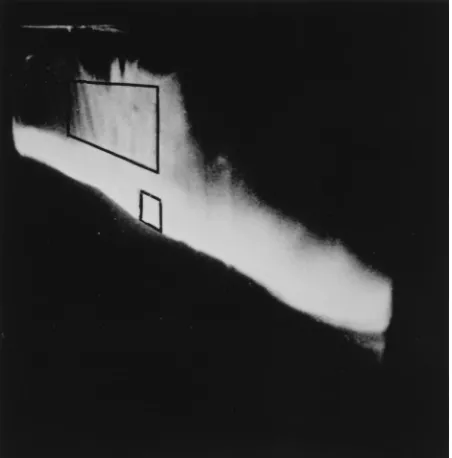
regions. As indicated by the arrows, this probe detected the FIG. 1. Southern blot of 2D gel electrophoresis of HSV-1 DNA which had been digested with BamHI. The blots were probed with32P-labeled HSV-1 DNA probes, and the X-ray films were exposed for 5 days at2708C with an intensifying screen. (A) High-molecular-weight HSV-1 DNA was separated from mature genomes by PFGE, digested with BamHI, and probed with a fragment which hybridizes with the inverted repeats found at the termini and at the junction between the ULand USregions of the HSV-1 genome. The single arrowheads indicate the 6.5-kb junction fragment and the 2.9-kb left terminus. The double arrowheads point to the arches due to Y junctions (rightmost) and X junctions (leftmost). (B) Same as blot A, but the DNA was digested with T7 endonuclease I prior to loading onto the 2D gel. (C) Monomeric, linear virus. The arrows indicate the 6.5-kb junction fragment, the 3.4-kb right terminus, and the 2.9-kb left terminus. (D) DNA from mock-infected cells. (E) High-molecular-weight HSV-1 DNA was digested with SalI prior to 2D gel electrophoresis. The blot was probed with the TK gene, which hybridizes to a 5.9-kb fragment. (F) Same as blot A, but the blot was probed with the HSV-1 alkaline nuclease gene, which hybridizes to a 12.6-kb fragment.
on November 9, 2019 by guest
http://jvi.asm.org/
6.5-kb fragment corresponding to the junction region and the 2.9-kb left terminus of the genome (fainter band). A hybrid-ization signal in the form of a full arch (double arrow on the right) is visible above the region where the linear fragments migrated. This arch originates at the 6.5-kb fragment and ends at a point which is about twice the size of the 6.5 kb fragment. It is characteristic of molecules with Y junctions (4). Above and on the left of the arch in Fig. 1A, a spot is visible. This spot is connected to the arch by a faint line of hybridization (double arrowhead on the left) and, according to published work on 2D gel electrophoresis, is characteristic of molecules with X junc-tions (4). There is also considerable background hybridization along the line of migration of the linear fragments. This is most probably due to shearing and trailing of the DNA fragments which occurred during sample preparation and electrophore-sis. Moreover, it was necessary to overexpose the signal of the linear fragments to visualize clearly the arches produced by the branched fragments.
Figure 1B shows the same preparation of DNA after incu-bation with phage T7 endonuclease I, an enzyme which spe-cifically cleaves X and Y branches to create linear fragments of DNA (9, 11). After digestion, the hybridization signal due to the arches disappeared, while that for the linear fragments remained essentially unchanged. The background smear was somewhat enhanced because of the generation of linear frag-ments of various sizes by the endonuclease.
Figure 1C shows a 2D gel of linear, monomeric HSV-1 DNA purified by PFGE and digested with BamHI. The Southern blot was hybridized with the junction probe that, in addition to the 6.5-kb fragment of Fig. 1A, also detects the two genomic termini of 3.4 kb (right terminus) and 2.9 kb (left terminus) (arrows). The film shown in Fig. 1C was overexposed to dem-onstrate the absence of arches due to branched structures.
The gel of Fig. 1D contains DNA from mock-infected Vero cells; it does not show any signal indicating hybridization with the junction probe.
The presence of branched structures was not limited to the
BamHI fragment which spans the junction region of the HSV-1
genome. In the 2D gel shown in Fig. 2E, HSV-1 DNA was digested with SalI and probed with a TK probe which hybrid-izes to a 5.9-kb fragment. Arches similar in shape to those in Fig. 2A were detected by this probe. A similar result, shown in Fig. 1F, was obtained with BamHI-digested DNA probed with the HSV-1 alkaline nuclease gene (UL12). This probe hybrid-izes with a 12.5-kb fragment situated in the ULportion of the
genome, away from the junctional region.
EM of branched DNA.To investigate the structure of the
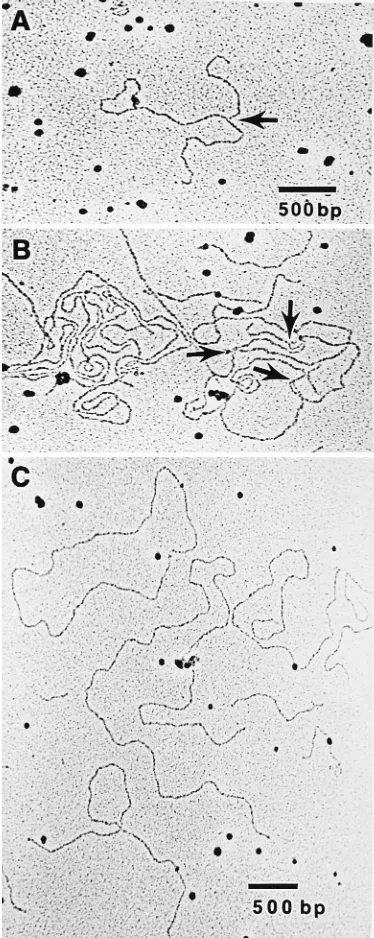
branches, HSV-1 DNA was separated from the cellular DNA by density centrifugation on an NaI gradient. The HSV-1 DNA was then digested with BamHI and electrophoresed at a high concentration on a 2D gel. Figure 2 shows the 2D gel stained with ethidium bromide. The restriction fragments migrated in a line and were poorly separated because of overloading of the gel. This was done to produce enough branched structures for visualization with ethidium and purification from the gel. A smear is visible above the line of the restriction fragments. This smear is very evident, especially in the high-molecular-weight region (left side of the gel), and it should contain the branched DNA fragments. DNA samples were collected from the re-gions outlined by the boxes, and each contained approximately the same amount of DNA.
Figure 3A and B shows EM images of the structures present in the DNA isolated from the region of the branches. Numer-ous Y junctions were observed, many of which had two arms of equal lengths (Fig. 3A), as would be expected from restriction enzyme cleavage of the homologous arms of a replication fork.
Many complex tangles of DNA were also observed. These tangles contained several strand ends and multiple Y junctions. Figure 3B shows such a tangle, in which three consecutive Y junctions are present in a single DNA molecule (arrows). Four-way crossovers were also observed, suggesting the pres-ence of Holliday junctions due to recombination (data not shown); however, it was not possible to distinguish between a genuine Holliday junction and a fortuitous physical crossing over of two DNA molecules. DNA obtained from the linear region contained neither Y junctions (only one Y junction was observed in all of the EM grids examined [approximately 400 molecules]) nor large tangles. Most of the molecules appeared to be linear fragments of DNA like those presented in Fig. 3C. A number of circular molecules were observed in the linear and branched samples of DNA, and one (of 6 to 7 kb long) is visible in Fig. 3C. They probably do not have particular signif-icance with respect to replication and are more likely to have resulted from pairing of the cohesive ends generated by
BamHI digestion.
Sucrose gradients.To investigate the effect of these branches
on the overall structure of replicating HSV-1 DNA, we per-formed the following experiment. DNA from HSV-1-infected cells was purified in agarose, and high-molecular-weight HSV-1 DNA was separated from the monomer HSV-1 by PFGE as described in Materials and Methods and reported previously (34). The high-molecular-weight DNA was then digested with
PacI, a restriction enzyme that cuts the HSV-1 PAC1DTK
genome only once, in the TK gene. Control samples were not treated with PacI. The DNA was electrophoresed a second time on PFGE to separate the linear fragments from the branched structures that remained at the origin of the gel (34). This ‘‘well’’ DNA was excised from the gel and cen-trifuged on a sucrose-NaClO4gradient. Figure 4A shows the
[image:3.612.323.549.72.301.2]distribution of the HSV-1 DNA in the gradient as detected by dot blot hybridization of the fractions collected. There was no FIG. 2. Preparative 2D gel electrophoresis used to purify HSV-1 DNA for electron microscopy. HSV-1 DNA from infected cells was separated from the cellular DNA by an NaI gradient, digested with BamHI, and separated on a 2D gel. The gel was stained with ethidium bromide. The boxes indicate the areas excised from the gel to prepare the linear (small box) and branched (large box) DNA samples.
on November 9, 2019 by guest
http://jvi.asm.org/
significant difference in DNA distribution before and after digestion with PacI. This indicates that digestion with PacI did not reduce the size of the replicating HSV-1 DNA, and most of the DNA was found at the bottom of the gradient. The effi-ciency of digestion with PacI was checked by a Southern blot of HpaI digests of the DNA probed with a TK gene frag-ment. This probe detects a 9.5-kb fragment containing the PacI site, as shown in Fig. 4B, lane 1, that was over 90% cleaved to produce two bands of 6.5 and 2.9 kb, as shown in Fig. 4B, lane 2.
DISCUSSION
Our previous work (34) indicated that intracellular HSV-1 DNA contains complex structures which impede its migration on PFGE, even after digestion with the restriction enzyme
PacI, a single cutter in our strain of HSV-1 (PAC1DTK). A
head-to-tail concatemeric arrangement was observed, but par-tial digestion with PacI failed to show the presence of a signif-icant amount of long, linear concatemers in samples of DNA up to 24 h after infection. Despite the absence of long con-catemers, the intracellular HSV-1 DNA has the mass of many genomes, as initially shown by sucrose gradient sedimentation (22) and confirmed by studying high-molecular-weight HSV-1 DNA purified by PFGE (34). This apparent contradiction can be readily explained by the presence of large branched struc-tures held together by intergenomic links.
[image:4.612.84.271.72.541.2]In this report, we have presented direct evidence of the presence of such branched structures in replicating HSV-1 DNA. (i) 2D gel electrophoresis showed typical arches indic-ative of the presence of Y structures and fainter indications of X structures, as defined by Brewer and Fangman, who per-fected this type of 2D gel electrophoretic analysis while study-ing the replication of the yeast 2m plasmid (4). The arches corresponding to the Y structures are formed as follows. A Y junction moving through a restriction fragment of a given unit length (for example, 1 kb) produces a series of fragments ranging from unit length to twice unit length (2 kb). In the first dimension, the fragments migrate in accordance with their molecular weights and form a smear ranging from 1 to 2 kb. In the second dimension, the fragments in which the arms of the Y junction are equal in length are retarded the most because they deviate the most from the structure of a linear molecule of DNA. This is the reason for the formation of the characteristic continuous arch shown in Fig. 1A. Fragments containing a double Y junction, formed by two replication forks chasing each other or running into each other, also form a continuous smear in the first dimension. In the second dimension, the larger fragments are retarded the most, since they have the longer arms. As a result, these fragments migrate on a line originating at the spot of migration of the linear, unbranched
FIG. 3. EM of HSV-1 high-molecular-weight DNA isolated from 2D gel electrophoresis. (A and B) DNA isolated from the branched region of the 2D gel of Fig. 2. Magnification,374,000. The arrows point to Y branches. (C) DNA isolated from the linear region of the 2D gel of Fig. 2. Magnification,363,000. A circular molecule is visible in the upper left corner.
FIG. 4. (A) NaClO4-sucrose sedimentation gradient of replicative interme-diates of HSV-1 separated on PFGE with (circles) or without (squares) digestion with PacI. The arrow shows the approximate position of the monomeric linear DNA. The HSV-1 DNA in the fractions collected from the gradient was detected by dot blot hybridization with an HSV-1 genomic probe. The intensities of the dots were measured with a PhosphorImager, and the density values are plotted as percentages of the total density of all of the fractions. (B) Samples of the DNA used for the gradients shown in panel A were digested with HpaI and run at a constant voltage on a 0.8% agarose gel. The blot was hybridized with a TK probe which detects a 9.5-kb fragment carrying the PacI site. Lane 1 shows the HSV-1 DNA prior to digestion with PacI (corresponding to the circles in panel A), and lane 2 shows the HSV-1 DNA after digestion with PacI (squares in panel A).
on November 9, 2019 by guest
http://jvi.asm.org/
[image:4.612.317.559.495.629.2]restriction fragment, stretching up above the zone of migration of the linear fragments (Fig. 1A; more evident in Fig. 1E). X structures formed as intermediates of recombination migrate in a narrow band in the first dimension, corresponding to double the size of the linear restriction fragment. In the second dimension, they migrate to a spot found at the end of the line formed by the double Y fragments (Fig. 1A, leftmost double arrow). It is not possible on a 2D gel to distinguish conclusively between X fragments formed by two replication forks and X fragments resulting from recombination events. Fragments containing bubbles due to internal initiation of replication form incomplete and interrupted arches. We did not observe arches compatible with the presence of bubbles. More details and examples of the behavior of branched DNA on 2D gels can be found in the original report by Brewer and Fangman (4).
The arches that we observed were selectively eliminated by digestion with phage T7 endonuclease I, a resolvase that has been shown to linearize Y and X branches (9, 11). Our 2D gel electrophoresis experiments also suggest that the branches are not confined to a specific region of the HSV-1 genome, since they were detected by using different probes. Ethidium bro-mide staining of a 2D gel overloaded with purified HSV-1 DNA detected slowly migrating arches and smears over the entire range of BamHI digestion products. Fewer branches were seen in the low-molecular-weight range, and this can be explained by the fact that there is a lower probability of finding a branch in shorter fragments.
(ii) EM of fragments of replicating HSV-1 DNA showed the presence of single molecules with Y junctions, as well as com-plex tangles containing several Y junctions in the space of a few kilobases. No conclusion could be reached about the pres-ence of recombination intermediates (Holliday junctions): four-way junctions were observed in micrographs, but they were not numerous and could not be distinguished from simple overlapping of two separate fragments of DNA. However, the techniques we used may not detect Holliday junctions effi-ciently. After digestion with BamHI, Holliday junctions would be flanked by termini that may freely rotate, allowing branch migration of the junction and generation of two linear mole-cules.
(iii) After digestion with PacI, the replicative intermediates still sedimented at the bottom of a sucrose gradient and failed to migrate in PFGE, showing that interruption of the linear continuity of the DNA molecules did not reduce the overall size of the DNA network. This indicates that the concatemers are linked by intergenome branches.
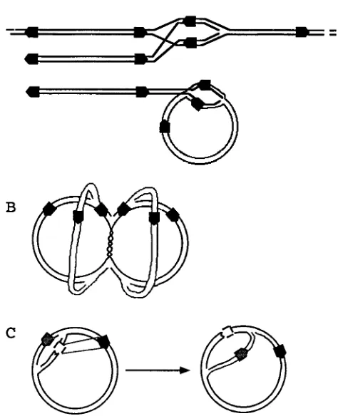
The network we have described is reminiscent of the net-work formed during late phage T4 replication. In the case of phage T4, genomic ends invade homologous sequences on oth-er genomes to produce a Y junction that soth-erves as the repli-cation origin (29). Frequent recombination leads to the forma-tion of a large network of branched concatemers. To add to the analogy to T4 phage, work by Sarisky and Weber (33) indicates that recombination of HSV-1 is accelerated by the presence of free genomic termini. We have already discussed how strand exchange at termini of DNA can lead to the formation of replication forks (28). Double-strand exchange between ho-mologous free ends can generate two replication forks that move along the genome in opposite directions. The same model applies to a recombination event between a free end and an internal homologous sequence. We have hypothesized how this mechanism might apply to the replication and recom-bination of HSV-1 and how it can explain the tight coupling observed between these two processes (28). This model of replication does not require initiation at a replication origin and, as for T4 phage, it would apply only to later stages of
replication, since origin-dependent initiation seems to be es-sential for the establishment of HSV-1 replication (49). Figure 5A is a schematic representation of the formation of a repli-cation fork by double-strand recombination between a termi-nal a sequence and an intertermi-nal a sequence of a linear concate-mer. Figure 5A also shows the same process occurring between a linear genome and a circular genome. Random strand ex-change between a sequences in opposite orientations can gen-erate branched concatemers in all of the possible orientations of the ULand USregions of the HSV-1 genome.
[image:5.612.315.552.88.380.2]Other models can explain the formation of the network we have observed. For example, the frequent Y junctions may be generated by continuous initiation of replication at the origins (Fig. 5B). Initiation would likely be bidirectional (theta struc-tures), since frequent origin-dependent initiation of a rolling circle would produce a free terminus at the origin(s) of repli-cation. These termini should produce additional restriction fragments, which, to our knowledge, have not been observed. Figure 5B shows a branched circular structure which consists of four partially replicated circular genomes held together by an unreplicated region of DNA. The structure contains multiple replication forks (Y junctions). Recombination between in-verted a sequences would produce all of the possible orienta-FIG. 5. Possible models for HSV-1 replication which could lead to the for-mation of a large, branched structure. The black boxes represent the a sequences (not to scale), and the arrowheads indicate relative orientation. Each HSV-1 genome has two terminal a repeats in the direct orientation and one internal a repeat in the inverse orientation. (A) Formation of branched concatemers by double-strand invasion by a terminal a sequence of an internal a sequence. (B) Theta replication of circular genomes, in which continuous initiation leads to the formation of partially replicated circles held together by unreplicated regions of DNA. (C) Recombination between a replicated (white) a sequence and an unreplicated (black) a sequence in the inverted orientation leads to the creation of two replication forks that follow each other.
on November 9, 2019 by guest
http://jvi.asm.org/
tions of the ULand USregions, while recombination between
direct a sequences would produce larger circular concatemers. Figure 5C illustrates the effects of recombination between a newly replicated a sequence and a still unreplicated a sequence within the same circular genome. The recombination results in inversion of the replication fork located between the recom-bining a inverted repeats. The two replication forks are now chasing each other on the circular genome, with the production of concatemers, as in a rolling circle. Equimolar amounts of genomes in opposite orientations will be produced if recombi-nation occurs at each a sequence after the passage of a repli-cation fork. This is the mechanism of replirepli-cation of the 2m plasmid in Saccharomyces cerevisiae (16), and it was proposed as a model for the replication of HSV-1 by Zhang et al. (50). However, it is likely that formation of concatemers is not dependent on an inversion event, since DNAs lacking inverted repeats can be replicated by the HSV-1 machinery with the formation of concatemeric intermediates. This is the case for defective viruses (45), HSV-1-induced replication of simian virus 40 DNA (6), and replication of plasmids containing an HSV-1 origin of replication (42). It is more probable that as in lambda phage, bidirectional replication can convert to a rolling circle at random positions on the genome, generating head-to-tail concatemers that will be packaged starting from the left end of the genome (7, 34, 44, 50). This is in agreement with our previous observation that a greater proportion of longer-than-unit length concatemers appear late in the infection (24 to 48 h) (34).
The presence of a branched network poses additional prob-lems for packaging of mature, linear genomes. While long lin-ear concatemers can be cut to size by a terminase, a branched network needs an additional debranching enzyme, as is the case for phage T4 (29). Studies on encapsidation-deficient mu-tants showed that cleavage of the genome occurs at the time of encapsidation and that mutations of the capsid proteins that abolish encapsidation also prevent the formation of genomic termini (10, 32, 38). However, mutations in at least five addi-tional noncapsid genes can produce a similar phenotype (1–3, 38). These data underscore the complexity of the cleavage of the replicative intermediates. The HSV-1 alkaline nuclease is a possible candidate for a debranching enzyme. This enzyme has single-stranded endonuclease and processive exonuclease ac-tivities (19, 20, 43). Homologous counterparts occur in other herpesviruses. Induction of activity occurs late in infection, and it coincides with the production of mature linear genomes in HSV-1, cytomegalovirus (our unpublished results), and Ep-stein-Barr virus (30). Alkaline nuclease has been found in a protein complex bound to the cleavage site of the HSV-1 ge-nome (5). An alkaline nuclease null mutant replicates its DNA to nearly normal levels but is severely deficient in the export of mature capsids, which remain in the nucleus (35, 48). On the basis of these data, it was proposed that the HSV-1 alkaline nuclease may function as a debranching enzyme (35), and there is evidence that alkaline nuclease null mutants may pro-duce more highly branched replicative intermediates (24a). Unpublished data from our laboratory indicate that extracts from HSV-1-infected cells contain a cruciform-resolving activ-ity that requires the presence of the alkaline nuclease.
In conclusion, the occurrence of branched structures in the replicative intermediates of HSV-1 DNA adds complexity to a model for its replication. The two steps of formation and res-olution of the branches require the presence of an enzymatic machinery that has not been elucidated. These enzymes are likely to be involved in the coupling between replication and recombination in herpesviruses, and they may be essential for production of infectious herpesvirus genomes.
ACKNOWLEDGMENTS
We thank A. Richard Morgan and Sandra K. Weller for suggestions and helpful discussion.
This research was supported by a grant from GlaxoWellcome Can-ada to D.L.J.T. and a grant from the Medical Research Council of Canada to D.G.S.
REFERENCES
1. Addison, C., F. J. Rixon, J. W. Palfreyman, M. O’Hara, and V. G. Preston. 1984. Characterisation of a herpes simplex virus type 1 mutant which has a temperature-sensitive defect in penetration of cells and assembly of capsids. Virology 138:246–259.
2. Addison, C. F., F. J. Rixon, and V. G. Preston. 1990. Herpes simplex virus type 1 UL28 gene is important for the formation of mature capsids. J. Gen. Virol. 71:2377–2384.
3. Al-Kobaisi, M. F., F. J. Rixon, I. McDougall, and V. G. Preston. 1991. The herpes simplex virus UL33 gene product is required for the assembly of full capsids. Virology 180:380–388.
4. Brewer, B. J., and W. L. Fangman. 1987. The localization of replication origins on ARS plasmids in S. cerevisiae. Cell 51:463–471.
5. Chou, J., and B. Roizman. 1989. Characterization of DNA sequence-com-mon and sequence-specific proteins binding to cis-acting sites for cleavage of the terminal a sequence of the herpes simplex virus 1 genome. J. Virol. 63: 1059–1068.
6. Danovich, R. M., and N. Frenkel. 1988. Herpes simplex virus induces the replication of foreign DNA. Mol. Cell. Biol. 8:3272–3281.
7. Deiss, L. P., L. Chou, and N. Frenkel. 1986. Functional domains within the a sequence involved in the cleavage-packaging of herpes simplex virus DNA. J. Virol. 59:605–618.
8. Delius, H., and J. B. Clements. 1976. A partial denaturation map of herpes simplex virus type 1 DNA: evidence for inversions of the unique DNA regions. J. Gen. Virol. 33:125–133.
9. de Massy, B., and R. A. Weisberg. 1987. Gene 3 endonuclease of bacterio-phage T7 resolves conformationally branched structures in double-stranded DNA. J. Mol. Biol. 193:359–376.
10. Desai, P., N. A. DeLuca, J. C. Glorioso, and S. Person. 1993. Mutations in herpes simplex virus type 1 genes encoding VP5 and VP23 abrogate capsid formation and cleavage of replicated DNA. J. Virol. 67:1357–1364. 11. Dickie, P., G. McFadden, and A. R. Morgan. 1987. The site-specific cleavage
of synthetic Holliday junction analogs and related branched DNA structures of bacteriophage T7 endonuclease I. J. Biol. Chem. 262:14826–14836. 12. Dunderdale, H. J., F. E. Benson, C. A. Parsons, G. J. Sharples, R. G. Lloyd,
and S. C. West.1991. Formation and resolution of recombination interme-diates by E. coli recA and ruvC proteins. Nature (London) 354:506–510. 13. Dutch, R. E., V. Bianchi, and I. R. Lehman. 1995. Herpes simplex virus type
1 DNA replication is specifically required for high-frequency homologous recombination between repeated sequences. J. Virol. 69:3084–3089. 14. Dutch, R. E., R. C. Bruckner, E. S. Mocarski, and I. R. Lehman. 1992.
Herpes simplex virus type 1 recombination: role of DNA replication and viral a sequences. J. Virol. 66:277–285.
15. Dutch, R. E., B. V. Zemelman, and I. R. Lehman. 1994. Herpes simplex virus type 1 recombination: the Uc-DR1 region is required for high-level a-se-quence-mediated recombination. J. Virol. 68:3733–3741.
16. Futcher, A. B. 1986. Copy amplification of the 2 micron circle plasmid of Saccharomyces cerevisiae. J. Theor. Biol. 119:197–204.
17. Garber, D. A., S. M. Beverley, and D. M. Coen. 1993. Demonstration of circularization of herpes simplex virus DNA following infection using pulsed field gel electrophoresis. Virology 197:459–462.
18. Hayward, G. S., R. J. Jacob, S. C. Wadsworth, and B. Roizman. 1975. Anatomy of herpes simplex virus DNA: evidence for four populations of molecules that differ in the relative orientations of their long and short components. Proc. Natl. Acad. Sci. USA 72:4243–4247.
19. Hoffman, P. J. 1981. Mechanism of degradation of duplex DNA by the DNase induced by herpes simplex virus. J. Virol. 38:1005–1014.
20. Hoffman, P. J., and Y.-C. Cheng. 1978. The deoxyribonuclease induced after infection of KB cells by herpes simplex virus type 1 or type 2. I. Purification and characterization of the enzyme. J. Biol. Chem. 253:3557–3562. 21. Jacob, R. J., L. S. Morse, and B. Roizman. 1979. Anatomy of herpes simplex
virus DNA. XII. Accumulation of head-to-tail concatemers in nuclei of infected cells and their role in the generation of the four isomeric arrange-ments of viral DNA. J. Virol. 29:448–457.
22. Jacob, R. J., and B. Roizman. 1977. Anatomy of herpes simplex virus DNA. VIII. Properties of the replicating DNA. J. Virol. 23:394–411.
23. Jongeneel, C. V., and S. L. Bachenheimer. 1981. Structure of replicating herpes simplex virus DNA. J. Virol. 39:656–660.
24. Lurz, R., M. Grote, J. Dijk, R. Reinhardt, and B. Dobrinski. 1986. Electron microscopy study of DNA complexes with proteins from the archaeobacte-rium Sulfolobus acidocaldarius. EMBO J. 5:3715–3721.
24a.Martinez, R., R. Sarisky, P. C. Weber, and S. K. Weller. Unpublished data. 25. McGeoch, D. J., M. A. Darlymple, A. J. Davinson, A. Dolan, M. C. Frame, D. McNab, L. J. Perry, J. E. Scott, and P. Taylor.1988. The complete DNA
on November 9, 2019 by guest
http://jvi.asm.org/
sequence of the long unique region in the genome of herpes simplex virus type 1. J. Gen. Virol. 69:1531–1574.
26. Mocarski, E. S., and B. Roizman. 1982. Herpesvirus-dependent amplifica-tion and inversion of cell-associated viral thymidine kinase gene flanked by viral a sequences and linked to an origin of viral DNA replication. Proc. Natl. Acad. Sci. USA 79:5626–5630.
27. Morgan, A. R., D. H. Evans, J. S. Lee, and D. E. Pulleyblank. 1979. Review: ethidium fluorescence assay. Part II. Enzymatic studies and DNA-protein interactions. Nucleic Acids Res. 7:571–594.
28. Morgan, A. R., and A. Severini. 1990. Interconversion of replication and recombination structures: implications for terminal repeats and concatem-ers. J. Theor. Biol. 144:195–202.
29. Mosig, G. 1987. The essential role of recombination in phage T4 growth. Annu. Rev. Genet. 21:347–371.
30. Ooka, T., M. de Turenne, G. de The, and J. Daillie. 1984. Epstein-Barr virus-specific DNase activity in nonproducer Raji cells after treatment with 12-O-tetradecanoylphorbol-13-acetate and sodium butyrate. J. Virol. 49: 626–628.
31. Poffenberger, K. L., and B. Roizman. 1985. A noninverting genome of a viable herpes simplex virus 1: presence of head-to-tail linkages in packaged genomes and requirements for circularization after infection. J. Virol. 53: 587–595.
32. Preston, V. G., J. A. V. Coates, and F. J. Rixon. 1983. Identification and characterization of a herpes simplex virus gene product required for encap-sidation of virus DNA. J. Virol. 45:1056–1064.
33. Sarisky, R. T., and P. C. Weber. 1994. Requirement for double-strand breaks but not for specific DNA sequences in herpes simplex virus type 1 genome isomerization events. J. Virol. 68:34–47.
34. Severini, A., A. R. Morgan, D. R. Tovell, and D. L. J. Tyrrell. 1994. Study of the structure of replicative intermediates of HSV-1 DNA by pulsed-field gel electrophoresis. Virology 200:428–435.
35. Shao, L., L. M. Rapp, and S. K. Weller. 1993. Herpes simplex virus 1 alkaline nuclease is required for efficient egress of capsids from the nucleus. Virology 196:146–162.
36. Sharples, G. J., and R. G. Lloyd. 1991. Resolution of Holliday junctions in Escherichia coli: identification of the ruvC gene product as a 19-kilodalton protein. J. Bacteriol. 173:7711–7715.
37. Sheldrick, P., and N. Berthelot. 1974. Inverted repetitions in the chromo-some of herpes simplex virus. Cold Spring Harbor Symp. Quant. Biol. 39: 667–678.
38. Sherman, G., and S. L. Bachenheimer. 1987. DNA processing in tempera-ture-sensitive morphogenic mutants of HSV-1. Virology 158:427–430. 39. Smiley, J. R., B. S. Fong, and W.-C. Leung. 1981. Construction of a
double-jointed herpes simplex viral DNA molecule: inverted repeats are required for segment inversion and direct repeats promote deletions. Virology 113: 345–362.
40. Smiley, J. R., M. J. Wagner, W. P. Summers, and W. C. Summers. 1980. Genetic and physical evidence for the polarity of transcription of the thymi-dine kinase gene of herpes simplex virus. Virology 102:83–93.
41. Spaete, R. R., and N. Frenkel. 1982. The herpes simplex virus amplicon: a new eukaryotic defective-virus cloning-amplifying vector. Cell 30:295–304. 42. Stow, N. D. 1992. Herpes simplex virus type 1 origin-dependent DNA
rep-lication in insect cells using recombinant baculoviruses. J. Gen. Virol. 73: 313–321.
43. Strobel-Fidler, M., and B. Francke. 1980. Alkaline deoxyribonuclease in-duced by herpes simplex virus type 1: composition and properties of the purified enzyme. Virology 103:493–501.
44. Varmuza, S. L., and J. R. Smiley. 1985. Signals for site-specific cleavage of HSV DNA: maturation involves two separate cleavage events at sites distal to the recognition sequences. Cell 41:793–802.
45. Vlazny, D. A., and N. Frenkel. 1981. Replication of herpes simplex virus DNA: localization of replication recognition signals within defective virus genomes. Proc. Natl. Acad. Sci. USA 78:742–746.
46. Walboomers, J. M. M., and J. T. Schegget. 1976. A new method for the isolation of herpes simplex virus type 2 DNA. Virology 74:256–258. 47. Weber, P. C., M. D. Challberg, N. J. Nelson, M. Levine, and J. C. Glorioso.
1988. Inversion events in the HSV-1 genome are directly mediated by the viral DNA replication machinery and lack sequence specificity. Cell 54: 369–381.
48. Weller, S. K., M. R. Seghatoleslami, L. Shao, D. Rowse, and E. P. Car-michael.1990. The herpes simplex virus type 1 alkaline nuclease is not essential for viral DNA synthesis: isolation and characterization of a lacZ insertion mutant. J. Gen. Virol. 71:2941–2952.
49. Wu, C. A., N. J. Nelson, D. J. McGeoch, and M. D. Challberg. 1988. Iden-tification of herpes simplex virus type 1 genes required for origin-dependent DNA synthesis. J. Virol. 62:435–443.
50. Zhang, X., S. Efstathiou, and A. Simmons. 1994. Identification of novel herpes simplex virus replicative intermediates by field inversion gel electro-phoresis: implications for viral DNA amplification strategies. Virology 202: 530–539.