0022-538X/96/$04.0010
Copyrightq1996, American Society for Microbiology
Evidence for a Second Function of the MA Sequence in the
Rous Sarcoma Virus Gag Protein
LESLIE J. PARENT,1,2* CAROL B. WILSON,2MARILYN D. RESH,3
ANDJOHN W. WILLS2
Department of Medicine1and Department of Microbiology and Immunology,2Pennsylvania State
University College of Medicine, Hershey, Pennsylvania 17033, and Department of Cell Biology and Genetics, Memorial-Sloan Kettering Cancer Center, New York, New York 100213
Received 27 July 1995/Accepted 19 October 1995
During retrovirus assembly, Gag proteins bind to the inner leaflet of the plasma membrane to initiate the budding process. The molecular basis of this protein-lipid interaction is poorly understood. For the human immunodeficiency virus type 1 Gag protein, we recently reported that the membrane-binding domain resides within the N-terminal 31 amino acids and consists of two components: myristate and a cluster of basic residues, which together promote membrane binding in vitro and budding in vivo (W. Zhou, L. J. Parent, J. W. Wills, and M. D. Resh, J. Virol. 68:2556–2569, 1994). The positively charged residues associate electrostatically with acidic phospholipids to stabilize membrane binding, while myristate provides membrane-binding energy via hydrophobic interactions. Here we demonstrate that the human immunodeficiency virus type 1 Gag membrane-binding domain can fully replace the membrane-targeting function of the N-terminal 100 residues of the non-myristylated Rous sarcoma virus (RSV) Gag protein. To further explore the importance of myristate and basic residues in membrane binding, we developed a gain-of-function assay whereby budding was restored to defec-tive mutants of RSV Gag. Detailed mutational analysis revealed that the position, number, and context of charged residues are crucial to budding. Myristate provides additional membrane-binding energy, which is critical when a Gag protein is near the threshold of stable membrane association. Finally, viruses with altered matrix (MA) proteins that are noninfectious, even though they produce particles with high efficiency, were identified. Thus, we present the first evidence that the RSV MA sequence plays two distinct roles, mem-brane binding during particle assembly and a second, as yet undefined function required for viral infectivity.
Retroviruses complete their replication cycle by budding from the plasma membrane of the host cell, and the Gag protein is the only viral gene product required for this process. Following synthesis on cytoplasmic ribosomes, Gag polypro-teins are transported to the plasma membrane, where 2,000 to 3,000 molecules (45) aggregate to initiate particle formation. Very late in or immediately after the budding process, the viral protease cleaves the Gag protein into mature products (Fig. 1A), which include the matrix (MA), capsid (CA), and nucleo-capsid (NC) proteins, as well as variable accessory proteins, which, in the case of Rous sarcoma virus (RSV), include p2a, p2b, p10, a spacer peptide, and protease (PR) (7, 14, 28, 29). Insights into the molecular mechanism of budding have been gained by mapping essential domains of the Gag polyprotein (49). We have characterized three assembly domains (ADs)of the RSV Gag protein that are required for budding: the mem-brane-binding domain (M; previously named AD1), the inter-action domain (I; previously AD3) and the late domain (L; previously AD2) (Fig. 1A). The M domain takes the Gag protein to the cytoplasmic face of the plasma membrane and is the focus of the present report. It is located at the N terminus of Gag, and its sequence is a subset of the MA sequence (24). All mutants that are defective in the M domain can be rescued by complementation with budding-competent Gag molecules, indicating that downstream assembly functions are not dis-rupted (51). The I domain maps to the NC region and is a major site of interaction between Gag proteins (1, 8, 47). The
L domain is a small, proline-rich motif required late in budding and is located in the p2b sequence (48). Collectively, the RSV assembly domains amount to only about 20% of the total Gag protein sequence.
Functional equivalents of the M, I, and L domains have also been found in the human immunodeficiency virus type 1 (HIV-1) Gag protein (1, 26, 55). The HIV-1 M domain consists of a bipartite signal: myristate, which is cotranslationally added to the glycine at position 2 after removal of the initiator me-thionine, and a cluster of basic amino acids positioned between residues 15 and 31 (55). Myristate provides a hydrophobic anchor for the Gag protein, but biophysical studies have dem-onstrated that alone, it is insufficient to partition proteins sta-bly onto the membrane (27). Electrostatic interactions be-tween basic residues and acidic membrane phospholipids, which are found on the inner leaflet of the plasma membrane (2, 25), further stabilize the protein-lipid association and can potentially contribute more binding energy than the hydropho-bic myristate moiety (16, 23). These basic residues in the HIV-1 M domain reside within two amphipathic beta sheets on an exposed surface of the protein, allowing the positive charges in the protein to engage the negatively charged phospholipid head groups (19, 20, 55). Furthermore, seven additional basic residues from other regions of the molecule appear to form a collar around the beta strands, increasing the net positivity of the region (19). Since the N-terminal regions of many Gag proteins are enriched in basic residues and many are myristy-lated, we speculated that other Gag proteins may utilize hy-drophobic and electrostatic interactions to bind the plasma membrane (55).
To test this hypothesis, we examined the M domain of the RSV Gag protein, which appears to be very different from the HIV-1 M domain in that it is large (86 residues [24]) and not
* Corresponding author. Mailing address: Department of Microbi-ology and ImmunMicrobi-ology, Pennsylvania State University College of Med-icine, 500 University Dr., P.O. Box 850, Hershey, PA 17033. Phone: (717) 531-3528. Fax: (717) 531-6522. Electronic mail address: lparent @cor-mail.biochem.hmc.psu.edu.
1016
on November 9, 2019 by guest
http://jvi.asm.org/
myristylated. Although the RSV M domain contains 11 basic residues, they are scattered throughout the sequence without any obvious clusters. Previous characterization of the RSV M domain has shown that even small deletions within it abolish budding (1, 24, 51). We therefore adopted a gain-of-function approach to test whether introducing myristate and basic res-idues within the N-terminal sequences of RSV Gag could re-store budding to assembly-incompetent deletion mutants of the M domain. Initially, we found that placement of the entire HIV-1 M domain (myristate plus basic residues) at the N terminus of RSV Gag suppressed deleterious deletions in the MA sequence; in fact, the HIV-1 M domain could fully replace the RSV M domain. Subsequent experiments revealed that a component of the HIV-1 M domain, a cluster of basic residues, could restore budding to an RSV M domain mutant in a myristate-independent manner. Other single-amino-acid sub-stitutions in the RSV MA sequence resulted in mutants that
were dependent on myristate for budding. When examined in the context of the proviral genome, a subset of gag mutations that produced noninfectious viruses was identified, providing the first evidence that in addition to its role in budding, this region of RSV MA has a critical function at another step in the viral replication cycle.
MATERIALS AND METHODS
Construction of the HIV-RSV M domain chimeras.All of the RSV gag mu-tants are derivatives of pATV-8, an infectious molecular clone of the RSV Prague C genome. HIV-1 sequences were derived from the pHXB2Dgpt infec-tious molecular clone. Cloning procedures and sequencing were performed by standard methods (37), and the presence of each mutation was confirmed by dideoxy sequencing with at least two independent clones tested in each experi-ment.
pSV.Myr4, in which the first 10 codons of HIV-1 gag were substituted for the first 10 codons of RSV gag, has been described previously (1). To make pSV.H32R, the first 32 codons of the HIV-1 gag gene were substituted for the 10 codons of the pp60v-src(Src) oncoprotein in pSV.Myr1 (50, 51). PCR amplifica-tion of the first 32 gag codons of the HXB2 plasmid was performed to create an
NdeI site at the initiator ATG and a MluI site following codon 32, using the
following upstream and downstream primers (restriction endonuclease recogni-tion sites are underlined): 59-GCTAGAAGGAGACATATGGGTGCGAGAG CGT (NdeI) and 59-ATAGGTTTTACACGCGTCTTTTAATTTATATTTTTT CTTT (MluI). The resulting product was digested only with NdeI and ligated with the 384-bp NdeI (nucleotide [nt] 380)-NcoI (nt 9016) fragment from pSV.Myr1N, a derivative of pSV.Myr1 which has an NdeI site overlapping the codon for the initiator methionine (Fig. 1A; made with the oligonucleotide 59-CCCGGTGG ATCACATATGGGATCCAGCAA). The ligated DNA was used as the tem-plate for a second round of PCR amplification with the same downstream primer and an upstream primer containing an SstI site (nt 255, in the 59untranslated region), 59-CTCAGAAGTCGACGAGCTCTACT. The product was digested with SstI and MluI (nt 408) and cloned into equivalent sites of pSV.Myr1 to make pSV.H32R, which has one foreign residue (Asp) introduced between the HIV-1 and RSV Gag sequences (Fig. 1A). Constructs pSV.H32R.DMA3, pSV.H32R.
DMA10, and pSV.H32R.B1c were made by replacing SstI-MluI fragments from pSV.Myr1.DMA3, pSV.Myr1.DMA10 (1), and pSV.Myr1.B1c (51), respectively, with the longer SstI-MluI fragment of pSV.H32R.
The RSV Gag M domain sequence was removed from the pSV. H32R coding sequence by digesting pSV.H32R.DMA10 with XhoI (nt 630) and BglII (nt 1630). The small fragment was exchanged with the XhoI-BglII fragment of pSV.DMA6S (24), which has a deletion from nt 637 to 677 and introduces an SpeI site at the junction. This intermediate plasmid was digested with MluI and SpeI, treated with Klenow fragment, gel purified, and ligated, resulting in pSV.H32R.DMB (Fig. 1A).
Oligonucleotide-directed mutagenesis of the RSV gag gene. pSV.Myr0, pSV.Myr2, and pSV.3h have been described previously (46, 50). Additional RSV
gag mutants were made by oligonucleotide-directed mutagenesis, performed by
the method of Kunkel et al. (17). The template used, unless otherwise noted, was MGAG.Myr2 (11, 50), an M13 derivative containing a point mutation in the second codon that changes Glu to Gly, thereby creating a functional myristyla-tion site. The following oligonucleotides were used to make the indicated gag alleles and to create the restriction endonuclease sites (shown in parentheses and underlined in the sequence of the oligonucleotide): myr2.4K,6V, 59-TCAAG CATGGGAGCCAAGATAGTGGTGATTTCGTCCGCG; myr2.4R, 59-CAAG CATGGGAGCCCGCATAAAGGTGATTTCG; myr2.HB12, 59-GTCATAAA GGTGATTTCGTCCG(C,G)GAAGAAAAAGTATAAGCTGAAAACC TCTCCTTCTAAG; myr2.HB23, 59-CCTCTCCTTCTAAAAAGAAATACAA GCTTAAGTTGTCCCTCTTAC (HindIII); myr2.HB33, 59-GCCATGTTGTC CCTCAAAAAAAAGTATAAGCTTAAGATGTCTCCCTCAGAC (HindIII);
myr2.HB43, 59-GGTTGCTTATGTCTCCGAAAAAGAAATATAAACTGAA GTCCTGGGATCCCAT; myr2.C12K, 59-GTGATTTCGTCCGCGAAAAAAA CCTATTGCGGG; myr2.T14K, 59-GTCCGCGTGTAAAAAATATTGCGGGA AAAC (SspI); myr2.C16K, 59-CGTGTAAAACCTATAAGGGGAAAACCT CT; myr2/C12R, 59-ATTTCGTCCGCGCGCAAAACCTATTGC (BssHII);
myr2.C12E, 59-ATTTCGTCCGCGGAGAAAACCTATTGC (SstII); myr2.
C12S, 59-ATTTCGTCCGCTAGCAAAACCTATTGC (NheI); myr2.C12N, 59 -ATTTCGTCCGCGAATAAAACCTATTGC; myr2.Y15F, 59-GCGTGTAAAA CCTTTTGCGGGAAAACC; and myr2.HB12,Y15F, 59-GCGAAGAAAAAGT TCAAGCTGAAAACC (XmnI). MGAG.Myr2.HB12 DNA was used as the tem-plate to make myr2.HB12, Y15F.
To make gag alleles of the above mutants that have the wild-type second codon (e.g., myr0.HB12, myr0.C12K, myr0.T14K, and myr0.C16K), the mutagenesis re-actions were repeated with the oligonucleotides indicated above and the wild-type MGAG DNA template (50). Mutants containing the MA10 deletion (1) were also made by oligonucleotide-directed mutagenesis with the MGAG.HB12 and MGAG.Myr2.HB12 DNA templates.
Each of the gag alleles made by M13 oligonucleotide-directed synthesis was transferred into pSV.Gag.SX (also named pDSV.Myrx[50]) for expression in FIG. 1. Alterations of the RSV MA sequence. (A) N-terminal chimeras. A
schematic representation of the RSV Gag polyprotein, Pr76gag, is shown at the
top, with its cleavage products indicated. The numbers shown above the protein indicate the position of each cleavage site measured as the number of amino acids from the N terminus. The relative positions of key restriction endonuclease sites in the gag gene are flagged with boxes and arrows. Solid black rectangles below Pr76gagindicate domains essential for membrane binding (M), a late event
in particle release (L), and interactions between Gag molecules (I). Myr0 is a designation for the wild-type Gag protein, which indicates that it is not myristy-lated. In Myr1, the cross-hatched box represents the first 10 residues of pp60v-src, which were substituted for the first 10 residues of RSV Gag. The squiggle at the N terminus represents the fatty acid myristate. In Myr2, a Glu-to-Gly mutation at position 2 results in myristylation. Gray boxes represent sequences derived from HIV-1 Gag. In Myr4, the first 10 residues of HIV-1 Gag replace those of RSV Gag, and in H32R, the first 32 residues of HIV-1 Gag were substituted for the first 10 residues of RSV Gag. In H32R.DMB, RSV MA residues 11 to 100 were deleted from the full-length H32R construct, with insertion of four foreign residues (D, A, L, and V) at the junction. (B) Deletions within the RSV MA region. The indicated MA3, MA10, and B1c deletions within the MA sequence were combined with various N-terminal chimeras as described in the text.
on November 9, 2019 by guest
http://jvi.asm.org/
COS-1 cells. M13 replicative-form DNAs of each gag mutant were digested with
SstI and XhoI, agarose gel purified, and ligated into identical sites within pSV.
Gag.SX. Initial screening by using loss of a ClaI site identified putative recom-binants, which were subsequently confirmed by dideoxy sequencing and/or re-striction endonuclease digestion.
Nucleotides encoding the downstream segment of the RSV M domain were removed from pSV.Myr2.HB12 to make pSV.Myr2.HB12.DMB by PCR with the following upstream and downstream primers, respectively: 59-CTCAGAAGTC GACGAGCTCTACT (SstI), and 59-CTTAGAAGGAACTAGTTTCAGCTTA TACTTTTT (SpeI). The amplified fragment was digested with SstI and SpeI and ligated into plasmid pSV.Myr1.DMA6S (24) which had been digested with the same enzymes.
The pSV.(K)7construct was made by annealing overlapping oligonucleotides, 59-TATGGGCAAGAAAAAAAAGAAGAAAAAGGA and 59-CGCGTCCTT TTTCTTCTTTTTTTTCTTGCCCA, to give a double-stranded DNA fragment with 59NdeI and 39MluI sticky ends. The oligonucleotide pair was cloned into
the NdeI and MluI sites of pSV.Myr1N.
The B1c deletion (removes nt 599 to 674) was introduced into the gag mutant alleles by exchanging the large SstII (nt 543 and 1806) fragment, which had been treated with shrimp alkaline phosphatase, with the small SstII fragment of pSV.Myr1.B1c (51).
Transfection, labeling, and immunoprecipitation in mammalian cells.COS-1 cells were transiently transfected by the DEAE-dextran-chloroquine method, as previously described (50, 51). In experiments in which a single construct was transfected, approximately 750 ng of each plasmid DNA was used to transfect cell monolayers in 35-mm-diameter plates. In the complementation experiments, 250 ng of the rescuing molecule (pSV.Myr2.3h) and 500 ng of the experimental DNA were mixed prior to transfection. In both cases, cells were labeled for 2.5 h withL-[35S]methionine 48 h after transfection. The growth medium was re-moved, centrifuged to remove any loose cells, and mixed with RIPA buffer containing protease inhibitors (50). Cells on the plates were lysed, and immu-noprecipitation of Gag proteins in the postnuclear cell lysates and medium samples was performed with polyclonal rabbit anti-RSV serum, as previously described (46, 50, 51). Immunoprecipitated proteins were resolved by sodium dodecyl sulfate-polyacrylamide gel electrophoresis (SDS-PAGE) and detected by fluorography and autoradiography. To calculate budding efficiency, each lane of the autoradiogram was scanned with a densitometer and the amount of Gag antigen in the medium was divided by the sum of that in the medium plus that in the lysates. The degree of suppression of an MA deletion was determined as the ratio of the budding efficiency of the deletion mutant to the budding effi-ciency of the full-length protein. Results of at least two separate experiments were averaged for each calculation.
Infectivity assays.Alleles of gag to be tested were transferred into a derivative of pBH.RCAN.HiSV (31), which carries a hygromycin resistance marker (9), by exchanging SstI (nt 255)-HpaI (nt 2731) fragments, and at least two independent clones of each mutant virus were analyzed. The wild-type control, pRC.V8, has been described previously (7, 8).
Quail (QT6) cells (22) were transfected in duplicate sets of 60-mm-diameter dishes by the calcium phosphate precipitation method (7, 8). For the first set of plates, the efficiency was analyzed 18 h later by labeling for 3.5 h withL-[35
S]me-thionine and collecting Gag proteins from the medium and cells by immunopre-cipitation with anti-RSV serum after preclearing the lysates with staphylococcal protein A to reduce background (8). In some experiments, 0.5ml of polyclonal rabbit serum directed against purified MA protein (kindly provided by V. Vogt, Cornell University, Ithaca, N.Y.) was added to the immunoprecipitates to en-hance detection of MA. The other set of transfected cells was subcultured for 3 days to allow viruses to spread. The medium was reserved and replaced with fresh medium containing 300mg of hygromycin (Sigma) per ml to kill any uninfected cells. After 9 days, hygromycin-resistant colonies were stained with methylene blue and quantitated with an automated counter. The previously reserved medium samples were centrifuged at 8003g, and the virus-containing
supernatant was placed on subconfluent monolayers of uninfected QT6 cells in 60-mm-diameter dishes. After incubation at 378C for 8 h, the medium was replaced with fresh medium containing hygromycin. Nine days later, colonies were stained and counted.
To assay for persistent virus production, immunoblot analysis of hygromycin-selected quail cells was performed. Hygromycin-containing medium was added 3 days after transfection, and cells were passed at 1:6 dilution every 3 to 5 days for 5 weeks. Resistant cells were obtained with infectious viruses but not with viruses having little or no infectivity. To confirm that the cells continued to produce virus, culture supernatants were pelleted through a layer of 25% sucrose, sepa-rated by PAGE as above, and transferred to nitrocellulose. Immunoblots were probed for 1 h with anti-RSV serum diluted 1:5,000, and antibody-protein com-plexes were detected by enhanced chemiluminescence (Amersham) as described previously (41).
To maximize the chances of identifying mutant viruses with very low infectivity (which may not be detected by the above assays), virus spread was permitted over a 3-week period in the complete absence of hygromycin. Following transfection, QT6 cells were passed at 3- to 5-day intervals to allow continued infection of new cells in the culture. Before each passage, the growth medium was removed and cleared of cells by low-speed centrifugation, and virus particles were pelleted through a 25% sucrose cushion at 100,0003g for 45 min, resuspended in 25ml
of phosphate-buffered saline, and stored at2208C. After 27 days, all of the pelleted virus samples were analyzed by immunoblotting as described above.
To determine if virus particles contain reverse transcriptase, particles were harvested from stable QT6 cell lines and assayed for their ability to synthesize DNA from an exogenous template and primer (6).
RESULTS
Understanding the molecular interactions between Gag pro-teins and the plasma membrane is fundamental to the study of retrovirus assembly. Progress has been made for the HIV-1 Gag protein, for which it has been shown that myristate and basic residues within the MA sequence form a bipartite mem-brane-binding signal that associates with the membrane via hydrophobic and electrostatic interactions (55). However, the mechanism of membrane binding for nonmyristylated Gag proteins, like that of RSV, has not been examined. In this study, we inserted basic residues into myristylated and non-myristylated versions of RSV Gag to determine how the num-ber, position, and arrangement of basic residues contribute to the efficiency of particle assembly. Since a role for the RSV MA sequence outside of budding had not yet been described, we also examined the effects of the gag mutations on viral infectivity.
Replacement of the M domain of RSV Gag with that of
HIV-1 Gag.To begin the analysis of the RSV M domain, we
examined whether the HIV-1 M domain could replace the RSV M domain, which lacks obvious clusters of basic residues and is insensitive to myristate (11, 50, 51). If so, perhaps the components of the HIV-1 sequence which are most important in conferring membrane binding to RSV Gag could be identi-fied. It seemed likely that this approach would be successful, given two previous findings from our laboratory. First, the membrane-binding domain of the Src protein (the N-terminal 10 residues), which, like the HIV-1 M domain, depends on myristate and basic residues (15, 38, 39), can restore particle production to all assembly-defective mutants of the RSV M domain when placed at the Gag N terminus (Myr1; Fig. 1A and 2); in fact, the entire RSV M domain can be deleted from the Src-Gag chimera without impairing particle release (51). Sec-ond, substitution of the first 10 amino acids of HIV-1 Gag for those of RSV (Myr4; Fig. 1A and 2) restores particle release to some budding-defective MA deletion mutants (B1c and
DMA10); however, not all deletions are tolerated (DMA3 [Fig.
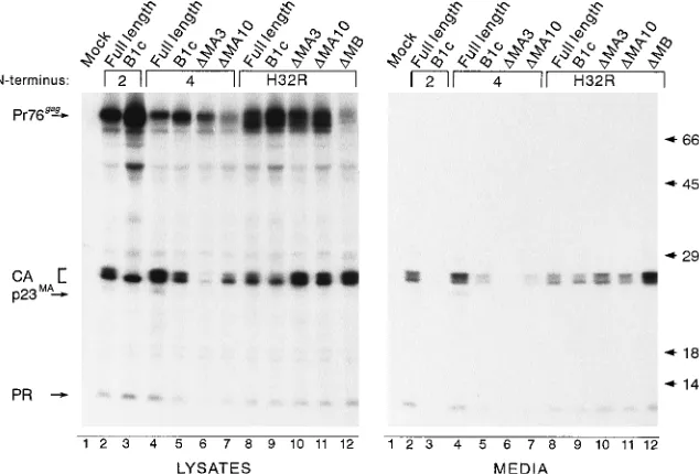
1B]) (1). Since this chimera contains only part of the HIV-1 M domain, we predicted that an HIV-RSV chimera containing the entire HIV-1 M domain could tolerate all deletions within the RSV MA sequence. Indeed, this was the case. Substitution of the first 32 residues of HIV-1 Gag for the first 10 of RSV Gag (H32R; Fig. 1A) suppressed the B1c and MA10 deletions (Fig. 3, lanes 9 and 11, respectively) reproducibly two to three times better than Myr4 suppressed the same two deletions (lanes 5 and 7, respectively). Suppression of the proximal MA3 deletion by the Myr4 sequence was barely detectable but was increased dramatically (nearly 14-fold in this experiment) by the H32R substitution (lanes 6 and 10, respectively). While the
Myr4.DMA3 protein is expressed at levels comparable to
wild-type levels in pulse-labeling experiments, it is more rapidly degraded (44). Addition of a stronger membrane-targeting/ binding signal (i.e., that of Src or HIV-1) results in protection of the protein from cellular proteases (compare lysate lanes 6 and 10), presumably because of its efficient targeting to the membrane and sequestration within the nascent viral particle. The full-length H32R substitution produced particles slightly less efficiently than the control did (Fig. 3, compare lanes 8 and 2). To our surprise, removal of the remaining RSV M domain sequence (RSV residues 11 to 100) from the
on November 9, 2019 by guest
http://jvi.asm.org/
mera reproducibly resulted in a three- to fourfold
enhance-ment of budding (H32R.DMB; Fig. 1A and 3, lanes 12
com-pared with lanes 8). We hypothesize that removal of the entire RSV membrane-binding domain from H32R eliminates se-quences which interfere to some degree with the folding of the HIV-1 M domain. Knowing that the HIV-1 M domain could replace the entire RSV M domain, we began testing our ideas
about membrane binding by systematically introducing com-ponents of the HIV-1 M domain into RSV to determine the minimal changes that would restore budding to mutants defec-tive in membrane binding.
[image:4.612.147.464.468.683.2]Basic substitutions within the first 10 residues of RSV MA. From previous studies, it is clear that the simple addition of myristate to the RSV Gag protein by changing glutamate to
FIG. 2. Substitutions within the N-terminal region of the RSV MA domain. The top line (Myr0) shows the N-terminal sequence of the wild-type (WT) Gag protein. The sequences of the mutants used in this study are shown below the wild type, with positions of identical residues indicated by dashes. The HIV-1 basic motif (25-KKKYKLK-31) is indicated by a solid box. A derivative of this motif, in which the tyrosine (Y) at position 29 was mutated to phenylalanine (F), is enclosed by a dashed box. The ability of the mutants to be labeled with [3H]myristate is summarized to the right, and mutants predicted to be positive are indicated by parentheses, as (1). The ability of each mutant to be released into the medium in the presence and absence of an inactivating MA deletion mutant (B1c) is also summarized. In this case,1indicates particle release at approximately the same level as the wild type,2indicates no particle release,1/2indicates a low but detectable level of particle release, m indicates that myristate was required for particle release, and ND means not done.
FIG. 3. Ability of HIV N-terminal sequences to replace the RSV M domain. COS-1 cells were transfected with the indicated plasmid DNAs and metabolically labeled 48 h later with [35
S]methionine for 2.5 h. Gag proteins in cell lysates and media were immunoprecipitated with polyclonal anti-RSV serum and analyzed by SDS-PAGE and fluorography as described in the text. The N-terminal sequence present in each construct is indicated, with Myr2 derivatives in lanes 2 and 3, Myr4 derivatives in lanes 4 to 7, and H32R derivatives in lanes 8 to 12. The positions of Pr76gag
and its cleavage products are indicated to the left. p23MA
is a processing intermediate consisting of p19MA
and the p2 sequences. Molecular mass standards (in kilodaltons) are shown on the right.
on November 9, 2019 by guest
http://jvi.asm.org/
glycine at position 2 (Myr2; Fig. 1A and 2) is not enough to suppress deletions in the M domain that destroy budding (Myr2.B1c; Fig. 4, lanes 3) (51). However, the myristylated Myr4 sequence, which has some suppressive activity, is quite similar to the glutamate-to-glycine mutant (Fig. 2). To deter-mine which residues of the HIV-1 substitution (Myr4) are important in suppressing the B1c deletion, systematic substi-tutions were made in the Myr2 sequence to make it more similar to that of Myr4.
In Myr2.4K,6V (Fig. 2), lysine and valine were exchanged to move a positively charged residue to position 4 (where it is found in HIV-1) without changing the amino acid composition of Myr2 and without impairing the ability of the molecule to direct budding (Fig. 4, lanes 6). When the B1c deletion was introduced, Gag proteins were detected in the medium at very
low levels, indicating partial restoration of budding (Fig. 4, lanes 7 and 8).
Mutant Myr2.4R (Fig. 2) has an arginine at position 4 (like HIV-1) in place of valine, adding a second positive charge within the first 10 residues. This mutant could not tolerate the B1c deletion (Fig. 4, lanes 10 and 11), but the full-length protein was also inefficiently released into the medium (Fig. 4,
lanes 9). Labeling with [3H]myristic acid revealed that the
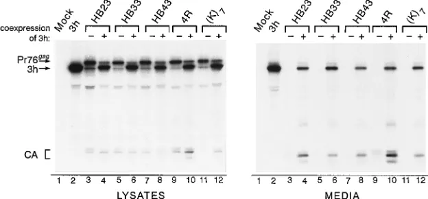
mutation blocked myristylation, which could explain its bud-ding defect (Fig. 2). The mutant protein is not globally dis-rupted by the basic substitution, since it can be incorporated into particles when coexpressed with a budding-competent Gag protein (Fig. 5, lanes 9 and 10).
We next examined whether it would be possible to repair the membrane-binding defect of the B1c mutant by adding a clus-ter of basic residues (7 Lys) to the Gag N clus-terminus. However, instead of enhancing membrane binding, budding was de-stroyed, in the context of both the full-length protein and the
B1c deletion [Fig. 2, (K)7; data not shown). Like Myr2.4R, the
(K)7mutant can be rescued into particles by complementation,
indicating that downstream domains needed for particle for-mation are intact (Fig. 5, lanes 11 and 12). Perhaps the lack of myristylation in this mutant explains its budding defect (Fig. 2). It is also possible that protein folding does not place the cluster of lysines on an accessible surface of the protein to enable membrane interactions.
Insertion of the HIV-1 basic motif into RSV MA.Basic
resi-dues must lie on an exposed surface of the protein to engage in electrostatic interactions with acidic membrane phospholipids; thus, the overall protein conformation is critical. In HIV-1, the cluster of basic residues (25-KKKYKLK-31) important for mem-brane binding appears to reside on the exterior surface of MA (19, 20). To test whether basic residues contribute to mem-brane binding in RSV, it would be desirable to make basic substitutions in regions that are accessible to the membrane. Unfortunately, targeted mutations cannot be made on an in-formed basis, since the structure of the RSV M domain is not yet known. However, a very hydrophilic sequence like the HIV-1 25-KKKYKLK-31 motif might adopt an external posi-tion when introduced into the RSV M domain. Furthermore, if the sequence is inserted just downstream of the myristate in Myr2, a bipartite membrane-binding signal like that of HIV Gag (i.e., myristate plus basic residues) might be created,
[image:5.612.62.295.73.245.2]re-FIG. 4. Effects of positive charges within the first 10 amino acids of RSV Gag. Gag proteins from COS-1 cell lysates and medium samples were radiola-beled and immunoprecipitated with anti-RSV and anti-MA sera. Each mutant was examined in the presence or absence of the B1c deletion, as indicated by2 or1signs at the top of each lane. Myr2 serves as the positive control for budding. Single lysine substitutions were made at each of three positions in MA. Two independent clones, designated A and B, were analyzed for Myr2.4K,6V. B1c and Myr2.4R.B1c. The positions of two MA species, p19MAand p23MA, are indicated.
FIG. 5. Rescue of M domain mutants by complementation. To determine whether budding-incompetent M domain mutants could be rescued into particles by complementation, COS-1 cells were cotransfected with a plasmid expressing the defective mutant and a plasmid encoding a protease-truncated Gag protein that makes particles very efficiently. Viral proteins were radiolabeled and detected by immunoprecipitation with anti-RSV serum. Lanes in which pSV.Myr2.3h was cotransfected with the M domain mutant plasmid are designated by1, while2indicates that the indicated plasmid was transfected alone. HB23, HB33, and HB43 have the Myr2 N-terminal sequence. The presence of cleavage products (e.g., CA) in the medium indicates that the defective mutants were rescued into particles. The positions of the Gag precursors, the truncated 3h precursor, and the CA species are indicated at the left.
on November 9, 2019 by guest
http://jvi.asm.org/
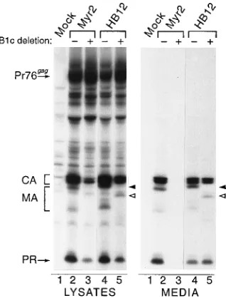
[image:5.612.152.464.531.676.2]sulting in a molecule that could tolerate downstream deletions in the RSV M domain. To test this, the 25-KKKYKLK-31 sequence was substituted for residues 12 to 18 in the myristy-lated mutant of RSV Gag (Myr2.HB12 [Fig. 2]). Strikingly, this mutant released particles very efficiently even when the B1c deletion was introduced (Fig. 6, compare lanes 4 and 5), sug-gesting that the membrane-binding defect was repaired by the basic motif. Moreover, it appears that this substitution influ-ences the conformation of the RSV MA domain (or at least the
major antibody epitope), since detection of the p23MAband,
eliminated by the B1c deletion in the parent (Myr2.B1c [Fig. 6, LYSATES, lanes 2 and 3]), was restored by the basic motif substitution (Myr2.HB12 [Fig. 6, lanes 5]).
If the position of basic residues is critical for membrane binding, the 25-KKKYKLK-31 motif might not work every-where that it is inserted within the RSV M domain. Alterna-tively, because this sequence is very hydrophilic, it seemed possible that it would appear readily on the surface of the molecule and hence promote membrane binding in many con-texts. With this in mind, the basic motif was substituted for residues 23 to 29 of RSV Gag, a similar position to its natural location in HIV-1 Gag; however, budding was abolished, even in the absence of the B1c deletion (Myr2.HB23 [Fig. 2; data not shown]). Placement of this sequence at positions 33 to 39 or 43 to 49 in Myr2 also abrogated particle release (Myr2. HB33 and Myr2.HB43 [Fig. 2; data not shown]). The global structure of the Gag protein was not destroyed by the basic substitution, since each budding-incompetent mutant could be rescued into particles by complementation (Fig. 5, lanes 3 to 8). In these mutants, the basic motif may lie on an exterior surface of the protein but may not be facing the membrane. Thus, the RSV M domain is sensitive to the position and context of basic residues within it, supporting the idea that conformation is critical to its function during budding.
Because the HB12 mutant was competent for budding in the
presence of the B1c deletion, we considered the possibility that it contains a novel and small membrane-binding domain which, like the H32R and Myr1 chimeras, can tolerate any disruption of the RSV M domain. To address this, the HB12 substitution was combined with the MA10 deletion mutant (Myr2.HB12.
DMA10), theDMB deletion (Myr2.HB12.DMB, missing
resi-dues 19 to 100), and the defective HB23 substitution mutant
(Myr2.HB121HB23). We found that it could not correct the
budding defect of any of these mutants (data not shown). As expected, the control with the Src substitution (Myr1) could
suppress all three mutations (Myr1.DMA10, Myr1.DMB, and
Myr1.HB23). These findings, together with the ability of HB12 to bud independently of myristate and to restore the MA epitope in immunoprecipitates, suggest that the basic motif alters the three-dimensional structure of the region, thereby recreating the original RSV M domain rather than creating a new, autonomous membrane-binding signal.
Suppression of the B1c deletion with single-amino-acid
sub-stitutions. The Myr2.HB12 mutant, which tolerates the B1c
deletion, has three additional basic residues (Lys at positions 12, 14, and 16) compared with the wild-type RSV Gag protein. We asked whether each lysine could individually suppress this deletion. To our surprise, mutants with a single lysine could tolerate the B1c deletion (Myr2.C12K.B1c, Myr2.T14K.B1c, and Myr2.C16K.B1c [Fig. 7, lanes 4 to 6, 7 to 9, and 10 to 12, respectively]). Therefore, the M domain defect caused by the B1c deletion must be fairly subtle.
If only a subtle change is required to repair the membrane-binding defect of B1c, substitutions of amino acids other than lysine might be corrective, and several were tested. To quan-titate the ability of each substitution to suppress the B1c de-letion, the budding activity of each full-length substitution mu-tant was compared with the particle release retained by its B1c derivative (see Materials and Methods). For example, averag-ing the results of five separate experiments, the Myr2. C12K.B1c deletion mutant was found to retain 25% of the budding activity of the full-length Myr2.C12K protein. Identi-cal comparisons were made for each of the single-amino-acid substitution mutants described below, and averages from at least two experiments were calculated. A representative auto-radiogram is shown in Fig. 8.
When a positively charged residue other than lysine was substituted at position 12 (Arg, C12R [Fig. 2]), suppression of the B1c deletion was nearly as effective as that by lysine (ap-proximately 15% of the full-length budding level was retained compared with 25% for the lysine derivative [Fig. 8, lanes 5 and 6]). In contrast, substitution of a small polar amino acid or a
negatively charged residue had little or no (,5%) suppressive
activity (Ser, C12S; and Glu, C12E [Fig. 2 and 8, lanes 9 and 10 and lanes 7 and 8, respectively]). Substitution of a large neutral residue (Asn, C12N [Fig. 2]) was intermediate in its ability to produce particles when the B1c deletion was present (approx-imately 10% budding activity was retained by the B1c deriva-tive [Fig. 8, lanes 11 and 12]). In addition to the lysine residues, the tyrosine in the 25-KKKYKLK-31 motif is conserved among different strains of the HIV-1 and simian immunodeficiency virus Gag proteins (13, 54). When tyrosine was changed to phenylalanine (HB12.Y15F [Fig. 2]) the molecule tolerated the B1c deletion without any loss of budding efficiency com-pared with the full-length mutant, indicating that the tyrosine residue was not critical for restoration of budding (Fig. 8, lanes 15 and 16). As a control, the tyrosine at position 15 in RSV Gag was also substituted with phenylalanine (Myr2.Y15F [Fig. 2]) and the budding defect of B1c was not repaired (Fig. 8, lanes 13 and 14).
[image:6.612.99.258.71.280.2]The results of the single-amino-acid substitutions suggest
FIG. 6. Effect of HIV-1 basic motif substitution on budding and tolerance of B1c. COS-1 cells were transfected with the indicated plasmids and lysed, and proteins were radioimmunoprecipitated with anti-RSV and anti-MA sera. The 25-KKKYKLK-31 sequence was substituted between residues 12 and 18 in Myr2. The presence or absence of the B1c deletion is indicated by1or2above each lane. The region where the mutant MA species are found is surrounded by a bracket. The solid arrowhead points to p23MA
in lanes 2 and 4. The open arrowhead shows the Myr2.HB12.B1c p23MA
band in lanes 5; this band migrates more rapidly because of the deletion.
on November 9, 2019 by guest
http://jvi.asm.org/
that the B1c deletion may disrupt the folding of the RSV M domain without removing any information essential for the function of this domain. Thus, single-amino-acid changes in the upstream sequences (positions 12, 14, and 16) may influ-ence the overall structure of the M domain enough to repair the minor defect (with dramatic consequences) caused by the B1c deletion.
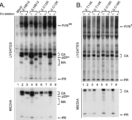
Contribution of myristate.The experiments above focused
on the role of basic residues in a myristylated mutant of RSV Gag. We also examined how basic residues influence the non-myristylated RSV Gag M domain. The basic motif (Myr2. HB12) and single-basic-residue substitution mutants (Myr2. C12K, Myr2.T14K, and Myr2.C16K) were reconstructed with the nonmyristylated (Myr0) RSV Gag N terminus, which dif-fers from Myr2 by a single amino acid at position 2 (Fig. 2). The nonmyristylated basic motif mutant still produced parti-cles, even with the B1c deletion (Myr0.HB12.B1c [Fig. 9A, compare lanes 4 and 5]), indicating that basic residues can promote membrane binding in a nonmyristylated M domain. In contrast, none of the nonmyristylated forms of the single-lysine-substitution mutants could tolerate the B1c deletion (Myr0.C12K.B1c [Fig. 9A, lanes 9]; Myr0.T14K.B1c and Myr0. C16K.B1c [Fig. 9B, lanes 4 and 8, respectively]). These mu-tants were absolutely dependent on myristate for budding.
Infectivity of MA mutants. No role for the RSV MA
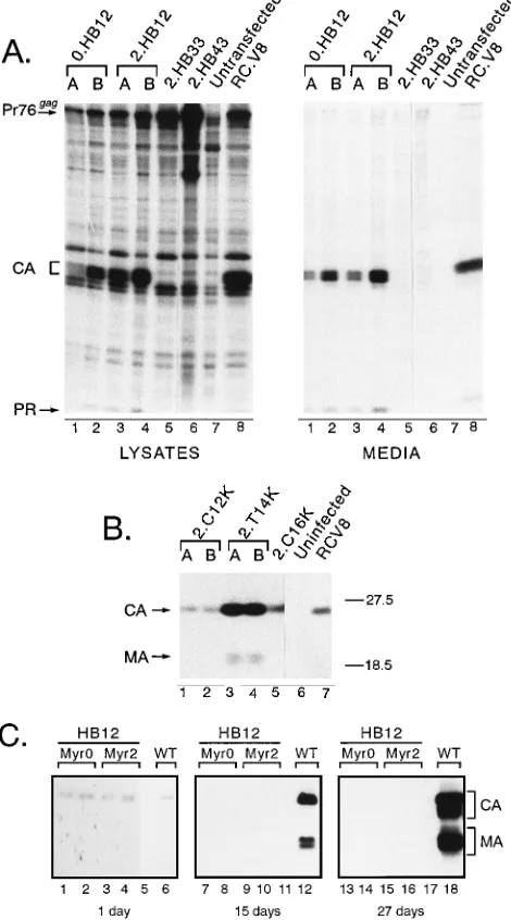
se-quence beyond membrane binding/targeting has been demon-strated, although there are precedents for multiple MA func-tions in HIV-1 (see Discussion). We investigated whether the RSV MA sequence might have a second function by transfer-ring many of the gag alleles described above into pRCAN, a proviral vector containing an infectious RSV genome and a selectable marker for hygromycin resistance. First, the effect of the mutations on particle assembly in quail (QT6) cells was determined in a transient-expression assay, in which Gag pro-teins in cell lysates and media were radiolabeled 18 h after transfection and detected by immunoprecipitation. In every case, the budding phenotype observed for MA mutant was identical to that seen in COS-1 cells (summarized in Table 1). For example, both the nonmyristylated and myristylated deriv-atives of the HB12 substitution made particles efficiently in quail cells (Myr2.HB12 and Myr0.HB12 [Fig. 10A, lanes 1 and 2 and lanes 3 and 4, respectively]) while Myr2.HB33 and Myr2. HB43 were defective (lanes 5 and 6, respectively). Also, the myristylated forms of the single-lysine substitutions (Myr2.
C12K, Myr2.T14K, and Myr2.C16K) transiently released virus particles into the medium, and Myr2.T14K was insensitive to the B1c deletion (Table 1).
[image:7.612.60.235.73.203.2]The effect of the gag mutations on infectivity was determined by taking advantage of the selectable hygromycin marker in pRCAN. Parallel plates of QT6 cells were transfected with plasmid DNAs bearing wild-type or mutant gag genes. In one set of plates, transient Gag expression was again measured to ensure that the transfection was effective (results not shown). Cells in the other set were cultured for 3 days to allow viruses that were capable of spreading throughout the culture to do so. The medium was then removed (but saved for further testing [see below]) and replaced with medium containing hygromy-cin. Nine days later, the cells were stained with methylene blue and hygromycin-resistant colonies were counted. For the wild-type virus (RC.V8) and Myr2.T14K, cell-to-cell spread was very efficient and nearly all cells became hygromycin resistant (Table 1). In contrast, no resistant colonies were seen in cul-tures expressing myristylated or nonmyristylated derivatives of the HIV-1 basic motif substitution (Myr0.HB12 or Myr2. HB12), even though particles were produced transiently. Like-wise, no colonies were detected on plates receiving Myr2. T14K.B1c, which also was competent for budding. The Myr2. C16K and Myr2.C12K mutants produced resistant colonies at 30 and 5% of wild-type levels, respectively. As expected, none of the mutants defective for particle release (Myr2.HB23, Myr2.HB33, and Myr2.HB43) gave any resistant colonies.
FIG. 7. Single lysine substitutions at positions 12, 14, and 16. Transfected COS-1 cells were radiolabeled, and viral proteins in the cell lysates and media were detected by immunoprecipitation with anti-RSV and anti-MA sera. C12K, T14K, and C16K each have the Myr2 N terminus. The presence or absence of the B1c deletion is designated as1or2above each lane. A and B represent two independently derived clones of the indicated mutant. Positions of the Gag precursor and cleavage products are shown at the left.
FIG. 8. Single-amino-acid changes at position 12. Viral proteins from trans-fected COS-1 cell lysates and medium samples were radioimmunoprecipitated with anti-RSV and anti-MA sera. Each of the constructs has the Myr2 sequence at its N terminus. The1and2signs at the top of each lane indicate the presence and absence, respectively, of the B1c deletion. Only the relevant portion of the fluorogram for the medium samples is shown below that of the cell lysates, as labeled on the left side of the figure. The Gag precursor and cleavage products are indicated to the right.
on November 9, 2019 by guest
http://jvi.asm.org/
[image:7.612.318.547.372.659.2]Cells transfected with each mutant were cultured in the presence of hygromycin for 3 weeks or more to examine the persistence of virus production. As shown by immunoblot anal-ysis of culture supernatants, Gag proteins continued to be released into the medium for Myr2.C12K, Myr2.T14K, and Myr2.C16K (Fig. 9B, lanes 1 and 2, lanes 3 and 4, and lane 5, respectively). However, the impaired infectivity of Myr0.HB12 and Myr2.HB12 prevented virus spread and the establishment of resistant monolayers in a single 60-mm-diameter plate, so they could not be tested in this way. Although it was not an efficient process, stable cell lines were established from the HB12 mutants by transfecting QT6 cells in two 100-mm-diam-eter dishes, allowing cells to grow for 3 days to allow random
integration of the plasmid, dividing the cells into six separate 100-mm-diameter plates, adding hygromycin, and expanding resistant colonies that arose over the ensuing 2-week period. In preliminary studies, virus particles from multiple clones of these cells were found to have reverse transcriptase activity (data not shown).
As a second means of assessing infectivity, media containing wild-type or mutant viruses collected for 3 days posttransfec-tion (see above) were added to uninfected QT6 cells. Follow-ing a 16-h incubation period to allow viral entry and integra-tion, as well as expression of the resistance gene, hygromycin was added to the medium. Resistant colonies were stained and counted 9 days later. Using this method, we confirmed that all three viruses with single-basic-residue substitutions (Myr2. C12K, Myr2.T14K, and RC.Myr2.C16K) in Gag were infec-tious, while Myr0.HB12 and Myr2.HB12 produced virus par-ticles with undetectable levels of infectivity (Table 1). At least two independent clones of each mutant were tested, and each transfection was repeated two or more times. Although the hygromycin selection methods are not always quantitative, we have found upon repeated testing of many different Gag mu-tants that we can consistently detect the replication of mumu-tants with 100-fold-lower infectivity than the wild type (8, 29).
For the Myr0.HB12 and Myr2.HB12 mutants, which had no detectable infectivity in the hygromycin-dependent assays, we used a more sensitive assay that will amplify viruses with low levels of infectivity. Following transfection, cells were passed every 3 to 5 days for 3 weeks in the absence of hygromycin to allow ample time for any infectious virion to spread to neigh-boring cells. Culture supernatants were collected before each passage, loose cells were discarded, and virus particles were pelleted through a sucrose cushion and frozen. At the end of the collection period, immunoblot analyses of all virus samples were performed simultaneously. For the wild type, spread oc-curred and increasing amounts of virus were detected after each passage (Fig. 10C, lanes 6, 12, and 18). For the HB12 mutants, which lacked infectivity in the previous assays, virus particles were transiently made at levels equal to wild-type levels (lanes 1 to 4 compared with lane 6). However, amplifi-cation did not occur, and by the end of the 3-week period, Gag proteins were no longer detected in the medium, indicating that they were noninfectious (lanes 7 to 10 and lanes 13 to 16). These results provide the first reported evidence that the RSV MA sequence is involved in replication at a step other than during membrane binding.
DISCUSSION
The experiments described in this report give insight into the molecular interactions between the RSV Gag MA sequence and the cytoplasmic face of the plasma membrane during par-ticle assembly. We found that basic residues can promote bud-ding in both nonmyristylated and myristylated mutants of RSV Gag, suggesting that positive charges may be important for membrane binding in viruses other than HIV-1. However, the finding that a single basic or neutral amino acid substitution in the MA sequence can enhance particle assembly suggests that the overall conformation of the RSV M domain is very sensi-tive to alterations and emphasizes the need for information about its three-dimensional structure. An unexpected but very important finding in these experiments is the discovery that the MA sequence of RSV Gag may have a previously unappreci-ated, second, critical role in viral replication.
A second role of RSV MA.In this report, we described MA
[image:8.612.60.292.72.282.2]mutants that are not impaired for budding but have lost their ability to infect avian cells, suggesting that MA may have a
FIG. 9. Contribution of myristate to particle assembly. Transfected COS-1 cells were radiolabeled, and cell lysates and media were immunoprecipitated with anti-RSV and anti-MA sera. Mutants labeled with 2 have myristylated Myr2 N termini, while those beginning with 0 have the Myr0, nonmyristylated (wild-type) N-terminal sequence. The presence and absence of the B1c deletion are indicated by1and2, respectively, above each lane. The relevant portion of the medium sample fluorogram is located below the lysate samples, as designated on the left. Bands corresponding to the Gag precursor and cleavage products are indicated to the right.
TABLE 1. Infectivity of Gag mutantsa
Gag protein Budding Infectivity
RC.V8 (wild-type) 1 11
RC.Myr2 1 11
RC.Myr2.HB12 1 2
RC.Myr0.HB12 1 2
RC.Myr2.HB23 2 2
RC.Myr2.HB33 2 2
RC.Myr2.HB43 2 2
RC.Myr2.C12K 1 1
RC.Myr2.T14K 1 11
RC.Myr2.C16K 1 1
RC.Myr2.T14K.B1c 1 2
a
Wild-type and mutant alleles were expressed in the context of the RCAN. BHiSV genome in QT6 cells. Virus particles were evaluated for their ability to confer hygromycin resistance to the transfected cells and to a separate popula-tion of uninfected QT6 cells. Identical results were obtained in each type of experiment.11,.500 hygromycin-resistant colonies/60-mm-diameter plate;1, 20 to 350 colonies;2,,2 colonies.
on November 9, 2019 by guest
http://jvi.asm.org/
[image:8.612.57.297.556.676.2]function in addition to that of membrane binding. These are the first mutants of this kind described for RSV, and a function other than membrane binding has not been demonstrated pre-viously for the RSV MA protein. Other studies in our labora-tory have shown that the second half of MA (residues 87 to 155) is dispensable for both budding and infectivity (24). Thus, it is clear that this new function of MA resides in the N-terminal half of MA (residues 1 to 86) and overlaps the region required for membrane binding. At the moment, we do not know when during the viral replication cycle this second func-tion is needed. It is not required for RNA incorporafunc-tion into virions, since replacement of the first 84 residues of MA with the small membrane-binding domain of Src does not prevent genome packaging (36); furthermore, MA binds RNA very weakly and nonspecifically (21). The block to infectivity prob-ably is not due to an inability of the mutants to incorporate the Gag-Pol fusion protein, since reverse transcriptase activity was detected in the noninfectious viruses. However, it is possible that a mutation in MA has a subtle effect on Gag-Pol packag-ing, as demonstrated for HIV-1 (40). It seems likely that the noninfectious particles contain glycoproteins, because wild-type RSV efficiently packages Env mutants lacking cytoplasmic tails (30) and heterologous glycoproteins (10, 52); moreover, a specific interaction between MA and Env has not been dem-onstrated for this virus. Nevertheless, this is a defect that has been reported for certain MA mutants of HIV-1 (53), and it may be that the interaction of our MA mutants with the mem-brane is sufficiently altered to influence glycoprotein incorpo-ration. Also, it remains possible that the mutant particles are incorrectly formed, so that they are noninfectious because of an indirect effect of MA. That is, perhaps misfolding of MA influences another structural protein which in turn has a role required for infectivity.
Although the second function of RSV MA must be investi-gated more thoroughly, we suspect that this new role may be one required during early stages of infection, including fusion, release of the viral core into the cytoplasm, transport of the complex to the host genome, and alteration of the activities of reverse transcriptase or integrase. We expect that the role of RSV MA early in infection would be different from the nuclear targeting function ascribed to HIV-1 MA. In that case, the function of MA appears to be limited to a small subset of molecules which direct the preintegration complex to the nu-cleus of nondividing cells (3–5, 12). In contrast to the lentivi-ruses, productive infection with RSV and other oncoretrovi-ruses requires cell division (35, 43), during which the nuclear envelope dissolves, allowing the provirus access to the host genome (18, 34). Further experimentation is under way to identify this second function of RSV MA.
Myristate and basic residues can promote budding of RSV
Gag.The N-terminal sequences of most Gag proteins are rich
[image:9.612.59.294.97.520.2]in basic residues, and we have suggested that they interact with concentrated regions of acidic phospholipids during mem-brane binding (55). In RSV Gag, there are 11 basic residues within the 86 amino acids that make up the M domain (24), but they are widely spaced and do not form an obvious motif as they do in HIV Gag. Moreover, myristate is not present on the RSV Gag protein. We found that introduction of a cluster of basic residues derived from HIV-1 into the RSV MA sequence (which added three new positive charges) restored budding to one M domain mutant, and in this case myristate was still not required. This is the first demonstration of basic residues re-storing function to a defective membrane-binding domain; however, we also found that the number, position, and ar-rangement of basic residues were crucial for repairing the budding defect. Basic residues potentiated budding only when
FIG. 10. Effect of basic substitutions on budding and infectivity in avian cells. QT6 cells were transfected with the indicated proviral constructs. RC.V8 carries the wild-type gag gene. (A) Transient-expression analysis. At 18 h posttransfec-tion, cells were labeled with [35S]methionine for 3 h and cell lysates and media were detected by immunoprecipitation with anti-RSV serum. Mutant 0.HB12 has the nonmyristylated, wild-type N terminus. Mutants 2.HB12, 2.HB33, and 2.HB43 have the Myr2 N-terminal sequence. Independent clones are denoted A and B. Positions of the Gag precursor, CA, and PR bands are shown to the left. (B) Immunoblot analysis of resistant cultures. Each of the indicated Gag mutants has the Myr2 myristylated N terminus. After transfection, cells were passaged every 3 to 5 days in medium containing hygromycin, and infectious genomes allowed establishment of resistant monolayers. After 5 weeks, culture superna-tants were collected, floating cells were discarded, and virus particles were pelleted through a sucrose cushion. Viral proteins were resolved by SDS-PAGE, immunoblotted, and detected with anti-RSV serum. The positions of CA and MA are indicated on the left. Molecular mass standards (in kilodaltons) are shown on the right. (C) Persistence and amplification of virus in long-term culture. QT6 cells were transfected with the indicated proviral DNAs and ana-lyzed for virus production by immunoblotting of the culture medium at the indicated times posttransfection. The cells were passed in fresh (hygromycin-free) medium every 3 to 5 days. Lanes 5, 11, and 17 are untransfected controls. The positions of the bands representing CA and MA are defined by brackets to the right of the figure.
on November 9, 2019 by guest
http://jvi.asm.org/
inserted at certain positions, whereas at others they destroyed particle formation. Thus, it is possible that the overall confor-mation of the M domain and hence the accessibility of basic residues to the membrane are more important for function than is the net positive charge of the domain. The importance of the conformation of the M domain is further supported by the finding that a single substitution of a neutral residue at position 12 in RSV Gag could promote budding.
Further understanding of how basic residues contribute to membrane binding in the RSV M domain will require three-dimensional structure information. Efforts are under way to obtain the nuclear magnetic resonance spectrum and crystal structure of this domain, and we ultimately hope to learn how its conformation may be altered by membranes. For now, we hypothesize that the folding of the RSV M domain causes basic residues that are separated in linear sequence to be brought together on an exposed surface of the molecule, forming a concentrated area of positive charges that interacts with the negatively charged plasma membrane. If the structural infor-mation does not support this model, another mechanism of membrane interaction will have to be considered for RSV.
Myristate-dependent mutants of RSV Gag. Although the
wild-type RSV Gag protein is insensitive to myristate, particle production can be made dependent on it by making minor changes in the MA sequence. A single lysine substituted at any of three positions (residue 12, 14, or 16) repaired the budding defect of an M domain mutant only if the protein was myri-stylated. This finding correlates well with results of biophysical studies in which binding energies for the hydrophobic interac-tion between myristate and neutral phospholipids and the elec-trostatic interaction between basic peptides and acidic phos-pholipids were measured. Myristate contributes 8 kcal of binding energy per mol (27), and each lysine residue interact-ing with an acidic phospholipid vesicle provides 1.4 kcal of
binding energy per mol (1 kcal54.184 kJ) (16). If these values
are applied to the RSV mutants, the single-lysine-substitution mutants should possess an additional 9.4 kcal of binding energy per mol when myristylated (compared with wild-type RSV Gag) but only 1.4 kcal/mol when not myristylated. For the HB12 substitution, which has three added basic residues, the protein is predicted to have an additional 4.2 kcal/mol when nonmyristylated (compared with the wild type) and 12.2 kcal/ mol when myristylated. Both forms of HB12 tolerate the B1c deletion, so presumably the energy contributed by myristate is not required. We propose that if a Gag protein is near the threshold for stable membrane binding, either additional pos-itive charges or hydrophobic interactions can provide the crit-ical binding energy to allow budding.
Membrane targeting of Gag proteins.The mechanism
where-by cytoplasmic proteins are targeted to different cellular com-partments remains poorly understood. In particular, it is not clear how proteins synthesized in the cytosol (like Gag) find their way to the plasma membrane and interact with mem-brane lipids. It is clear that myristate alone is not a sufficient membrane-targeting signal, since many myristylated proteins are found at nuclear, mitochondrial, endoplasmic reticulum, and Golgi membranes and free in the cytosol. Moreover, many proteins that are targeted to the plasma membrane, like RSV Gag, are not myristylated. Clearly, signals within the amino acid sequences of these proteins must contribute to membrane targeting as well as membrane binding (32, 33, 42). However, the interaction between acidic phospholipids and basic resi-dues is not sufficient to account for the specific targeting of a protein to the cytoplasmic face of the plasma membrane, since acidic lipids are present at many membrane sites (2, 25). Nev-ertheless, for HIV-1 Gag, it appears that the targeting
infor-mation is contained within the first 31 amino acids, which include myristate and the positively charged residues needed for membrane binding. This segment of HIV-1 Gag, the M domain, is an independent targeting signal capable of directing Src, dihydrofolate reductase (55), and RSV Gag (see above) to the plasma membrane. Because downstream HIV-1 sequences are not present in any of these chimeras, they cannot contrib-ute to targeting. Rather than being directly involved in target-ing, distal sequences may be important for the proper folding of the M domain and for stabilizing Gag at the membrane through cooperative protein-protein associations. It is reason-able to postulate that Gag proteins take advantage of a pre-existing targeting pathway in the cell. This hypothesis suggests that the Gag M domain may interact with a host factor(s), for example, a transport/shuttle protein or even a Gag ‘‘receptor’’ at the plasma membrane (49), and that in some Gag proteins, basic residues may play a role in this process.
ACKNOWLEDGMENTS
We thank the members of our laboratory for support and helpful discussions, particularly Becky Craven and Tim Nelle, who also pro-vided critical reviews of the manuscript.
Support for this research was provided by grants from the NIH to L.J.P. (K11 AIO1148), J.W.W. (CA52405), and M.D.R. (CA52405) and from the American Cancer Society to J.W.W. (FRA-427) and M.D.R. (VM4F).
REFERENCES
1. Bennett, R. P., T. D. Nelle, and J. W. Wills. 1993. Functional chimeras of the Rous sarcoma virus and human immunodeficiency virus Gag proteins. J. Virol. 67:6487–6498.
2. Bishop, W. R., and R. M. Bell. 1988. Assembly of phospholipids into cellular membranes: biosynthesis, transmembrane movement, and intracellular translocation. Annu. Rev. Cell Biol. 4:579–610.
3. Bukrinskaya, A. G., G. K. Vorkunova, and Y. Y. Tentsov. 1992. HIV-1 matrix protein p17 resides in cell nuclei in association with genomic RNA. AIDS Res. Hum. Retroviruses 8:1795–1801.
4. Bukrinsky, M. I., S. Haggerty, M. P. Dempsey, N. Sharova, A. Adzhubel, L. Spitz, P. Lewis, D. Goldfarb, M. Emerman, and M. Stevenson.1993. A nuclear localization signal within HIV-1 matrix protein that governs infec-tion of non-dividing cells. Nature (London) 365:666–669.
5. Bukrinsky, M. I., N. Sharova, T. L. McDonald, T. Pushkarskaya, W. G. Tarpley, and M. Stevenson.1993. Association of integrase, matrix, and reverse transcriptase antigens of human immunodeficiency virus type 1 with viral nucleic acids following acute infection. Proc. Natl. Acad. Sci. USA 90: 6125–6129.
6. Craven, R. C., R. P. Bennett, and J. W. Wills. 1991. Role of the avian retroviral protease in the activation of reverse transcriptase during virion assembly. J. Virol. 65:6205–6217.
7. Craven, R. C., A. E. Leure-duPree, C. R. Erdie, C. B. Wilson, and J. W. Wills. 1993. Necessity of the spacer peptide between CA and NC in the Rous sarcoma virus Gag protein. J. Virol. 67:6246–6252.
8. Craven, R. C., A. E. Leure-duPree, R. A. Weldon, and J. W. Wills. 1995. Genetic analysis of the major homology region of the Rous sarcoma virus Gag protein. J. Virol. 69:4213–4227.
9. Dong, J., J. W. Dubay, L. G. Perez, and E. Hunter. 1992. Mutations within the proteolytic cleavage site of the Rous sarcoma virus glycoprotein define a requirement for dibasic residues for intracellular cleavage. J. Virol. 66:865– 874.
10. Dong, J., M. G. Roth, and E. Hunter. 1992. A chimeric avian retrovirus containing the influenza virus hemagglutinin gene has an expanded host range. J. Virol. 66:7374–7382.
11. Erdie, C. R., and J. W. Wills. 1990. Myristylation of Rous sarcoma virus Gag protein does not prevent replication in avian cells. J. Virol. 64:5204–5208. 12. Gallay, P., S. Swingler, C. Aiken, and D. Trono. 1995. HIV-1 infection of
nondividing cells: C-terminal tyrosine phosphorylation of the viral matrix protein is a key regulator. Cell 80:379–388.
13. Gonzalez, S. A., J. L. Affranchino, H. R. Gelderblom, and A. Burny. 1993. Assembly of the matrix protein of simian immunodeficiency virus into virus-like particles. Virology 194:548–556.
14. Hunter, E., J. C. Bennett, A. Bhown, R. B. Pepinsky, and V. M. Vogt. 1983. Amino-terminal amino acid sequence of p10, the fifth major Gag polypep-tide of avian sarcoma and leukemia viruses. J. Virol. 45:885–888. 15. Kaplan, J. M., G. Mardon, J. M. Bishop, and H. E. Varmus. 1988. The first
seven amino acids encoded by the v-src oncogene act as a myristylation
on November 9, 2019 by guest
http://jvi.asm.org/
signal: lysine 7 is a critical determinant. Mol. Cell. Biol. 8:2435–2441. 16. Kim, J., M. Mosior, L. A. Chung, H. Wu, and S. McLaughlin. 1991. Binding
of peptides with basic residues to membranes containing acidic phospholip-ids. Biophys. J. 60:135–148.
17. Kunkel, T. A., J. D. Roberts, and R. A. Zakour. 1987. Rapid and efficient site-specific mutagenesis without phenotypic selection. Methods Enzymol. 154:367–382.
18. Lewis, P. F., and M. Emerman. 1994. Passage through mitosis is required for oncoretroviruses but not for the human immunodeficiency virus. J. Virol. 68: 510–516.
19. Massiah, M. A., M. R. Starich, C. Paschall, M. F. Summers, A. M. Chris-tensen, and W. I. Sundquist.1994. Three-dimensional structure of the hu-man immunodeficiency virus type 1 matrix protein. J. Mol. Biol. 244:198– 223.
20. Matthews, S., P. Barlow, J. Boyd, G. Barton, R. Russell, H. Mills, M. Cunningham, N. Meyers, N. Burns, N. Clark, S. Kingsman, A. Kingsman, and I. Campbell.1994. Structural similarity between the p17 matrix protein of HIV-1 and interferon-gamma. Nature (London) 370:666–668. 21. Miernicki, C., and V. M. Vogt. 1990. RNA-binding properties of the matrix
protein (p19gag
) of avian sarcoma and leukemia viruses. J. Virol. 64:847–855. 22. Moscovici, C., M. G. Moscovici, H. Jimenez, M. M. C. Lai, M. J. Hayman, and P. K. Vogt.1977. Continuous tissue culture cell lines derived from chemically induced tumors of Japanese quail. Cell 11:95–103.
23. Mosior, M., and S. McLaughlin. 1992. Binding of basic peptides to acidic lipids in membranes: effects of inserting alanine(s) between the basic resi-dues. Biochemistry 31:1767–1773.
24. Nelle, T. D., and J. W. Wills. A large region within the Rous sarcoma virus matrix protein dispensable for budding. Submitted for publication. 25. Op den Kamp, J. A. F. 1979. Lipid asymmetry of membranes. Annu. Rev.
Biochem. 48:47–71.
26. Parent, L. J., R. P. Bennett, R. C. Craven, T. D. Nelle, N. K. Krishna, J. B. Bowzard, C. B. Wilson, B. A. Puffer, R. C. Montelaro, and J. W. Wills.1995. Positionally independent and exchangeable late budding functions of the Rous sarcoma virus and human immunodeficiency virus Gag proteins. J. Virol. 69:5455–5460.
27. Peitzsch, R. M., and S. McLaughlin. 1993. Binding of acylated peptides and fatty acids to phospholipid vesicles: pertinence to myristoylated proteins. Biochemistry 32:10436–10443.
28. Pepinsky, R. B., R. J. Mattaliano, and V. M. Vogt. 1986. Structure and processing of the p2 region of avian sarcoma and leukemia virus gag precur-sor polyprotein. J. Virol. 58:50–58.
29. Pepinsky, R. B., I. A. Papayannopoulos, E. P. Chow, N. K. Krishna, R. C. Craven, and V. M. Vogt.1995. Differential proteolytic processing leads to multiple forms of the CA protein in avian sarcoma and leukemia viruses. J. Virol. 69:6430–6438.
30. Perez, L. G., G. L. Davis, and E. Hunter. 1987. Mutants of the Rous sarcoma virus envelope glycoprotein that lack the transmembrane anchor and cyto-plasmic domains: analysis of intracellular transport and assembly into viri-ons. J. Virol. 61:2981–2988.
31. Petropoulos, C. J., and S. H. Hughes. 1991. Replication-competent retrovi-rus vectors for the transfer and expression of gene cassettes in avian cells. J. Virol. 65:3728–3737.
32. Resh, M. D. 1993. Interaction of tyrosine kinase oncoproteins with cellular membranes. Biochim. Biophys. Acta 1155:307–322.
33. Resh, M. D. 1994. Myristylation and palmitylation of Src family members: the fats of the matter. Cell 76:411–413.
34. Roe, T., T. C. Reynolds, G. Yu, and P. O. Brown. 1993. Integration of murine leukemia virus DNA depends on mitosis. EMBO J. 12:2099–2108. 35. Rubin, H., and H. M. Temin. 1959. A radiological study of cell-virus
inter-action in the Rous sarcoma. Virology 7:75–91.
36. Sakalian, M., J. W. Wills, and V. M. Vogt. 1994. Efficiency and selectivity of
RNA packaging by Rous sarcoma virus Gag deletion mutants. J. Virol. 68:5969–5981.
37. Sambrook, J., E. F. Fritsch, and T. Maniatis. 1989. Molecular cloning: a laboratory manual, 2nd ed. Cold Spring Harbor Laboratory Press, Cold Spring Harbor, N.Y.
38. Sigal, C. T., W. Zhou, C. A. Buser, S. McLaughlin, and M. D. Resh. 1994. The amino-terminal basic residues of Src mediate membrane binding through electrostatic interactions with acidic phospholipids. Proc. Natl. Acad. Sci. USA 91:12253–12257.
39. Silverman, L., and M. D. Resh. 1992. Lysine residues form an integral component of a novel NH2-terminal membrane targeting motif for myristy-lated pp60v-src. J. Biol. Chem. 119:415–425.
40. Srinivasakumar, N., M.-L. Hammarskjo¨ld, and D. Rekosh.1995. Charac-terization of deletion mutations in the capsid region of human immunode-ficiency virus type 1 that affect particle formation and Gag-Pol precursor incorporation. J. Virol. 69:6106–6114.
41. Stewart, L., and V. M. Vogt. 1991. trans-acting viral protease is necessary and sufficient for activation of avian leukosis virus reverse transcriptase. J. Virol. 65:6218–6231.
42. Towler, D. A., J. I. Gordon, S. P. Adams, and L. Glaser. 1988. The biology and enzymology of eukaryotic protein acylation. Annu. Rev. Biochem. 57: 69–99.
43. Varmus, H. E., and R. Swanstrom. 1984. Replication of retroviruses, p. 369–512. In R. Weiss, N. Teich, H. E. Varmus, and J. Coffin (ed.), RNA tumor viruses. Cold Spring Harbor Laboratory Press, Cold Spring Harbor, N.Y.
44. Verderame, M. F., T. D. Nelle, and J. W. Wills. The membrane-binding domain of the Rous sarcoma virus Gag protein. Submitted for publication. 45. Vogt, V. M., R. Eisenman, and H. Diggelmann. 1975. Generation of avian myeloblastosis virus structural proteins by proteolytic cleavage of a precursor polyprotein. J. Mol. Biol. 96:471–493.
46. Weldon, R. A., Jr., C. R. Erdie, M. G. Oliver, and J. W. Wills. 1990. Incor-poration of chimeric Gag protein into retroviral particles. J. Virol. 64:4169– 4179.
47. Weldon, R. A., Jr., and J. W. Wills. 1993. Characterization of a small (25-kilodalton) derivative of the Rous sarcoma virus Gag protein competent for particle release. J. Virol. 67:5550–5561.
48. Wills, J. W., C. E. Cameron, C. B. Wilson, Y. Xiang, R. P. Bennett, and J. Leis.1994. An assembly domain of the Rous sarcoma virus Gag protein required late in budding. J. Virol. 68:6605–6618.
49. Wills, J. W., and R. C. Craven. 1991. Form, function, and use of retroviral Gag proteins AIDS 5:639–654. (Editorial.)
50. Wills, J. W., R. C. Craven, and J. A. Achacoso. 1989. Creation and expression of myristylated forms of Rous sarcoma virus Gag protein in mammalian cells. J. Virol. 63:4331–4343.
51. Wills, J. W., R. C. Craven, R. A. Weldon, Jr., T. D. Nelle, and C. R. Erdie. 1991. Suppression of retroviral MA deletions by the amino-terminal mem-brane-binding domain of p60src. J. Virol. 65:3804–3812.
52. Young, J. A. T., P. Bates, K. Willert, and H. E. Varmus. 1990. Efficient incorporation of human CD4 protein into avian leukosis virus particles. Science 250:1421–1423.
53. Yu, X., X. Yuan, Z. Matsuda, T. Lee, and M. Essex. 1992. The matrix protein of human immunodeficiency virus type 1 is required for incorporation of viral enveloped protein into mature virions. J. Virol. 66:4966–4971. 54. Yuan, X., X. Yu, T. Lee, and M. Essex. 1993. Mutations in the N-terminal
region of human immunodeficiency virus type 1 matrix protein block intra-cellular transport of the Gag precursor. J. Virol. 67:6387–6394.
55. Zhou, W., L. J. Parent, J. W. Wills, and M. D. Resh. 1994. Identification of a membrane-binding domain within the amino-terminal region of human immunodeficiency virus type 1 Gag protein which interacts with acidic phos-pholipids. J. Virol. 68:2556–2569.