COMPARISON OF MODIFIED FLUORESCENT
METHOD AND CONVENTIONAL ZIEHL NEELSEN
METHOD IN THE DETECTION OF ACID FAST
BACILLI IN LYMPH NODE ASPIRATES.
Dissertation submitted to
Tamil Nadu Dr. M.G.R. Medical University Chennai
for
MD (PATHOLOGY)
April 2012
Under the guidance of Dr. P. ARUNALATHA, M.D.
Professor,
Department of Pathology
Govt. Stanley Medical College Chennai
THE TAMIL NADU Dr. M.G.R. MEDICAL
UNIVERSITY
This is to certify that this dissertation titled
“
COMPARISON OF MODIFIED FLUORESCENT
METHOD AND CONVENTIONAL ZIEHL NEELSEN
METHOD IN THE DETECTION OF ACID FAST
BACILLI IN LYMPH NODE ASPIRATES”
is the originaland bonafide work done by Dr.T.UMASANKAR under the guidance
of Dr. P. Arunalatha, M.D., Professor, Department of Pathology at the
Government Stanley Medical College & Hospital, Chennai – 600 001,
during the tenure of his course in M.D. Pathology from May-2009
to April-2012 held under the regulation of the Tamilnadu Dr. M.G.R.
Medical University, Guindy, Chennai - 600032.
PROF. S. MARY LILLY, M.D., Professor and Head
Department of Pathology Government Stanley Medical College
Chennai- 600 001.
PROF. R. SELVI, M.D.,
Dean-In-Charge
Government Stanley Medical College Chennai- 600 001.
Place : Chennai
Date : .12.2011
Place : Chennai
I take this opportunity to express my heart felt gratitude to
Dr. S. Mary Lilly, M.D., Professor and Head of the Department of
Pathology, Stanley Medical College, Chennai for her keen interest, constant
encouragement, guidance and valuable suggestions throughout this study.
I would like to express my sincere gratitude and appreciation for my
guide, Dr. P. Arunalatha, M.D., Professor of Pathology, Stanley Medical
College, for her kind and able guidance, immense help and timely advices
towards completion of my study. Her constant motivation and drive were the
key factors for the construction of this study. I am extremely grateful to her.
My heart felt thanks to Dr. R. Geetha, M.D., Former Professor and
Head of the Department of Pathology, Stanley Medical College, for the
constant encouragement and guidance offered by her during the study.
I owe my humble thanks to Dr. S. Chitra, M.D., Phd., Professor of
Pathology, Stanley Medical College who has extended her encouragement,
guidance and valuable suggestions during the study.
I am extremely thankful to Dr. V. R. Ramamoorthy, M.D., Professor
of Pathology, Stanley Medical College, who has extended his encouragement
Pathology, Stanley Medical College, for her immense help and support for the
completion of this study.
My heart felt thanks to Dr. Nalli R. Sumithra Devi, M.D., Professor
of Pathology, Stanley Medical College, for the constant encouragement and
guidance offered during the study.
I express my sincere thanks to Dr. S.Y. Jagannathan, M.D., DPH,
Assistant Professor, Department of Pathology, Kilpauk Medical College who
has extended his guidance and valuable suggestions for statistical analysis in
the study.
Last but not the least, I am grateful to all the faculty members, my
colleagues and the technical staff members of the Department of Pathology,
Stanley Medical College, my family members and my friends for their
This is to certify that this dissertation titled
“
COMPARISON OF MODIFIED FLUORESCENT
METHOD AND CONVENTIONAL ZIEHL NEELSEN
METHOD IN THE DETECTION OF ACID FAST
BACILLI IN LYMPH NODE ASPIRATES”
is the originaland bonafide work done by Dr. T. UMASANKAR under my guidance
and supervision at the Government Stanley Medical College & Hospital,
Chennai – 600 001, during the tenure of his course in M.D. Pathology
from May-2009 to April-2012 held under the regulation of the
Tamilnadu Dr. M.G.R. Medical University, Guindy, Chennai - 600032.
PROF. P. ARUNALATHA, M.D., Professor
Department of Pathology
Government Stanley Medical College Chennai- 600 001.
Place : Chennai
I solemnly declare that this dissertation titled
“
COMPARISON OF MODIFIED FLUORESCENT
METHOD AND CONVENTIONAL ZIEHL NEELSEN
METHOD IN THE DETECTION OF ACID FAST
BACILLI IN LYMPH NODE ASPIRATES”
is the originaland bonafide work done by me under the guidance of Dr. P.Arunalatha,
M.D., Professor, Department of Pathology at the Government Stanley
Medical College & Hospital, Chennai – 600 001, during the tenure of
my course in M.D. Pathology from May-2009 to April-2012 held under
the regulation of the Tamilnadu Dr. M.G.R. Medical University,
Guindy, Chennai - 600032.
Place : Chennai Signature by the candidate
S.NO. TITLE PAGE NO
1. INTRODUCTION 1
2. AIMS AND OBJECTIVES 4
3. REVIEW OF LITERATURE 5
4. MATERIALS AND METHODS 35
5. OBSERVATION AND RESULTS 43
6. DISCUSSION 53
7. SUMMARY AND CONCLUSION 60
8. MASTER CHART
9. PROFORMA
AFB - Acid Fast Bacilli
AO - Auramine O
FNAC - Fine Needle Aspiration Cytology
H&E - Hematoxyllin & Eosin
MTB - Mycobacterium Tuberculosis
Pulmonary Tuberculosis (TB) is a contagious bacterial infection that
involves the lungs, but may spread to other organs. The great pioneer of
bacteriology, Robert Koch, discovered a bacterium which he called bacillus
tuberculosis and is now known as Mycobacterium tuberculosis, is the
etiological agent of tuberculosis[1]. Tuberculosis continues to be a major
health problem in developing countries due to poor sanitation, overcrowding
and lack of knowledge about the disease. AIDS patients show coinfection
with Mycobacterium tuberculosis. More than 50% of Tuberculosis cases are
from Asia and Africa alone. It has been estimated that one patient die every
minute due to MTB in India. Symptoms of pulmonary TB include cough ,
coughing up blood(hemoptysis), excessive sweating especially at nights,
fatigue, evening rise of temperature, unexplained weight loss. Other
symptoms that may occur with this disease are dyspnea, chest pain,
wheezing., clubbing of the fingers or toes (in people with advanced disease),
enlarged or tender lymph nodes in the neck or other areas, pleural effusion,
unusual breath sounds (crackles)[2,3].
TB is a preventable and potentially curable disease provided it is
detected earlier in the course of the disease. Microscopy is the most common
case detection test in use. Microscopy is inexpensive, relatively rapid to
The average sensitivity of sputum microscopy for pulmonary tuberculosis in
immunocompetent individuals is less than 60% compared with culture, even
in research settings[5]. Microscopy can also be done in other pathological
materials like lymph node aspirates and body fluids including CSF.
The diagnostic value of FNAC of lymphnodes in tuberculous
lymphadenitis has been emphasized by several workers[6,7,8]. FNAC
examination is simple, and a relatively painless and less cumbersome
procedure introduced first by Martin[9], can be adopted in lieu of biopsy. This
can be carried out as an O.P.procedure and preparation of the smears can be
carried out in the laboratories even at the peripheral hospitals. This will serve
as an effective adjuvant in arriving at an appropriate diagnosis.
Identification of AFB in lymph node aspirates by the routine H&E
stain is difficult. We give a diagnosis of granulomatous lesion based on the
presence of epitheloid cell,giant cells and caseous necrosis.As the etiology of
granulomatous lesions are varied we need to arrive at a definitive diagnosis
for the cause of the granulomatous lesions MTB is the commonest cause of
granulomatous lesion in our country.In order to give a definitive cytological
report that the cause of granulomatous lesion is due to MTB,we have to
employ special stains like Ziehl Neelsen .Ziehl Neelsen method is the current
gold standard test employed for the identification of AFB on FNAC smears.
longer time for the growth of the organism. The tests based on PCR have
shown promise for the detection of mycobacteria in clinical samples[15].
However, several different PCR systems that have been described for the
diagnosis of tuberculosis have produced widely differing results with regard
to the sensitivity of the assay with different types of clinical samples .PCR
although a sensitive test detects both viable and non viable bacilli[16].
Peripheral blood appears to be the clinical material of choice for PCR,
especially in cases of disseminated and extrapulmonary forms of the
disease[17]. Hence we need a test which should be sensitve, rapid, easy to
perform and cost effective as well.
Auramine O has been used in fluorescence acid-fast microscopy since
it was first introduced by Hageman in 1938 and reported by Richards et
al[11]. This method is said to be more sensitve than the Ziehl Neelsen method
and can be performed on lymph node aspirates as well.
In this study the sensitivity, specificity, efficacy and other advantages
of using AO stain over the conventional gold standard Ziehl Neelson stain in
AIMS AND
AIMS AND OBJECTIVES
1. To correlate the modified fluorescent method with the conventional ZN
method, and also to compare the results with routine cytology on
lymph node aspirates reported as granulomatous lesions for the
detection of AFB.
2. To provide a definitive cytological diagnosis that the cause of
granulomatous lesion is due to AFB.
3. To study the efficacy and advantages of using Auramine O stain under
fluorescence microscope.
REVIEW OF
REVIEW OF LITERATURE
Bhatia VN, et al, compared Auramine staining with Ziehl-Neelsen
staining of Mycobacterium leprae in skin smear slide. The auramine method
was found to be more sensitive than Ziehl-Neelsen method and is useful in
detecting small number of Mycobacterium leprae in skin smears. The
inter-observer variance was found to be minimal with auramine staining[18].
Myrna T. Mendoza, M.D. and Cristino P. Narciso, R.M.T et. al.,
found that use of fluorescent acid fast staining with sputum
microscopy[FAM] gives better overall accuracy.Out of the total 2183
specimens 159 smears were positive for acid-fast bacilli by the FAM and 132
were positive by Z-NT bright field microscopy. By either method the positive
yield from the total specimens was 159 or 7.0%. Both techniques were in
agreement that 2024 (92.7%) were negative smears. The total positive yield
from the 2183 specimens was slightly higher by FAM 159 (7.0%) as against
132 (6.0%) positive by the Z-NT method. Higher grades of positive smears
were noted by the FAM technique. 24 more positive AFB smears (1.09%)
were picked up by FAM which were completely missed by the Z-NT method.
Higher positive yield by FAM was noted which suggest greater sensitivity of
the technique. Higher grades of positivity were also observed with the FAM,
similar to the H.L. David study cited by Toman. Expectedly, due to the lower
able to view a much larger area of the smear. In addition, fluorescing yellow
orange bacillus in a dark background is easier for the eye to detect. These
findings showed that it is the more reliable technique for AFB detection
especially in laboratories with a heavy work load for sputum microscopy[19].
F. Ba, H. L. Rieder et. al., compared fluorescence and bright-field
microscopy for acid-fast bacilli. Two smears from 2630 consecutive sputum
specimens between January 1996 and June 1998 were prepared for
examination of one smear each by the Ziehl-Neelsen technique and
fluorescence microscopy at 1000x magnification. The time required to screen
and declare a slide as negative was determined for both techniques in a
sample of 68 slides. Concordance was 96.9% and 92.3% for diagnostic and
follow-up examinations, respectively. The result was similar with both
techniques for specimens with at least 10 bacilli per 100 fields, but higher
with fluorescence microscopy in those with fewer than 10 bacilli per 100
fields. The mean time required by fluorescence microscopy before declaring a
slide as negative with the same magnification was 3 minutes 34 seconds,
compared to 7 minutes 44 seconds with the Ziehl-Neelsen technique. The
results obtained with one technique are highly reproducible by the other.
Fluorescence microscopy appears to be more likely to detect bacilli in
paucibacillary cases than bright-field microscopy, and it more than halves the
B.W. Oromcan et. al., designed a study to compare the performance
of Ziehl Neelsen(ZN) staining and Auramine fluorescent microscopy staining
techniques in detecting the presence of Mycobacterium tuberculosis in
sputum. This was a cross sectional study design conducted at Mengo hospital
laboratory that is located in Rubaga division Kampala district. All those
patients whose smear results by routine ZN stain were reported as negative
were selected for the study. 124 sputum samples were studied which included
122 sputum samples that were negative for AFB by routine ZN smear
technique and 2 sputum samples which were of low positivity that is + AFBs
seen with routine ZN. Four slides were labeled for each sample as ZN,
auramine-o preparation (AP), The finding of this study revealed that 0.8%
that was negative for ZN was positive when auramine-O technique was
performed. This study also found that 1.6% negative cases when ZN
concentrates were used was positive with Auramine-O concentrates
preparation. Also 4.0% positive cases when ZN concentrates were used where
negative with Auramine-O concentrates preparation. 3.2% of positive cases
when Auramine concentrates were used were negative with ZN concentrates
preparation. 0.8% negative cases with auramine-O were positive with ZN
concentrates preparation. However both preparations showed 5.6% of positive
cases with a significant relationship between both techniques (P= 0.0000).
This study also found that 20% of cases showed false positive with Ziehl
Auramine-O concentrate preparation. In conclusion, there was a highly
significant relationship in the performance of ZN and Auramine O techniques
in the detection of AFB, although auramine-O showed a greater false positive
than ZN method in the detection of AFB. Both ZN and Auramine-O
techniques can be used in the detection of AFB in this study population.
However, auramine-O should remain a method of choice in this study
population whenever dealing with few samples because it showed a greater
sensitivity than ZN method in the detection of AFB[21].
Amir Hossein Jafarian et. al., designed a study to compare the
sensitivity and specificity of Acid fast and Auramine-Rhodamine staining and
Multiplex PCR for the detection of Mycobacterium tuberculosis complex and
non tuberculosis Mycobacteria on formalin fixed paraffin embedded tissues
(FFPE). Forty cases of FFPE pleural and bronchial tissue with chronic
granulomatous inflammation and caseous necrosis and 10 cases with
bronchogenic carcinoma as controls were investigated. They designed a
Multiplex PCR DNA amplification method with two targets: 123bp DNA
fragment from IS6110, which is present only in mycobacterium tuberculosis
complex and 162bp DNA encoding Ag 85complex which is present in all of
mycobacteria. The FFPE also stained by Acid fast and Rhodamine-Auramine
staining method. In 26 samples (65%) 123 bp and 162 bp DNA fragments
were detected together (12 in bronchial samples and 14 in pleural samples).
and the specificity was 100%. Eleven cases were positive for Acid fast
staining. There was 27.5% sensitivity and 100% specificity. Thirteen cases
were positive for Auramine-Rhodamine staining; there was 32.5% sensitivity
and 100% specificity. All of the 10 controls were negative for 123 bp, 162 bp
DNA fragments, for Acid fast and Auramine-Rhodamine staining. This study
shows that the sensitivity of auramine fluorescent staining is more sensitive
than acid fast staining[22].
Hemalatha Krishnaswami and C.K.Job et. al., designed a study to
demonstrate the advantage of fluorescent staining in the diagnosis of
tuberculosis in tissue sections.265b consecutive lymph node biopsy
specimens were studied over a ten month period. One half of the lymph node
was kept in a sterile container for culture studies and the other half was fixed
in 10 percent formalin. Several 5 µ sections were made, embedded in paraffin,
stained using haematoxylin and eosin stain and studied. All the sections which
were histologically diagnostic of tuberculosis were stained for
Mycobacterium tuberculosis by the Ziehl-Neelsen stain and Fluorescent stain
using auramine and rhodamine. Ziehl-Neelsen stain was done on tissue
sections according to the method described in the Armed Forces Institute of
Pathology Manual of Histologic and Special Staining Techniques (1937) and
acid fast bacilli were looked for by searching the whole section under oil
immersion lens using a 10x objective. The fluorescent staining was done by
staining using a 10x ocular and a low power (10x) objective lens.
Characteristic features typical of Mycobacterium tuberculosis were confirmed
with a higher power (45x) objective lens. Dark field illumination was
preferred as it was less fatiguing and also in its fluorescence, the contrast
between organisms and the background was more prominent. Control sections
that were known to contain organisms, were prepared with each group of
unknowns. An additional negative control of 10 different lymph node sections
without granulomatous lesions were stained with the fluorescent dye and were
found to be negative. Histopathologically 128 lymph nodes were diagnostic of
tuberculosis out of 265 lymphnodes. Mycobacterium tuberculosis was found
in 91 specimens (71.1 per cent) using Ziehl-Neelsen stain and in 102
specimens (79.7 per cent) using fluorescent stain. With fluorescent stain the
bacilli fluoresced a reddish golden yellow while the tissue appeared a dark
pale green and the background appeared black. Artefacts tended to appear
hazy yellow or grey green and lacked the reddish tinge and were poorly
delineated. Although the organisms tended to appear larger than expected due
to fluorescent glow, they retained their slightly curved rod like structure. Out
of 265 lymph nodes subjected to culture for acid fast bacilli, mycobacterium
tuberculosis was grown in 101 specimens. Of the 128 histopathologically
positive biopsies, 101 were identified by culture, 102 by fluorescent method
and 91 by Ziehl-Neelsen staining method. Hence it was stated that where
simple acid fast staining method according to Ziehl-Neelsen, yielding fairly
comparable results. The fluorescent stain has a definite advantage in speed in
identification of small numbers of mycobacteria in tissues[23].
Kumar N et al, compared Ziehl-Neelsen and fluorescent staining
methods in cytodiagnosis of tuberculosis without classical features. Fine
needle aspirates (FNA) were obtained from lymph node and other sites in 250
suspected cases of tuberculosis. Twenty-four cases proved to be
non-tubercular on FNA smears and served as negative controls. Of the smears
obtained from the remaining 226 cases, 233 were classified into five groups
based on cytomorphological features, i.e. presence of necrosis and
granulomas, necrosis alone or acute inflammatory exudate (AIE) with or
without granuloma. Cases with AIE alone formed the largest group (n = 123).
Staining for AFB was done by Ziehl-Neelsen (ZN) and fluorescent methods
in all 250 cases. A correlation of AFB positivity and its semiquantitative
scoring (1+ to 3+) with the cytomorphological spectrum was done. Overall
AFB positivity by ZN staining was 33.5% and by fluorescent staining 45.4%.
When the two methods were combined, AFB positivity was 58.7%.
Fluorescent staining was superior to the ZN stain in the presence of a low
bacterial load as seen in smears with diagnostic cytomorphological features of
tuberculosis. In problem areas like AIE alone or with occasional granulomas,
AFB positivity by ZN staining is nearly as good as the fluorescent method,
Masood Ziaee et al, compared the diagnostic value of fluorochrome
microscopy (FM) with Ziehl-Neelsen (ZN) staining in the diagnosis of
tuberculosis. In this study, 920 consecutive patients suspected of having
pulmonary TB were selected. A total of 2760 sputum specimens were
collected from them between April 1996 and April 2004. All samples were
smeared and stained using both Ziehl-Neelsen and auramin-phenol methods
as recommended by WHO. Two independent experts examined smears
microscopically. The American Thoracic Society recommendation was used
for reporting the results of smears examination. All positive smears by
fluorescent microscopy were over-stained by ZN technique for confirmation.
Positive and negative control slides were included with each staining batch
for internal quality control of the staining methods. Active TB was diagnosed
in a patient when two sputum specimens were positive for AFB by smear.
Smear negative patients with history of prolonged fever, weight loss and/or
cough, and radiological evidence suggestive of TB, were also considered to
have clinically diagnosed TB. The results obtained from ZN smears and the
auramine-phenol methods were compared together. Sensitivity, specificity,
and positive and negative predictive values for each method, with their 95%
confidence intervals, were calculated using SPSS 10 software. A total of 102
out of 920 study subjects had pulmonary TB, and among them 68 (66.66%)
patients were smear positive by either staining method while others were
compared. The agreement in grading between the two methods was 93.2%.
The proportion of positive smears detected was 51%and 57% for the ZN and
auramine phenol staining methods, respectively. ZN method missed 16
(27.6%) of the 58 slides found positive by the auramine phenol method while
auramine phenol method missed only 10 (19.2%) of the 52 slides found
positive by the ZN method. The performance of the ZN method and auramine
phenol method were evaluated using a combination of smear result and
clinical picture of patients as the “gold standard.” The proportion of smear
negative patients was higher with the ZN (49.02%) than with auramine phenol
method (43.14%). The ZN method missed 6 more patients than the auramine
phenol method did. The sensitivity, specificity, positive predictive value and
negative predictive value were 51%,100%,100%,94% and
57%,100%,100%,95% for the ZN and auramine phenol staining methods,
respectively. They also compared the sensitivity of ZN staining in different
contamination conditions. The results showed that in 1+, 2+ and 3+
contamination, the sensitivity of ZN staining was 70%, 67% and 83%,
respectively. In conclusion, it was said that, because of the higher sensitivity
and rapidity of the fluorochrome technique compared to ZN, in clinical
laboratories with large specimen numbers, at first it is preferred to evaluate
smears with fluorochrome staining and then positive specimens should be
Laifangbam S et al, did a comparative study of fluorescent
microscopy with Ziehl-Neelsen staining and culture for the diagnosis of
pulmonary tuberculosis. This comparative study was conducted on the sputum
specimens of 102 patients suspected of pulmonary tuberculosis. Patients
attending the Respiratory Diseases Department OPD and the DOTS Centre,
RIMS, and having fever, night sweats, cough for more than 3 weeks with
sputum, loss of appetite, loss of weight, chest pain, haemoptysis and/ or
radiological evidence of tuberculosis were included.
Those cases who have not taken a course of antibiotics, known cases of
carcinoma lung and paediatric cases were excluded. Those unable to produce
at least 5 ml of mucopurulent sputum were also excluded. 3 sputum samples
were collected on 2 consecutive days from each patient - spot specimen on the
first day, one early morning and one spot specimen on the second day.
Samples were collected in clean, sterile, leak-proof, wide-mouth containers.
The processings of the samples were carried out in a biosafety cabinet. Each
sample was processed by the Petroff’s method and subjected to
Ziehl-Neelsen (ZN) staining, Fluorescent Auramine-O (AO) staining and culture on
modified Lowenstein-Jensen medium. Smear reporting is done according to
Forbes BA et al. Out of 102 clinically diagnosed pulmonary tuberculosis
patients, 45, 73 and 72 cases were found to be positive for AFB by ZN
staining, AO staining and culture techniques respectively. The ZN smear
(45/102) and 71.6% (73/102) respectively. The combined smear positivity
using both the staining techniques was 72.5% (74/102). Scores were definitely
higher by fluorescence microscopy: 73 (20+28+16+9) positive as against 45
(3+19+16+7) positive by the ZN method. The difference in the case-yields
was found to be highly significant ( p < 0.001). Disregarding the scores, 72
(28+44) of 102 smears gave identical results. In other words, there was 70.6%
agreement or 29.4% disagreement between ZN and AO. In ZN stained
smears, 42 (19+16+7) multibacillary and 3 paucibacillary cases were detected
whereas in fluorochrome stained smears 53 (28+16+9) multibacillary and 20
paucibacillary cases were detected. 43 out of 72 culture positive cases were
diagnosed by ZN stained smear microscopy. 29 cases missed by ZN were
detected by culture. There was agreement in 71/102 cases (69.6%) and
disagreement in 31/102 (30.4%). There was agreement in 97/102 cases
(95.1%) and disagreement in 5/102 (4.9%) between fluorescence microscopy
and culture. On comparison against culture, the gold standard in the detection
of the tubercle bacilli, the sensitivity and the predictive value of negative test
of the ZN stain was much lower than those of AO stain. The false positive
results of the AO stain were slightly higher than that of ZN. The false
negative results of the ZN stain were much higher than that of AO.
Combining the results of ZN and AO, the efficiency was significantly
increased than that of the individual stains. It was concluded that
sputum, especially the paucibacillary cases. Since screening is done under
lower power of magnification (400x), fluorescence microscopy has been
found to be less time consuming as compared to ZN method (1000x) in the
diagnosis of tuberculosis. Hence, it has been advocated to be a method of
choice where a large number of sputum smears are to be examined. The
fluorescing bacilli are easily identifiable and cause less eye strain. Culture
examination is more reliable but is time consuming, expensive and requires
trained technical hands. The efficacy of fluorescence microscopy proved to be
much higher than conventional light microscopy and is comparable to that of
culture[26].
Niaz Mohammad Sulaiman Khail et al., did a morphological study
of tuberculous lymphadenopathy using both Ziehl-Neelsen and fluorescent
stains. Two hundred patients with lymphadenopathy were screened and one
hundred and one patients with tuberculous lymphadenopathy were studied.
Their ages ranged from 2-70 years. Maximum numbers of cases were in age
groups 10-29 years. Females(69.31%) were more affected than
males(30.69%). The common presenting symptom was fever. Out of 101
patients 83 had affected cervical lymph nodes, 7 had axillary lymph nodes
and 11 had multiple sites of lymph node involvement. Fluorescent staining of
histopathological sections from 103 chronic granulomatous lymphadenitis
gave positive results in 76 out of 103(73.78%) cases, however Ziehl-Neelsen
mycobacteria on fluorescent staining was highly significant (p<0.001) as
compared to Ziehl-Neelsen staining thereby providing the superiority of
fluorescent stain[27].
Khagi AR et al, did a cross sectional study to demonstrate comparison
of different diagnostic method for mycobacterium tuberculosis in suspected
patients. A total of 250 samples were included in the study. Ethical approval
and consent of patient was taken. Sputum is the sample of choice in this
study. The samples were divided into group A by sputum smear positive by
Auramine fluorochrome stain(n=150), one from each patient and group B by
sputum smear negative by Auramine fluorochrome stain (n=100), one from
each patient. The sputum sample was subjected to direct microscopy
examination by Ziehl-Neelsen and Auramine fluorochrome method. In ZN
staining the Acid fast bacteria (AFB) appear red and they were reported and
recorded according to the Bulletin of the International Union Against
Tuberculosis (IUATLD) 1978 WHO. Similarly in auramine method the
reporting and recording was done according to the ALA scale (American
Lung Association, USA). Another part of the sample was used for primary
culture by Lowstein-Jension method. The inoculated slants were placed in the
incubator at 37°C. The caps were closed tightly when the surface of the media
dried; incubation was continued up to at least 8 weeks. The culture was
observed at the 7 day for the rapid growers and at fourth week for slow
Presence of colonies on the medium was checked when colonies were
present at each stage (at 7th day or 4th week); their acid fastness was
determined by ZN staining. If the colonies do not appear at the time
mentioned above, observe weekly until 8 weeks before giving decision as
negative. Recording and reporting by was done according to WHO guidelines.
Chest X-ray was taken by radiographer technologist of NTC and reading of
X-ray was done by expert of NTC. In National Tuberculosis Centre, mainly
mass miniature Xray film was taken for tuberculosis of lung, which could be
used to diagnose PTB with certain characteristics of chest radiograph. In the
study group A (n=150) all the specimens were positive in Auramine
fluorochrome stain and all of them show positive in X-ray but only 134
showed positive in Ziehl-Neelsen stain and 136 showed positive in culture. In
the study group B (n=100), all the specimens were negative in Auramine
fluorochrome stain and all of them show negative in Ziehl-Neelsen stain but
14 of them were positive in culture and 24 were positive in chest X-ray. The
study was not verified by PCR. The group A includes 150 direct Auramine
fluorochrome smear positive sputum samples, 90.66% of them were positive
in cultural examination in LJ medium, 89.33% were positive in Ziehl-Neelsen
stain and all the specimens i.e.100% were positive in radiological
examination.
Among the studied 150 Auramine fluorochrome stain positive cases,
the highest number was seen in the age group 41 to 50 (26.66%), followed by
31 to 40 (24%).
Out of 150 Auramine fluorochrome positive cases 123 (82%), were
male and 27 (18%) were female. This greater occurrence in male than female
is statistically significant(x2=61.44). Among the studied 150 Auramine
fluorochrome stain positive cases, only 136 cases were positive in culture, in
which 82% (n=111) were male and 18% (n=25) were female. This study
showed that the highest number was seen in the age group 31 to 40 (26.47%),
and 41 to 50 (26.47%), followed by 21 to 30 (18.38%) . Among the studied
150 Auramine fluorochrome stain positive cases, all 150 cases were positive
in X-Ray, in which 82% (n=123) were male and 18% (n=27) were female.
This study showed that the highest number was seen in the age group 41 to 50
(26.66%). Out of 150 Auramine Fluorochrome positive cases 123 (80%) were
male and 27 (18%) were female. This greater occurrence in male than female
is statistically significant (x2=61.44). Among the studied 150 Auramine
fluorochrome stain positive cases, all 134 cases were positive in
Ziehl-Neelsen stain, in which 82% (n=123) were male and 18% (n=27) were
female. This study showed that the highest number was seen in the age group
41 to 50 (27.61%), followed by 31 to 40 (26.86%), 21 to 30 (15.67%), 11 to
20 (14.92%) and so on. Out of 134 Ziehl-Neelsen stain positive 110 (82%)
were male and 24 (18%) were female. This greater occurrence in male than
Auramine fluorochrome smears negative sputum samples, 24% (24/100) were
found to be positive by X-ray and the remaining 76 % (76/100) were negative,
where all fluorochrome stain positive samples showed positive in ZN stain.
Among 100(100%) Auramine fluorochrome staining negative sputum
samples collected from these cases, 14% (14/100) samples were positive by
culture in LJ medium where as the remaining 86% were negative. Since all
the Auramine fluorochrome stain positive cases showed positive in X-ray and
all the Auramine fluorochrome stain negative cases showed negative in ZN
stain, the Auramine fluorochrome stain positive cases do not require to do
X-ray examination and Auramine negative cases do not require to do ZN
stain, it saves time and money but some of the Auramine negative cases
showed positive in culture. Hence the study concluded that the diagnosis of
PTB could make by Auramine fluorochrome microscopy and culture. Not all
X-ray shadows, collapse, cavities etc should be treated for PTB unless sputum
is positive. Depending upon other supporting symptoms further investigations
are recommended[28].
Aggarwal P et al., did a clinico-bacteriological study of peripheral
tuberculous lymphadenitis. A total of 138 patients with tuberculous
lymphadenitis were included in the study. Diagnosis of tuberculosis was
established on the basis of fine needle aspiration cytology, histopathology,
stain, or aspiration of pus with negative Gram's stain and pyogenic culture
with radiologic evidence of pulmonary tuberculosis. Mycobacterial cultures
were performed on aspirated material and species identified using standard
methods. Of 138 patients, single lymph nodal enlargement was found in
48.6% patients while others had more than one lymph nodes. Lymph nodes
were matted in 26.8% cases while fluctuation could be elicited in 12.3%
patients. Chest X-ray showed evidence of active pulmonary lesions or
mediastinal lymphadenopathy in 28.3% cases. The fine needle aspiration
cytology was positive for tuberculous lymphadenitis in 41.3% cases while it
revealed granulomas or necrosis in another 13% cases. The Ziehl-Neelson and
the auramine-rhodamine staining were positive in 19.6% and 26.8% patients,
respectively. On culture, the lymph node aspirate was positive for
Mycobacterium species in 40.6% patients. In all but two cases, the culture
revealed presence of Mycobacterium tuberculosis. The other two cultures
revealed growth of Mycobacterium fortuitum chelonae complex. Of the two
HIV-positive patients, Mycobacterium tuberculosis could be isolated in one
case. This study also showed that sensitivity of auramine staining is more than
that of ziehl-neelsen staining[29].
R Pahwa et al., did an assessment of possible tuberculous
lymphadenopathy by PCR compared to non-molecular method like various
staining and culture. The objective of this study was to compare the various
and to find a suitable, cost-effective but sensitive and specific method for
diagnosis. A total of 100 cases were recruited for the study. Fine needle
aspiration cytology was done in all cases and the smears prepared were
processed for Giemsa, Ziehl-Neelsen's, Kinyoun and Papanicolaou stains.
Parts of the aspirated materials were assessed by fluorescent staining, culture
and PCR. Seventy-four percent of aspirates were positive by fluorescent stain
while only 22 % were positive by culture. PCR could be performed in 55
cases, out of which 22 (40 %) were positive. When compared to culture, the
sensitivity and specificity of PCR were found to be 89.5 % and 86.1 %,
respectively. Fluorescent stain was found to be the most sensitive (81.8 %) of
the conventional methods but showed poor specificity (28.2 %). Interestingly,
PCR detected 80 % of smear-negative but culture-positive cases[30].
Kumar VA and Chandra PS et. al., did a study on auramine phenol
staining of smears for screening acid fast bacilli in clinical specimens. The
aim of this study was to find out the value of auramine phenol (AP) staining
technique in diagnosis of the suspected tuberculosis cases. A total of 2000
samples which included sputum (746), gastric aspirates (380), urine (336),
endometrial biopsy (150), pleural fulids (146), synovial fluids (67), ascitic
fluids (35), cerebrospinal fluids (43), bone marrow (18), lymph node biopsy
(11), pericardial aspirates (6), skin biopsy (4), peritoneal fluids (2), and stool
(1) were included in the study. Sample were subjected for decontamination
stained by auramine phenol (AP stain) and Ziehl-Neelsen staining (ZN stain)
and specimens were cultured for Mycobacterium tuberculosis. Of the total
positive isolates 69.23% were having pulmonary tuberculosis and 30.76 had
extrapulmonary tuberculosis. Genitourinary tuberculosis was the most
common diagnosis among the extrapulmonary tuberculosis followed by
chronic synovitis, bursitis, meningitis, septic arthritis and pericardial effusion.
Out of 130 positive samples 70 by culture, 66 smears were positive by
auramine phenol stain and 62 were positive by ZN stain. A total of 27
samples were tested positive only by AP staining technique, which included
(12) pulmonary and (15) extrpulmonary samples. The endometrial biopsy and
pericardial fluid samples showed positive for acid fast bacilli by AP stain
only, whereas ZN stain and culture technique failed to demonstrate any bacilli
in the same sample. Auramine stain showed high sensitivity (47.14%) and
specificity (96.58%). Result of the present study showed that the auramine
stain is a better method for screening samples from the suspected cases of
tuberculosis sample especially pulmonary and extrapulmonary cases where
bacilli count is usually low[31].
Hussain Gad ElKarim Ahmed et al., did a retrospective descriptive
study on to investigate the morphological pattern of tuberculous
lymphadenitis, as well as to assess the reliability measures of (ZN)
Ziehl-Neelsen and fluorescent methods in identification of Mycobacterium
lymph node biopsies, which were previously obtained from patients with
enlarged lymph nodes. All specimens were formalin-fixed paraffin wax
processed tissues. Information regarding each patient was obtained from each
patient's file. The specimens were fixed in 10% formalin and then processed
by tissue processing machine using the following schedule adopting 24-hour
scheduling. Three 5-microns thickness sections were obtained from each
patient's block using Rotary Microtome. Of the 3 sections, each one was
stained with one staining procedure (haematoxylin and eosin, ZN, or
fluorescence). In this descriptive study, we assessed the histopathological
pattern of TB in 100 tuberculosis patients, their ages ranging from 7 to 86
years with a mean age of 29 years old. The great majority of the specimens
were obtained from cervical lymph node followed by axillary lymph node
representing 74 (74%) and 9 (9%), respectively. The remaining sites include
mesenteric, inguinal, mediastinal, and submandibular, constituting 6 (6%), 5
(5%), 4 (4%), and 2 (2%) correspondingly. In this study, 100 patients with
enlarged lymph nodes were diagnosed as having lymph node tuberculosis by
histopathology. These patients were further divided into two groups according
to the presences of strong and weak tuberculosis histopathological evidences.
Those showing histopathological pattern containing giant cells + granuloma
+ caseation were considered as strong evidence, and the other showing less
evidences (e.g., ill-defined aggregates of epithelioid histiocytes only,
as weaker evidence. Accordingly, the strong evidence (positive) was used as a
gold standard for comparing the other variables. Accordingly, of the 100
patients, 68 were categorized as having strong evidences (positive) and the
remaining 32 were detected with weaker evidences (positive), cases. Of the
100 studied lymph nodes, only 3 (3%) were demonstrated as positive in ZN
(in all cases more than 5 bacilli were seen). The entire 3 ZN positive were
previously found as strong evidence positive. On staining of the lymph node
by fluorescent method, 9 (9%) were found positive for M. tuberculosis. Out of
the 9 positive, 7 (7%) were identified as strong positive and the remaining two
were at negative level. This study also concluded that fluroscent staining is
more sensitive than ziehl-neelsen[32].
N P Singh, S C Parija et. al., have compared light microscopy of ZN
stained smears with that of fluorescence microscopy of sputum smears stained
by auramine-phenol flurochrome dye for detection of AFB in sputum
specimens. Sputum specimens from a total of 2,600 clinically suspected and
diagnosed cases of pulmonary tuberculosis were examined by both the
methods. Sputum specimens from a total of 1,104 patients were found to be
positive for AFB. These included sputa from 975 (37.5%) patients positive for
AFB by both ZN and auramine staining methods and sputa from an additional
129 (4.96%) patients positive for AFB by auramine staining only. Thus, it
was concluded that auramine staining of sputum smears in comparison to that
AFB in sputum specimens. Fluorescence microscopy is relatively more
sensitive and has the added advantage of allowing a large number of sputum
specimens to be examined in a given time, in laboratories equipped with a
fluorescent microscope[33].
Alan G Cheng et al., evaluated the effectiveness of the auramine
orange (AO) stain in diagnosing mycobacterial cervical adenitis (MCA) from
fine needle aspiration (FNA) cytology. A retrospective review of 19 patients
evaluated at 2 urban hospitals from 2000 to 2003 for suspected MCA. FNA
specimens were inoculated to culture media and had direct smears stained by
the auramine acid fast method. Mycobacteria were identified in 16 (84.2%) of
19 AO-stained FNA specimens, with results available within 4 hours.
Corresponding cultures were positive for mycobacteria in 12 specimens,
9 tuberculous and 3 nontuberculous, and grew Mycobacterium tuberculosis
from the 3 AO-negative specimens. Three of the 4 patients with negative
cultures had previously taken anti-mycobacterial medications. This study also
concluded that the AO stain with fluorescence microscopy is a sensitive and
rapid method for detecting tuberculous and nontuberculous mycobacteria. It is
a valuable tool for the otolaryngologists and pathologists in the diagnosis of
MCA[34].
Gülnur Tarhan et al., compared auramin-rhodamine (A-R) and
tuberculosis. Of 311 sputum samples collected from active pulmonary
tuberculosis patients and tuberculosis-suspected patients, 103 (33%) were
found culture positive. In the direct microscopic examination of EZN stained
smears, 86.4%(89/103) of culture positive samples, and 3.8%(8/208) of
culture negative samples yielded asido-resistant bacteria, while these rates
were 74.8%(77/103) and 11.5%(24/208) for A-R staining method. When
culture was accepted as reference method, the specificity and sensitivity of the
staining techniques were found as 88.5% and 74.8% for A-R, and 96.2% and
86.4% for EZN, respectively. As a result, it was concluded that, the use of
A-R staining alone, could not be an alternative method to EZN staining[35].
Trusov, A., R. Bumgarner, et al., did cross-sectional studies in
Russia (n = 502) and Macedonia (n = 205), with fluorochrome-stained sputum
examined by 1) the new Lumin light emitting diode (LED) fluorescent
attachment on a light microscope, and 2) conventional fluorescent microscope
(CFM) available in each laboratory, and compared to 3) Ziehl-Neelsen (ZN)
restaining/reading of the same smears. They did comparison of all the above
three methods. In Macedonia, the sensitivity of the Lumin and CFM were
87.8%, and that of restained ZN smears with conventional light microscope
was 78.0%. In Russia, sensitivity was as follows: Lumin 72.8%, CFM 52.5%;
re-stained ZN smears 28.5% and directly ZN stained smears 55.6%. It was
concluded that fluorescence microscopy is more sensitive than conventional
provided results equal to or better than the CFMs. Smear restaining for ZN
showed a 12% advantage for Lumin and CFM in Macedonia, in line with
other meta-analyses. Restaining for ZN gave poor results in Russia for
unknown reasons. Retrospective analysis of directly ZN-stained smears
showed 55.6% sensitivity compared to the Lumin (72.8%) [36].
Kommareddi, S., C. R. Abramowsky et al., did a study to compare
the fluorescent auramine-O and Ziehl-Neelsen techniques in tissue diagnosis
of Nontuberculous mycobacterial infections. Biopsy specimens from 22
patients with clinical histories highly consistent with nontuberculous
mycobacteriosis in which part of the tissue was cultured were selected for
study. Coded tissue blocks and control specimens were stained by the
Ziehl-Neelsen (ZN) or auramine-O (A0) fluorescent technique and examined
blindly for the presence of characteristic organisms. Results of these studies
were compared with the culture results, and predictive values were calculated.
This experience showed that the AO technique is technically simpler,
allowing faster screening at lower power and showing greater sensitivity and
predictive value of a negative result although less specificity than the
ZN technique[37].
Hooja, S., N. Pal et al., compared Ziehl Neelsen & Auramine O
staining methods on direct and concentrated smears in clinical specimens.
Petroff's concentration and examined by fluorescent microscopy and Ziehl
Neelsen method. The concentrated material was also cultured on Lowenstein
Jensen media and the results of the two microscopy methods were compared
with the culture results taken as the gold standard. Mycobacterial growth was
detected in 137 (35.77%) specimens, out of which three were non-tubercular
mycobacteria. Using culture as the reference method, the sensitivity of direct
staining was 55.55% for ZN and 71.85% for AO. Direct fluorescent
microscopy detected 9.29% paucibacillary sputum samples that were missed
on ZN staining. On concentration, the sensitivity increased by 6.67% for ZN
and 11.11% for AO. The sensitivity of AFB smear microscopy increased by
27.41% and was statistically significant (p = < .001) when both methods were
combined. The specificity was 99.19% for both ZN and AO. Thus,
fluorescent microscopy has higher sensitivity and comparable specificity
which is further enhanced by concentration[38].
N. Greenwood and H. Fox et. al., compared Ziehl-Neelsen stain
with three alternative methods of staining tubercle bacilli in paraffin sections:
Fite's method; a modification by Armstrong and Price of Fite's method; and a
fluorescent method using the auramine-phenol stain. Seventy cases which had
been diagnosed as tuberculosis were selected at random from the files of the
Department of Pathology, University of Manchester. Sections from all these
cases showed epithelioid cell granulomata and areas of caseation. The tissues
(9), gastrointestinal tract (6), epididymus (5), liver (4), omentum (4),
synovium (3), brain (1), vagina (1), heart (1), spleen (1), and skin (1).
Sections from each case were cut and stained by all four methods. All sections
were examined initially using a 40x objective and 10x eyepieces. They were
then reexamined using 100x oil-immersion objective and 10x eyepieces.
Sections stained by the fluorescent method were also examined using a 25x
objective and 10x eyepieces. The yield of positive results was much greater
with the fluorescent method than with the other techniques. The sections in
which a positive result was obtained solely with the fluorescent method
showed only a very few bacilli. The fact that none of the fluorescent negative
cases were found to be positive by any of the other methods suggests that it is
a reliable technique and is unlikely to give false negative results. The
fluorescent method, with its low incidence of false-negative results and its
ease of performance, can be used as a screening test. Fluorescent-negative
sections can be reported as such, but positive sections can be confirmed by
staining the same sections using the Ziehl-Neelsen method and concentrating
on the area of the section shown to be positive by the fluorescent method,
using, if necessary, the 100x oil immersion objective lens. This study
concluded that there is a very strong case for the introduction of the auramine
phenol fluorescent technique as a routine tool in diagnostic
Seth W. Gilkerson And Oscar Kanner et. al., in two sets of
experiments, tested the sensitivity and the specificity of the proposed method.
In experiment 1, a random group of 400 sputa from tuberculosis wards were
processed by both the Kinyoun’s acid fast and the phenol-auramine stains.
Culture and guinea pig inoculations were made from each specimen. The
results of the acid-fast staining were unknown to the observer until the result
of the fluorescent stain had been reported. The fluorescent staining yielded
105 positives compared with only 60 by the acid-fast staining. All the 105
fluorescent positives yielded positive cultures, positive guinea pigs, or both.
To confirm or refute this apparent specificity, a second experiment was
performed with a view to an increased exposure to the danger of false
positives. In this series, sputa from tuberculosis and nontuberculosis wards
were used. The sequence was randomized according to a table of random
digits, leaving the investigators ignorant of the origin of any of the specimens
as well as of the results of the acid-fast staining until they had reported their
results. The results of the second series of 400 show, with respect to the 207
specimens from the tuberculosis wards, results in keeping with the results of
the first series. The fluorescent staining revealed 63 positives against 41 by
the acid-fast staining. However, in four cases the fluorescent stain was
reported negative whereas Kinyoun's stain showed acid-fast bacilli. The
results with the specimens from the nontuberculosis wards lend further
both by acid-fast and fluorescent staining. This was the first indication of
subsequently confirmed tuberculosis. In conclusion, the proposed fluorescent
staining method is more sensitive and not less specific than Kinyoun's
acid-fast staining.
Thus, from the above studies it is understood that fluorescent stains are
more rapid and more sensitive in the detection of acid fast bacilli in various
body fluids when compared with routine ziehl neelson method which is
commonly employed. The number of fields scanned in the detection of AFB
using fluorescent stains is considerably less and the ease of detection of AFB
is more when compared with conventional ziehl nelson stain.
In this study, the detection of AFB in suspected cases of tuberculous
lymphadenitis by fine needle aspiration cytology is determined using ziehl
nelson method and fluorescent method. The sensitivity in the detection of
AFB by ziehl neelson and auramine stains in TB lymph node aspirates are
compared. The study also helps to provide a definitive and confirmatory
cytopathological diagnosis of tuberculous lymphadenitis in H&E proved
S.
NO. NAME OF STUDY SPECIMEN
TOTAL NO OF CASES ZN +VE AR
+VE RESULTS
1 MYRNA T.
MENDOSA ET AL
SPUTUM 2183 132 159 FAM GIVES
BETTER OVERALL ACCURACY
2 AMIR HOSSAIN
JAFARIAN ET AL
L.N.-BIOPSY
40 11 13 FAM IS MORE
SEMSITIVE THAN AURAMINE 3 HEMALATHA KRISHNASAMY AND C.K.JOB L.N.-BIOPSY
128 91 102 FAM IS FASTER IN
IDENTIFICATION OF SMALL NUMBER OF MYCOBACTERIA
4 KUMAR N ET AL FNAC
NODT
250 33.50% 45.40% FAM IS MORE SENSITIVE AND RAPID THAN ZN
5 MASOOD ZIAEE
ET AL
SPUTUM 2760 51% 57% FAM IS
SUPERIOR THAN ZN
6 LAIFANGBAM S
ET AL
SPUTUM 102 45 73 EFFICACY OF
FAM IS HIGHER THAN ZN 7 NAIZ MOHAMMAD SULAIMAN KHAIL ET AL L.N.-BIOPSY
103 29 76 YEILD OF
MYCOBACTERIA ON
S.
NO. NAME OF STUDY SPECIMEN
TOTAL NO OF CASES ZN +VE AR
+VE RESULTS
8 KHAGI AR ET AL SPUTUM 250 134 150 DIAGNOSIS OF
MTB IS MADE EASIER BY FAM
9 AGGARWAL P ET AL
FNAC- NODE
138 19.60% 26.80% SENSITIVITY OF AURAMINE IS MORE THAN ZN
10 KUMAR VA; CHANDRA PS
FNAC-NODE
130 62 66 AURAMINE IS A BETTER
METHOD FOR SCREENING MTB
11 HUSSAIN GAD ELKARIM ET AL
L.N.-BIOPSY
100 3 9 FLUORESCENT
STAINING IS MORE
SENSITIVE THAN ZN
12 NP SINGH ET AL SPUTUM 2600 975 1104 FAM IS MORE SENSITIVE THAN ZN
13 SETH W. GILKERSON; OSCAR KANNER
SPUTUM 400 105 60 FAM IS MORE
SENSITIVE THAN ROUTINE ZN 14 VAMSEEDAR ANNAM FNAC-NODE
102 45 83 FAM MORE
MATERIALS AND
MATERIALS AND METHODS
Patients suspected clinically of lymphadenopathy with symptoms of
TB referred from various departments for FNAC to the Department of
Cytology, Stanley Medical College from May 2009 to November 2011 were
included in this study. A total of 212 cases of clinically suspected TB
lymphadenitis cases were subjected to FNA.
INCLUSION CRITERIA
Inclusion criteria included lymphadenopathy with classical symptoms
of TB like evening rise of temperature,chronic cough, unexplained weight
loss and PUO.
EXCLUSION CRITERIA
Exclusion criteria included patients without classical features. Patients
initially suspected clinically of TB lymphadenitis but later found to be due to
reactive lymphadenitis, metastatic secondary deposits and
SUPPORTIVE TESTS
Relevant investigation details like total leucocyte count absolute
lymphocyte count, ESR, Mantoux, HIV status, chest radiogram were
reviewed in these patients.
PROCEDURE OF FNA
FNAC was performed for all the 212 cases as a OP procedure. Written
consent was obtained from the patients and the nature of study was also
explained .The patients, after verifying their identification were asked to lie
down comfortably and the procedure was explained. After following all the
universal safety precautions,examination of the enlarged lymphnodes was
done and noted.
FNA was done using sterile disposable syringes. Needle size of 23 for
adults and 25 for children were used. A total of 3 smears were taken and
smeared on a clean standard glass slide.Cytology running number was marked
on the glass slide with a glass marking pencil.Out of the 3 smears taken one
was fixed in iso-propyl alcohol for H&E, and the other 2 were heat fixed and
All the aspirates by FNAC were processed for direct microscopy using
the routine H&E, the conventional ZN staining for the detection of AFB and
compared with the findings of the fluorescent method.
THE PRINCIPLE OF Z-N AND FLOURESCENT STAINS.
Acid fast organisms have mycolic acid in their cell walls which are
thought to bind fuchsin or fluorochrome stains tightly making them difficult
to decolourise with acid alcohol.
Flourochromes are dyes which make the organism fluoresce.The dyes
are called fluorophores.
ZIEHL – NEELSEN METHOD:
Reagents- strong carbol fuchsin, acid decolouriser, methylene blue as
counter stain.
Procedure
1. Cover the smears with carbol fuchsin.
2. Heat the smear from the lower side of the slide until steam
arises for about 8-10 minutes. Do not allow the stain to boil or
3. Wash with distill water and decolourise for 2 minutes.
4. Wash with distilled water and counter stain with methylene blue
for 30 seconds.
5. Wash with distilled water, dry and observe under oil immersion.
The smears were screened under oil immersion (1000x) and
graded as follows:
0 no AFB seen
doubtful/repeat 1-2 bacilli/300hpf
1+ 1-9 bacilli/100hpf
2+ 1-9 bacilli/10 hpf
3+ 1-9 bacilli/1hpf
4+ 10-100 bacilli/1hpf
Results: Acid fast bacilli appear as pink rods in a light blue background.
FLOURESCENT METHOD
PhenolicAuramine O stain commercial preparation with the following
Phenolic Auramine O, conc.Hcl in 70% ethanol, potassium
permanganate as counter stain.
PROCEDURE
1. The heat-fixed smears were stained with the phenolic Auramine stain
for 15 minutes.
2. The slides were rinsed with distilled water for 2 minutes.
3. Decolorization was performed with 0.5% hydrochloric acid in 70%
ethanol for 2 minutes.
4. The slides were rinsed with distilled water for 2 minutes.
5. Counterstaining was performed with 0.5% aqueous potassium
permanganate for 2 minutes.
6. The slides were rinsed with distilled water for 2 minutes, and air dried
and examined under low power (200x) and confirmed under oil
immersion (1000x).
Results:
AFB appeared as green-yellow, slender, rod-shaped bacilli under
Report
No of AFB observed
20x 40x
No AFB seen 0 0
Doubtful:repeat 1-2/30F 1-2/70F
1+ 1-9/10F 2-18/50F
2+ 1-9/F 4-36/10F
3+ 10-90/F 4-36/F
4+ >90/F >36/F
Mycobacterium Tuberculosis culture positive smears were obtained
and stained for both ZN & Fluorescent stains and kept as positive controls.
H&E stained lymph node aspirate smears that were reported as reactive
lymphadenitis were kept as negative controls.
Controls were stained simultaneously during every batch of staining of
FLOURESCENCE MICROSCOPE SPECIFICATIONS.
A NIKON make fluorescence microscope with the following
specifications was used.
Light source --- mercury vapour lamp.
Filter set --- B-2A- BLUE filter.
Excitation --- 450-490 nm.
Emission --- > 520 nm.
PRECAUTIONS
The following precautions were strictly followed during the staining
procedure.
1. The smears were heat fixed.
2. The smears were covered fully with the stain and was not allowed to
get dry.
3. Distilled water was used for rinsing
4. Slides were stained individually taking care that no cross
5. Counter staining was done strictly for two minutes since longer time
may quench the fluorescence of AFB.
6. Stains were stored in a dark place and the smears were also examined
OBSERVATION AND
OBSERVATION AND RESULTS
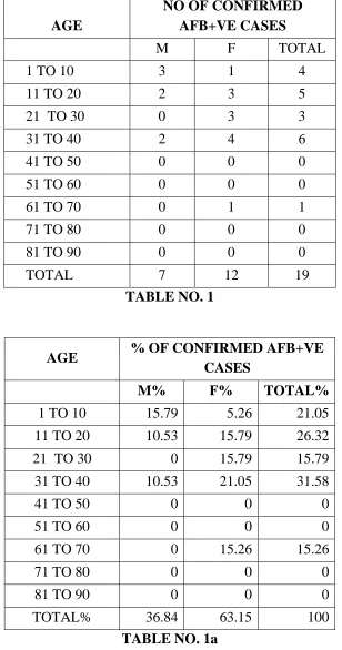
AGE AND SEX DISTRIBUTION OF TB LYMPHADENITIS CASES
AGE
NO OF CONFIRMED AFB+VE CASES
M F TOTAL
1 TO 10 3 1 4
11 TO 20 2 3 5
21 TO 30 0 3 3
31 TO 40 2 4 6
41 TO 50 0 0 0
51 TO 60 0 0 0
61 TO 70 0 1 1
71 TO 80 0 0 0
81 TO 90 0 0 0
TOTAL 7 12 19
TABLE NO. 1
AGE % OF CONFIRMED AFB+VE
CASES
M% F% TOTAL%
1 TO 10 15.79 5.26 21.05 11 TO 20 10.53 15.79 26.32 21 TO 30 0 15.79 15.79 31 TO 40 10.53 21.05 31.58
41 TO 50 0 0 0
51 TO 60 0 0 0
61 TO 70 0 15.26 15.26
71 TO 80 0 0 0
81 TO 90 0 0 0
[image:56.595.155.456.165.446.2]TOTAL% 36.84 63.15 100
[image:56.595.152.460.168.754.2]AGE DISTRIBUTION OF TB LYMPHADENITIS CASES
0
5
10
15
20
25
1 TO 10 11 TO
20
21 TO
30
31 TO
40
41 TO
50
51 TO
60
61 TO
70
71 TO
80
81 TO
90
AGE
%
O
F
C
A
SES
M%
F%
Age range of patients with tuberculous lymphadenitis was from 3 to 70
years. The commonest age group affected was between 31 to 40
years(31.58%). Females were most commonly affected than males(63.15%).
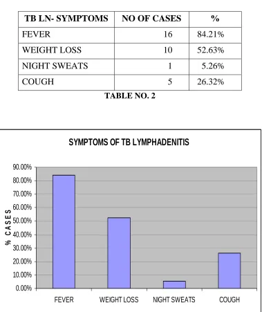
SYMPTOMS OF TB LYMPHADENITIS
0.00% 10.00% 20.00% 30.00% 40.00% 50.00% 60.00% 70.00% 80.00% 90.00%
FEVER WEIGHT LOSS NIGHT SWEATS COUGH
%
C
A
SES
CLINICAL PRESENTATION OF TB LYMPHADENITIS CASES
TB LN- SYMPTOMS NO OF CASES %
FEVER 16 84.21%
WEIGHT LOSS 10 52.63% NIGHT SWEATS 1 5.26%
[image:58.595.117.496.136.587.2]COUGH 5 26.32%
TABLE NO. 2
Most common symptom of tuberculous lymphadenitis was found to be
fever(84.21%)(evening rise of temperature). Other symptoms were weight
LYMPHNODE GROUPS INVOLVED IN TB LYMPHADENITIS
CERVICAL
SUBMANDIBULAR
SUPRACLAVICULAR
INGUINAL AXILLARY
LYMPH NODE GROUPS INVOLVED IN TB
LYMPHADENITIS
LN GROUP NO OF CASES %
CERVICAL 11 57.90%
SUBMANDIBULAR 0 0
SUPRACLAVICULAR 4 21.05%
INGUINAL 4 21.05%
[image:59.595.110.501.217.656.2]AXILLARY 0 0
TABLE NO. 3
Most common lymphnode group involved in tuberculous
lymphadenitis was the cervical group (57.90%). Other lymphnodes found to
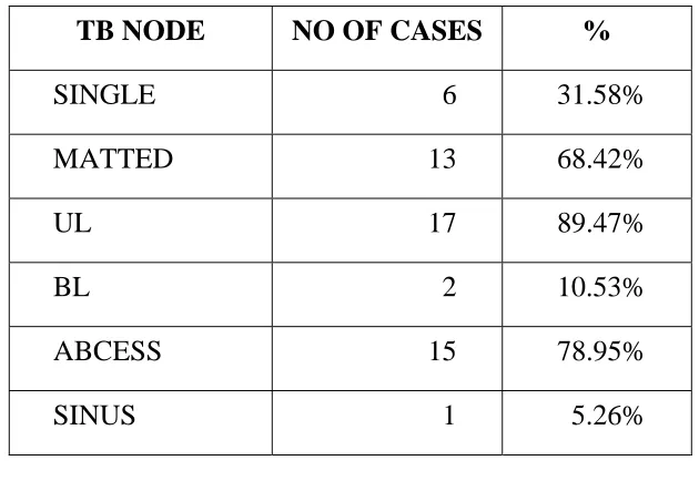
CLINICAL CHARACTERISTICS OF A TB LYMPH NODE
TB NODE NO OF CASES %
SINGLE 6 31.58%
MATTED 13 68.42%
UL 17 89.47%
BL 2 10.53%
ABCESS 15 78.95%
[image:60.595.147.462.152.369.2]SINUS 1 5.26%
TABLE NO. 4
Most of the cases showed unilateral involvement of nodes(89.47%).
Involved lymphnodes were found to be matted in 68.42% of cases. Abcess
LUNG IMAGING FINDINGS IN TB LYMPHADENITIS
IMAGING NO OF CASES %
NORMAL 10 52.63%
PNEUMONITIS 6 31.57%
FIBROSIS 1 5.26%
[image:61.595.120.508.116.592.2]BRONCHIECTASIS 2 10.52%
TABLE NO. 5
X – ray chest showed no significant findings in most of the
cases(52.63%). Findings like pneumonitis(31.57%), fibrosis(5.26%) and
bronchiectasis(10.52%) were found in few cases.
LUNG IMAGING FINDINGS IN TB LYMPHADENITIS
NORMAL PNEUMONITIS FIBROSIS
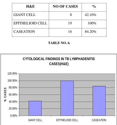
CYTOLOGICAL FINDINGS IN TB LYMPHADENITIS CASES(H&E)
0.00% 20.00% 40.00% 60.00% 80.00% 100.00% 120.00%
GIANT CELL EPITHELIOID CELL CASEATION
% C
A
S
E
S
CYTOLOGICAL FINDINGS IN TB LYMPHADENITIS (H&E).
H&E NO OF CASES %
GIANT CELL 8 42.10%
EPITHELIOID CELL 19 100%
[image:62.595.101.504.130.561.2]CASEATION 16 84.20%
TABLE NO. 6.
Epithelioid cells were found in fine needle aspiration smears of all the
cases of tuberculous lymphadenitis(100%). Giant cells(42.10%) and caseation
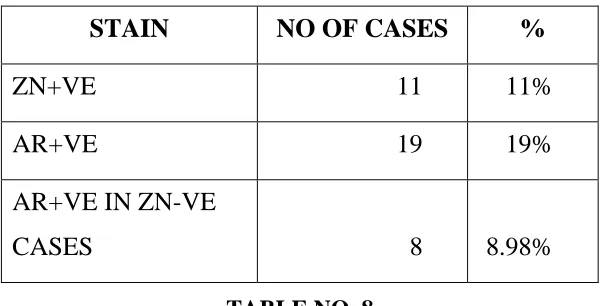
NO OF ZIEHL-NEELSEN AND AURAMIINE POSITIVE
CASES
STAIN NO OF CASES %
ZN+VE 11 11%
AR+VE 19 19%
AR+VE IN ZN-VE
[image:63.595.155.456.162.315.2]CASES 8 8.98%
TABLE NO. 8
Ziehl Neelsen detected acid fast bacilli in 11 out of 100 cases. Auramine stain showed positivity in 19 cases which included all the 11 ZN positive cases.
0 5 10 15 20
NO OF
CAS
ES
ZN+VE AR+VE
0 1 2 3 4 5 6 7 8
NO OF CASES
GIANT CELL EPITHELIOID CELL CASEATION
CYTOLOGY (H & E)
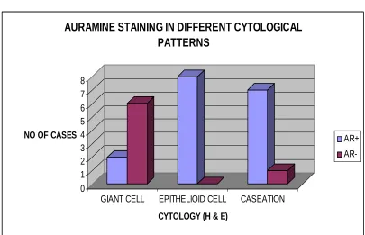
AURAMINE STAINING IN DIFFERENT CYTOLOGICAL PATTERNS
AR+
AR-AURAMINE STAINING IN DIFFERENT CYTOLOGICAL
PATTERNS
CYTOLOGY (H & E) AR+ AR-
GIANT CELL 2 6
EPITHELIOID CELL 8 0
[image:64.595.102.507.335.596.2]CASEATION 7 1
TABLE NO. 7
Caseation and epithelioid cells were often associated with positive
COMPARISON OF FLUORESCENT STAIN WITH THE
GOLD STANDARD ZN STAIN
ZN POSITIVE ZN NEGATIVE TOTAL
AR POSITIVE 11 8 19
AR NEGATIVE 0 81 81
[image:65.595.108.503.195.341.2]TOTAL 11 89 100
TABLE N0. 9
All the 11 Ziehl Neelsen positive cases were also positive for
Auramine stain. Auramine stain also showed positivity in 8 cases which were
EVALUATION OF ZN AND FLUORESCENT STAIN
ZN AO
SENSITIVITY(TRUE+VE) 58% 100%
SPECIFICITY(TRUE-VE) 89% 91%
POSITIVE PREDICTIVE VALUE 100% 58%
NEGATIVE PREDICTIVE
[image:66.595.127.486.151.359.2]VALUE 91% 100%
TABLE NO. 10
The sensitivity of Auramine O stain was more (100%) when compared
with ZN stain (58%). The false negativity of ZN stain (42.10%) was
considerably more when compared with Auramine stain which means that ZN
Fig-1- 10X- showing epithelioid cells and lymphocytes. (H&E)
[image:67.595.118.495.85.380.2]Fig-3- AFB seen as pink rods under oil immersion ( ZN stain )
Fig-5- pink slender rod shaped AFB under 100X ( ZN stain )