0022-538X/94/$04.00+0
Copyright C) 1994, American Society for Microbiology
Extensive
Envelope
Heterogeneity of Simian
Immunodeficiency
Virus
in
Tissues from
Infected
Macaques
BARBARA J. CAMPBELLANDVANESSA M. HIRSCH*
Immunodeficiency VirusesSection, Laboratoryof Infectious Diseases, NationalInstitute
of
Allergy
andInfectious
Diseases,
Rockville,
Maryland
20852 Received 1 September 1993/Accepted 7 February 1994Theextentofvirus genetic variation within tissues and peripheralblood mononuclear cells (PBMC) from twosimianimmunodeficiency virus(SIV)-infectedmacaques wasanalyzed. The products ofPCRamplification
oftworegions,region 1 (SIVVi region) and region 2 (region correspondingtothe humanimmunodeficiency
virus V3 cysteineloop andpartof the C3 region immediately downstream), oftheSIVenvelopewereexamined
for single-stranded conformation polymorphism followed by sequence analysis of selected clones. The Vi
regionof theSIVenvelope of virusespresentwithin lymphoid tissues displayed extensive heterogeneity,while viral populations within the PBMC and brain appeared tobe less variable. Region 2 heterogeneity in both animalswasgenerallyconfinedtothree residues inatissue-specificmanner.In addition, virusfrom the brains
of bothanimalsappearedtobedistinct compared with virusespresentin other tissues and PBMC ofthesame
animal,both in thepatternof PCR-single-stranded conformation polymorphism SCP and in thesequenceof
region 2. These studiesrevealedthat the tissues ofSIV-infectedmacaques were a reservoirfor viral variants
distinct fromthose seeninPBMC.
The genetic and biologic diversity of human immunodefi-ciencyvirus(HIV) is thoughttobeoneof the major
determi-nants for enhanced survival of the virus within an infected
individual; this diversity is characterized by thepresence ofa
virus quasispecies within infected peripheral blood
mononu-clearcells(PBMC) (1, 15, 18, 36). HIV existsas aquasispecies
invivo most likelyas a result of the high mutation frequency
and lack of an error correction mechanism of reverse
tran-scriptase (6, 16, 17). Asa consequence, the existence ofthis largepopulation pool allows for the selection of variants that have thepotentialtoescapefrom the host immuneresponse or
grow in different cell types and environments. The most
extensive variation has been observed within the envelope surfaceglycoprotein (17),where several epitopesseem tobe keytargetsfor the immunesystem(7, 35, 50).Infact, the major neutralization domain of the HIVenvelope is the hypervari-able V3 loop (35, 50). Neutralization escape mutants have beenobservedfollowinginfectionwith HIV(1, 39, 48, 49).In additionto the potential production of immune escape vari-ants, HIV envelope heterogeneitymay alter cellulartropism. Forexample,variationofonlyafewamino acidswithin the V3 loop can affect the ability of HIV to infect macrophages in culture(26, 40, 45, 46, 52). Themajorityof these studies have examined HIV envelope variation within PBMC of infected individuals. However, lymphoid and other tissues such asthe
brainaremajorreservoirs of HIV (12, 14,43, 44). The brain
appearstoharbor viral variants of distinctgeneticandbiologic properties (14, 43). However,littleis known about thetypeand extentofvariation inlymphoid tissues, although analysisofV3
sequences from splenic and brain viruses suggests that the separate evolution oftissue-specificvariantsoccurs (14).
Simianimmunodeficiencyvirus(SIV)infectionofmacaques
is a useful and relevant model for understanding the patho-genesisandgeneticvariation ofHIV infections (2, 11, 25, 33).
*Correspondingauthor. Mailingaddress: LID/NIAID/NIH,
Twin-brook II, 12441 Parklawn Dr., Rockville, MD 20852. Phone: (301)
496-2976. Fax: (301)480-2618.
SIV, like HIV, infects both macrophages and CD4 lympho-cytes and induces a similar immunodeficiency syndrome in
experimentallyinfected monkeys. Importantly, SIVexistsas a
quasispecies in the infected animal (29) and also evolves during the course of infection, generating neutralization
es-cape variants (8). Also as for HIV, while there has been extensive research of SIV variation in PBMC from infected
macaques(3, 9, 29, 41, 42),little is known aboutspecificviral
variants found intissues. An apparently high degreeof
enve-lope heterogeneityhas beenobservedduringanalysisof DNA derived directly from the spleen of an SIV-infected pigtail macaque (Macaca nemestrina) (25). Incontrast to the
exten-siveheterogeneityobserved in thisstudyof the spleen, varia-tion inlymphnodesfrom anotherstudyofinfectedmacaques
generally paralleledthat observed from PBMC at lessthan 1
yearpostinfection (41, 42). As in humans infected with HIV,
macrophages harborSIV in infectedtissuessuch asthebrain
(5, 32, 38).ViralDNA fromseveral tissues(includingthe brain and gut) of rhesus macaques infected with SIVmac239 had significantamounts of variation inat least tworegionsof the envelope gene (32). However, the extent of virus variation between tissues in an infected macaque has notbeen clearly established. Although SIV is generally considered an ideal model for humanAIDS,thereare someimportantdifferences betweenSIV and HIV.First,while thereareseveralanalogous hypervariable regions in the SIV envelope (Vl, V2, V4, and V5),none specifically correspondsto aneutralizationepitope
(8, 28, 31). Second,unlikethe HIV V3loop,theanalogousSIV V3regionis muchmoreconservedinsequence (9, 29, 41)and hasnotbeen showntocorrespond toaspecificlinear neutral-ization epitope (8, 28, 31). However studies in thislaboratory demonstrate that thisregion is involved in cellulartropismof SIVsm
(24a)-The goals of this study were to investigate SIV envelope heterogeneityinlymphoidandnonlymphoid tissues, compared with the variation in the peripheral blood, and to determine whether the variation observedwastissuespecific.Inaddition, the extent and type of variation may suggest whether such
pressuresascellulartropismorimmuneselectionplayarole in 3129
on November 9, 2019 by guest
http://jvi.asm.org/
thegeneticdiversity of the SIVenvelope in tissues other than PBMC. In this study, SIV envelope variation was analyzed
directly from specific tissues by using PCR followed by both
single-stranded conformation polymorphism (SSCP) and
se-quence analysis. Comparisons focused on the most variable region (V1) of the SIV envelope and region2 (region corre-spondingtothe HIV V3loopand part of the conservedregion
immediately downstream).
MATERIALSAND METHODS
Virus and Animals. Two macaques were inoculated with related
SIVSm
strains.Rhesus macaque(Macaca mulatta)E543 was inoculated withSIVSm/F236,
which has been describedpreviously(25).Briefly, SIVwasisolated from rhesus macaque
F236by cocultivationwith H9 cells andfiltered culture
super-natantusedtoinfectRhE543.Pigtailmacaque
(M.
nemestrina)
Pt56 wasinfected with cell-freevirus isolated from CEMX174 cells incubated with PBMC from rhesus macaque 660
(Rh660)
(25). This donor animal, Rh660,wasinfected previouslywith lymph node(LN) cells from RhE543. Related sequence infor-mation aboutthese virus inocula ispresentedbelow.
Diseaseprogressioninmonkeys.Althoughbothanimals had
SIV-induced
meningoencephalitis
(47)
withmultiple
foci ofsyncytial cells, they differed notably in clinical course of
disease. RhE543 was persistently viremic and had a strong
antibody response against all major SIV proteins throughout
the course ofinfection. High titers of
neutralizing
antibodieswerepresentthroughoutthe course ofinfection
(unpublished
data). The monkeywas sacrificed 42 months after infection
principally as aresult ofSIV-inducedmeningoencephalitis. In contrast,whilePt56wasalsopersistently viremic, this
monkey
developed onlyatransient antibodyreactivity against theSIV
envelope surface protein, as detected by Western
immuno-blotting (25), andneverdeveloped antibodiestothe SIVGag
protein. Pt56 serum was not tested for the presence of
neu-tralizingantibodies. PtS6wassacrificed 14months after
inoc-ulationprimarily because ofopportunisticinfections
resulting
from profound immunosuppression (25). In addition to
me-ningoencephalitis, pathological findings at necropsy of PtS6
included interstitial histiocytic pneumonia and disseminated
mycobacterium infection. Various tissues and PBMC were
obtainedatnecropsyfrom thetwomacaques, snapfrozen,and stored at -70°Casdescribedpreviously
(25).
DNA isolation. DNA was isolated from PBMC, brain,
spleen, axillary LN, mesenteric LN, and ileum from RhE543,
usingstandard techniques (23) in a separate room used only
for PCR to limit possible SIV plasmid contamination. DNA wasisolatedpreviously from various tissues fromPtS6: PBMC,
brain, spleen, mesenteric LN, lung, and ileum (25).
PCR amplificationandcloning.DNA(100 ng)fromsamples preparedasdescribed abovewasused in nested PCRs contain-ing primersenv 1F/env 1R(outer) andenv2F/env2R(inner) toamplify theenvgeneasdescribedpreviously (29). The PCR conditionswere30cycles of 94°C for 1
min,
55°C for 1.5 minut, and 72°C for 3 min. Ten microliters from the first-round reactionwastransferred to new tubes with fresh reagents for second-round reactions. Perkin-Elmer Cetus reagents andequipmentwereused in all PCRs asspecified by the
manufac-turer. A fraction of the product was analyzed on a 0.9% agarosegel, and the remainder was digested withCsp45Iand
SphI and cloned into a pGEM-7Zf (+) vector (Promega).
Twenty clones from each tissueorfrom PBMC were identified
by colony hybridizationwitharadiolabeled probe representing
the entireenvgene andwereused in PCR-SSCP and
sequenc-ingreactions.
PCR-SSCP. Two well-characterizedvariable
regions
in theenvgene, Vl and
region
2(corresponding
toamino acids 320 to399of theSIVSmH4
envelope),
aswellas ahighly
conservedregion
of theintegrase
(Int)
domain ofpol,
wereanalyzed
by
PCR-SSCP.PCR-SSCPinvolves
incorporation
of radiolabeled32p
into PCRproducts,
followedby
analysis
ofthedenatured,
single-stranded products
on a neutralpolyacrylamide
gel
(19,
20,
27).
Two rounds ofamplification
of 25cycles
each wererequired
for sufficientsignal
for PCR-SSCPanalysis
of tissue DNA.However,
only
oneround ofamplification
of 25cycles
wasrequired
for the detection of PCR-SSCPproducts
whenplasmid
cloneswere used. The PCR conditions for the first round ofamplification
of the entiregpl20
gene ofenv weredenaturationat
94°C
for 1min,
annealing
at55°C
for 1.5min,and extension at
72°C
for 3 min. For the first round of theintegrase
region,
as well as all second-roundreactions,
thetimes of the PCRweremodifiedto30sfor eachstep,while the
temperatureswerenotaltered.
Sequences
of theprimers
usedin the PCRs are asfollows:
Env
Int
Rl F TGGGATGTCTTGGGAATCAGCTGCTTA (6588)
R CTTTTCTTGCTGAATTTGTGCTTCTTC (8596)
R2 VI F CTCACCCCACTATGTATAGCAATGAGA (6896)
R AATTACAACCTATCATGGGCTCCTGTT (7098)
Reg2 F GATCAAGCTTAATAAGTATTATAATCTAAC (7493)
R GATCCTCGAGTCTACAATTTGTCCACAT (7774)
Rl F CTATTACAGAGAAGGCAGAGACCAGCTGTG (5191)
R GCTATGCCACCTCTCTAGCCTCTCCGGTATCCT (5399)
R2 F ACTAAGCTTTGTCCAAAGGGGAAGGAGCAGTCA (5246)
R ACTCTCGCGACTATCCAATTCTTTTCCTCCTCCATA (5354) Actual genome coordinates from
SIVpSmH4
of theunderlined nucleotidearegiveninparentheses.Theextra5'nucleotides in someof theprimers
were introduced for restriction enzymecloning.
Ri,
R2, F, R, and Reg2 denote round 1, round2,
forward,reverse, andregion 2,
respectively.
Inadditiontotherecommended concentrations of
deoxynucleoside
triphos-phates,0.5mCi of
[ot-32P]dCTP
wasused in each second-roundreaction (or onlyround for
plasmid
clones)
to radiolabel thePCR products. Three microliters of a 25-,u reaction was
diluted 1:1 with Sequenase stop buffer
(U.S.
Biochemical),
denatured by boilingfor 5 min, and loaded on a 10%
glycer-ol-6% hydrolink gel
(AT Biochem, Malvern,
Pa.)
in 0.6xTris-borate-EDTArunningbuffer. The gelswere run at con-stant power (30
W)
for 5 h at room temperature,dried,
andexposedovernighttoX-rayfilmat -70°C.Plasmid cloneswere
scored asidentical in the target region whenidentical
migra-tion patternswere observedonthe autoradiograph
(comigra-tion of both double-stranded and single-stranded
bands).
PCR-SSCP reactions were performed in duplicate on all tissues and clones.
Sequencedetermination andanalysis.The virus stocks used
for inoculation of the two study animals were partially
se-quenced previously (24, 25, 29). Briefly, smH4i and smH3,
full-length clonesderived from the F236 chronically infected
H9cell line
(RhE543
virusinoculum),
differ inonly five aminoacids (nine nucleotide differences) in the envelope gene
(24,
29). There were no differences in either theVi
region orregion 2of smH4i and smH3. Sequences ofeight additional
clones of the
Vi
regionofvirusDNAderived from thesame cell line failed to indicate significant heterogeneity(unpub-lisheddata). Littleor novariation(twoorfewer aminoacids)
was observed in the
Vi
region from 10 clones derived from CEM cells infected with the 660virus(PtS6
virus inoculum) (25)or from three clones inregion 2(unpublished data).The multiple sequence alignment program Clustal V was
used forcomparisonsof sequences derived from several clones
on November 9, 2019 by guest
http://jvi.asm.org/
RhE543 Pt56
t
(a3
a
VI
R2
FIG. 1. PCR-SSCP analysis of DNA from tissues and PBMC of
RhE543and Pt56.Two regions of the SIV envelope, Vi and region 2 (R2), from the indicated tissues of the two infected macaqueswere
examinedforvariability. The bandsintheuppertwo-thirds of each Vi autoradiogram show specific single-stranded conformations of the double-strandedamplified product (strong bandsatbottom of each Vl gel).Thedouble-strandedproducts of region 2wereomitted in these
photographs because there was no detectable variation in the sizes
observedonthegels. Ax.LN, axillary LN; mes.LN, mesenteric LN.
fromeach animal(21, 22). Alignments of the predictedamino acids of individual clones of various regions of theenv gene were alsoperformed by usingClustal V.
Nucleotide sequence accession numbers. All sequences
listed in Fig. 4 to 6 have been submitted to GenBank and assignedaccession numbersU05049to U05191.
RESULTS
Direct PCR-SSCPanalysisofSIV heterogeneityin tissues. Thegeneralpatternof virusvariation within tissues and PBMC ofRhE543 and Pt56wasexamined;PCR-amplified productsof
the entire gpl20 gene were subjected to a second round of
PCR (PCR-SSCP), using nested primers spanning either the Vl regionorregion2of the SIVenvgene.ControlPCR-SSCP
reactions of the integrase region were performed to
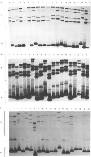
demon-strate the fidelity of the PCR. As expected, no detectable variation within the integrase region was observed (data not shown). In contrast, considerable heterogeneitywasobserved intheVl regionofviruses withinmosttissues of either animal (Fig. 1), as evidenced by the multitude of single-stranded
[image:3.612.65.278.74.347.2]productsin theuppertwo-thirds of the spleenandLN lanesin bothanimals. Double-stranded products also varied consider-ablyinsize,especiallyin thesamplesfromRhE543,suggesting that insertions and deletions occurred within the VI region. While therewas extensive heterogeneity of virus in the lym-phoid tissues, limited heterogeneity was observed within the
TABLE 1. Analysis of nucleotide changes in tissue-derived clones
%
Transitions'a
N Nonsynonymous Monkey Region AG CT TC substitutions'RhE543 Vi 29 12 1 0 96
R2 20 27 1 33 76
Pt56 Vi 28 22 25 0 95
R2 32 16 28 0.5 79
aDerived from the numbers of each type oftransition in the region/total numberof transitions foreach tissue from individual monkeys.
bSee footnoteafor derivation offrequencies. Anonsynonomoussubstitution resultsinanamino acidchange.
brain of RhE543 and the brain, lung, and PBMC of Pt56; one or two species predominated, with the presence of limited numbersof single-stranded products, generally fewer than five major bands (greater than 50% of the products).
In contrast to the extensive variability in the Vi region, considerably less variation of the single- or double-stranded PCR-SSCP products was observed within region 2. However, distinct migration patterns of PCR-SSCP products were de-tected from both brain samples and also the lung of Pt56,
suggestingthatthese tissues harborunique genotypes.
PCR-SSCP
analysis
of env clones. Twentyfull-lengthenve-lope genescloned from each tissueof RhE543 andPt56were analyzed by PCR-SSCP to determine the frequencies of indi-vidual Vl region and region 2 variants. The extent of
hetero-geneity of all clones was analyzed on individual gels, and
PCR-SSCP reactions of the Vl region of clones from three RhE543 tissues (brain, mesenteric LN, and PBMC) are illus-trated in Fig. 2. There were clear differences in the SSCP patternsbetween thetissues. For instance, while clones derived from the brain (Fig. 2A) were relatively homogeneous
(iden-ticalmigration patternsof clones2,4,6, 8, 9, 10,14 to 17, 19,
and 20), a wide array of virus variants were observed in
lymphoid tissues such as the mesenteric LN (Fig. 2B; one
group of four [clones 2, 12, 13, and 19], one groupof three
[clones 1, 5, and17],andtwogroupsof two [clones10and 15
andclones 4 and 9]; all of the other cloneswereunique). In
general, thevariability seen amongenvelope clones reflected
the heterogeneity observed in the PCR-SSCP reactions
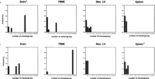
per-formed directly with tissue-specific genomic DNA. For in-stance, the virus clones from the brain of RhE543 and the PBMC ofPt56had the least amount of variation inVl(80and
90%, respectively, consisted of two or one major species)
comparedwith the virus clones from the LN orspleen from
eitheranimal, in which no variants represented greater than
25% ofthespecies (Fig. 3).The virus cloned from thebrain,
lung, and PBMC of either animalwasgenerallylesscomplex
than that cloned from the LN and spleen. However, clones
amplified from the PBMC of RhE543 and the lung of Pt56
(data not shown) displayed a larger degree of
heterogeneity
than that observed during analyses of other
nonlymphoid
tissues and the PBMC ofPt56.Interestingly,the
heterogeneity
of the virus from all RhE543 tissues and PBMCwaslessthan that fromPt56 in region2 (datanotshown).
Sequenceheterogeneity oftheVi regionand region2.The
Vl region and region 2 from several of the PCR-derived
envelopecloneswere
sequenced
todetermine(i)
the accuracyofPCR-SSCPanalysisin
predicting
nucleic acidheterogeneity,
(ii) the types of nucleic acid changes observed in different
tissues, and (iii) whether any of these
changes
could beexplained byselective pressures suchasimmuneselection
(3,
8,
42) orpresumed changesin cell
tropism
(37,
38).
on November 9, 2019 by guest
http://jvi.asm.org/
[image:3.612.313.556.88.159.2]A. 1 2 3 4 5 6 7 8 9 10 11 12 13 14 15 16 17 18 19 20
_- e - - - Q _- 'ul wwqp _
ss _ _.'so
__ 0 * - - _ -....-._t e
__
- -a-ds [
B. 1 2 3 4 5 6 7 8 9 10 11 12 13 14 15 16 17 18 19 20
ds
C. 1 2 3 4 5 6 7 8 9 10 11 12 13 14 15 16 17 18 19 20
ds[
FIG. 2. PCR-SSCPanalysisof clones from RhE543 tissues and PBMC.Twentyclonesfrom the brain
(A),
mesentericLN(B),
and PBMC(C)
were analyzedbyPCR-SSCP on individual gels. Regionscontainingeithersingle-stranded
products
(ss)
ordouble-strandedproducts
(ds)
areindicatedbythe bracketsonthe left. Therewere extrasingle-strandedbands
(other
thanthoseexpected)
in allVI PCR-SSCPgels
of tissue-orPBMC-derived clones. Webelieve that theprominentshadow bandswere asecond stableconformation of theDNA,which have also been noted inother PCR-SSCP reactions(19).
V
iA,A" .4
-- . M-
tr;..
a M.ftAwlmkA.
on November 9, 2019 by guest
http://jvi.asm.org/
[image:4.612.155.472.103.650.2]Brain"
. n r l /
0.8-10 678910111213141S1*17181920
numberofclones/group
Brain
PBMC
*.0- 0.8-o.. 0.4
0.2
123 *5678910111213.4tSiGI7181920
numberofclones/group
PBMC
n ef ng
.A
0.6 t ... ...
23*567901!7131411617'81920
number ofclones/group
Mes. LN
1..-a's
23A 5 6 78-9 10,11121314I151617-181920
number ofclones/group
Mes. LN
0n6
00
...
I23- 5678910'11 2131411IS 181970SIq?
number ofclones/group
Spleen
n r l /
0.6
0.0. .1...
1234 56789I010lll213 i6 i7, 9 20
numberofclones/group
Spleen'
n0.ubo.,,,,nes/group
1:134 56789 'I'1?31 * 5 lbI1181920
numberofclones/group
FIG. 3. Frequency distribution ofgroupsof unique clones(columns 1)orsetsof identical clones (columns 2to20) oftheVl regionfrom the indicated tissue of RhE543 (A)orPt56(B). Frequency (f) = number of clonespergroupdivided by the total number of clones analyzed.For
example,onesetof 12identicalclones(f= 0.6),onesetof 3identical clones (f= 0.15), and 4 unique clones (f=0.2)werefound by PCR-SSCP
analysis of the brain of RhE543; and therewerefivesetsof 2identical clones (f=0.5) and 10 unique clones (f= 0.5)found in the mesenteric
(Mes) LN of Pt56. aNineteenclonesexamined. Thereweresignificantlymore groupscontainingtwoormoreidentical clones found in the brain
of RhE543 and the PBMC of Pt56 than in theother tissuestested (chi-square analysis, P= 0.05).
PCR amplification generally introduces nucleotide errors
intoamplified products atthe rateof 1 in 1,000to 1in 4,000
bases (13, 30). We have found in previousstudies using the
same reagents and conditions a PCR error rate of approxi-mately 1 in 1,383 bases (29). Since ourPCR-SSCP reactions were repeated and the same resultswere obtained (data not shown),variation introducedbyTaq polymerasewaslessthan
the heterogeneity expected or observed in our amplified
products.
(i)ComparisonwithPCR-SSCPanalysis. Sequence analysis of theVlregionof selected SIVenvelope-derivedclonesfrom RhE543 and Pt56 confirmed PCR-SSCP results. Allgenerated setsofuniqueoridentical clones(identified by PCR-SSCP)of the brain, mesenteric LN, and PBMC from RhE543 were
grouped similarly bysequence analysis (Fig. 2, 4,and5;some
PCR-SSCP resultsnot shown),thusdemonstrating that PCR-SSCPwas a reliable predictorofgenetic heterogeneity in the Vl region. However, two groups of clones derived from RhE543samples (Fig. 6A)andonegroupof clones from Pt56 (Fig. 6B),allof whichappearedtobe identicalbyPCR-SSCP of region 2 (data not shown), contained minor sequence
variations which consisted of three or fewer nucleic acid differences. Thereareatleasttwopossiblerelatedreasonsfor thisdiscrepancy. First,thesensitivityofPCR-SSCP isinversely proportional to the size of the fragment analyzed by PCR-SSCP (19). Since region 2 islarger than the Vl region (284
versus 219bp), thesensitivity of the Vl SSCP reactionsmay
havebeensignificantlygreaterthan forV3.Second,therewere
fewer nucleotide changes in region 2 than in the Vl region. Thus, the multiple differences among Vi regions would
in-creasethe likelihood ofdetectingthis variation.
(ii) Typesof nucleic acid andpredictedaminoacidchanges.
Previous investigators have observed that the polymerases of SIV and HIV have a propensity for G-to-A transitions in clonesderived fromeither PBMC(SIV-infected macaques) (9, 29, 42) or HIV-1 clones derived from cultured cells (51) or
from the brain of infected individuals(34). Neithermacaquein thisstudywasinfected withamolecular clonebutwasinfected withan essentially homogeneous virus population (see
Mate-rials and Methods). Therefore, comparisons were based on
nucleotidechangesrelativetoaconsensussequence (sequenc-esusedwerefromFig.5and6). Unlike the results of previous investigators, nucleic acid sequences of the virus from all
tissues of Pt56 containedapproximately thesame percentage of C-to-Ttransitions as G-to-A transitions (Table 1; nucleic
acid datanot shown).
Envelope clones were examined for potential N-linked glycosylation sites within the Vl region by comparisonwith a
representative sequence of the virus inoculum (smH4i for RhE543 and 660 for Pt56). As shown in Fig. 4 and 5A, glycosylation patternsfromthePBMC andsomeofthelymph node and spleen clones of RhE543 were similar to those reportedpreviously in which N-linked glycosylation sites
ap-peared to evolve, especially late in the disease course (42).
However, the clones from the brain andsomeof thetissues lost potentialN-linkedglycosylationsitesortheir patterns didnot change. In contrast to the results with RhE543, most clones derived from the brain, lung, mesenteric LN, and spleen of Pt56 had lost the C-terminal N-linked glycosylation site and hadnotgainedanother in theVl region (Fig. SB).
We also determined whetheranyof thechangesobservedin
theVl regionorregion 2weretissuespecific.Therewere no
consistent deduced amino acid changes in the Vl region between different tissues from either animal. However, in
C
0~
e
3
B.
C
cr
0
n
23 56e7891011c12e3141Sig7ro 920
number ofclones/group
on November 9, 2019 by guest
http://jvi.asm.org/
[image:5.612.59.558.77.331.2]aa 121 166
A. 1-2 WGLT GTTKAPTTKTSTTTrTPRTAEKVINEGDPCIKNNSCAGIEQEP
1-4 -1-9 --1-10 ---1-14 ---1-16 --1-17 ---1-19 ---1-15 *---1-20 ....
1-5 ----RNAGT--TKS-ASST---V--SVT--- -D---1-7 ----RNAGT--TKS-ASST---V--SVT---
D---1-13 ----RNAGT--TKS-ASST---V--SVT-Q ---
D---1-18
----RNATT--TTT-ASSPS--V---VT---S---Y-N-T---1-1 ----RNATT--TIT-ASST----.-.
VT---S---Y-N-T---1-11 ----GNART--T-TPAP-I--A-P-SVT-..---NS---RSD
---B. 4-10 WGLTRNATT TTTTASSPSTTVTPRVrEKVINEGDSCIKYNNCTGLEQEP
4-15
--4-2 ---...T---TT--T...
--P---4-12 ---...T---TT--T.
--P---4-9 ...--- T---TT
---4-14 ---...
T---A---DE---4-1 ----GE-G--K-PT-IKPTGPST---S-.... ---P---N-S-A---4-5 ----GE-G--K-PT-IKPTGPST---S-....
---P---NS-A---4-17 ----GE-G--K-PT-IKPTGPST---S-....
---P---NLS-A---
----GE-G--K-PT--KS-A-ST--T--S---K-S-P---4-6 ----G---...T--A-KT-TT--T... -TP---P---N-S-A---4-8 ---...T---ST---....S .---PS--N-S-A---4-11 ----G---T---KTP-T
-IP--V--D-P---iNS.-A.---4-16 ---...T---TTT--T...--P---K---P--- NX=-A---4-3 ----G--G--...
-T-ATT---A-S---N----SNP---N-S-A-S----4-20 ---G-... AT-VTT---S-A-N---SNP---N-S-A---4-18 ----GT-K-PTAKTPA---.
T---S---P---N---C. 6-1 WGLTRNUTTTTTTTASSTTTTTVTP----KVINEGDSCIKYNNCrGLEQEP
6-2
6 4 ---
-6-11
6-12
---6-19
*---6-18
.-6-10 ---T----.... -K---N-S
---6-14 ---T----.----.... ---K---P---tN.---6-15 -5----.-I-....S----N-P
.---6-13 ---
STV-SPS--I--KVTE.---6-20 ---- ...---PS--.
---RVTG---N-P---6-5 ----G.
TA---TA-SPS---RVTE---6-17 ----G.
TA---TA-SPS---RVTE---6-6
---R-P-SS--T.T--S---SVTE---P---N---6-3 ----G--.. ----A-TKTS----I--RVKA---P---NzS-A---6-7 ----G--...-T--KAPS-ITVTP.
...S---P---N-S-A---6-8 ---- -AK-S-ASPTT-ASSPS--TVTPS
...P---N-S---6-16 ---K-GA-N-S-...
---A-NVAEN---P---Y---FIG. 4. Deducedamino acid heterogeneity of clonesfrom theVl
region of RhE543 brain (A), mesenteric LN (B), and PBMC (C)
envelopes. Alignmentsof the VIregionto themostfrequent typeof clone in each tissue or PBMC are shown. Amino acid residues
(correspondingtopsmH-4) of thebeginningand end of thesequence are indicated. Potential N-linked glycosylation sites (NXS/T) are
underlined. Dashes denote identity to the consensus, while dots representgapsintroduced intothesequenceforoptimal alignments. Clones marked with asterisks were not actually sequenced but are
included on the basisof their PCR-SSCP results.
agreementwith PCR-SSCPanalysis, twoor three amino acid
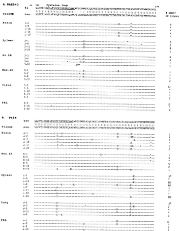
residueswere consistentlyvariable in region 2 ofSIV clones from the brainorlungof either animal(Fig. 6; R[aminoacid {aa} 375] and T [aa 383] in macrophage-rich tissues from
RhE543;P[aa 335],R[aa 375],and R[aa 384]in macrophage-rich tissues from Pt56). In addition, alysine (aa352) residue
wasobserved immediatelydownstream of the cysteine loopin the majority of the envelope clones of the virus from the spleen, axillary LN, and mesenteric LN of RhE543 but from
none of the other tissuesor PBMC. DISCUSSION
By usingPCR-SSCPas ascreeningassayforvariation within
a tissue, we were able to reasonablyestimate the number of envelopeclones thatitwasnecessarytosequencetoaccurately
A. RhE543 aa12; 166
4i WGLTGNAGTTfT...AiTTT....ATPSVAENVINESNPCIKNSSCAGLEQEP 1111 11
T. 1111 111111 111111
TISSUE con. WG;LTERNATTTTTTTTASST...TTTTTTVTPKVINEGDPCIKNNNCTGLEQEP Brain 1-2 1-9 1-14 1-19 Spleen 2-1 2-8 2-11 Ax.LN 3-3 3-4 3-9
Mes Ln 4-1 4 -2
Al,s
18 Ipleum 5-2 5-4 LP S-19 e-2A B. Pt56 Tissue conBrain a-' a-2 a-1R Mes.Ln
b-b-'2 b-18 b-1 9 Spleen c-1 c-o c-'9 c-P ci-3 d-126 e-9 e-8 e-20
----GTTKA...PT.-KTST---- PRTAEA---S-A---GTTKA...PT
.-KTST----PRTAE---ooS-A---GTTKA...PT.-KTST----PRTAE
---l-A---GTTKA... PT.-KTST----PRTAE---A---GTTKA...
PT.-KTST----PRTAE*---S-A---__
---=---P..
.S--V-PK--E---Y---K---I!A--PA.SSTTTA-V-PS--E---D---S
---#SSCP/
20clones 12 I, .. 2 2 2 5 2 2 2 2 3 2
-- ---S---Y
.--- .-K--P...---S--Kt
---GE-G--K-P--IKP-....GPS---S--- -A
---S
---.A--- ..A....
SSS---S---S----$-RR--- P...S--V-PR--E. ---S---Y---GARG---SS-TA-.----.---S----
D-A.---G-.AA----i'--R...-....S--- -a-A---AGT.A-P---KST
..-....AS---R---A---S-E---...
ATTPAG-PSGKE---.R-A---K.AEA----KST
..-....S---AS---G---S---PP---
AV---S---Y---.
TA-P...
S---S---Y---...---P...S--V-PRV-G----*N--t--Y
---WGLTAGAATTTT...AITTTATPSVAENVINESNPCILNNSCAELEQEP
ASilA
l i CI 11 11 11 1 1 1 WGLTGNAGTTTTS....ATTTTATP SVAENV INESNPC IKNNNCAGLEQEP ----R-- K-..----R---K-.-.
----R---A--....V----VAA
---R--R --- ---AA
---.---K...
P...-T---
---... T--R-V---
S---- S----S---- S---2P...---T--R-V---- ---
---A-...
S----TA---I---
A--- ----S -...--V-- --'.'-K--D--
---R---....
z---A---AD---....
A---N---.... A---
N---.---K-....
K-- K--K--K-- K--K--K-- K--K-- K--K--K-- 8
-D-S---______---- 6
2 __-_1-2 _ __---__ 2 --___--- 2
_ _ _ _ __-- 2
1 S2 -D-S--- 3
-D--S--- 2
---S--- 3
--- ---- 1
---D--- 5
--D---__ ,, __ _______-- 3
__ _ __ _ _--- 2
----S--- 3
-S--- 18
----AR-- ---A--...- A-....A---VV- ---V-
--
------*---...I
---FIG. 5. Deducedaminoacidheterogeneityof theVl regionofthe
RhE543 (A)and Pt56(B) envelopes.Acomputer-assisted alignment
ofpredicted amino acid sequences from the VI region ofselected RhE543 (or Pt56) envelope clones compared major and selected
minorspecies (determined by PCR-SSCP)witheach otherandwith botha consensussequence(con.)andarepresentativesequencefrom
the inoculum(SIVpsmH4imolecularclone[4i]orSIV660clone[660]).
Amino acid residues(correspondingtopsmH4)of thebeginning and
endof thesequenceareindicated.The numbersof identical clonesby
PCR-SSCP are listed at the right. Avertical line indicates thatthe
clone is identical to the clone abovebyPCR-SSCPanalysis.Potential
N-linked glycosylation sites(NXS/T) areunderlined. Dashesdenote identitytothe consensus, whiledotsrepresentgapsintroducedintothe sequenceforoptimal alignments.Aminoacidchangesareindicatedby letters, and asterisks represent nucleic acid changes without amino
acid alteration. t, presence of a stop codon. Ax.LN, axillary LN; Mes.LN,mesentericLN; PBL, peripheralbloodleukocytes.
predict virus heterogeneity. We did not detect minor nucle-otidechangeswhenanalyzing region 2, probablybecauseofthe size limitation of PCR-SSCP (19). Other alterations of the technique used here, such as restriction digestion before resolution on an acrylamide gel (19), may be necessary to further resolve single strands. Therefore, as in our previous studies using PCR-SSCP (27), this technique is useful as a
on November 9, 2019 by guest
http://jvi.asm.org/
[image:6.612.326.561.80.478.2] [image:6.612.69.310.81.457.2]A.RhE543 aa 320 Cysteine loop 399
4i VLPVrTMSEGLVFHSOPTNF.RPKOAWCWFEGSWKKAIQEVKETLVKHPRYTGTNDTRXINLTAPAGGDPEVTFFIWTNCRGE TISSUE con. VLPVTIMGLVHSQPINEPKQAWRGGNEAIQEVETLVHPRYTGTDTKKINLTAGGDPE CRGE
Brain 1-2 ---R---T---1-9 ---T---1 ---T---10 --- --- T---1-14
---R---T---1-19 ---R---T---Spleen 2-1---i---_______________-K---__*__________________
2-8 ---_K---_---___ *_________-2 -11 ---K--- R---T---2 -R---T---20 --- - --- ----W--- ---E-- --- ---- ----
---Ax LN 3-3 ---K---________
3-4---*--K---
--___---______________---3-9 ---*---S-K---_---_______
3 20 - --SV
-Mes.LN 4-1 --- K---R---4 -2 --- ---- --- --- ---- -*--K--- --- ---
---4 -8 ---________________K---__-________
4 110 --- --_-_-_-_
Ileum 5-2
---5566
-5 1 9 --- _--- _---_---_-__-__-_-___-_-___-_-_ _-_-_ 5-4 --- --_----_-_-_---__-_--_--_-__-__-_--_--_-__-__-_--_-* _-_--_-__-_-PBL 6-9
---6-18 ---L---
---620--. B. Pt56
660
Tissue con.
Brain a-1
a-2
a-3
a-18 a-17 a-20
Mes.LN b-1
b-4
b-12 b-5 b-18 b-19
Spleen c-i
c-6
c-19
c-8
c-7 c-11 c-12
c-16
Lung d-2
d-3
d-5
d-12
d-8
PBL e-1
e-9
e-8
e-20
VLPVTIMSGLVFHSOPINsERPKOAWCWF'GGSWKEAIQEVKETLVKH4PRYTGTNDTKKINLTAPAGGDPEVTFMWTNCRGE
VLPVTIt GLVF SQP RPKAWCWFGGSW A OEVKETLVKHPRYTGTNDTK LTAPAGGDPEVTFMW1NCRGE 3 3
..
1 2
1
2 1
5a
ND
ND
3
ND 1 ND
3b 2 4
11
11
[image:7.612.134.488.77.530.2]1
FIG. 6. Alignmentsofpredictedaminoacids inregion2ofRhE543 (A)and Pt56 (B) envelopes.Annotations are asdescribed forFig.5; in
addition,the SIV V3cysteineloop(correspondingtothe HIV V3loop)is underlined.Only17of 20clonesfrom thespleen(a)and18of 20 clones
from thelungs (b)ofregion2of the virus derived from Pt56were examined.
rapid screening tool that, in addition to sequence analysis, providesanoverallimpressionofgeneticheterogeneitywithin
anindividual sample.
Inthisstudy,PCR-SSCP andsequenceanalysiswereused to
examine the degree ofenvelope heterogeneity in tissues and
PBMC of SIVsm-infected macaques. Extensive Vl region variationwasobserved inenvelopeclones from thespleensand
LN of bothmacaques, while clones from thebrain orPBMC were generally more homogeneous. Additionally, unique
re-gion 2 variants were observed in the brain and lung, tissues
which would be expected to be rich in virus-infected
macro-phages. This region has been demonstrated in anotherstudy from this laboratory to be a critical determinant of tropism (24a). Our analysisof virusheterogeneity suggeststhat
varia-tionwithin PBMCmayvastlyunderrepresent thespectrum of viralgeneotypeswithin tissuesof thesameanimal,sincenone
of theSIV variantscloneddirectlyfrom RhE543PBMCwere
identical to clones from any examined tissueby either PCR-SSCPorsequenceanalysis.Therefore,tissuesofSIV-infected
macaques appear to constitute a reservoir for genetically
distinct viruses with potentially differentbiologic and patho-genic properties. The extent of virus heterogeneity within lymphoid tissues was an unexpected finding. This contrasted with the rather limitedheterogeneityobserved in thebrainand PBMC which has beenreportedforHIV-1-infectedindividuals (14, 43).Moreover,thetypeof variants alsoappearedtovary, dependingonthe tissue;thecentral nervoussystemharbored distinct virus variants.Thisfindingis consistent withprevious
#SSCP/
20 clones
9
I..
I..
8 1
8
4
1
2
11
2
12
..
2
on November 9, 2019 by guest
http://jvi.asm.org/
studies of brain variants ofboth SIVmaC239 and HIV-1
(5,
14,43). An arginine residue (aa 380) in region 2was detected in the majority of the clones from the macrophage-containing
tissues in our study. In addition, preliminary results of trans-fections with infectious molecular clones containing the SIV envelope from the brain of RhE543 (B-9 and B-10) indicate that this arginine residue may be required for replication in macrophages but not CEMX174 cells (10). Sequence compar-isons of the entire envelope revealed that the residues which were brain specific were located throughout the envelope (unpublished results), much like the altered amino acids in macrophage-tropic
SIVmac239
(5, 37). As previously noted for HIV (40) and SIV (5, 37), this finding suggests that it is the overall envelope conformation, not specific sequence motifs,which is important in tissue or cell tropism.
The type and extent of virus variation in tissues of the two SIV-infected macaques differed in a number of respects. Analysis of variants within tissues of the strongly seropositive RhE543 demonstrated that the overall virus population had evolved considerably from the original inoculum represented bysmH4. This was most evident within the first variable region of envelope, where the consensus sequence of clones from tissues differed by 39% from the inoculum. Similar evolution of region 2 was not observed. Another study of SIV-infected macaques which had strong antibody responses also indicated little heterogeneity in the V3 region of the SIV envelope (contained in region 2) (9). Amino acid substitutions, inser-tions, and deletions were characteristic within theVl region of RhE543 clones. Additionally, some of the substitutions re-sulted in alterations in many of the potential N-linked
glyco-sylationsites. This finding is similar to those of previous studies
in SIV-infected macaques thatdemonstrated the acquisition of new glycosylation sites during progression to AIDS (42). In contrast to RhE543, little evolution of tissue clones of the
seronegative macaque, Pt56, was observed; the consensus
sequence of the Vl region differed by only 11% from the
SIVsm/E660
inoculum. In addition, the pattern of potential N-linkedglycosylation sites was relatively conserved. Although the virus did not appear to have evolved, there was still considerable random variation in both the Vl region and region 2 of clones derived from tissues of this animal. This may represent the variation that would occur as a result of error-prone reverse transcription and massive uncontrolled virus replication in the absence of immune selection. These findings support previous suggestions that the Vl region is an immu-noreactive epitope and possibly a portion of a neutralizing epitope (3, 42). An additional difference between the two animals was variation at the Pro-335 residue within region 2. This residue was variable only in tissues of the seronegative macaque, Pt56. Interestingly, this residue was found to be an important determinant in cellular tropism in another study in ourlaboratory (24a). In addition, recent evidence of extensiveheterogeneity in the V3 region of SIV isolated directly from
several seronegative macaque tissues also suggests that key
changes that may be important in cell tropism occur in the V3 region (32). Even though the virus was
SIVmac239,
one of the particular amino acid changes was also at the Pro-335 residue (32).Inconclusion, tissues ofSIV-infected macaques harbor virus variants that aregenetically and potentiallybiologically distinct from those observed in the peripheral blood. In addition, the brain of SIV-infected animals contain discrete viralsequences. These variants may serve as a continual reservoir of virus, to increase the viral burden of the host, while remaining relatively undetected by the humoral immune response. Other studies, such as a prospective analysis of the generation of
tissue-specific virus variantsduring diseaseprogressionor ananalysis
of the infectivityoftissue-specific viruses, would further define the role of envelopevariation within tissues in disease
patho-genesis and ultimate progression to AIDS. ACKNOWLEDGMENTS
We thank Jennifer Martin for technical assistance andPhilip Zack for pathological studies. William T. London, William Elkins, and Russell Byrum were essential in their assistance with animal studies. WealsothankRobert Chanock and John Gerin for continued support andJim Williams forhelpful discussions.
REFERENCES
1. Albert, J., B. Abrahamsson, K. Nagy, E. Aurelius, H. Gaines, G. Nystrom, and E. Fenyo. 1990. Rapid development of isolate-specific neutralizingantibodies after primary HIV-1 infection and consequent emergenceof virusvariants which resistneutralization by autologous sera. AIDS 4:107-112.
2. Allan, J. 1991. Pathogenic properties of simianimmunodeficiency viruses in nonhuman primates. p. 191-206. In W. Koff, F. Wong-Staal, and R. Kennedy (ed.), AIDS research reviews,vol. 1.Marcel Dekker, Inc., New York.
3. Almond, N., A. Jenkins, A. B. Heath, and P. Kitchin. 1993. Sequence variation in the env gene of simian immunodeficiency virus recovered from immunized macaques is predominantly in the Vl region. J. Gen. Virol. 74:865-871.
4. Almond, N., A. Jenkins, A. Slade, A. Heath, M. Cranage, and P. Kitchin. 1992. Population sequenceanalysis of a simian immuno-deficiency virus (32H reisolateof
SIVmac25I):
avirus stock usedfor international vaccine studies. AIDS Res. Hum. Retroviruses8:77-88.
5. Anderson, M. G., D. Hauer, D. P. Sharma, S. V. Joag,0. Narayan, C. Zink, and J. C. Clements. 1993. Analysis of envelope changes acquired by SIVmac239 during neuroadaptation in rhesus ma-caques. Virology 195:616-626.
6. Biebricher, C., M. Eigen, and W. Gardiner, Jr. 1985. Kinetics of RNA replication: competition and selection among self-replicat-ing RNA species. Biochemistry 24:6550-6560.
7. Boeri, E., A. Giri, F. Lillo, G. Ferrari, 0. E. Varnier, A. Ferro, S. Sabbatani, W. C. Saxinger, and G. Franchini. 1992. In vivo genetic variability of the human immunodeficiency virus type 2 V3 region. J. Virol. 66:4546-4550.
8. Burns, D., C. Collignon, and R. Desrosiers. 1993. Simian immu-nodeficiency virus mutants resistant to serum neutralization arise during persistent infection of rhesus monkeys. J. Virol.
67:4104-4113.
9. Burns, D., and R. Desrosiers. 1991. Selection of genetic variants of simian immunodeficiency virus in persistently infected rhesus monkeys. J. Virol. 65:1843-1854.
10. Campbell, B., and V. Hirsch. Unpublished observations. 11. Desrosiers, R. 1990. The simian immunodeficiency viruses. Annu.
Rev. Immunol. 8:557-578.
12. Embretson, J., M. Zupancic, J. L. Ribas, A. Burke, P. Racz, K. Tenner-Racz, and A. T. Hasse. 1993. Massive covert infection of helper T lymphocytes and macrophages by HIV during the incubation period of AIDS. Nature (London) 362:359-362. 13. Ennis, P., J. Zemmour, R. Salter, and P. Parham. 1990. Rapid
cloning of HLA-A,B cDNA by using the polymerase chain reac-tion: frequency and nature of errors produced in amplification. Proc. Natl. Acad. Sci. USA 87:2833-2837.
14. Epstein, L., C. Kuiken, B. Blumberg, S. Hartman, L. Sharer, M. Clement, and J. Goudsmit. 1991. HIV-1 V3 domain variation in brain and spleen of children with AIDS: tissue-specific evolution within host-determined quasispecies. Virology 180:583-590. 15. Fenyo, E. M., and E. Norrby. 1991. Biological variation of human
immunodeficiency viruses and evolution of the late pathogenic infection in vivo, p. 149-164. In W. Koff, F. Wong-Staal, and R. Kennedy (ed.), AIDS research reviews, vol. 1. Marcel Dekker, Inc., New York.
16. Goodenow, M., T. Huet, W. Saurin, S. Kwok, J. Sninsky, and S. Wain-Hobson. 1989. HIV-1 isolates are rapidly evolving quasispe-cies: evidence for viral mixtures and preferred nucleotide
on November 9, 2019 by guest
http://jvi.asm.org/
tutions. J. Acquired Immune Defic. Syndr. 2:344-352.
17. Goudsmit, J., T. F. W. Wolfs, and C. L. Kuiken. 1991. Biological significance of human immunodeficiency virus envelope variabil-ity, p. 35-46.In W. Koff, F. Wong-Staal, and R. Kennedy (ed.), AIDS research reviews,vol. 1. Marcel Dekker, Inc.,New York.
18. Hahn, B., G. Shaw, M. Taylor, R. Redfield, P. Markham, S. Salahuddin, F. Wong-Staal, R. Gallo, E. Parks, and W. Parks.
1986. Genetic variation in HTLV-III/LAV over time in patients with AIDS. Science 232:1548-1553.
19. Hayashi, K. 1991. PCR-SSCP: a simple and sensitive method for detection of mutations in the genomic DNA. PCR MethodsAppl. 1:34-38.
20. Hayashi, K. 1992. PCR-SSCP: a method for detection of muta-tions. GATA 9:73-79.
21. Higgins, D., A. Bleasby, and R. Fuchs. 1991. Clustal V: improved
software for multiple sequence alignment. Comput. Appl. Biol. Sci. 8:189-191.
22. Higgins, D., and P. Sharp. 1989. Fast and sensitive multiple
sequence alignments on a microcomputer. Comput. Appl. Biol.
Sci. 5:151-153.
23. Hirsch, V., G. Dapolito, C. McGann, R. Olmsted, R. Purcell, and P. R. Johnson. 1989. Molecular cloning of SIV from sooty
mangabey monkeys. J. Med. Primatol. 18:279-285.
24. Hirsch, V., R. Olmsted, M. Murphey-Corb, R. Purcell, and P. R.
Johnson. 1989. An African primate lentivirus (SIVsm) closely related to HIV-2. Nature (London) 339:389-392.
24a.Hirsch, V. M., J. E. Martin, G. Dapolito, W. R. Elkins, W. T. London, S. Goldstein, and P. R. Johnson. 1994. Spontaneous
substitutions in the vicinity of the V3 analog affect cell tropism and pathogenicity of simian immunodeficiency virus. J. Virol. 68:2649-2661.
25. Hirsch, V. M., P. M. Zack,A. P. Vogel, and P. R. Johnson. 1991.
Simian immunodeficiency virus infection of macaques: end-stage disease is characterized by widespread distribution of proviral DNA in tissues. J. Infect. Dis. 163:976-988.
26. Hwang, S. S., T. J. Boyle, H. K. Lyerly, and B. R. Cullen. 1991. Identification of the envelope V3 loop as the primary determinant
of cell tropism in HIV-1. Science 253:71-74.
27. Hynes, N. A., D. Adger-Johnson, G. Dapolito, and V. M. Hirsch.
1993. Rapid screening for simian immunodeficiency virus variants using single-strand conformation polymorphism ofPCR-amplified
DNA fragments. AIDS Res. Hum. Retroviruses 9:803-806. 28. Javaherian, K., A. Langlois, S. Schmidt, M.Kaufmann, N. Cates,
J. Langedijik, R. Meloen, R. Desrosiers, D. Burns, and D.
Bolognsi. 1992. The principal neutralizationdeterminantofsimian immunodeficiency virus differs from that of human immunodefi-ciency virus type 1. Proc. Natl. Acad. Sci.USA89:1418-1422.
29. Johnson, P. R., T. E. Hamm, S. Goldstein, S. Kitov, and V. M.
Hirsch. 1991. The genetic fate of molecularly cloned simian immunodeficiency virus in experimentally infected macaques. Vi-rology 185:217-228.
30. Keohavong, P., and W. Thilly. 1989.Fidelity ofDNA polymerases in DNA amplification. Proc. Natl. Acad. Sci. USA86:9253-9257.
31. Kodama, T., D. Burns, D. Silva, F. Veronese,and R.C. Desrosiers. 1991. Strain-specific neutralizing determinant in the
transmem-brane protein of simian immunodeficiency virus. J. Virol. 65:2010-2018.
32. Kodama, T., K. Mori, T. Kawahara, D. J. Ringler, and R. C.
Desrosiers. 1993. Analysis of simian immunodeficiency virus
se-quence variation in tissues of rhesus macaques with simian AIDS. J. Virol. 67:6522-6534.
33. Letvin, N., and N. King. 1990. Immunologic and pathologic manifestations of the infection of rhesus monkeys with simian immunodeficiency virus of macaques.J. Acquired ImmuneDefic.
Syndr. 3:1023-1040.
34. Li,Y., H. Hui, C. J. Burgess, R. W. Price, P. M.Sharp, B. H. Hahn, and G. M. Shaw. 1992. Complete nucleotide sequence, genome organization, and biological properties of human immunodefi-ciency virus type 1 in vivo: evidenceforlimited defectiveness and complementation.J. Virol. 66:6587-6600.
35. McKeating, J., J. Gow,J.Goudsmit, L. Pearl,C. Mulder, and R.
Weiss. 1989. Characterization of HIV-1 neutralization escape mutants. AIDS 3:777-784.
36. Meyerhans, A., R. Cheynier, J. Albert, M. Seth, S. Kwok, J. Sninsky, L.Morfeldt-Manson, B. Asjo, and S. Wain-Hobson. 1989.
Temporal fluctuations in HIV quasispecies in vivo are not re-flected by sequential HIVisolations. Cell 59:901-910.
37. Mori, K., D. J. Ringler, and R. C. Desrosiers. 1993. Restricted replication of simian immunodeficiency virus strain 239 in macro-phages is determined byemllbut is not due to restricted entry. J. Virol. 67:2807-2814.
38. Mori,K., D. J. Ringler, T. Kodama, and R. C. Desrosiers. 1992. Complex determinants of macrophage tropism in enm' of simian immunodeficiency virus. J. Virol. 66:2067-2075.
39. Nara, P., L. Smit,N. Dunlop,W. Hatch, M. Merges, D. Waters,J. Kelliher, R. Gallo, P. Fischinger, andJ. Goudsmit. 1990. Emer-gence of virusesresistant to neutralization by V3-specific antibod-ies in experimental human immunodeficiency virus type I IIIB infection of chimpanzees. J. Virol. 64:3779-3791.
40. O'Brien, W., Y. Koyanagi, A. Namazie, J. Zhao, A. Diagne, K. Idler, J. Zack, andI.Chen. 1990. HIV-1tropism formononuclear phagocytes can be determined by regions of gpl20 outside the CD4-binding domain. Nature (London) 348:69-73.
41. Overbaugh, J., and L. M. Rudensey. 1992. Alterations inpotential sitesfor glycosylationpredominate during evolution of the simian immunodeficiency virus envelope gene in macaques. J. Virol. 66:5937-5948.
42. Overbaugh, J., L. M. Rudensey, M. D. Papenhausen, R. Ben-veniste, and W. R. Morton. 1991. Variation in simian immunode-ficiencyvirusenv isconfined to VI andV4 duringprogression to
simian AIDS. J. Virol. 65:7025-7031.
43. Pang, S., H. V. Vinters, T. Akashi, W. A. O'Brien, and 1. S. Y.
Chen. 1991. HIV-1 Env sequence variation in brain tissue of patients with AIDS-related neurologic disease. J. Acquired
Im-mune Defic. Syndr. 4:1082-1092.
44. Pantaleo, G., C. Graziosi, J. Demarest, L. Butini, M.Montroni,C.
Fox, J. Orenstein, D. Kotler, and A.Fauci. 1993. HIVinfection is active and progressive in lymphoid tissue during the clinically latent stage of disease. Nature (London) 362:355-358.
45. Shioda, T., J. A. Levy, and C. Cheng-Mayer. 1991. Macrophage and T cell-line tropisms of HIV-1 are determined by specific regions of the envelope gpl20 gene. Nature (London) 349:167-169.
46. Shioda, T., J. A. Levy, and C. Cheng-Meyer. 1992. Small amino acid changes in the V3hypervariableregion of gpl20 can affect the T-cell-line and macrophage tropism of humanimmunodeficiency virus type 1. Proc. Natl. Acad. Sci. USA89:9434-9438.
47. Simon, M., L.Chalifoux, and D. Ringler.1992. Pathologicfeatures
of SIV-induced disease and the association ofmacrophage infec-tion with disease evoluinfec-tion. AIDS Res. Hum. Retroviruses 8:327-337.
48. Trembley, M., K. Numazaki,X. Li, M.Gornitsky, J. Hiscott,and
M. Wainberg. 1990.Resistance to infection by HIV-1 ofperipheral blood mononuclear cells from HIV-1-infected patients isprobably mediated by neutralizingantibodies. J. Immunol. 145:2896-2901. 49. Trembley, M., and M. Wainberg. 1990.Neutralization ofmultiple HIV-1 isolates from asingle subject by autologoussequential sera.
J. Infect. Dis. 162:735-737.
50. van Tijin, D., C. Boucher, M. Bakker, and J. Goudsmit. 1989. Antigenicityof linearB-cellepitopes inthe Cl,Vl, and V3region of HIV-1 gpl20. J. Acquired Immune Defic. Syndr. 2:303-306.
51. Vartanian, J.-P., A. Meyerhans, B. Asjo, and S. Wain-Hobson.
1991. Selection, recombination, and G-A hypermutation of human immunodeficiency virus type 1 genomes.J.Virol. 65:1779-1788.
52. Westervelt, P., H. E.Gendelman, and L. Ratner. 1991.
Identifica-tion ofa determinant within the humanimmunodeficiencyvirus I
surface envelope glycoprotein critical for productive infection of primary monocytes. Proc. NatI. Acad. Sci. USA 88:3097-3101.