A STUDY ON
SEETHAKAZHICHAL
Dissertation submitted to
THE TAMILNADU DR. M.G.R MEDICAL UNIVERSITY
Chennai-32
For the partial fulfillment of the requirements to the Degree of
DOCTOR OF MEDICINE (SIDDHA)
(Branch IV - Kuzhanthai Maruthuvam)
DEPARTMENT OF KUZHANTHAI MARUTHUVAM
GOVERNMENT SIDDHA MEDICAL COLLEGE
PALAYAMKOTTAI – 627 002.
ACKNOWLEDGEMENT
First of all, I thank God for giving the loving parents and expresses
my whole hearted gratitude to my parents for their valuable support and
encouragement and blessings in my career, from the very beginning.
I gracefully record my indebtedness to the revered Vice Chancellor,
The Tamilnadu Dr. M.G.R. Medical University, Chennai and Special
Commissioner, Commissionerate of Indian Medicine and Homeopathy and
Joint Director of Indian Medicine and Homeopathy Chennai.
I express my honorable gratitude to Dr.R.Devarajan M.D(S).,
Principal and Dr.S.Soundarajan M.D(S)., Vice Principal, Govt Siddha
Medical College, Palayamkottai for granting permission to do this
dissertation work in the college premises.
Its my unique pleasure to express my whole hearted thanks to
Dr.N.Chandra Mohan Doss M.D(S)., Head of the Department of
Kuzhanthai Maruthuvam Govt Siddha Medical College, Palayamkottai. I
consider myself extremely fortunate to have him as my guide. I am very
thankful for his excelled care, continuous support and optimistic approach,
which influenced me to accomplish this work successfully. I could never
I express my profound gratitude to Dr.R.Patturayan M.D(S)., former
Head of the Department of Kuzhanthai Maruthuvam, Govt Siddha Medical
College, Palayamkottai for his encouragement and valuable suggestions in
this work.
I express my profound gratitude to Dr.D.K.Soundarajan M.D(S).,
Lecturer,P.G. Department of Kuzhanthai Maruthuvam, Govt Siddha Medical
College, Palayamkottai for his valuable suggestions.
It gives me great pleasure to thank Dr.K.Shyamala M.D(S).,
Assistant Lecturer,P.G. Department of Kuzhanthai Maruthuvam, Govt
Siddha Medical College, Palayamkottai for her effective guidance and
constant encouragement in my dissertation.
I express my sincere thanks to Dr.Kathir Subramaniyam M.D.,
DCH., HOD and Dr.Mathivanan M.D.,DCH., Asst Professor, Department
of Pediatrics, Tirunelveli Medical College, Palayamkottai for their kind
opinions in this dissertation work.
I am very much indebted and thankful to Mr.M.Kalaivanan M.Sc.
Lecturer and all the staffs of Pharmacology, Post graduate study centre,
I am also thankful to Prof N.Nagaprema M.Phil Head of the Dept
and all the staffs of Biochemistry,GSMC,Palayamkottai for their help in
Biochemical analysis.
I convey my thanks to Dr.S.Bageerathi M.B.B.S, M.D.,
and all the laboratory staffs and other staffs of GSMC, Hospital
Palayamkottai.
I gladly thank Dr.V.S. Padma M.B.B.S, D.M.R.D, and the staffs of
Radiology Dept attached to GSMC, Hospital Palayamkottai.
I am grateful to the Librarian Mrs T.Poonkodi M.A., B.Lib.Sc., and
the staff of library attached to GSMC, Palayamkottai.
I immensely thank Dr.Napolean B.Sc.,M.D., and the staffs of Malar
Diagnostic Centre, Tirunelveli, for their help in conducting drug sensitivity
studies.
I express my gratitude to the patients who were the back bone of the
clinical trail.
With profound sense of gratitude and appreciation, I recall the
constant support and kind co-operation recorded by the members of the
family and friends in the successful completion of this work.
Finally I express my thanks to Broad Band Net Café (BBNC) and its
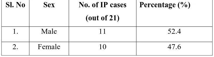
52.4% 47.6%
Distribution
according
to
the
sex
Male
Female
85.7%
14.3%
0%
0 10 20 30 40 50 60 70 80 90
Hindu Christian Muslim
Pe
rc
e
n
ta
ge
0 10 20 30 40 50 60 70 80
Poor Middle class Rich
71.4% 28.6%^ 0% Pe rc e n ta ge
Distribution
according
to
the
Socio
economic
status
0 10 20 30 40 50 60
1 day 2 days 3 days 4 days
0% 19% 57.2% 23.8% Pe rc e n ta ge
0 10 20 30 40 50 60 70 80 90 100
Saaram Chenneer Oon Kozhuppu Enbu Moolai Sukkilam /Suronitham 100% 100%
0 0 0 0 0
PE rc e n ta g e
Distribution according to Ezhu Udal kattukal
0 10 20 30 40 50 60
Vaatha neer Pitha neer Kaba neer 33.4% 57.1% 9.5% Pe rc e n ta ge
0 10 20 30 40 50 60
Less than 60 61 to 65 66 to 70 Above 70 4.8% 38% 52.4% 4.8% Pe rc e n ta ge
Distribution
according
to
Haemoglobin
content
0 10 20 30 40 50 60 70 80 90 100
Good Fair /Moderate Poor
95.2% 4.8% 0 Pe rc e n ta ge
INTRODUCTION
Medicine is an art of fundamental importance to the healthy
survival of humanity. Siddha, a Medical science is very ancient in origin,
as old as the ancient civilization.
“fy;Njhd;wp kz; Njhd;whf; fhyj;Nj
thndhL Kd;Njhd;wpa %j;jf;Fb”
Though it is believed that lord Siva was the first to teach the
Siddha system of Medicines and then the system were followed by
Siddhars like Tirunandhi thaevar, Agathiar, Pulathiar, Bohar, Tirumoolar,
Thaeraiyer, Yugimuni etc, the history of Siddha Medicine dates back to
the prehistoric period. Its origin and development is a matter of very
remote antiguity.
The word Siddha comes from ‘Siddhi’ which means perfection or
healthy bliss. It generally refers to the ‘Astama Siddhi’ i.e the eight
supernatural powers. Those who attained these powers are known as
Siddhars.
The basic principle of Siddha system is 96 thathuvas of which
panchapootha theory and mukkutra theory was very important. The
pathology in Siddha system depends upon the mukkutra theory viz,
Vatha, Pitha and Kaba. The normal order of Vatha, Pitha Kaba is in
This is stated in the following verses.
“toq;fpa thjk; khj;jpiu nahd;whfpy;
joq;fpa gpj;je; jd;dp yiuthrp
moq;Fq; fge;jhdlq;fpNa fhNyhby;
gpwq;fpa rPtHf;Fg; gprf;nfhd;W kpy;iyNa”
- Fzthfl ehb
Imbalance in this results in disease this can be inferred from the
following Thirukkural.
“kpfpDk; FiwapDk; Neha; nra;Ak; E}NyhH
tsp Kjyh vz;zpa %d;W”.
-jpUts;StH
Siddhars treated the body as well as mind and have also formulated
the ways for the prevention of diseases. Siddhars defined medicine as
follows,
“kWg;gJ cly;Neha; kUe;njd yhFk;
kWg;gJ csNeha; kUe;njd rhYk;
kWg;gJ ,dpNeha; thuhjpUf;f
kWg;gJ rhitA kUe;njdyhNk”
The clinical methods through which the correct diagnosis made out
are Envagai thervugal. They are Naadi, Sparissam, Naa, Niram, Mozhi,
According to Chattamuni:
“mz;lj;jpy; cs;sNj gpz;lk;
gpz;lj;jpy; cs;sNj mz;lk;
mz;lKk; gpz;lKk; xd;Nw
mwpe;J jhd; ghHf;Fk; NghNj”
Every minor change in the universe will immediately reflect on the
human body. This is evident from the fact that the incidence of
seethakazhichal is more common during the periods of change in seasons
in India.
Balavagadam is a branch of medical science of siddhars which
deals with the diseases and treatment of the children. Children are the
future citizens. Hence their health is of paramount importance to our
nation. The other names of medical care of children are Balamaruthuvam,
pillaipini maruthuvam.
In Balavagadam, the diseases of children are broadly classified into
Agakarana noigal and pura karana noigal.
Among the pura karana noigal, Seethakazhichal is a commonly
occurring disease in infants and children. It is a disorder of gastro
intestinal tract caused by micro organism due to poor personal hygiene
and sanitation ultimately leads to derangements in tridosas and disease
It has been clearly depicted in Gurunadi Nool (Shanmuga Velu
1987) that Seethakazhichal is caused by kirumigal and explained the
pathogenesis of the disease. The aetiological factors, pathogenesis,
clinical features of the disease explained in Siddha literature are more or
less related to amoebic and bacillary dysentery described in modern
system of medicine.
Even though there are many more medicines described in Siddha
system for Seethakazhichal, ‘Madhulam pinju Choornam’ was selected
for the present study which is purely an herbal medicine, easily available
and harmless to infants and children. The ingredients of ‘Madhulam pinju
Chooranam’ have the property of controlling Seethakazhichal without
AIM AND OBJECTIVES
Prevention and cure are the basic aims of all system of medicine.
The basic emphasis of Siddha system is on positive health viz to prevent
diseases by careful dieting and proper relaxation of the mind to achieve a
totality of health.
Seethakazhichal in children is a major health hazard in the
developing countries like India, a common disease in the tropics and
subtropics. If proper attention has not been given it may lead to many
complications like dehydration, rectal prolapse, septicemia, etc.
India, being densely populated with people of different socio
economic status, children with poor sanitary facilities, lack of personal
and environmental hygiene are the common victims of this disease. It
forms one of the major causes of sickness among infants and children
which causes a heavy economic burden to health services. At the present
time, the dysentery causing bacteria are resistant to many antibiotics,
polyresistant strains are widely spreading.
As a Siddha paediatrician, an extra personal interest in the study of
new drug for this common paediatric disease has been aimed. With this
aim in mind, Madhulam pinju chooranam was tried in the patients
OBJECTIVES :
¾ To explore the most efficacious drug for Seetha Kazhichal.
¾ To collect the literal evidences regarding the disease seethakazhical
as per Siddha System.
¾ To have a comparative study of the disease in Siddha and Modern
aspect. (Amoebic dysentry and Bacillary dysentery)
¾ To evaluate the disease Seethakazhical clinically by careful
examination on aetiology, clinical features, differential diagnosis,
investigations, diagnosis, treatment, diet, prognosis, complications etc.
¾ To find out whether any adverse effects caused by Madhulam Pinju
Choornam.
¾ To evaluate the biochemical and pharmacological analysis of the
drug.
¾ To evaluate the efficacy of trail medicine on anti – microbial
activity in vitro studies.
¾ To have a clinical trail on the Seethakazhical affected children with
Madhulam Pinju Chooranam.
¾ To highlight the factors like hygienic condition, diet, climate on the
incidence of this disease.
REVIEW OF SIDDHA LITERATURE
“Seethakazhichal” is a disease which occurs both in children and
adults. In various siddha literatures, it is described as a type of kazhichal
noi.
In Siddha Maruthuvam, it is described separately. But in
Balavagadam, it is classified under Kazhichal Vaguppu.
fopr;ry;: ,ay;:
cz;l czT nrhpj;jJk; nrhpahjJkhfTk; foptJk;> rpy Ntis fPo;f;Fly; ntJk;gpapUg;gpd;> mq;Fj; jq;fKbahikahy;> clNd fope;JtpLtJk;> clw;F Cl;lk; jUtjw;fhf cz;Zk; czT Flypw;wq;fhJ ntspahfptpLjYk;> clypd; Cl;lk; Fiwe;J cly; nkyptiltJk;> cz;l nghUs; midj;Jk; mbf;fb gyKiw
foptJkhd ,ay;Giljy; fopr;rnyd toq;fg;gLk; (rpj;jkUj;Jtk;).
gpqs<sz<!Ofib<!ujggt< (Classifications):
Various classifications of Kazhichal noi, which have been
described in several Siddha texts, are given below.
1. In Balavagadam (Pon Gurusironmani), three types of Kazhichal noikal
have been described.
At the same time,
1. ntg;Gf;fopr;ry; 2. ,uj;jf;fopr;ry; 3. mjprhuf;fopr;ry; 4. fLg;Gf;fopr;ry; 5. nghUky; fopr;ry; 6. gr;rpiyf; fopr;ry; 7. tplhf;fopr;ry;
have also been mentioned in the treatment of kazhichal noikal in
Balavagadam.
2. Sambasivampillai have described the following Kazhichal noikal.
2. sQkg<gpqs<sz<! 3. -vk<kg<gpqs<sz< 4. szg<gpqs<sz<! !
5. Osihg<gpqs<sz< 6. out<Tjmg<!gpqs<sz<
7. ubqx<Xg<gpqs<sz<! 8. sr<givg<gpqs<sz<!
3. In Uyir Kakkum Siddha Maruthuvan also called Vaidya Sara
Sangirakam, fifteen types of Kazhichal noikal have been described.
“nrhy;YfpNwd; fopr;ry;tif Njhle; jd;idr;
Ropke;jf; fopr;rnydr; nrg;g yhFk;
nty;YfpNwd; ghw;fopr;ry; tul;f opr;ry;
tPwhd the;jpapd;wd; fopr;r yhFk;
Gy;YfpNwd; fzf;fopr;ry; khe;jf; fopr;ry;
Gfohd Mkj;jpd; fopr;r yhFk;
nfhy;Yfpd;w ryf;fopr;ry; ntJg;Gf; fopr;ry;
$whd uj;jj;jpd; fopr;r yhNk.”
“MNkjhd; mjprhuf; fopr;r yhFk;
mg;gNd nghUkypd; fopr;r yhFk;
NghNkjhd; rPjuj;jf; fLg;G khFk;
jhNkjhd; gr;rpiyf; fopr;r yhFk;
rhHthd tplhf;fopr; ry;rhw; wyhFk;
ehNkjhd; nrhd;NdhNk fopr;ry; khHf;fk;
etpd;wpl;lhH ghyUf;F etpd;wpl;lhNu”
1. Ropkhe;jf; fopr;ry; 2. ghw;fopr;ry; 3. tul;fopr;ry; 4. the;jpfopr;ry; 5. fzf;fopr;ry; 6. khe;jf; fopr;ry; 7. Mkf; fopr;ry; 8. ryf;fopr;ry; 9. ntJg;Gf;fopr;ry; 10. ,uj;jf; fopr;ry; 11. mjprhuf; fopr;ry; 12. nghUky; fopr;ry; 13. rPjuj;jf; fLg;G 14. gr;rpiyf;fopr;ry; 15. tplhf; fopr;ry;
4. In Noi Nithanankal, ten types of “Kazhichal noikal” are given.
1. %yf; fopr;ry; 2. thj fpuhzp 3. gpj;j fpuhzp 4. rPj fpuhzp 5. thj gpj;j fpuhzp 6. gpj;j rpNyj;Jk fpuhzp 7. thj rPj fpuhzp 8. njhe;j fpuhzp 9. tapw;Wf; fLg;G 10. tapw;Wf; nfhjpg;G
5. According to “Agathiyar vaidya kaaviyam 1500” “Kazhichal” is
classified into six types.
“fopr;rnyd;w fpuhzpapNy tpjkh wg;gh
fz;lgpj;jk; mdy;thjk; thA thFk;
mopr;rnyd;w IaePH %d;Wq; $b
njopr;rnyd;w thAjhd; Nkf Ngjp
jpwkhd %yj;jpd; Njhl Ngjp
gopr;rnyd;w rq;fhd Ngjp nahd;W
ghug;gh thAnthd;W MW khr;Nr”.
1. thj fopr;ry; 2. gpj;j fopr;ry; 3. fg fopr;ry; 4. %yf; fopr;ry; 5. rq;fhd fopr;ry; 6. Nkf fopr;ry;
6. Same classification has been given in “Thirumoolar Vaidhyam
Karukkidai 600”.
“fopr;ry; fpuhzpf; fopAk; tpjq;NfS
mopr;rpa gpj;jk; mdy;thjk; Iakhk;
nrOr;rpa thA NrHe;jpit %d;why;
gopr;nrdg; Ngjpf;Fk; ghHngyk; NghFNk”.
‘ngykhd Nkfj;jpy; gpwe;j njhUNgjp
Fykhd %yj;jpy; nfhbaJ xUNgjp
rykhd thAthy; rq;fpj;jJ xUNgjp
Tykhd MWk; tFj;j KiwahNk.”
The present topic “Seethakazhichal” has been selected from
Balavagadam.
SEETHAKAZHICHAL:
,ay; (Definition):
rPjf;fopr;ry;> ghythflk; E}ypy; Foe;ijfSf;F cz;lhFk; fopr;ry; Neha;fspy; xd;whff; $wg;gl;Ls;sJ. ,JNt Mkf;fopr;ry; vd;Wk; $wg;gl;Ls;sJ. (ghy thflk;); Mkk; vd;gJ tapW. ,J Iak; my;yJ rPjk; vd;gijAk; Fwpf;Fk;. vdNt ,e;Neha; Flypy; Iak; ghjpg;gjdhy; cz;lhFk; fopr;ry; NehahFk;.
Seethakazhichal means the dysentery due to specific inflammation
and ulceration of the mucus lining of large intestine resulting in
evacuation of stools mixed with mucus and blood (Sambasivampillai)
NtW ngaHfs; (Synonymes in various texts):
Balavagadam!
1. Mkf;fopr;ry; 2. rPjf;fopr;ry; 3. rPj Ngjp
Siddha Maruthuvam!
1. fLg;Gf; fopr;ry; 2. rPj Ngjp 3. rPj uj;j Ngjp
Pararasasekaram Balaroga Nithanam!
1. tapw;Wf;fLg;G 2. tapw;WisT
Noi Nithanankal!
1. tapw;Wf; fLg;G 2. tapw;Wf; nfhjpg;G
Sambasivampillai!
1. rPjf; fLg;G 2. ,uj;jf; fLg;G 3. Mk Ngjp
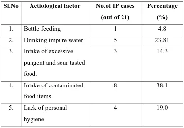
Neha; tUk; top (Aetiology):
The common causes for Seethakazhichal mentioned in various
Siddha texts are as follows.
1. Intake of excess amount of any food.
2. Intake of food stuffs which are not easily digestable.
3. Intake of excessive pungent and sour tasted food stuffs.
4. Taking large amount of sweets, carbohydrate rich foods and
mutton
5. Taking improperly cooked food stuffs.
6. Taking medicines which are having poisonous effects (Kara
Marunthugal).
7. Drinking impure water like sunai neer (stagnant water) and
karchunna neer.
8. Wandering in hot sun and exposure to cold air.
9. Living in over crowded areas.
10. Community and personal unhygienic conditions.
11. Suffering from “Seethasuram”.
12. Improper treatment for “Athisara Noi”.
The above mentioned causes are stated in the following verses.
“khndd;w tapw;wpd;ke;j kpUf;Fk; NghJ
khg;gz;l kJuq;fs; kq;if Nfh\;b
cz;ljhw; fpuhzpte;Jw; gtpf;Fq; fz;lha;.”
. A+fp itj;jpa rpe;jhkzp!!!
‘jhdhf cz;lhFk; tpjj;ijf; Nfsha;
juzpjdpw; FspHr;rpAld; tplr;rj;Jj; jhDk;
Njdhf kpFjPdp Grpj;j yhYk;
jpuz;lrdf; $l;lj;jpy; Nght jhYk;
khdhd rPjRuq; fhZk; NghJk;
kfj;jhd ,e;Neh Az;lh nkd;W
Nfhzhd E}y;jdpNy nghpNahH nrhd;dhH
nfhw;wtNd ajpDila Fzj;ijf; NfNs.”
“fhzlh ke;jf;Fly; rt;Tf; Fe;jhd;
fjpg;ghd c\;ze;jhd; nry;Y kg;gh
Czlh tpahjpjhd; mjpf khdhy;
cl;Flypy; JthuKz; lhFk; ghU
NgzpNa tt;NghJ RuKz; lhFk;
ngykhd jpNufe;jhd; jsHr;rp fhZk;
Nfhzlh ngUq;Flypd; ,uze;jh dg;gh
nghq;fKld; rpWFlypw; nry;Ye; jhNd”.
- mfj;jpaH Fzthflk;
The above stanza states that chronic Seethakazhichal is called as
Girani, which occurs due to the formation of ulcers in the large intestine
and rectum due to excessive heat. This affects the mucous membrane of
the colon and rectum and causes Seethakazhichal.
‘rq;ifapNy tp\fug;ghd; tUkhNwJ
rhuKld; fpUkp tpOe;jd;ikNaJ
nghq;fpaq;Nf A+ZUFk; fpuhzpNaJ
………
fUj;JlNdape;j tiff; fUkq;$Nw.”
“Gurunaadi Nool” explains the causative organisms and the
pathogenesis of this disease.
‘NfSkpdpf; fpUkpahy; te;jfpuhzpiaj;jhd;
fpUigAld; %yj;jpy; NtTnfhz;L
ehSkJ fpUkpajpd; Fliyr; Rw;wp
uj;jKz;lhQ; RNuhzpjj;jhy; kyKq;fl;b
kPStJ tha;T nrd;W tputpj;jhDk;
tputpaq;Nf fye;jpUf;fpy; fpUkpnay;yhk;
NfhSkJ gytpjkha;f; fopAk; ghyH
FbnfLj;j fpUkpnra;j fpuhzpjhNd”.
“Gurunaadi Saasthiram -235” also explains the same.
Due to excessive heat, the pathogenic micro organisms (Kirumikal)
multiply in large numbers in the intestine. They may make the stools
solid (Erugal) and decomposed producing foul smelling gases (Vayu).
Kirumikal multiplied from the decomposed stools are responsible for
Seethakazhichal. Thus Kirumikal is an important cause for
Kw;FwpFzq;fs;!( premonitory symptoms):
Head ache, nausea, pain in the abdomen, burning sensation in the
anus, tenesmus due to increased peristaltic movement are the symptoms
produced in the initial stage of the disease (Noi Naadal).
nghJ FwpFzq;fs< (General signs and symptoms):
Following the above premonitory symptoms, passing of loose
stools containing small amount of mucus and blood is noticed. Later all
these symptoms are aggravated.
Besides passing of mucus and blood, frequent scanty stools are
evacuated. During that time intense abdominal pain is observed. Due to
severe pain, the patient will be always in sitting posture. The patient may
pass loose stools many times a day. If it is not controlled by proper
treatment, the patient gets severe discomfort, naadi appears weak and
perspiration is seen. Eyes will be sunken, tongue becomes dry and
symptoms of muppini will occur and may be fatal. The above mentioned
features are stated in “Siddha Maruthuvam”
General signs and symptoms of “Seethakazhichal” are also
mentioned in ‘Agathiyar Gunavagadam’
“Nfslh Kjypy;jhd; tapw;wp yg;gh
Fwpg;GlNd ke;je;jhd; cz;lhk; ghU
thslh Fly;KWf;Fk; NtjidAq; fhl;Lk;
Njslh NghdTlNd Ntjidjhd; jPUk;
njspthd tpahjpjhd; gykhFk; Ntis
ghslh Flypy;jhd; ,uzKz; lhfpg;
gz;ghf mbf;fbjhd; Ngjp NghNk”.
“Ngjpjhd; Nghdgpd;G ,isg;Gz; lhFk;
ngykhd kyj;jpy;uj;jQ; rPjq; fhZk;
Mjpnad;w kye;jhDk; cz;il fl;b
mg;gNd jPa;e;JmJ GOf;if Nghyhk;
thijAld; kye;jhD kpwq;F kg;gh
td;ikAld; fLg;GlNd Kf;fy; fhZk;
thjpkdk; miytJNghy; kdJ nkj;j
td;ikAld; Ntjidfs; gLthd; ghNu”.
“ghulh kykJjhd; JHf;fe;j khfp
gz;ghff; fWg;GlNd gr;irepwq; fhl;Lk;
CWfpd;w %j;jpug;ig nfhjpg;Gf; fz;L
cj;jkNd %j;jpue;jhd; nrte;J nkj;j
NjNueP abf;fbjh dpwq;Fk; ghU
njspthfr; rpyNtis ePHr;RUf;Ff; fhZk;
rPuhd ehbaJ jPtukha;r; nrd;W
rpWj;jpUf;Fk; NeHikjhd; fz;L $Nw”.
- mfj;jpaH Fzthflk;
Initially there is dysfunction in the colon which is followed by
increased frequency of motion with small amount of mucus and blood,
unbearable gripping pain and irritation of the anal region. Fatigue and
According to Pararasasekaram, the following signs and symptoms
occur in Vayettru kaduppu.
“,Lg;Gf; fLj;J tapWise;jpl; bsfpr; rPj kw;wPe;J
KLf;fpj;Jau Kld;%ye; Njhd;wp kyKq; fope;jpUf;Fk;
mLj;Njh ud;d kUtUf;F kwNt aq;f nkype;JtUk;
njhLf;Fk; tapw;Wf; fLg;ngd;W nrhy;Yq; Fzq;fspitahNk.”
- guuhr Nrfuk;-ghyNuhf epjhdk;
Patients have gripping pain in the lower abdomen, with irritation in
and around the anal region, rectal tenesmus with loose stools, loss of
appetite, and weakness of the body due to excessive blood loss in stools.
The same features have been described in
“,Lg;Gf;fLj;J tapWise;J ,sFr;rPj kj;jpj;J
KLf;Fe;Jau kjpfKkha; Kd;dkyNk fope;jlq;F
kLj;Njhud;de; jidj;Njlh awNtaq;f nkype;JtUe;
njhLf;Fk; tapW fLg;gpnjd;W nrhd;NdhQ; nra;Ae;JaH fz;Nl.”
- mfj;jpah; 2000
Patients have fever with abdominal pain, loss of appetite,
sleeplessness, loose motion with mucus, joint pain, general weakness and
shivering.
In chronic stage, regurgitation of milk, anaemia (due to blood loss
with motion), fever, chillness of extremities and low pitched voice are
“cz;lgh nyjpnuLf;Fk; cly;gy Kof;fq; fhl;Lk;
Rz;LNk uj;j rhyr; Rukpfpe;jpUf;Fk; Nkdp
fz;LNrH nkhopAe; jho;e;J fhnyhL ifAePj;J
tpz;bl yhnkd;W tpsk;gpdH KdptHjhNd.”
-ghythflk;
Lg<Gx<x!OuXhiMgt< (Pathology):
According to Siddhars, diseases are produced due to derangements
in Thridosham (i.e) Vaatham, Pitham, and Kabam.
The following is the Siddha concepts of pathology of Seetha -
kazhichal, described in “Thirumoolar Vaidhya Karukkidai 600”
“fopr;ry; fpuhzpf; fopAk; tpjq;NfS
mopr;rpa gpj;jk; mdy;thjk; Iakhk;
nrOr;rpa thA Nrh;e;jpit %d;why;
gopr;nrdg; Ngjpf;Fk; ghHngyk; NghFNk.”
-jpU%yh; itj;jpak; fUf;fpil 600.
In “Seethakazhichal” occurring due to various causes, the Pitha
dosham is vitiated from its normal condition. This in turn stimulates
Abaanan (keezhnokkukkal) a type of Vaatham. Chenner (blood) and
Kabam are also affected.
Vitiated Pitham causes nausea, vomiting and burning sensation in
the rectum. Vitiated Pitham along with Kabam causes ulceration in the
intestine and produces passage of loose stools with blood and mucus.
produced mainly by vitiated Vayu (Sambasivampillai)
Finally, all thridoshas are deranged from their normal position and
produces “Muppini Noi” (Siddha Maruthuvam)
gpzpawpKiwik (Diagnosis):
According to Siddha Medicine, diagnosis of a disease is done by
using the following principles.
1. Poriyaal arithal (Inspection)
2. Pulanaal arithal (palpation)
3. Vinaathal (Interrogation)
The physician should observe, interrogate and palpate the patient.
Pori are the five organs of perception namely Mei, Naa, Mookku, Kann,
and Sevi. Pulan are the five objects of senses namely Manam, Suvai,
Roopam, Saptham and Sparisam.
Poriyaalarithal and Pulanaalarithal go hand in hand with the
concept of examining the patient’s pori and pulan with that of the
physician’s pori and pulan.
By Vinaathal (asking questions), the physician knows about the
patient’s name, age, native place, socioeconomic status, complaints and
duration, past history, dietetic habits, history of Pica, history of
immunisation etc. If the patient is infant or child or unable to speak (deaf
and dumb or having some other diseases) the informations may be
Poriyaalarithal, Pulanaalarithal and Vinaathal are done with the
help of Envagai thervugal and Ezhu udarthathukkal.
vz;tifj; Njh;Tfs;!)Envagai thervukal):!
“ehbg;ghprk; ehepwk; nkhoptpop
kyk; %j;jpukpit kUj;JtuhAjk.;”
- NjiuaH
Envagai thervugal are considered as the physician’s instruments.
By using them the physicians come to a correct diagnosis.
2/ ehb (Pulse)
3/ ];ghprk<!)Palpation)
4/ eh (Tongue)
5/ epwk< (Colour of the skin)
6/ nkhop (Speech)
7/ tpop (Eyes)
8/ kyk; (Stools)
9/ %j;jpuk; (Urine)
Naadi (Pulse):
Naadi is an important observation for diagnosis, treatment and
prognosis. It represents the function of heart and circulation of blood in
the body. Thus naadi is responsible for the existence of life. It can be felt
one inch below the wrist on the radial side by means of palpation with the
tips of the index, middle and ring finger corresponding to vatham, pitham
Normally the three humours namely vatham, pitham and kabam,
exist in the ratio 1: ½: ¼. Derangement in these ratio leads to various
disease entities and are best diagnosed by feeling the naadi.
Naadi nadai in Seethakazhichal or Girani:
According to Sathaga Naadi, the vitiated pitham with heat produces
the symptoms of Seethakazhichal.
“jiog;ghd gpj;jj;jpYl;bzq; nfhz;lhy;>
rakj;jp Ruk; ntJg;G rj;jpFd;kk;>
fisg;ghd nghUj;J isTtjprhuq;fs;
fLg;GlNd tapw;Wtyp %ythA
,isg;ghfp A+z;kWj;jy; ehf;frg;G
,utpy; fdTlNd rq;fhu Njhlk;
giog;ghd gapj;jpa NehnahpTjhfk;
te;jZfpy; gy gpzpf;Fk; tifajhNk.”
- rjfehb
Further, Sathaga Naadi explains that due to Pitha Vaatham naadi,
Girani is produced.
“rpwg;ghd gpj;jj;jpy; thjehb
NrhpYWe;jhJ el;lKju gPil> ciwg;ghfr; nrhpahikf;Fd; kQ;#iy
Aw;wRuq;fpuhzp tapw;wpiur;ry; ke;jk; miwg;ghd Xq;fhu GwePh;f; Nfhit>
Mahrkpuf;f nkhL kaf;f %Hr;ir> Kiwf;fha;T tp\ tPf;fk; %ytha;T>
Kulhd Neha; gyT KLFk; gz;Ng.”
In addition, Sathaga Naadi also describes that Thonthamana kabam
with Vayu produces motion mixed with mucus.
“njhe;jpj;j Nrj;Jkj;jpy; thA $bj; njhlHe;j
Fd;kk; neQ;rilg;G Rthrfhrk;>
te;jpj;j Fuy;jdpNy cWj;jyPis
tOtOg;G eP&wy; kyj;jpy; rPjk;>
nte;jpj;jy; nfhOj;jy; Fj;Je; jpkpH tpahjp
tPr;RlNd typ nal;Le;jpul;rp ghz;L>
me;jpj;j fpWfpWg;G kaf;fk; tpf;fy;>
Mdgy gpzpfSNk te;jl Ue;jhNd.”
- rjfehb
The same Sathaga Naadi also makes it clear that the aggravated
Vaatha naadi will produce the disease girani.
“thjnkDk; ehbaJ Njhd;wpy;
rPjke;jnkhL tapW nghUky; jpul;rp tha;T
rPjKWq;fpuhzp kNfhjuk; ePuhik
jpus;tha;T #iy typfLg;Gj; jPiu
ePjKWq;fp UkpFd;kk; mz;lthjk;
epiyAk; ePHfphpr;ruq;fs; je;J Nkfk;
Ngjfkh Kjugpzp %yNuhfk;
NgrntFgpzpfSNk nghUsjhNk.”
- rjf ehb
Sparisam (Palpation):
hardness, sweat, dryness, swelling, tenderness, ulcers, pigmentation and
anterior fontanelle can be examined by sparisam.
In Seethakazhichal dryness of the body, raised body temperature,
tenderness in the abdomen, sometimes enlargement of liver are present.
Naa (Tongue):
By the examination of tongue, the colour, coating, moisture or
dryness, excessive salivation, deviation in movements, fissures, variation
in taste, condition of teeth and gums are carefully noted.
In “Seethakazhichal”, coated tongue shows loss of appetite and
indigestion. Pallor tongue shows the anaemic condition. Dryness shows
dehydration.
Niram (Colour):
Colour of the skin indicates Vaatham, Pitham, Kabam, and
Thontham, cyanosis, pallor, yellowish discolouration and
hyperpigmentation.
In chronic condition of Seethakazhichal, the body is pale due to
loss of blood in motion.
Mozhi (Speech):
In the examination of mozhi, high or low pitched voice, crying,
laughing, slurring, speech in hallucination, nasal or hoarseness of voice,
incompleteness while talking and breathlessness may be noted.
appetite, poor intake of food, severe abdominal pain, malabsorption (due
to frequent loose stools) and dehydration.
Vizhi (Eye):
Both sensory and motor disturbances are noted during
Seethakazhichal. Colour, irritation, inflammation, ulceration, lacrimation,
sharpness of vision, response of the pupil to stimuli are also being noted
carefully.
In the case of Seethakazhichal, sunken eyes and pallor of eyes may
be noted in severe condition.
Malam (Faeces):
In the examination of malam (stools), Niram (colour), Nurai
(froth), Erugal (solid), Elagal (semisolid or liquid), quantity (increased or
decreased) and smell (foul smell, offensive odour) can be noted. Other
examinations like presence of blood, mucus and undigested matter in the
stools should also be noted.
In Seethakazhichal, the malam may be semisolid or liquid, bulky or
scanty in quantity, bright red or dark brown in colour. Sometimes
offensive odour may be present due to the presence of blood and mucus.
Moothiram (Urine):
Colour, odour, quantity of urine, presence of froth, deposits, blood
and pus, abnormal constituents like sugar, protein etc., and frequency of
In Seethakazhichal, the quantity of urine is slightly diminished and
yellow in colour.
According to Siddha aspect, moothiram (urine) may be examined
in two ways.
a) Neerkuri and b) Neikuri
a) fQIg<Gxq!(Neerkuri):
“te;j ePHf;fhpvil kzk; Eiu vQ;rnyd;
iwe;jpaYstit aiwFJ KiwNa”.
- rpj;j kUj;Jthq;fr; RUf;fk;
According to this verse, the general features of urine noted are
Niram (colour), Edai (Specific gravity), Manam (smell), Nurai (froth) and
Enjal (quantity).
b) nea;f;Fwp!)Neikuri):
Collection of urine for neikuri:
“mUe;JkhwpujKk; mtpNuhjkjha;
m0fy; myHjy; mfhyT+z; jtpHe;jow;
Fw;wstUe;jp cwq;fp itfiw
Mbf;fyrj; jhtpNa fhJ nga;
njhUK$Hj;jf; fiyf;Fl;gL ePhpd;
epwf;Fwp nea;f;Fwp epUkpj;jy; flNd.”
- NjiuaH
Prior to the day of examination, the patient is asked to take regular
patient is allowed to have a good sleep. In the next early morning, the
urine first voided is collected in a glass container for analysis. The
analysis should be carried within one and half hours.
“epwf;Fwpf; Fiuj;j epUkhz ePhpw;
rpwf;f ntz;nza;NahH rpWJsp eLtpLj; njd;Wwj; jpwe;njhyp Nafhjikj;jjp
dpd;wjptiy Nghk; newptpopawpTk; nrd;wJ GfYQ; nra;jpia AzNu.”
- Neha; ehly; Kjy; ghfk;
A wide vessel containing urine is kept under the bright sunlight in
a calm place without shaking. A drop of gingelly oil is dropped on the
surface of the urine. The derangement of three thathus is studied by
observing the nature of spreading of the oil on the surface of the urine.
“muntd ePz;bbd; n0Ok thjk;
MopNghw; gutpd; m0Nj gpj;jk;
Kj;njhj;J epw;fpd; nkhoptnjd;fgNk.”
- Neha; ehly; Kjy; ghfk;
If the oil spreads like a snake, it indicates vaatham.
If the oil spreads like a ring, it indicates pitham.
If the oil remains like a pearl without spreading, it indicates kabam.
In “Seethakazhichal”, oil spreads like a snake or ring indicating the
vitiation of vaatham and pitham.
Complications:
“cz;lhFk; Ngjpjhd; cf;fpukha;f; fz;lhy;
ed;whd Fly;rt;Tj; jhgpjNk fhZk;
eykhd <uypy;jhd; rPf;fl;bf; nfhs;Sk;
gz;lhd ,uzKyHe;J Flw;RUq;fp dhf;fhy;
gspr;nrd;W kyge;jk; cz;lh kg;gh
rpz;lhd rpNyl;Lkr;rt;T mOfpg; Nghdhy;
rpwg;GlNd Rug;Gf;fz;L ,wg;ghd; fhNz.”
“fhZfpd;w Nuhfe;jhd; gofp tpl;lhy;
fz;bjkha;r; rPf;fpuj;jpy; rpl;irnra;jh Ye;jhd;
G+Zfpd;w Nuhfe;jhd; trg;g lhJ
nghy;yhj Flw;rt;T RUq;fpg; Nghdhy;
MzpNgh yjpUf;Fk; fpue;jpNeha; nfhy;Yk;
mg;gNd jpNufe;jh dpisj;Jg; NghFk;
rhzhd jpNufe;Njh Yhpe;J NghFk;
rjpuhf xUFzkha; epy;yh njd;Nw.”
- mfj;jpaH Fzthflk;
From the above verses, it is clear that severe pethy leads to
perforation and inflammation of the colon, liver abscess, intestinal
obstruction and constipation. If the mucous membrane is destroyed,
oedema will be formed. Sometimes it may end fatally.
According to Kannusamy
“ghz;L gpuNkfk; gd;thj #iyFd;kk;
Ntz;lh raQ;re;ep ntz;Nrhig - ePz;l
mjpePNu fhkhiy ahdgpzp jk;K
sjprhukh fhjwp.”
If the above diseases are associated with girani, it may lead to a
fatal outcome.
He also adds that,
“re;ep ajprhuQ; rhUq; fpuhzpFd;kk;
cd;dpa rafhrk; cl;fha;r;ry; - Jd;dpNa
Nghf;Fk; tplNrhig nghy;yhj ePhpoptpy;
tPf;fq;$ lhnjd;Nw tps;.”
- fz;Zr;rhkpak;
Further he says,
“re;ep tplNrhig rhHFd;kk; ePhpopT
Jd;Dq; fpuhzp Ruk;Ngjp -gd;Dgpu
Nkfk; rakptw;Ws; %r;Rtpf;fy; Nky;tPf;fk;
Mfp YapHNgh kwp”.
! ! ! ! .!g{<[s<silqbl<!!
jPUk;jPuhepiy ( Prognosis):
“Seethakazhichal” is a curable one with proper medicine at proper
time. If it is not treated with proper medicine, it leads to severe
discomfort, ulceration of colon causing passage of excessive amount of
blood and mucus. Pulse appears weak; perspiration (excessive sweating)
is seen. Eyes become sunken and there is dryness of tongue. Pallor of the
body due to excessive loss of blood may leads to muppini. Finally end in
Neha;f;fzpg;G tpthjk; (Differential diagnosis):
Seethakazhichal should be differentiated from other kazhichal
noikal.
2/ khe;jf;fopr;ry< (Maanthakazhichal):
“the;jp gpuhe;jp %Hr;irajha; tha;e;J FuYQ; rPuzpj;Jf;
fha;e;J Nkdp ntJntJg;gha;f; iffhy; Fsph;e;J typAz;lhk;
NrHe;J fopA kye;jhDk; rPHnfl; bUf;Fk; gytpjkha;g;
Nghe;j khe;jf; fopr;rypJ nghy;yh njdNt Gfd;wdNu”.
- ghythflk;
Vomiting, loss of consciousness, hoarseness of the voice, dryness
of the skin, fever, coldness of the limbs, convulsions and different types
of loose stools are seen in Maanthakazhichal.
3/!fzf;fopr;ry<!)Kanakazhichal):!
Stools may be mucus or curdy milk or curry water, chillness of the
hands and legs, deafness, fever, and restlessness are seen.
“rPjq; fopA kyq;fopAk; jpUk;gpf; nfl;l ghy;NghNy
Nghjf; fopAq; fwpj;jz;zPH NghYq; ifAq; fhy;FspHe;J
fhij ailf;Fk; ntJg;Gz;lhk; ifapw; gps;is jq;fhJ
Nfhjh ape;jf; fzq;fz;lhy; FyT kpjd;NgH fopfzNk”.
- ghythflk;
Seethakazhichal should also be differentiated from
Vaathakazhichal, Pithakazhichal, Kabakazhichal, Mukkuttrakazhichal,
kUj;Jtk< (Treatment):
In Siddha system of medicine, the principle of treatment is bringing
back the vitiated thathus to their normal position by giving proper
medicine !
“%d;wpnyhd;WaHe;jij Kd;duwpe;J>
Ke;jpajid nahopj;jpL kUe;jpL
jzpAk; Nehapd; je;jpukpJNt
Ngzpf; fzpj;jpbd; gpwtha; gpd; Fzk;.”
- Neha; ehly; Kjy; ghfk;
kUj;Jt topKiw (Line of treatment):
1. rPjf;fopr;ry; Nehapy; jd;dpiy gpwo;e;j moy;Fw;wj;ijAk; thjf; Fw;wj;ijAk; (fPo;Nehf;Ff;fhy;) jd;topg;gLj;j Ntz;Lk;.
3/ Specific medicine for arresting the passage of loose stools with
blood and mucus should be given.!
A large number of medicines are stated in different literatures.
Among them an economical and efficacious medicine is madhulam pinju
chooranam administrated three times a day with buttermilk.
Dose: 250 mg – 1gm (dose varies with age adjusted according to the
condition of the patient and severity of the disease)
gj;jpah gj;jpak;!(Diet regimen):
Yugimunivar Vaidya Chinthamani restricts “Langhanam” for
Seethakazhichal which is understood in,
kpFe;j nfHg;gpzp ];jphpf;Fk; fpotdhHf;Fk;
,sk;gNt apisj;jtHf;Fq; Foe;ijfl;Fk;
,Lf;fz;Nzha;f; fhuUf;Fk; raNuhfpf;Fk;
csk;gNt cgthrkhd NgHf;Fk;
cWgpj;j Njfpf;Fq; fpuhzpNahHf;Fe;!
Jsk;gNt njhe;jkhk; NuhfpaHf;Fk;
#LKl; fha;r;ryhQ; RNjfHf;Nf
...
kpfthFk; yq;fzkhk; tpjpahfhNjh.”
- A+fpKdp itj;jpa rpe;jhkzp
In infants breast feeding should be appreciated. It prevents
dehydration also.
“,Ue;Njh\k; Nghf;F kpfw;fphpr;rue; jPHf;Fk;
mUe;J kUe;jpd Dghdk; -nghUe;Jk;
mQ;rdj;jpw; fhF kdy;twl;rp ePf;fptpLk;
gQ;rpdb khjH Kiyg; ghy.;”
- gjhHj;jFz rpe;jhkzp.
As per Pathartha Soodhamani
“khjHjk; Kiyg;ghy; Nrlk; tul;rpNa apUk whfk;
fhjpL %y Nuhfq; fizr;Ruk; gapj;jp ak;Nghk;.”
The advised diet as per “Pathartha Guna Chinthamani” are,
Vaazhai pinchu(Vaazhai kachchal), Athi Pinchu, Twice cooked
rice, Kaar Kuruvai kanji, Manakaththai rice kanji and kanji of arrow root
“khNdtapW fopthHf;F thiof;fr; ryj;jpf;fha;
fhNd jphpA%Hf;FUtp fhilcLk;G fTjhhp
jhNdahF KaYyhpw; wf;Fsj;Jf; fUthlh
kPNdtpUk;gpj; jpd;gPuhy; tPNzAapiu apoe;jPNu.”
Flesh of Oorkuruvi, kaadai, udumbu and gowthari are the
non-vegetarian foods which are pathiyam. Ulari, rabbit, dryfishes and fishes
should be avoided and these are all considered to be apathiyam.
As per Pathartha Guna Chinthamani, cow’s butter milk, buffalo’s
butter milk and goat’s milk are useful in Seethakazhichal.
“tPf;f kNfhjuKs; tPWFd;kk; ghz;Lgpj;je; jhf;F kUe;jpl;ljjp rhunkhL - $f;FuNy khwhj; jphpNjh\ ke;jkdw; whfk;Nghk; tPwhtpd; NkhUf;F nka;.”
- gjhHj;jFz rpe;jhkzp.
“jhfq; fpuhzp ryq;fopr;ry; fhkhiy
Mfq; FilGOT kw;Wg;Ngh - NkhfKWe;
Njth kpUjKkhQ; rPHkhdp lHjkf;F
%th kUe;njUik NkhH”.
- gjhHj;jFz rpe;jhkzp.
Nelpori kanji or Nelpori water is useful for “Seethakazhichal”. It
also prevents dehydration (Pathartha Guna Chinthamani).
“new;nghhpiaj; jpd;wh ndLe;jhfk; the;jpke;j
kw;gpj;j thj kj%Hr;ir - gw;gythk;
Ngjp aNuhrpif NgUtif tpl;nlhopAQ;
Rhjp klkapNy rhw;W”.
In Pararasasekaram, the following stanza mentions the diet regimen
of vayettrulaivu, and explains the benefits of buttermilk and also about
other of additional foods. The same is explained in Chikitcha Ratha
Deepam.
“tuFNrh Wldy; nyz;nza; itj;jePHr; NrhWNkhUk;
jukpF kpur thio jq;fpa fdpA ed;whk;
GukpF KRl;ilf; fPiu nghUe;jpa fwpA ed;whk;
cukpF NkhUq; $l;b Az;bb YisT NghNk.”
In Pathartha Guna Soodhamani, describes that drinking water
should be boiled and then cooled properly,
“tz;ikaha;f; fha;r;rp ahw itj;jeP Uoiy jhfk;
mz;zpa %Hr;ir tpf;f yjprhue; jphpNjh \k;Nghk;.”
Pathartha Guna Chinthamani stated that, the following diet such as
karamani keerai, kattu parangi leaves, leaves of perumpayaru, Agathi
leaves, katharikai and fishes should be avoided.
“fhuh kzpf;fPiu fhl;Lg; gwq;fpapiy
Nguhk; ngUk;gaw;wpd; Nghpiyfs; - rPuh
ufj;jp ngUq;fj;jphpf;fh ahapioNa kPd;fs;
gifj;jjpf NgjpjUk;g ghH”.
- gjhHj;jFz rpe;jhkzp
Prophylaxis:!
1. Personal hygiene should be maintained. It plays an important
ozhukkam, kaala ozhukkam and Noi illaneri which are
mentioned in the Siddha texts, Seethakazhichal can be
prevented. (Noi Illa Neri)
Some more preventive measures are given below.
2. Uncooked and half cooked foods should be avoided.
3. Drinking water should be boiled and cooled.
4. Fruits and vegetables should be eaten after washing them.
5. Hand washing before eating, nail cutting, use of foot wears etc.
should be appreciated.
6. Toilet should be used for defaecation.
7. In infants breast feeding should be appreciated.
8. Excessive sweets should be avoided.
9. Exposed food items, sold in shops or markets should be
REVIEW OF MODERN LITERATURE
DYSENTERY:
It is the term used for diarrhoea with visible mucus and blood.
Dysentery is also often associated with fever and tenesmus. Common
clinical features of dysentery include anorexia, rapid weight loss and
complications such as renal failure and encephalopathy.
Dysentery results from “Entero invasive” microorganisms that
penetrate through the mucosa and cause inflammation of intestinal wall.
Some of the entero invasive organisms are
I. Bacteria : Shigella (S. sonni, S.flexneri, S.boydii, S. dysenteriae)
Escherichia coli (Enterotoxigenic, Enteropathogenic)
Salmonella sp.
Staphylococcus sp.
Campylobactor sp.
Yersinia sp.
II. Protozoa : Entamoeba histolytica, Giardia lamblia., etc.
III. Virus : Rota virus, Norwalk and allied viruses.
The two main forms of dysentery are,
1. Bacterial (bacillary) dysentery and
2. Protozoal (amoebic) dysentery.
BACILLARY DYSENTERY:
Synonym: Shigellosis, Shigella colitis.
Definition:
Bacillary dysentery is an acute infection of the bowel caused by the
organisms belonging to the genus Shigella. The disease is more common
in infants than in adults.
Aetiology:
The causative agents are Shigellae. Shiga bacilli was named after
Kiyoshi Shiga who isolated the bacilli during a severe epidemic of
dysentery in Japan in 1896, S.dysenteriae bacilli. They are small, gram
-negative, non-motile rods (1 to 3µm in diameter) and non-encapsulated
with curved ends, that grow on the usual culture media. They belong to
the family Enterobacteriaceae and divided into four groups based upon
serologic similarity and biochemical reactions.
1. S. dysenteriae (group A, 12 serotypes).
2. S. flexneri (group B, 6 serotypes),
3. S. boydii (group C, 18 serotypes) and
4. S. sonnei (group D, 1 serotype).
Epidemiology:
Bacillary dysentery is endemic all over the world. The source of
infection is the dysentery patient or carrier.
second in importance. S.dysenteriae serotype 1 tends to occur in massive
epidemics. It shows special predilection for child population. In most of
the developing countries S. flexneri is more common than S. sonnei.
The age specific attack rate is highest in the first four years of life.
Infants in the first few months of life are rarely symptomatically infected
with Shigella because breast milk contains antibodies to both virulence
plasmid coded antigens and lipopolysaccharides. There appears to be no
sex predilection.
Susceptibility to dysentery is very high, particularly in the second
and third year of life. Various factors that reduce a patient’s resistance,
such as acute or chronic infections (immunodeficiency), artificial feeding
(non breast fed babies), hypotrophy, hypovitaminosis, etc., increase
susceptibility. After an attack, immunity is type-specific and short lived,
fading apparently within a few months. The immunity is especially
unstable after S. sonnei dysentery.
S.dysentriae occurred in south India in the years 1974-78 and in the
eastern parts of India in mid 1980.
Increased attack rates occur in nursing home patients, children in
day-care centers, residents of facilities for the mentally ill.
Mode of transmission:
In developing countries, it is usually acquired by ingesting
Direct person-to-person spread by contaminated hands
(dysentery has therefore been called ‘the disease of dirty hands)
or other indirect vectors are common.
Through fomites such as door handles, water taps, lavatory
seats, eating utensils, toys etc.
House flies can have asymptomatic gut colonization, excreting
Shigella in faeces and carrying the organisms on their legs and
foot hairs. Cockroaches also transfer the cysts to food articles.
Closed environments, especially of people with underlying
disease, low sanitation standards in populated localities, over
crowding, poor sanitary habits in the population, inadequate
medical services and poor nutritional states, all predispose to
epidemic Shigellosis.
Crowding, poor personal hygiene and lack of sanitary facilities
significantly increase the likelihood of intrafamilial spread of
infection.
Day-Care centers are an important focus of Shigella outbreaks
due to the gathering of young, susceptible, children who
practice poor personal hygiene, they readily contaminate their
environment.
The organism can survive in water and buffered food (such as
Incidence:
Highest during the summer and autumn months due to increased
breeding of flies, over heating, a change of diet.
Pathological anatomy:
The most characteristic pathological changes in dysentery are in
the large intestine, chiefly in its distal portion. In severe forms, the whole
large intestine and the adjacent portion of the ileum may become
involved.
The inflammatory process in the intestinal mucosa is due to the
action of both the dysentery bacteria themselves and their endotoxins.
The Shigellae penetrate the epithelial cells and multiply in them.
The mucus membrane of the intestine is swollen and brightly
hyperaemic in places, with areas of microhaemorrhages. Zones of
necrosis and epithelial desquamation, hyperaemia, oedema and round-cell
infiltration, not only of the mucous membrane but also partly of the
submucosa, are seen.
In the chronic form, regeneration is sluggish or absent. The mucous
membrane of the large intestine is pale, focal catarrhal, catarrhal follicular
inflammation and sometimes catarrhal-erosive processes are revealed.
Pathogenesis:
The main virulence property of Shigella is the ability to invade
Following ingestion, Shigella adhere to intestinal epithelial
cell-surface receptor sites, generally the retrosigmoid and distal colon, causing
an alteration of the microvillus surface of the brush border that leads to
vesicle formation on the cell membrane. The organisms that enter the cell
cytoplasm via., direct vesicular penetration and by pinocytosis, initially
surrounded by a membranous vacuole. Disrupting this barrier, the
bacteria become free within the cytoplasm of the epithelial cells and
subsequently spread into the cytoplasm of the adjacent epithelial cells.
Overt disease is produced by Shigella multiplication within host tissue
cells, causing eventual ulceration of gut mucosal surfaces. Host gut
epithelial cell penetration by Shigella is usually limited to the mucosa,
with the sites of most intense inflammation correlated with the highest
local bacterial inocula. Fibrinous purulent exudative membranes are often
apparent on the involved mucosal surfaces.
S. dysenteriae(Shiga bacillus) produces cytotoxins that kill cells in
tissue culture and stop protein synthesis in primary human epithelial
cell-line cultures. This toxin may account for the severity of infection by S.
dysenteriae serotype 1 relative to other Shigella strains. There is also
evidence that this toxin is involved in the pathogenesis of the haemolytic
uremic syndrome, a complication of infection with S.dysenteriae serotype
Incubation period:
1 to 7 days after ingestion of the organisms.
Clinical manifestations:
Onset of fever to 400C, generalized malaise and crampy abdominal
pain are common, over third of infected children have an associated brief,
uncomplicated generalized seizure. Diarrhoea occurs several hours later.
Initially the stools are bulky, with a semiliquid - to-liquid consistency, but
without apparent blood or mucus evolving into frequent small volume
bloody mucoid stools due to colonic invasion. Tenesmus, faecal urgency,
diffuse abdominal pain, prostration and bloody diarrhoea are frequent. S.
dysenteriae produces a more severe clinical illness than other Shigella
strains.
Physical examination of acutely ill patients reveals systemic
toxicity, often with pronounced fever. The patient’s abdomen is often
diffusely tender, usually without rebound, and occasionally intensely
tender, in the lower quadrants. Bowel sounds are generally hyperactive. If
dysentery with repeated episodes of stooling has preceded, an atonic
portion of the rectum may be prolapsed. Proctoscopy, especially it
performed after several days of dysentery, reveals friable, oedematous,
hyperaemic mucosa. Areas of intense erythema, with focal ulcerations
Neurologic symptoms such as convulsions, lethargy, head ache,
confusion and hallucination occur in 10 to 35% of children with
Shigellosis. Seizure frequency indicates the production of a neurotoxin by
Shigella most important complication is dehydration with its attendant
risk of renal failure and death.
Complications:
The complications associated with the dysenteric process itself are
encephalitis, neuritis, prolapse of the rectum and they occur chiefly in
severe forms. The complications may be caused by secondary infections
like bronchopneumonia, stomatitis, gingivitis, thrush, otitis, pyodermitis,
furunculosis, pyuria and nephritis.
The important complications of dysentery are dehydration,
convulsion, haemolytic uremic syndrome, sepsis, disseminated
intravascular coagulation, rectal prolapse, toxic megacolon,
pseudomembranous colitis, cholestatic hepatitis, conjunctivitis, iritis,
corneal ulcer, arthritis, Reiter’s syndrome, cystitis, myocarditis, vaginitis
and Ekiri syndrome. Severe dystrophy, avitaminosis and anaemia may
result from a prolonged course of dysentery and inadequate therapy.
Diagnosis:
Essentials of diagnosis:
Abdominal colic with bloody diarrhoea
Fever and malaise
Faecal Leukocytes
Peripheral blood leukocytosis
Isolating the bacillus from faeces
Stool culture is considered to be the golden standard
Rectal swab.
Examination of stools:
Macroscopic examination:
The colour of the faeces is often pink with no foul smell, blood and
mucus intimately mixed.
Microscopic examination:
A fresh stool specimen or rectal swab should be promptly
inoculated into selective and non-selective media. Isolation of Shigella
will be substantially reduced with an inappropriate delay in processing.
Stool should be plated on MacConkey’s xylose-lysine-deoxycholate
(XLD), Eosin-methylene blue and Salmonella-Shigella agars.
Colitis due to Shigella can be diagnosed presumptively by
microscopic examination of stool. A small quantity of stool is placed on a
added. The sample is then mixed and covered with a glass coverslip.
Shigella infections produce faecal leukocytes.
Serological tests:
Blood culture should be obtained in patients with bacteremia.
The agglutination test, indirect haemagglutination test, luminescent
serum method, the carbon agglomeration test are useful serological tests
to diagnose Shigella infection.
Coprology, i.e., microscopic study of faeces for pathological
admixtures (mucus, leukocytes, erythrocytes), is widely employed as an
auxiliary method. Detection of a considerable number of leukocytes, in
particular with an admixture of erythrocytes, has some diagnostic value.
Rectosigmoidoscopy, which often helps to reveal pathological
changes in the rectum and sigmoid colon, is of great assistance in the
diagnosis of dysentery. It is not recommended in children under a year
old, and is contra-indicated in patients with marked signs of an acute
intestinal process.
Differential diagnosis:
1. Mild dysentery in infants under 1 year should be differentiated
from simple dyspepsia.
2. Intestinal infection caused by pathogenic serological types of group
I Escherichiae.
3. Salmonellosis.
4. Staphylococcal enterocolitis
Outcome and prognosis:
The outcome of dysentery depends on a number of factors like the
state of the organism’s protective forces, the patient’s age. The gut
mucosal injury heals, and only rarely do abnormalities of intestinal
function persist. The disease is aggravated by nutritional disturbances,
avitaminosis, concomitant diseases (acute and chronic infections, rickets
etc), and helminthiasis, by the presence of bacterial intestinal infections,
and by the supervention of complications such as pneumonia, otitis etc.
Correct medical care (careful nursing, timely active therapy and a
proper diet) improves prognosis.
Treatment and diet:
The most important conditions for successful treatment of
dysentery patients are a properly organized regimen, careful nursing, and
suitable diet.
Diet:
The diet sheet is drawn up taking into account the patient’s age,
state, nutrition, the type of dysentery, and the phase of the disease.
The best for a baby under a year old is mother’s milk, either from
the breast or expressed. When that is not possible, acidophilous milk
(yoghourt) should be prescribed. Older children are kept on a mixed
carbohydrate-protein diet. Diet must be such as to leave a minimum of
residue in the bowel, and should not contain much fat. The diet must be
nutritious, with all essential ingredients (proteins, fats, carbohydrates,
Prevention:
As bacillary dysentery is exclusively human infection transmitted
by faeco-oral route, control consists essentially in improving
environmental sanitation. Health education with an emphasis on
washing hands with soap after each defaecation is of paramount
importance.
Improvement in food sanitation practices, effective sewage
removal and chlorination of water lessen the prevalence of enteric
infection.
Sanitary disposal of excreta, washing hands with soap before
eating, safe water supply, food hygiene practices like washing raw
vegetables and fruits (grapes) before eating them and health
education all can help in the prevention of the disease.
Breast feeding decreases the risk of symptomatic Shigellosis and
lessens its severity in infants who acquire infection despite breast
feeding.
Enteric isolation for hospitalized and institutionalized patients,
recognition and removal of infected children from the day-care
setting, strict personal hygiene and antibiotic therapy are
mandatory for reducing the secondary attack rate.
Control of the reservoiour is done by diagnosing all cases early,
AMOEBIC DYSENTERY:
Synonym:
Amoebiasis, Amoebic colitis
Definition:
The term amoebiasis is used here to denote the disease caused by
Entamoeba histolytica. This organism, which ordinarily inhabits the
lumen of the large intestine, is capable of invading and destroying the
bowel wall. The amoeba can spread to the liver as well as to other
extraintestinal sites and cause severe disease if not fatal.
Aetiology:
The disease, amoebic dysentery is caused by E.histolytica, which is
a protozoan that has trophozoite, precyst, cyst and metacyst stages. The
cyst is the infectious stage cysts are oval or round, asymmetrical with
four nuclear and destroyed easily by most disinfectants and by heating to
550C. Ordinarily trophozoites reside, feed and multiply in the lumen of
the colon. Cysts are never found in the tissues.
Cysts are usually not found in loose or diarrhoeic stools that
contain only trophozoites. In semiformed stools all stages of encystment
are encountered; in formed stools, usually only mature cysts are
recovered. These relatively resistant bodies can survive in faeces or water
Epidemiology:
Amoebiasis occurs world wide with highest frequency in tropics.
The prevalence of the infection varies inversely with degree of sanitation.
Poor sanitation is not limited to moist warm regions of the globe. In areas
with permafrost, satisfactory drainage is difficult to establish and
amoebiasis has been found in Eskimo communities. High infection rates
of amoebiasis are often encountered in institutions for the mentally
retarded. Certain communities in which there are recent immigrants from
highly endemic areas may have a substantially higher incidence.
Amoebiasis is usually more common in adults than in children.
Mode of transmission:
The cyst of E.histolytica is most frequently transmitted by
ingesting faecal contaminated food or water or as a result of food
contaminated by soiled fingers or other objects that have been in
contact with faecal material.
Food handlers carrying amoebic cyst play a role in spreading
the infections. Since cyst survive for over 45 minutes under the
finger nails.
Flies, cockroaches and rodents may mechanically transmit the
cysts to food.
Individuals who have vacationed in tropical and sub-tropical
amoebiasis after drinking local untreated water or eating
uncooked vegetables that were fertilized with human faeces
Pathology:
Recent studies have suggested that glycoproteins in normal colonic
mucus may block trophozoite adherence and attachment to mucosal
enterocytes. In addition, in vitro experiments have shown that direct
contact between mammalian cells and trophozoites is necessary for
mammalian cell lysis.
The caecal area is the most frequently involved anatomic site, and
the rectosigmoid the next. However, any or all parts of the colon
including the appendix may be involved. Occasionally disease may
include the terminal ileum. Initially, there may be only a few points of
mucosal invasion with little or no host reaction. Soon a tiny ulcer appears
on the surface and leads to a gradually expanding underlying necrotic
ulcer, the so-called flask-shaped lesion. As the tissues of the mucosa are
progressively destroyed by lytic necrosis, the amoebae continue to
multiply in tissues by binary fission. Frequently, multiple microscopic
lesions anastomose laterally, and ulcerations, initially confined to the
mucosa extend through the muscularis mucosa into the submucosa.
Organisms may spread out radially, and secondary sites of invasion may
occur at other levels in the colon, especially in the rectosigmoid portion
the host may not be seen until bacterial invasion of the ulcer occurs.
Often, the mucosa between ulcers appears normal, although it is not
unusual to see a diffusely inflammed mucosa resembling that of a
non-specific ulcerative colitis. Occasionally, the pathologic process extends
through the serosa and leads to perforation.
Amoebomas occur most frequently in the caecum, although they
have been reported in all parts of the colon. The basic lesion is
granulomatous thickening of the colon as a result of lytic necrosis
followed by secondary pyogenic inflammation leading to fibrosis,
proliferative granulation tissue and focal abscesses. The lesion may be
well localized or the colonic wall may be extensively involved.
Hepatic lesions are caused by E. histolytica that reach the liver
after invading blood vessels of the intestinal wall and initiate lytic
destruction of the hepatic parenchyma with abscess formation.
Incubation period:
Varies from about 4 days to possibly years, and is usually about 3
weeks to 4 months.
Clinical manifestations:
More than one half of people infected with E.histolytica are
without symptoms, few have severe disease, but many have mild to
moderate complaints. Asymptomatic individuals are referred to as
Severe diseases may be characterized by the sudden onset of
frequent copious diarrhoea usually containing mucus and blood, but more
often the symptoms develop gradually with irregular bouts of diarrhoea,
abdominal pain, nausea and loss of appetite. Tenesmus is frequently
reported associated with the evacuation of only one or two drops of bright
erythrocyte-stained mucus; under the microscope, the mucus usually
reveals large numbers of erythrophagous trophozoites. Low-grade fever
and leukocytosis are present in about one half of the patients. If the
febrile reaction is marked or there is a considerable
polymorphonucleocytosis, an amoebic liver abscess should be
considered. In severe intestinal disease, palpation of the abdominal wall
will reveal exquisite tenderness along the portion of the involved large
bowel. Sigmoidoscopy often reveals discrete ulcers that vary in size from
a pinhead to large coalesced lesions with overhanging necrotic edges.
When ulcers are still discrete, the intervening mucosa is often normal, but
occasionally a diffuse inflammed, friable mucosa, indistinguishable from
that of idiopathic ulcerative colitis, is encountered. Evidence of ulceration
can sometimes be discerned and 3% to 5% of patients have a localized
tumor-like lesion, an amoeboma, particularly in the caecum. It is often
misread as carcinoma of the colon.
Most patients with clinical disease have mild to moderate
alternating with periods of constipation. The stools will be streaked with
blood-tinged mucus. Ill-defined abdominal pain or abdominal distension
may be the only complaint, although there may be fatigue, low-grade
fever and backache. Physical examination may reveal mild to severe
tenderness in the right lower quadrant of abdomen on deep palpation.
In children, severe amoebiasis may present as an acute febrile
illness with bloody diarrhoea, or mild disease may occur as abdominal
pain with infrequent bouts of diarrhoea, poor appetite, pallor, occasional
mild fever, and failure to thrive. In infants, amoebiasis has presented as
fatal toxic dilatation of the colon. The liver is often palpable and tender
although an abscess may not be present.
In mild disease, sigmoidoscopy is often unrewarding. Blood count,
sedimentation rate, and liver function tests are normal. Patients with an
amoeboma, however, may have peripheral eosinophilia.
Complications:
The common complications of amoebic dysentery are massive
haemorrhage, perforation and peritonitis, toxic megacolon in fulminant
cases, post-dysenteric colitis, hepatic amoebiasis, pleuropulmonary
amoebiasis, amoebic pericarditis, cerebral abscess, and cutaneous
amoebiasis.
Diagnosis:
Essentials of diagnosis:
Diarrhoea with blood and mucus.
Evidence of colitis.
Pain and tenderness.
Detecting the organism in stool samples for trophozoites and cyst.
Sigmoidoscopy.
Endoscopy and biopsy when stool samples are negative.
Indirect haemagglutination test.
Even in those patients with typical ulcers seen by sigmoidoscopy,
diagnosis should be confirmed by identifying parasites in stool or from
scrapings obtained from an ulcer because lesions are not pathognomonic.
Identifying amoebae requires considerable experience.
Polymorphonuclear leukocytes, stool macrophages and nonpathogenic
amoebae are frequently misinterpreted as E.histolytica.
Examination of stools:
Stool examination is most useful when a series of three specimens
is collected on alternate days. Stool samples should be placed promptly
in preservative [Polyvinyl alcohol (PVA), Merthiolate-iodine-formalin
(MIF)].
Freshly passed unpreserved liquid stools should be examined
promptly, within 20 to 30 minutes, because they usually contain
trophozoites that are labile and degenerate quickly; if they cannot be
formed stools are less labile and can often be recognized even after 24
hours if the specimen is placed in the refrigerator.
Intestinal roentgenographic examinations that use barium often
make stool examinations for amoebae unsatisfactory for a week or more.
Examination of atleast three stool specimens with direct, concentration,
and staining techniques (Annexture V) raises the probability of diagnosis
to more than 90% of those infected. The finding of cysts, however,
should not be interpreted to mean non invasive disease or a “carrier
state”. Cysts as well as trophozoites are diagnostic of infection. Culture
and serologic methods done by a skilled laboratory may further increase
the diagnostic yield.
Liver abscess is usually diagnosed by the outcome of serologic,
radiologic, radioisotope and ultrasound studies. Diagnostic aspiration
under CT or ultrasound guidance may yield the typical red-brown
“anchovy paste” material, although the aspirate is more often yellow or
grey-green. Typically, the aspirate is sterile, i.e., no bacteria and no
odour. This finding should strongly suggest an amoebic aetiology of the
abscess.
Serological Test:
Serologic methods can be most helpful in diagnosing amoebiasis.
In patients with extraintestinal disease, the indirect haemagglutination


![[Residency in family medicine: problems and solutions].](data:image/gif;base64,R0lGODlhAQABAIAAAP///wAAACH5BAEAAAAALAAAAAABAAEAAAICRAEAOw==)