Copyrightq1997, American Society for Microbiology
Expression of Human Endogenous Retrovirus Type K Envelope
Glycoprotein in Insect and Mammalian Cells
RALF R. TO¨ NJES,* CHRISTIANE LIMBACH, ROSWITHA LO¨WER,ANDREINHARD KURTH
Paul-Ehrlich-Institut, D-63225 Langen, Germany
Received 1 October 1996/Accepted 31 December 1996
The human endogenous retrovirus type K (HERV-K) family codes for the human teratocarcinoma-derived retrovirus (HTDV) particles. The existence of the envelope protein (ENV) of HERV-K encoded by the sub-genomicenvmRNA has not yet been demonstrated. To study the genetic requirements for successful expression of ENV, we have constructed a series of recombinant HERV-K env expression vectors for infection and transfection experiments in insect cells and mammalian cells, respectively. Six baculovirus constructs bearing full-length or truncated HERV-Kenvwith or without homologous or heterologous signal peptides were used for infections of insect cells. All recombinant baculoviruses yielded ENV proteins with the expected molecular masses. The full-length 80- to 90-kDa HERV-K ENV protein including the cORF leader sequence was glyco-sylated in insect cells. In addition, the 14-kDa cORF protein was expressed due to splicing of the full-length
envmRNA. The ENV precursor protein is not cleaved to the surface (SU) and transmembrane (TM) glyco-proteins; it does not appear on the surface of infected insect cells and is not secreted into the medium. For ENV expression in COS cells, plasmid vectors harboring the cytomegalovirus immediate-early promoter/intron A element and the tissue plasminogen activator (t-PA) signal peptide or the homologous HERV-K signal peptide upstream of theenvgene were employed. Glycosylated and uncleaved ENV was expressed as in GH teratocar-cinoma cells but at higher levels. The heterologous t-PA signal sequence was instrumental for expression of HERV-K ENV on the cell surface. Hence, we have shown for the first time that the HERV-Kenvgene has the potential to be expressed as a full-length envelope protein with appropriate glycosylation. In addition, our data provide explanations for the lack of infectivity of HERV-K/HTDV particles.
The genomes of many organisms, from yeasts to humans, contain numerous endogenous retroviruses (ERV) as vertically inheritable genetic elements. In most species, all or almost all ERV are noninfectious but some retain open reading frames (ORFs) capable of encoding retroviral proteins. A pathogenic potential of intact ERV has been demonstrated in mice, in which tumors and immunological disorders are induced (6, 28). The human genome harbors up to 1% of endogenous retrovi-ral (HERV) sequences, virtually all of which are defective (reviewed in references 19, 23, and 51). Although HERV ele-ments are occasionally transcribed, mRNA seems to be rarely translated. No HERV with a complete exogenous life cycle have been detected so far, and there is basically no information on the pathophysiological function of these retroviral se-quences (23, 51).
The HERV-K family of retroviral sequences comprises 30 to 50 members, some of which have long ORFs (33, reviewed in references 23 and 44). Recently, the phenotype of human ter-atocarcinoma-derived retrovirus particles observed in terato-carcinoma cell lines (17, 22) has been correlated with complex mRNA expression of HERV-K sequences in those cells (21,
24). HERV-Kgag sequences encode HTDV GAG core
pro-teins (3, 21). The activity of HERV-K protease required for effective processing of the GAG precursor leading to the major HERV-K GAG protein has been reported (21, 31).
The existence of HERV-K proviruses is restricted to the lineage of Old World monkeys and hominoids (33, 42), with
extensive conservations ofpolandenvgene sequences among
monkeys, apes, and humans (43). The major difference
be-tween individual HERV-K proviral sequences studied so far is the presence or absence of a segment of 292 nucleotides
com-prising the amino-terminal part of theenvgene (25). HERV-K
type 1 genomes lacking this segment display fusedpolandenv
genes and predominantly express unspliced mRNA (25). Full-length transcripts derived from HERV-K type 2 proviruses are
spliced to subgenomicenvand two smaller mRNAs (21, 25).
One of the doubly spliced mRNAs encompasses most of the 292-bp segment and codes for the cORF protein, which accu-mulates as a 14-kDa protein in the nucleolus (25). Translation
of the subgenomicenvmRNA, which also contains the 292-bp
sequence, has not been demonstrated so far. Recently, we have
reported the cloning of a HERV-K type 2env cDNA clone
(pcK30env) from human teratocarcinoma cells displaying an
ORF for the surface protein (SU) and a membrane-spanning protein (TM) (25).
In this communication, we report the generation of anti-serum against the prokaryotically expressed 40-kDa HERV-K SU-ENV and eukaryotic expression of cloned HERV-K
full-lengthenvcDNA and subclones thereof by using the
baculo-virus expression system (BVES) and expression of ENV in teratocarcinoma cells and in transiently transfected COS cells. The BVES has been instrumental in the study of potential posttranslational modifications like signal peptide cleavage, proteolytic cleavage, and N and O glycosylation of highly ex-pressed heterologous proteins (34). Here we show that
recom-binant HERV-K env baculoviruses produced ENV proteins
with the expected molecular masses and that full-length 80- to 90-kDa HERV-K ENV protein including the cORF leader sequence was glycosylated in insect cells. In teratocarcinoma cells and in COS cells, signal peptide sequences are cleaved from the glycosylated ENV protein, which, as in the BVES system, is not cleaved to the SU and TM glycoproteins.
* Corresponding author. Mailing address: Paul-Ehrlich-Institut, Paul-Ehrlich-Strasse 51-59, D-63225 Langen, Germany. Phone: 49 6103 77-5304. Fax: 49 6103 77-1255. E-mail: [email protected] furt.de.
2747
on November 9, 2019 by guest
http://jvi.asm.org/
MATERIALS AND METHODS
Probes, cDNA, and genomic clones.Plasmid and bacteriophage DNA was digested with appropriate restriction endonucleases (New England BioLabs) and separated by gel electrophoresis. A HERV-Kenv-specific probe was excised from pcK30env(25) withXhoI andBamHI (nucleotides [nt] 7251 to 7755). The numbering of sequences is based on the HERV-K type 2 sequence including three single nucleotide insertions as described previously (25). The gel-purified envfragment was labelled by random priming with [a-32P]dCTP (Amersham,
Braunschweig, Germany) and used for library screening. A full-length HERV-K envcDNA clone (pcE12env; nt 5996 to 9315) was isolated from a GH cDNA library (25). Genomic sequence pgP23R1.9 was subcloned from al bacterio-phage harboring a HERV-K proviral sequence (42a). This bacterio-phage (FP23/ HERV-K) was isolated from a human genomic placenta DNA library cloned in lFixII (Stratagene) by using a HERV-Kpol-specific probe (21). A 1.9-kbEcoRI restriction fragment bearing part of the HERV-Kpolandenvgenes (nt 6079 to 7991) was cloned in pBluescript KS1(Stratagene). Plasmids pcE12env and pgP23R1.9 were characterized by restriction enzyme analysis and sequenced by the dideoxy chain termination method. All primers were commercially purchased from Eurogentec (Belgium) or produced on an ABI 380B DNA synthesizer by Martin Selbert in our institute.
Expression and purification of HERV-K SU-ENV and generation of anti-serum.For prokaryotic expression of HERV-K ENV, the pET15b system (No-vagen) was used as specified by the manufacturer. Fragment Ex23N, comprising HERV-K SUenv(nt 6805 to 7828), was derived from genomic clone pgP23R1.9 by PCR amplification with synthetic oligonucleotides Mut Nde V (59-GGCAG TCACACATATGGATAATCC-39; nt 6792 to 6815) and Mut Nde II (59-CTTT TGGATCCATATGAAACAC C-39; nt 7844 to 7822). Fragment Ex30N, com-prising the entire HERV-Kenvgene (nt 6451 to 8565), was PCR amplified with cDNA clone pcE12envas the template and Mut Nde 12 I (59-GAAGTTCTCA TATGAACCCATCAG-39; nt 6440 to 6463) and Mut Nde 12 II (59-CAAAAT GGAGTCTCATATGTCTA C-39; nt 8580 to 8557) as primers. All four oligo-nucleotides containNdeI restriction sites (underlined). PCR amplifications with a cycle scheme of 5 min at 948C followed by 45 s at 948C, 2 min at 558C, and 3 min at 728C for 10 cycles and then by 45 s at 948C, 2 min at 608C, and 3 min at 728C for 15 cycles and finally by 10 min at 728C were performed with AmpliTaq DNA polymerase (Perkin-Elmer Cetus) on a Biometra thermocycler.
TheNdeI-digested HERV-Kenvfragments were cloned in frame into theNdeI restriction site downstream of the histidine hexamer in pET15b. The construc-tion of both expression vectors was confirmed by sequencing.
The fusion protein of HERV-K SU ENV was purified from the membrane fraction of 1 liter of transformed and induced bacteria [Escherichia coli BL21(DE3); Novagen] transformed with Ex23N. Membrane proteins were sol-ubilized in buffer A (8 M urea, 20 mM Tris-HCl [pH 7.9], 1 mM EDTA) and subjected to fast protein liquid chromatography purification on Q-Sepharose Fast Flow columns (Pharmacia, Freiburg, Germany). Proteins were eluted with a 0 to 1 M NaCl gradient with buffer B (1 M NaCl) and buffer A. Subsequently, the protein was dialyzed against 6 M urea in phosphate-buffered saline overnight at 48C. Eventually, the fusion protein was purified by Ni-chelate affinity column chromatography under denaturing conditions as specified by the manufacturer (Novagen). Rabbits were immunized with 100mg of protein in Freund’s incom-plete adjuvant. The animals received boosters twice before antiserum was drawn.
Construction of recombinant baculoviruses. For cloning of recombinant HERV-Kenvbaculovirus transfer vectors pBacEx23 and pBacEx30, the pro-karyotic expression vectors Ex23N and Ex30N were partially digested withNcoI andEspI to remove the histidine hexamer along with the HERV-Kenvsequence. Protruding ends of purified restriction fragments were filled in with Klenow enzyme, ligated with aBglII linker (New England BioLabs), and cloned into the BglII restriction site of pVL1392 (Pharmingen).
All otherenvfragments were derived from plasmid pcE12envby PCR ampli-fication. Fragment EOP (nt 6805 to 8555) was generated with oligonucleotides Mut Bgl II EOP (59-GGGCAGTCAGATCTATGGATAAT-39; nt 6791 to 6813) and Mut Bgl II ENVRa(59-CCTATGTCTAGATCTTTCTACAC-39; nt 8567 to
8545), and fragment ESP (nt 6701 to 8555) was amplified with oligonucleotides Mut Bgl II ESP (59-CAGCCTTGAAGATCTTATCAATGG-39; nt 6686 to 6709) and Mut Bgl II ENVRa. Both amplification products were digested with
BglII, and fragments were cloned into theBglII restriction site of pVL1392 to produce pBacEOP and pBacESP, respectively. In addition, EOP was cloned in frame into theBamHI restriction site of vector pAcGP67B (Pharmingen). This transfer vector was designated pgp67SPBacEOP. Recombinant vector pPBa-cEOP was generated with oligonucleotides Mut Sma EOP (59-GATGGATAA CCCGGGAGAAGTATATG-39; nt 6804 to 6829) and Mut Bgl II ENV Rb
(59-TCTCTCAGATCTTTAATGATGATGATGATGATGGCCATCGTCATCA TGGC-39; nt 8477 to 8461). Both primers containSmaI orBglII restriction sites (underlined). Primer Mut Bgl II ENV Rbalso bears a stretch of six
histidine-encoding triplets (in italics) for future purification purposes. The PCR amplifi-cation product was digested withBglII andSmaI, and the fragment (nt 6814 to 8477) was subsequently ligated in frame into theBamHI andSmaI restriction sites of the baculovirus transfer vector pPBac (Stratagene). The appropriate construction of all recombinant HERV-Kenvbaculovirus vectors was confirmed by sequencing.
The insect cell lines SF9 and High Five (Invitrogen) were grown in TNM-FH
medium (Sigma) supplemented with 10% fetal calf serum (Seromed). A sub-cloned cell line of High Five (S-High Five) was grown in SF900 medium (Life Technologies) without serum for ease of analysis of secreted ENV proteins after infections.
Recombinant HERV-K envbaculoviruses were generated in SF9 cells by cotransfections of recombinant baculovirus transfer vectors and linearized Au-tographa californicanuclear polyhedrosis virus (AcNPV) genomic DNA (Baculo-Gold DNA; Pharmingen) as specified by the manufacturer. Recombinant bacu-loviruses were plaque purified by standard techniques (34) and were designated BacEx23, BacEx30, BacEOP, BacESP, gp67SPBacEOP, and PBacEOP, respec-tively.
Construction of mammalian expression vectors.Plasmid CEx30 harboring the complete HERV-Kenvgene was created by cloning a PCR-amplified fragment of pcE12envinto theHindIII site of pRc-CMV (Invitrogen) downstream of the cytomegalovirus (CMV) immediate-early promoter. The 59primer was CMV12a (59-GACATTTGAAGCTTTACAATGAAC-39; nt 6433 to 6456), and the 39 primer was CMV12b (59-CCTATGTCTAAAGCTTTCTACAC-39; nt 8567 to 8545) (theHindIII sites are underlined). Constructs Mut I and CEx30-Mut II bearing mutated splice donor (SD) and splice acceptor (SA) sites in the envgene (20a, 24, 25) were generated by recombinant PCR (10). Splice consen-sus sequences (30) were mutated to SD I (GG to AC at 6654 and 6655) and SA (AG to GC at 8409 and 8410) and were created by generation of three overlap-ping amplicons. Primers T3 (Stratagene) and Mut SD Ib (59-GGTTTGTGTCA GTTTTGTGTTCTC-39; nt 6666 to 6643), Mut SD Ia (59-GAGAACACAAAA CTGACACAAACC-39; nt 6643 to 6666) and Mut SA b (59-GTACACCTGCA GAGCAACAACAGAC-39; nt 8423 to 8399), and Mut SA a (59-GTCTGTTG TTGCTCTGCAGGTGTAC-39; nt 8399 to 8423) and T7 (Stratagene) with pcE12envas the template were used for initial amplifications. Subsequently, the assembledenvfragment was generated with primers CMV12a and CMV12b by using the three amplicons as the templates and cloned into pRc-CMV. Accord-ingly, mutated sequences SD II (GG to CC at 6711 to 6712) and SA were produced with primers Mut SD IIa (59 -GTATCAATGGTCCTAAGTCTCCC-39; nt 6700 to 6722) and Mut SD IIb (59-GGGAGACTTAGGACCATTGATA C-39; nt 6722 to 6700), and the assembled amplicon was cloned into pRc-CMV. The three mutations caused valine-to-leucine amino acid exchanges in all three cases regarding theenvORF.
Expression plasmids JW-Ex30 and JW-tPA-SP/EOP were constructed by clon-ing HERV-K envinto the pJW4303 vector (26), which is based on vectors described by Chapman et al. (5). Fragment Ex30 comprising the entireenvgene was PCR amplified with pcE12envas the template and CMV12a and Mut Nhe R (59-GGAGTCTCCGCTAGCTACTTCTTTC-39; nt 8574 to 8550) as primers (theNheI restriction site is underlined). TheHindIII-NheI restricted fragment was cloned into pJW4303 downstream of the CMV immediate-early promoter/ intron A element replacing the tissue plasminogen activator (t-PA) leader se-quence. The fragment EOP was generated with oligonucleotides Mut Nhe F (59-CATGGATGGCTAGCCCTACAGAAG-39; nt 6800 to 6823) and Mut Nhe R and was placed in frame with the t-PA leader using theNheI site.
Analysis of recombinant HERV-K ENV proteins.High Five cells were infected with recombinant HERV-Kenvbaculoviruses (multiplicity of infection, 0.5 to 1.0) and harvested 48 to 144 h postinfection (p.i.) depending on the maximum protein expression of each recombinant virus. Infected cells were lysed for 15 min on ice in phosphate-buffered saline containing 1% sodium dodecyl sulfate (SDS) and 0.5% Nonidet P-40. Cell lysates were centrifuged at 16,0003gand 48C for 30 min and analyzed by SDS-polyacrylamide gel electrophoresis (PAGE) (18) and immunoblotting (45) with polyvinylidene difluoride membranes (Millipore). The blots were first incubated with 1:250 to 1:1,000 dilutions ofa-HERV-K SU-ENV (a-ENV) ora-HERV-K cORF (a-cORF) antisera (25) or correspond-ing preimmune sera, respectively, and subsequently incubated with protein A-conjugated horseradish peroxidase (1:5,000; Bio-Rad). Immunoreactive proteins on membranes were stained indirectly with diaminobenzidine. Glycosylation of recombinant proteins was determined either by digestion of cell lysates with N-glycosidase F (Boehringer) at 378C for 20 h in the presence of protease inhibitors or by treatment of infected cells with tunicamycin (Sigma; 4mg/ml of medium) throughout the period of infection.
COS7 cells and GH teratocarcinoma cells were transfected with 2 to 5mg of recombinant plasmid DNA or vector control by using Lipofectamine (Life Tech-nologies). At 24 to 48 h posttransfection, the cells were analyzed by radioimmu-noprecipitation assays witha-ENV anda-cORF antisera or preimmune sera, respectively, essentially as described previously (25). Inhibition of glycosylation was mediated by cultivation of transfected cells in the presence of tunicamycin (0.1mg/ml of medium).
Immunofluorescence. Indirect immunofluorescence for the analysis of HERV-K ENV protein expression was performed as described previously (3). High Five cells in the logarithmic growth phase were infected with recombinant baculoviruses. Wild-type baculovirus-infected cells and uninfected cells served as controls. Insect cells and COS7 cells were either fixed in 2% paraformaldehyde or kept native on ice before being incubated with antibodies and following fixation. Preparations were analyzed with a laser scan microscope (MCR [Bio-Rad] attached to a Zeiss Axiovert fluorescence microscope).
Nucleotide sequence accession number. The EMBL accession number of pgP23R1.9 is X92887.
2748 TO¨ NJES ET AL. J. VIROL.
on November 9, 2019 by guest
http://jvi.asm.org/
RESULTS
Characterization of HERV-Kenvsequences and generation of anti-HERV-K SU-ENV antiserum. The recombinant
HERV-Kenvvectors used for prokaryotic and eukaryotic
ex-pression are based on the genomic HERV-K env subclone
pgP23R1.9 and cDNA clone pcE12env. The latter spans from
nt 5996 to 9315 and is 100% homologous to pcK30env(nt 6015
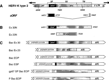
to 9381 [25]). These cDNAs are derived from an expressed HERV-K type 2 provirus as they contain the 292-bp (“plus”) segment (25) (Fig. 1A) and show an ORF from nt 6451 to 8548. The corresponding ENV protein has a theoretical molecular mass of 80 kDa. The amino acid sequence comprises an un-usual long (95-amino-acid [aa]) signal peptide (SP) that is closely related in hydrophobicity profiles and length and struc-tures of SP to mouse mammary tumor virus ENV (9, 25).
Genomic clone pgP23R1.9 harbors part of the HERV-K pol
andenvgenes (nt 6079 to 7991), showing 98.8% homology to
HERV-K type 2 sequences (pcE12envand pcK30env) and
ex-hibiting an ORF from nt 6451 to 7991.
For expression in Escherichia coli, the pgP23R1.9-derived
SU-env1,024-bp fragment Ex23N (nt 6805 to 7828 [Fig. 1B])
and the pcE12env-derived full-lengthenv2,115-bp ORF
des-ignated Ex30N (nt 6451 to 8565 [Fig. 1B]) were cloned in frame into pET15b. Protein expression was induced with 1 mM
isopropyl-b-D-thiogalactopyranoside (IPTG) at 378C for 3 h.
Cytosolic fractions and membrane proteins of lysed bacteria were analyzed separately by SDS-PAGE. A prominent 40-kDa SU-ENV protein was found exclusively in the membrane frac-tion of bacteria transformed with Ex23N but could not be observed in the cytosolic fraction of induced bacteria (data not shown). Expression of the entire HERV-K ENV protein with a predicted molecular mass of 80 kDa was not detectable in the
cytosolic or membrane fraction of Ex30N-induced BL21(DE3) (data not shown).
Purified 40-kDa HERV-K SU-ENV protein served as the antigen for the immunization of rabbits. The production of specific antibodies was confirmed by immunoblotting of SU-ENV protein from induced bacteria versus controls (data not
shown). For further investigations, thea-ENV antiserum was
used at 1:500 to 1:1,000 dilutions. Epitope mapping with syn-thetic decapeptides of HERV-K ENV protein (7) revealed three major epitopes (aa 161 to 180, nt 6931 to 6990; aa 281 to 310, nt 7291 to 7380; and aa 421 to 450, nt 7711 to 7800)
recognized by thea-ENV antiserum (data not shown).
Expression of HERV-K ENV proteins in insect cells.The BVES enables expression and processing of foreign proteins in insect cells (34). We have generated six recombinant baculo-viruses to study eukaryotic expression of the envelope protein of HERV-K. Viral vectors BacEx30 and BacEx23 bear the
entire env gene (AUG at nt 6451) or the SU-env segment
(AUG at nt 6805) of HERV-K type 2 (Fig. 1C). BacEOP harbors the SU/TM segment (AUG at nt 6805), and BacESP displays an N-terminal extension thereof containing 10 aa of the minimum putative signal peptide (AUG at nt 6706) and the first 22 aa of the SU sequence, which are not present in EOP (25, 33) (Fig. 1C). Viral vector gp67SPBacEOP bears the SU/TM sequence downstream of the heterologous signal pep-tide of acidic baculovirus glycoprotein gp67. Similarly, the re-combinant virus PBacEOP carries the SU/TM segment down-stream of the signal peptide of human alkaline phosphatase but is missing 23 aa at the carboxy terminus of TM-ENV due to the scheme of construction (Fig. 1C).
Recombinant HERV-K env baculoviruses and wild-type
[image:3.612.129.480.70.328.2]baculovirus were used for infections of High Five insect cells.
FIG. 1. HERV-Kenvgene sequences used for construction of prokaryotic expression vectors and recombinant baculoviruses. (A) Section of HERV-K type 2 provirus (upper graph) (25); cORF is encoded by two exons (lower graph) (25); the AUG codon (nt 6451) and stop codon (nt 8548) are indicated as vertical lines. (B) Ex30N (full-lengthenvderived from pcE12env) and Ex23N (SU-envderived from pgP23R1.9) were cloned into the prokaryotic expression vector pET15b. (C) Recombinant HERV-Kenvbaculoviruses: BacEx30, full-length HERV-Kenv; BacEx23, SU-env; BacEOP, SU and TM portion ofenv; BacESP, putative signal peptide (SP) and SU/TM portion ofenv; gp67SPBacEOP, SP from baculovirus gp67 protein and SU/TM portion ofenv; PBacEOP, SP from human alkaline phosphatase (alk.Ph.) gene and SU/TM portion ofenv. Promoter and poly(A) addition sequences are indicated.
on November 9, 2019 by guest
http://jvi.asm.org/
The time-dependent maximum of ENV protein expression af-ter infection was deaf-termined for each recombinant virus by
Western blot analyses with a-ENV antiserum. All viruses
evoked the expression of HERV-K ENV proteins with pre-dicted molecular masses as revealed by immunoblotting. BacEx23 elicited the expression of a 40-kDa protein which appeared to be the unglycosylated form of SU-ENV (Fig. 2A, lane 1). Recombinant virus BacEx30 produced an 80- to 90-kDa immunoreactive protein, and BacEOP (SU/TM-ENV) and BacESP (SP/SU/TM-ENV) elicited proteins of approxi-mately 66 and 70 kDa, respectively (lanes 2 to 4). A 66- to 70-kDa ENV protein was obtained after infection with gp67SPBacEOP, and PBacEOP produced a 60- to 80-kDa protein (lanes 5 and 6). None of the recombinant proteins showed nonspecific reactions with preimmune serum (data not shown). However, the highly expressed polyhedrin protein of approximately 32 kDa encoded by wild-type baculovirus dem-onstrated cross-reactivity (data not shown). In
PBacEOP-in-fected cells, a band of;120 kDa (lane 6) was also revealed by
the preimmune serum, indicating its nonspecificity. Lysates of
uninfected High Five cells showed no immunoreactivity (lane 7).
The data show that recombinant ENV proteins were not cleaved to SU and TM polypeptides irrespective of whether the cORF leader sequence (BacEx30), the minimum putative ho-mologous SP (BacESP), a heterologous SP (gp67SPBacEOP, PBacEOP), or no SP (BacEOP) was present. Cleavage of the cORF leader sequence (95 aa) from the full-length 80- to 90-kDa HERV-K ENV protein which would yield an immu-noreactive product of approximately 66 kDa did not occur. Also, the minimum putative SP of the BacESP product did not appear to be cleaved. On the other hand, cORF protein itself is expressed as a 14-kDa polypeptide in BacEx30-infected
in-sect cells (Fig. 2B) due to splicing of the HERV-KenvmRNA
(25). As the uncleaved full-length ENV comprises the cORF leader sequence including the “plus” region (25) (Fig. 1A) the
80- to 90-kDa protein does indeed react with a-cORF
anti-serum (Fig. 2B).
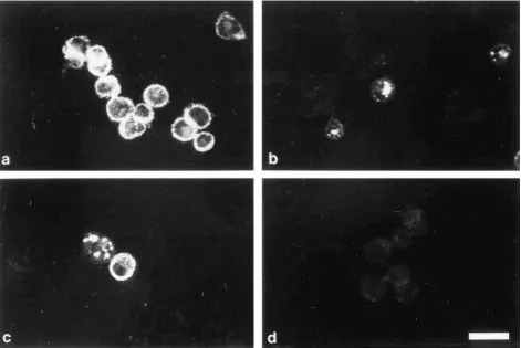
In the supernatants of insect cells infected with recombinant baculoviruses, no ENV proteins could be detected irrespective of the SP sequence used in the viral construct (data not shown). Indirect immunofluorescence analysis of insect cells
infected with BacEx30 by usinga-ENV antiserum after fixation
revealed a diffuse cytoplasmic staining pattern (Fig. 3a). Sim-ilar results were obtained with the other recombinant
HERV-K env baculoviruses (data not shown). The cellular
localization of cORF in BacEx30-infected High Five cells ap-peared to be more restricted (Fig. 3b) but also coincided with ENV (Fig. 3c), corroborating the results of immunoblotting (Fig. 2B). Indirect immunofluorescence analysis demonstrated a pattern reminiscent of teratocarcinoma cells, where cORF accumulates in nucleoli (25). When insect cells infected with BacEx30 or PBacEOP were incubated under native conditions
witha-ENV antiserum or preimmune serum, no specific
reac-tivity was observed, indicating that ENV protein does not ap-pear on the cell surface (data not shown).
Glycosylation of HERV-K ENV in insect cells.Glycosylation of expressed ENV proteins was investigated either by treat-ment of infected insect cells with tunicamycin or by digestion of
cell lysates withN-glycosidase F. As a control, samples were
compared with untreated lysates of infected cells.
The three ENV proteins elicited by BacEx23, BacEOP, and BacESP are not glycosylated (data not shown). Furthermore, the product of gp67SPBacEOP appears not to be glycosylated (data not shown). By contrast, full-length HERV-K ENV ex-hibits a size reduction from 80-90 kDa to 80 kDa and shows a second band of approximately 66 kDa after treatment with tunicamycin (Fig. 2C, lanes 1 and 2). This 66-kDa protein migrates at the level of EOP (data not shown) and does not
react with a-cORF antiserum, whereas the uncleaved 80- to
90-kDa ENV protein does (Fig. 2D, lanes 1 and 2). In addition, glycosylation of the ENV protein generated by PBacEOP could clearly be demonstrated (Fig. 2C, lane 3). The size of the glycosylated protein (60 to 80 kDa) was reduced to 60 kDa after treatment with tunicamycin (Fig. 2C, lane 4). Digestion
withN-glycosidase F confirmed these results for both
recom-binant viruses (data not shown). The data demonstrate that the entire HERV-K ENV protein is glycosylated in insect cells but is exclusively cleaved into SP, i.e., cORF leader and SU/TM, when being unglycosylated.
Expression of HERV-K ENV proteins in mammalian cells.
Heterologous expression of the full-length HERV-Kenvgene
under CMV promoter control was examined by using tran-siently transfected COS7 cells with different constructs (Fig. 4).
Initially, construct CEx30 was used to express the env gene.
[image:4.612.60.297.70.289.2]This plasmid vector evoked the expression solely of cORF
FIG. 2. Expression of HERV-K ENV and cORF proteins in insect cells (A and B) and analysis of glycosylation (C and D). (A) Immunoblots of insect cells infected with recombinant HERV-Kenvbaculoviruses. High Five cells were infected with BacEx23 (6 days p.i.) (lane 1), BacEx30 (2 days p.i.) (lane 2), BacEOP (3 days p.i.) (lane 3), BacESP (4 days p.i.) (lane 4), gp67SPBacEOP (3 days p.i.) (lane 5), PBacEOP (3 days p.i.) (lane 6), and uninfected cells (lane 7). Lane M, prestained molecular weight marker. Lysates of High Five cells were separated by SDS-PAGE (10% polyacrylamide). The Western blot was incu-bated witha-HERV-K SU-ENV antiserum. (B) Immunoblot of insect cells infected with recombinant HERV-Kenvbaculovirus BacEx30. Lanes: 1, unin-fected cells; 2, cells 2 days p.i. Cell lysates were separated by SDS-PAGE (15% polyacrylamide). The Western blot was incubated witha-cORF antiserum. (C) Immunoblots of tunicamycin-treated insect cells infected with recombinant bacu-loviruses BacEx30 (lanes 1 and 2) and PBacEOP (lanes 3 and 4). Lysates of High Five cells 48 h p.i. were separated by SDS-PAGE (10% polyacrylamide) (lanes 1 and 2) or SDS-PAGE (7% polyacrylamide) (lanes 3 and 4). Lanes: 1 and 3, infected cells untreated; 2 and 4, infected cells treated with tunicamycin. The Western blot was incubated witha-ENV antiserum. (D) Immunoblot of insect cells infected with recombinant HERV-Kenvbaculovirus BacEx30. Lysates of High Five cells 48 h p.i. were separated by SDS-PAGE (7.5% polyacrylamide). Lanes: 1, infected cells untreated; 2, infected cells treated with tunicamycin. The Western blot was incubated with a-cORF antiserum. Solid triangles denote glycosylated 80- to 90-kDa HERV-K ENV protein; open triangles indicate un-glycosylated ENV proteins (lanes 2 [C and D], lane 4 [C]); the arrow indicates the 14-kDa cORF protein (B).
2750 TO¨ NJES ET AL. J. VIROL.
on November 9, 2019 by guest
http://jvi.asm.org/
protein (Fig. 5a) located in nucleoli of COS7 cells analogous to the situation in teratocarcinoma cells (25). Neither indirect immunofluorescence nor Western blot analyses could reveal the ENV protein. To suppress the splicing of transcribed
full-length env mRNA which produces cORF-encoding mRNA
[image:5.612.73.544.70.385.2](25), both splice donor sequences (SD I, SD II) and the splice acceptor (SA) sequence were mutated, yielding constructs CEx30-Mut I and CEx30-Mut II (Fig. 4). COS7 cells trans-fected with these plasmids expressed neither cORF nor ENV protein as analyzed by indirect immunofluorescence (data not
FIG. 3. Immunofluorescence analysis of insect cells infected with recombinant HERV-Kenvbaculovirus BacEx30. High Five cells were fixed 72 h p.i. (a to c) Cells incubated witha-ENV (a) anda-cORF (b and c) antisera; (d) cells incubated with preimmune serum. Bar, 25mm.
FIG. 4. Mammalian HERV-Kenvexpression vectors. CEx30, full-length HERV-Kenvdownstream of CMV promoter; CEx-Mut I,envincluding mutated splice donor I and splice acceptor; CEx-Mut II,envwith mutated splice donor II and splice acceptor; JW-Ex30, full-length HERV-Kenvdownstream of the CMV immediate-early promoter/intron A element; JW–t-PA–SP/EOP, SU/TM portion ofenvin frame with the t-PA leader sequence. A section of HERV-K type 2 (25) is given for comparison.
on November 9, 2019 by guest
http://jvi.asm.org/
[image:5.612.145.464.502.692.2]shown). Reverse transcriptase PCR analysis of mRNA isolated from CEx30-Mut I- and CEx30-Mut II-transfected cells re-vealed alternatively spliced products relative to CEx30 expres-sion (data not shown). However, these experiments showed
minor levels ofenvmRNA expressed from all CEx constructs,
indicating that the level of expression was not sufficient to produce ENV protein.
For this reason, a CMV intron A-containing vector was chosen, as it has been shown that secretion of several glyco-proteins was significantly higher in the presence of this
se-quence (5) and that intron sese-quences located 59to the cDNA
insert prevent the utilization of possible cryptic 59donor splice
sites within the cDNA sequence (11, 12). Constructs JW-Ex30
bearing full-length HERV-K env downstream of the CMV
immediate-early promoter/intron A element and JW–t-PA–
SP/EOP containing the SU/TM portion ofenvplaced in frame
with the t-PA leader in pJW4303 (Fig. 4) were used for ex-pression experiments. Transient transfections of COS7 cells with JW-Ex30 revealed expression of ENV in the cytoplasm and of cORF in the nucleoli in 2 to 3% of the cells (Fig. 5b and c). However, the ENV protein could not be detected on the cell surface (data not shown). By contrast, ENV protein ex-pressed from construct JW–t-PA–SP/EOP was efficiently
trans-ported to the outer cell membrane in 2 to 3% of the cells (Fig. 5d). As this plasmid harbors the t-PA leader in place of the “plus” sequence of HERV-K (Fig. 4), no cORF expression was found (data not shown). In addition, in some JW-Ex30-trans-fected cells, a cytoplasmic fluorescence was revealed by
a-cORF serum, which might be due to the existence of cleaved
signal peptide corresponding to the cORF sequence and/or a perturbation due to overexpression (Fig. 5c; see below).
Molecular masses of expressed proteins derived from JW-Ex30 were determined by radioimmunoassays. Most of the ENV protein appeared to be present as a 75- to 80-kDa band and was detected as soon as 30 min after labelling with a maximum level 2.5 h after labelling (Fig. 6A, lanes 2, 4, and 6), suggesting that proteolytic cleavage of ENV at the SU/TM boundary was inefficient in COS7 cells. No specific band in the expected molecular mass range of 40 kDa (unglycosylated SU-ENV) to approximately 50 kDa (glycosylated SU-SU-ENV) could be observed. Negative controls merely showed nonspecific pre-cipitation products (lanes 7 and 8). Similar results were ob-tained with construct JW–t-PA–SP/EOP (data not shown). The
expressed ENV protein did not precipitate witha-cORF
anti-body (Fig. 6B, lanes 2, 4, and 6), indicating that the signal peptide encoded by the “plus” region had been cleaved from
FIG. 5. Immunofluorescence analysis of HERV-K ENV and cORF expression in transfected COS7 cells. Cells were fixed and stained 48 h posttransfection. (a) Cells were transfected with CEx30 and incubated witha-cORF antisera; (b and c) cells were transfected with JW-Ex30 and incubated witha-ENV (b) ora-cORF (c) antiserum; (d) cells were transfected with JW–t-PA–SP/EOP and incubated witha-ENV on ice prior to fixation. Arrows denote cORF located in nucleoli. Bar, 25mm.
2752 TO¨ NJES ET AL. J. VIROL.
on November 9, 2019 by guest
http://jvi.asm.org/
the precursor. Bands of 10 to 12 kDa possibly representing this leader sequence were detected in the same experiment (lanes 2, 4, and 6). In addition, the cognate 14-kDa cORF protein was expressed (lanes 2, 4, and 6). A similar situation was found in GH teratocarcinoma cells, where a 75- to 80-kDa ENV protein was precipitated after transfection of JW-Ex30 (Fig. 6C, lane 4). In untransfected GH cells, a low level of production of ENV was hardly detectable after 3 days of labelling (lane 2) and could not be revealed by indirect immunofluorescence (data not shown). However, due to the complex pattern of precipitated products, it cannot be excluded that some of the smaller bands represent degraded ENV protein.
Glycosylation of HERV-K ENV in COS7 cells.The glycosyl-ation status of expressed HERV-K ENV protein was investi-gated by treatment of JW-Ex30 transfected COS7 cells with tunicamycin. It could be shown in radioimmunoprecipitation assays that after 5 or 20 h of treatment, the 75- to 80-kDa ENV band almost completely disappeared (Fig. 7). Instead, an ap-proximately 66-kDa band exists which was already present in the absence of tunicamycin (Fig. 7, lanes 2, 4, and 6) and corresponds to the SU/TM portion (EOP) of ENV. As tuni-camycin was toxic to GH cells even at lower doses, the glyco-sylation status of the hardly detectable ENV protein could not be studied.
DISCUSSION
We have used eukaryotic systems to successfully express full-length segments and subsegments of HERV-K envelope protein, whose existence has not been demonstrated up to now.
Expression of glycosylated and uncleaved ENV protein in insect cells.As efficient expression of processed and
[image:7.612.71.544.69.331.2]glycosy-FIG. 6. Radioimmunoprecipitation analysis of COS7 cells and GH teratocarcinoma cells transfected with HERV-Kenvexpression vector JW-Ex30. (A and B) Transfected COS7 cells examined witha-ENV (A) anda-cORF (B) (lanes 2, 4, 6, and 8) antisera or corresponding preimmune sera (lanes 1, 3, 5, and 7). Labelling times were 30 min for lanes 1 and 2, 2.5 h for lanes 3 and 4, 4.5 h for lanes 5 and 6, and 4.5 h for COS7 cells transfected with vector control (lanes 7 and 8). (C) GH cells untransfected (lanes 1 and 2; labelling time, 3 days) and transfected (lanes 3 and 4; labelling time, 1 day);a-ENV antibody. Arrows denote HERV-K ENV protein (A and C); the solid triangle indicates the cORF protein, and the open triangle denotes putative signal peptide bands (B).
FIG. 7. Glycosylation of HERV-K ENV proteins expressed in COS7 cells. Cells were transfected with JW-Ex30 and incubated in the absence (lanes 1 and 2) or presence (lanes 3 and 4, 20 h; lanes 5 and 6, 5 h) of tunicamycin prior to radioimmunoprecipitation analysis. Lanes: 1, 3, and 5, preimmune serum; 2, 4, and 6,a-ENV. The solid arrow denotes glycosylated ENV, and the open arrow denotes putative unglycosylated ENV.
on November 9, 2019 by guest
http://jvi.asm.org/
[image:7.612.355.517.416.673.2]lated viral proteins of different origin in insect cells has been repeatedly described (1, 32, 37, 41), we have adopted this versatile tool for HERV-K ENV protein expression. Our data demonstrate that the cORF leader sequence is essential for expression of the entire HERV-K ENV glycoprotein in insect cells. This protein of 80 to 90 kDa is slightly larger than its unglycosylated form (80 kDa). The predicted amino acid se-quence of HERV-K ENV shows 11 potential sites for glyco-sylation of the “N-x-S/T” type, 7 in the SU-ENV and 4 sites in the TM-ENV portion. Theoretically, glycosylation of all sites could cause an increase of 22 to 25 kDa in the molecular mass. On average, one-third of all possible N-glycosylation sites are effectively modified (14). Hence, the observed size of glycosy-lated HERV-K ENV in insect cells is in the expected range. Glycosylation was also mediated by the heterologous SP of human alkaline phosphatase with high efficiency, leading to an ENV glycoprotein of 60 to 80 kDa.
We conclude that the 292-bp (“plus”) region and 50 bp of upstream sequence which encode the major part of cORF (25) (Fig. 1A and Fig. 2) encompass the SP sequence required for functional expression of HERV-K ENV. Indeed, the ENV reading frame and cORF are colinear and possibly start with an identical codon (AUG at nt 6451 [Fig. 1A]). The cORF coding region consists of two exons comprising the N-terminal ENV sequence (aa 1 to 87) and 18 aa in a different reading
frame of the 39 portion of the env gene (25) (Fig. 1A). In
BacEx30 (full-length env)-infected insect cells, the 14-kDa
cORF protein was also expressed due to splicing of the env
mRNA.
The size of the unglycosylated 80-kDa HERV-K ENV pro-tein may suggest that translation starts at nt 6451. Alterna-tively, a possible initiation site is at nt 6538 according to the consensus sequence (A/C)-x-x-A-U-G-G established by Kozak (15, 16). The first signal peptidase cleavage site C-terminal to the hydrophobic core of HERV-K ENV SP that follows the
“23,21” rule (47–49) is at position 96 (25). Thus, cleavage of
the ENV precursor would yield an SP of approximately 11 kDa and a SU/TM product of 66 to 69 kDa. In fact, the latter
protein shows no immunoreactivity witha-cORF antibody but
was revealed only after inhibition of glycosylation with tunica-mycin, which blocks the first step in the biosynthesis of aspar-agine-linked glycoproteins (8, 20). Obviously, the SP was not cleaved from the glycosylated 80- to 90-kDa ENV precursor encoded by BacEx30. It has been reported that in some cases the nonglycosylated proteins are more susceptible to proteases than are their normal counterparts (29, 40), possibly explaining our observation of SP cleavage under those conditions. Fur-thermore, the carbohydrate moiety may be important in pro-tein folding and conformation. This result suggests that the endoplasmic reticulum (ER)-associated peptidase responsible for SP cleavage (35) was activated in this deglycosylation assay. Further processing of ENV precursors during transport to the plasma membrane would involve di- or multimerization of ENV prior to transition into the Golgi apparatus, where ENV is usually cleaved to SU and TM glycoproteins with further modifications before the protein is eventually translocated to the cell membrane (27). Our data show that the glycosylated HERV-K ENV precursor is not cleaved to SU and TM
pro-teins in insect cells, although theenvsequence used displays an
amino acid stretch of R-S-K-R at the junction of SU and TM. This motif is closely related to the conserved basic cleavage site R-x-(K/R)-R of envelope proteins of many other retroviruses, all of which undergo identical types and sequences of process-ing (38). However, the same effect of nonprocessprocess-ing was ob-served when the human immunodeficiency virus envelope gene was expressed in insect cells where the 160-kDa precursor was
not cleaved to glycoproteins gp120 and gp41 (32, 39). Further-more, for the ENV protein of mouse mammary tumor virus it was shown that a single glycine-to-glutamic acid mutation at position 54 was responsible for nonmaturation of the precursor Pr74 to glycoproteins gp52 and gp33 (2).
Expression of glycosylated and uncleaved ENV protein in COS cells. The advantage of expression of glycoproteins in mammalian cells is the complex processing of glycosylated polypeptides, which does not occur in insect cells (13, 14, 32). Basically, the data that we obtained for expression of HERV-K ENV protein in COS7 cells are almost identical to the results which were generated with the BVES. Efficient transcription of
envcontrolled by the CMV promoter/intron A element caused
significant expression of ENV protein with an apparent mo-lecular mass of 75 to 80 kDa. This protein is slightly smaller than its unglycosylated form (80 kDa) and does not contain the N-terminal cORF sequence, suggesting that the corresponding SP has been cleaved. Furthermore, the existence of a 66-kDa ENV protein representing the intact SU/TM portion before and after treatment with tunicamycin suggests cleavage of the SP. In fact, bands of 10 to 12 kDa putatively relating to the leader sequence have been found. However, the homologous SP does not appear to be as efficient as the heterologous t-PA leader sequence which enabled transport of HERV-K ENV to the cell surface. These results suggest that at least the first step of cotranslational translocation of HERV-K ENV SP-medi-ated targeting catalyzed by the signal recognition particle and its ER membrane-localized receptor (reviewed in references 36 and 50) occurred in ENV-expressing COS7 cells.
As in insect cells and in GH teratocarcinoma cells (25), the cORF protein is expressed in transfected COS7 cells and is located in nucleoli. The major feature, as in the BVES, is the missing proteolytic cleavage of the SU/TM portion. The same phenomenon has been described recently for vaccine DNAs based on vector pJW4303 expressing the gp130 form of ENV
of simian immunodeficiency virus from macaques (SIVmac) in
COS cells (26). In this respect, it is conceivable that the ex-pression system and/or intrinsic sequence properties are re-sponsible for the unprocessed phenotype of HERV-K ENV observed in our experiments. Moreover, it cannot be excluded
that an HERV-K type 2 mRNA different from pcE12env or
pcK30env sequences has the capacity to code for fully
func-tional ENV protein. However, for GH teratocarcinoma cells
where the subgenomic HERV-KenvmRNA is expressed (21),
radioimmunoprecipitation assays revealed ENV protein ex-pression at barely detectable levels which could be increased by transfection of JW-Ex30 (Fig. 6C). Thus, the failure to transmit HERV-K/HTDV in cocultivation studies (22) could be due to at least three reasons regarding HERV-K ENV production in teratocarcinoma cells: (i) the possible inefficiency of the cORF-related signal peptide; (ii) the quantitative splicing of
the env subgenomic into cORF mRNA, leading to efficient
cORF protein expression with related extremely low levels of ENV protein expression; and (iii) the probable nonprocessing of the precursor into SU and TM portions.
The defective human endogenous retrovirus ERV-3 (HERV-R) (reviewed in reference 51) represents the only other example for an ENV-encoding HERV which is selec-tively expressed during differentiation of placental syncytiotro-phoblasts (4) and which elicits mass production of a nonglyco-sylated and unprocessed 65-kDa ENV protein in this tissue (46). While HERV-R does not encode a GAG protein, only HERV-K has the potential to code for virus-like particles as observed in teratocarcinoma cells (3, 21). These particles share morphological features with retrovirus-like particles observed in the placenta (51). In contrast to the complex HERV-K
2754 TO¨ NJES ET AL. J. VIROL.
on November 9, 2019 by guest
http://jvi.asm.org/
mRNA expression pattern in teratocarcinoma cells, other hu-man tissues including the placenta seem to express only HERV-K full-length mRNA (unpublished data), a conditio sine qua non and sufficient for particle formation if there is a
gag ORF, as in the situation of the GH teratocarcinoma cell
line (21). However, direct proof that placenta particles are indeed encoded by HERV-K proviruses has not been pro-vided. Experiments involving the use of full-length HERV-K proviral sequences for expression in heterologous systems to study the formation of recombinant particles are under way in our laboratory.
ACKNOWLEDGMENTS
This study was supported by a grant from the European Union (GENE-CT 93-0019) to R.K. and R.R.T. and by a donation from the Heinz Kuthe de Mouson Foundation to R.K.
The expert technical assistance of G. Braun, Y. Buckendahl, and Heike Hasche is gratefully acknowledged. We are indebted to K. Cichutek, J. Denner, J. Lo¨wer, and S. Norley for many stimulating discussions. We greatly appreciate the help of H. Ko¨nig during prep-aration of SU-ENV antigen, and we thank K. Boller for help with laser scan analyses. Plasmid pJW4303 was kindly provided by H. L. Robin-son, Worcester, Mass.
REFERENCES
1.Arp, J., C. M. Ford, T. J. Palker, E. E. King, and G. A. Dekaban.1993. Expression and immunogenicity of the entire human T cell leukaemia virus type I envelope protein produced in a baculovirus system. J. Gen. Virol.
74:211–222.
2.Bedgood, R. M., L. D. Snider, and M. R. Stallcup.1992. Maturation of mouse mammary tumor virus envelope protein is blocked by a specific point mutation. Virology189:393–396.
3.Boller, K., H. Ko¨nig, M. Sauter, N. Mueller-Lantzsch, R. Lo¨wer, J. Lo¨wer, and R. Kurth.1993. Evidence that HERV-K is the endogenous retrovirus sequence that codes for the human teratocarcinoma-derived retrovirus HTDV. Virology196:349–353.
4.Boyd, M. T., C. M. R. Bax, B. E. Bax, D. L. Bloxam, and R. A. Weiss.1993. The human endogenous retrovirus ERV-3 is upregulated in differentiating placental trophoblast cells. Virology196:905–909.
5.Chapman, B. S., R. M. Thayer, K. A. Vincent, and N. L. Haigwood.1991. Effect of intron A from human cytomegalovirus (Towne) immediate-early gene on heterologous expression in mammalian cells. Nucleic Acids Res.
19:3979–3986.
6.Coffin, J.1982. Endogenous retroviruses, p. 1109–1203.InR. Weiss, N. Teich, H. Varmus, and J. Coffin (ed.), RNA tumor viruses. Cold Spring Harbor Laboratory, Cold Spring Harbor, N.Y.
7.Denner, J., R. C. Phelps, J. Lo¨wer, R. Lo¨wer, and R. Kurth.1995. Expression of the human endogenous retrovirus HERV-K in tumor and normal tissues and antibody response of pregnant women, tumor and AIDS patients against HERV-K Gag and Env peptides. AIDS Res. Hum. Retroviruses11(Suppl. 1):103.
8.Elbein, A. D.1981. The tunicamycins—useful tools for studies on glycopro-teins. Trends Biochem. Sci.8:219–221.
9.Henderson, L. E., R. Sowder, G. Smythers, and S. Oroszlan.1983. Terminal amino acid sequences and proteolytic cleavage sites of mouse mammary tumor virusenvgene products. J. Virol.48:314–319.
10. Horton, R., H. Hunt, S. Ho, J. Pullen, and L. Pease.1989. Engineering hybrid genes without the use of restriction enzymes: gene splicing by overlap ex-tension. Gene77:61–68.
11. Huang, M. T., and C. M. Gorman.1990. The simian virus 40 small-t intron, present in many common expression vectors, leads to aberrant splicing. Mol. Cell. Biol.10:1805–1810.
12. Huang, M. T., and C. M. Gorman.1990. Intervening sequences increase efficiency of RNA 39 processing and accumulation of cytoplasmic RNA. Nucleic Acids Res.18:937–947.
13. Jarvis, D., and M. Summers.1989. Glycosylation and secretion of human tissue plasminogen activator in recombinant baculovirus-infected insect cells. Mol. Cell. Biol.9:214–223.
14. Kornfeld, R., and S. Kornfeld.1985. Assembly of asparagine-linked oligo-saccharides. Annu. Rev. Biochem.54:631–664.
15. Kozak, M.1981. Possible role of flanking nucleotides in recognition of the AUG initiator codon by eukaryotic ribosomes. Nucleic Acids Res.9:5233– 5252.
16. Kozak, M.1991. An analysis of vertebrate mRNA sequences: intimations of translational control. J. Cell Biol.115:887–903.
17. Kurth, R., R. Lo¨wer, J. Lo¨wer, R. Harzmann, R. Pfeiffer, C. G. Schmidt, J. Fogh, and H. Frank.1980. Oncovirus synthesis in human teratocarcinoma
cultures and an increased anti-viral immune reactivity in corresponding pa-tients, p. 835–846.InM. Essex, G. J. Todaro, and H. zur Hausen (ed.), Viruses in naturally occurring cancers. Cold Spring Harbor Laboratory, Cold Spring Harbor, N.Y.
18. Laemmli, U. K.1970. Cleavage of structural proteins during the assembly of the head of bacteriophage T4. Nature (London)227:680–685.
19. Larsson, E., N. Kato, and M. Cohen.1989. Human endogenous proviruses. Curr. Top. Microbiol. Immunol.148:115–132.
20. Leavitt, R., S. Schlesinger, and S. Kornfeld.1977. Tunicamycin inhibits glycosylation and multiplication of Sindbis and vesicular stomatitis viruses. J. Virol.21:375–385.
20a.Lo¨wer, R.Unpublished data.
21. Lo¨wer, R., K. Boller, B. Hasenmaier, C. Korbmacher, N. Mueller-Lantzsch, J. Lo¨wer, and R. Kurth.1993. Identification of human endogenous retrovi-ruses with complex mRNA expression and particle formation. Proc. Natl. Acad. Sci. USA90:4480–4484.
22. Lo¨wer, R., J. Lo¨wer, H. Frank, R. Harzmann, and R. Kurth.1984. Human teratocarcinomas cultured in vitro produce unique retrovirus-like viruses. J. Gen. Virol.65:887–898.
23. Lo¨wer, R., J. Lo¨wer, and R. Kurth.1996. The viruses in all of us: character-istics and biological significance of human endogenous retrovirus sequences. Proc. Natl. Acad. Sci. USA93:5177–5184.
24. Lo¨wer, R., J. Lo¨wer, C. Tondera-Koch, and R. Kurth. 1993. A general method for the identification of transcribed retrovirus sequences (R-U5 PCR) reveals the expression of the human endogenous retrovirus loci HERV-H and HERV-K in teratocarcinoma cells. Virology192:501–511. 25. Lo¨wer, R., R. R. To¨njes, C. Korbmacher, R. Kurth, and J. Lo¨wer.1995.
Identification of a rev related protein by analysis of spliced transcripts of the human endogenous retroviruses HTDV/HERV-K. J. Virol.69:141–149. 26. Lu, S., J. Arthos, D. C. Montefiori, Y. Yasutomi, K. Manson, F. Mustafa, E.
Johnson, J. C. Santor, J. Wissink, J. I. Mullins, J. R. Haynes, N. L. Letvin, M. Wyand, and H. L. Robinson.1996. Simian Immunodeficiency virus DNA vaccine trial in macaques. J. Virol.70:3978–3991.
27. Luciw, P. A., and N. J. Leung.1992. Mechanisms of retrovirus replication, p. 159–298.InJ. A. Levy (ed.), The Retroviridae, vol. 1. Plenum Press, New York, N.Y.
28. Marrack, P., E. Kushmir, and J. Kappler.1991. A maternally inherited superantigen encoded by a mammary tumour virus. Nature (London)349:
524–526.
29. McDowell, W., and R. T. Schwarz.1988. Dissecting glycoprotein biosynthesis by the use of specific inhibitors. Biochimie70:1535–1549.
30. Mount, S.1982. A catalogue of splice junction sequences. Nucleic Acids Res.
10:459–472.
31. Mueller-Lantzsch, N., M. Sauter, A. Weiskircher, K. Kramer, B. Best, M. Buck, and F. Gra¨sser.1993. The human endogenous retroviral element K10 (HERV-K10) encodes for a full length gag homologous 73kD protein and a functional protease. AIDS Res. Hum. Retroviruses9:343–350.
32. Murphy, C. I., M. Lennick, S. Lehar, G. Beltz, and E. Young.1990. Temporal expression of HIV-1 envelope proteins in baculovirus-infected insect cells. Genet. Anal. Tech. Appl.7:160–171.
33. Ono, M., T. Yasunaga, T. Miyata, and H. Ushikubo.1986. Nucleotide se-quence of human endogenous retrovirus genome related to the mouse mam-mary tumor virus genome. J. Virol.60:589–598.
34. O’Reilly, D., L. Miller, and V. Luckow.1992. Baculovirus expression vectors: a laboratory manual. W. H. Freeman & Co., New York, N.Y.
35. Pancino, G., H. Ellerbrok, M. Sitbon, and P. Sonigo.1994. Conserved frame-work of envelope glycoproteins among lentiviruses. Curr. Top. Microbiol. Immunol.188:77–105.
36. Rapoport, T. A., B. Jungnickel, and U. Kutay.Protein transport across the eukaryotic endoplasmic reticulum and bacterial inner membranes. Annu. Rev. Biochem., in press.
37. Rasmussen, L., J. D. Greenwood, and M. A. Gonda.1992. Expression of bovine immunodeficiency-like virus envelope glycoproteins by a recombinant baculovirus in insect cells. Virology186:551–561.
38. Redmond, S. M. S., and C. Dickson.1983. Sequence and expression of the mouse mammary tumour virus env gene. EMBO J.2:125–131.
39.Rusche, J. R., D. L. Lynn, M. Robert-Guroff, A. J. Langlois, H. K. Lyerly, H. Carson, K. Krohn, A. Ranki, R. C. Gallo, D. P. Bolognesi, S. D. Putney, and T. J. Matthews. 1987. Humoral immune response to the entire human immunodeficiency virus envelope glycoprotein made in insect cells. Proc. Natl. Acad. Sci. USA84:6924–6928.
40.Schwarz, R. T., and R. Datema.1982. The lipid pathway of protein glyco-sylation and its inhibitors: the biological significance of protein-bound car-bohydrates. Adv. Carbohydr. Chem. Biochem.40:287–379.
41.Sisk, W., R. Bradley, R. Leipold, A. Stoltzfus, M. de Leon, M. Hilf, C. Peng, G. Cohen, and R. Eisenberg.1994. High-level expression and purification of secreted forms of herpes simplex virus type 1 glycoprotein gD synthesized by baculovirus-infected cells. J. Virol.68:766–775.
42.Steinhuber, S., M. Brack, G. Hunsmann, H. Schwelberger, M. P. Dierich, and W. Vogetseder. 1995. Distribution of human endogenous retrovirus HERV-K genomes in humans and different primates. Hum. Genet.96:188– 192.
on November 9, 2019 by guest
http://jvi.asm.org/
42a.To¨njes, R. R.Unpublished data.
43. To¨njes, R. R., and R. Kurth.1994. Comparison of primate cellular sequences homologous to HERV-K reverse transcriptase. J. Cell. Biochem. Suppl.
18:41.
44. To¨njes, R. R., R. Lo¨wer, K. Boller, J. Denner, B. Hasenmaier, H. Kirsch, H. Ko¨nig, C. Korbmacher, C. Limbach, R. Lugert, R. C. Phelps, J. Scherer, K. Thelen, J. Lo¨wer, and R. Kurth.1996. HERV-K: The biologically most active human endogenous retrovirus family. J. Acquired Immune Defic. Syndr. Hum. Retrovirol.13(Suppl. 1):S261–S267.
45. Towbin, H., T. Staehelin, and J. Gordon.1979. Electrophoretic transfer of proteins from polyacrylamide gels to nitrocellulose sheets: procedure and some applications. Proc. Natl. Acad. Sci. USA76:4350–4354.
46. Venables, P. J. W., S. M. Brookes, D. Griffiths, R. A. Weiss, and M. T. Boyd.
1995. Abundance of an endogenous retroviral envelope protein in placental
trophoblasts suggests a biological function. Virology211:589–592. 47. von Heijne, G.1994. How signal sequences maintain cleavage specificity. J.
Mol. Biol.173:243–252.
48. von Heijne, G.1985. Signal sequences, the limits of variation. J. Mol. Biol.
184:99–105.
49. von Heijne, G.1986. A new method for predicting signal sequence cleavage sites. Nucleic Acids Res.14:4683–4690.
50. Walter, P., and A. E. Johnson.1994. Signal sequence recognition and protein targeting to the endoplasmic reticulum membrane. Annu. Rev. Cell Biol.
10:87–119.
51. Wilkinson, D. A., D. L. Mager, and J. A. C. Leong.1994. Endogenous human retroviruses, p. 465–535.InJ. A. Levy (ed.), The Retroviridae, vol. 3. Plenum Press, New York, N.Y.
2756 TO¨ NJES ET AL. J. VIROL.