A DISSERTATION ON
A STUDY OF 100 CASES
OF
GROIN HERNIA REPAIR
M.S. Degree
(Branch I)
GENERAL SURGERY
THE TAMILNADU
DR. M.G.R. MEDICAL UNIVERSITY
CHENNAI, TAMILNADU
CERTIFICATE
This is to certify that this dissertation entitled “
A STUDY OF
100 CASES OF GROIN HERNIA REPAIR
” submitted by
DR.R. SIVA PRIYA to the faculty of General Surgery, The Tamil
Nadu Dr. M.G.R. Medical University, Chennai, in partial fulfilment of
the requirement in the award of degree of M.S.Degree, Branch – I
(GeneralSurgery), for the SEPTEMBER 2006 examination is a
bonafide research work carried out by her under our direct supervision
and guidance.
Prof. Dr. S. VIJALAKSHMI. M.S., Prof. Dr. M. KALYANA SUNDARAM. M.S.,
Prof. of Operative Surgery Prof. and Head of the Department
Department of General surgery Department of General Surgery,
Govt. Rajaji Hospital & Govt. Rajaji Hospital &
Madurai Medical College, Madurai Medical College,
DECLARATION
I, Dr.R. Siva Priya
solemnly declare that the dissertation titled
“
A STUDY OF 100 CASES OF GROIN HERNIA REPAIR
” has
been prepared by me.
This is submitted to The Tamil Nadu Dr. M.G.R. Medical
University, Chennai, in partial fulfillment of the requirement for the
award of M.S. (General Surgery), degree Examination to be held in
September 2006.
Place : Madurai
ACKNOWLEDGEMENT
I am deeply grateful to my Unit chief Prof. Dr. S. Vijayalakshmi. M.S.,
who inspired me to take this topic of “A study of 100 cases of GroinHernia
repair”, as my dissertation.
I am also very grateful to my Professor & Head of the Department of
Surgery, Prof. Dr. Kalyanasundaram. M.S., and Senior Professors for their
encouragement & teaching for the preparation of my work.
I am also thankful to our Dean for the kind permission to utilize the
Clinical materials for the study.
I express my gratitude to my Assistant Professors
Dr. Somasundaram, M.S. Dr. Sankara Mahalingam, M.S. Dr. Rajasekaran.M.S.
and Dr. Murugan. M.S., for their kind encouragement and valuable guidance to
complete this project.
Without thanking the patients who willingly gave their kind cooperation
CONTENTS
PAGE No.
1.
INTRODUCTION 1
2.
AIM OF STUDY
2
3.
HISTORY
3
4.
REVIEW OF LITERATURE
5
5.
ANATOMY OF THE INGUINAL
REGION
16
6. ETIO-PATHOGENESIS
OF
GROIN HERNIAS
22
7.
DIAGNOSIS & CLASSIFICATION OF INGUINAL HERNIA 25
8.
FEMORAL
HERNIA
30
9.
TREATMENT FOR INGUINAL HERNIA
35
10.
COMPLICATION OF GROIN HERNIA REPAIR
49
11.
MATERIALS
AND
METHODS
74
12.
PROFORMA
13.
OBSERVATIONS
&
ANALYSIS
75
14.
CONCLUSION
78
INTRODUCTION
“No disease of the human body, belonging to the province of the surgeon
requires in its treatment a better combination of an anatomical knowledge with
surgical skill than Hernia in all its varieties.”
- Sir Astley Paston Cooper, 1804.
Hernia is derived from the latin word - Rupture
A hernia is defined as a abnormal protrusion of an organ or tissue through a
defect in surrounding walls.
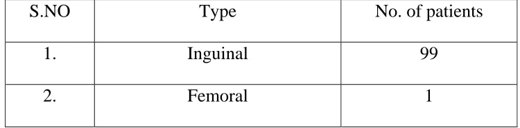
Among the abdominal wall hernias, groin hernia which includes inguinal and
femoral hernia are the commonest which occurs due to the defect in the inguinal
and femoral canal region.
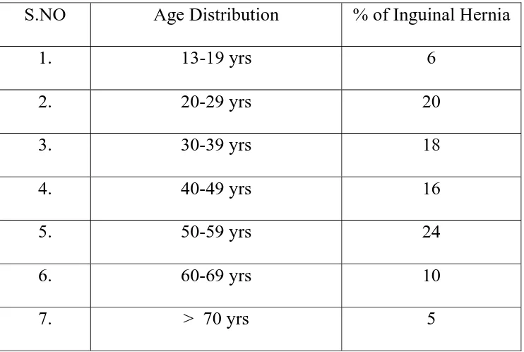
My study focuses on the Groin hernias it occurrence, age distribution, Sex
distribution, associated conditions, type of management for groin hernia, its
AIM OF THE STUDY
To study the Age distribution in Groin hernias
To study the prevalence of Direct, Indirect & Femoral hernias most
common side of occurrence.
To study the predisposing factors like benign prostatic hypertrophy,
chronic bronchitis, obesity, strenuous labourers, previous surgery
To study the different types of repair done
To study the post operative complications of groin hernia repair
To study the recurrent hernia operated
HISTORY
The earliest record of Inguinal Hernia dates back to approximately 1500 BC.
Greek word Hernia - offshoot, a budding, or bulge.
Latin word hernia - rupture or tear.
Trusses and bandages generally were used to control the herniation.
In the earlier part of the first century AD, Celsus described the operation in
vogue at that time in the Greco-Roman area.
Through an incision in the neck of the scrotum, the hernial sac was dissected
off the spermatic cord and transected at the external inguinal ring.
The testis usually was excised as well. The incision was generally left open.
Later, a mass ligature of the sac and cord at the external ring was recommended
with excision of the sac, cord, and testis distal to the ligature, as described by Paul
of Aegina in 700 AD.
Guy de chauliac, in 1363. differentiated between inguinal and femoral hernia
and described the technique of reduction for strangulation.
In 1556, Frenco illustrated the use of a grooved director to cut the
strangulating neck of the hernia while avoiding the bowel.”
In 1559, Casper Stromayr, distinguished direct from indirect hernia and
From beginning of the 18th to early 19th century the anatomy of the inguinal
region was described
The dawn of modern surgery began in 1865 when Joseph Lister introduced
his method of antisepsis by carbolic spray.
By the beginning of the 20th century, Koch had developed methods of asepsis,
REVIEW OF LITERATURE
TISSUE REPAIRS
Marcy, an American surgeon and a pupil of Lister, was the first to introduce
antiseptic techniques in the repair of hernia. He was also the first to recognize the
importance of the tranversalis fascia and of closing the internal ring.
In 1871, he published his report of two patients operated on in the previous
year in whom he used carbolized catgut to suture the ring.
A French pupil of Lister, Lucas-Championniere, brought antisepsis to France.
In 1881 he reported that the first case in which the aponeurosis of the external
oblique muscle was slit to reveal the canal, which allowed dissection and ligation of
the sac at the internal ring under direct vision. The depressing fact at this time was
that the best surgical centers in both Europe and North America were reporting
mortality rates of up to7%for hernia operations. The recurrence rate after 1 year was
30% to 40% and almost all hernias had recurred by the end of 4 years.
The greatest contribution to hernia surgery was that of the Italian surgeon
Edorado Bassini. His clear in sight into the anatomy and physiology of the inguinal
region enabled him to dissect and reconstruct the inguinal canal to preserve the
functional anatomy.
He laid the inguinal canal open widely by splitting the aponeurosis of the
external oblique. He next opened the transversalis fascia from the pubic tubercle to
the retroperitoneal space. He realized the importance of repairing the transversalis
fascia and of reinforcing the posterior wall of the canal: using interrupted sutures of
silk, he sutured the internal oblique and transversus abdominis muscles, as well as
the upper leaf of the transversalis fascia in one lower to the lower leaf of the
transversalis fascia and with inguinal ligament. The rectus sheath was incorporated
into the medial end of the repair. The aponeurosis of the external oblique muscle
was resutured in front of the spermatic cord.
Bassini first performed this operation in 1884 and reported it in 1887, 1888,
1889,1890, and finally, in 1894,
206 operations with no operative mortality. The patients varied from young
children to elderly men. Includes Bilateral repairs, strangulation, cryptorchidism, 5
years 100% followup, 11 wound infections and 8 recurrences.
These phenomenal results earned him the title of “Father of Modern
Herniorrhaphy.”
During the next 100 years, most inguinal hernias were repaired by the Bassini
method or variations of it. Some of the variations were unsuccessful Some were
improvements and reduced the incidence of recurrent hernia.
In 1953, multilayered repair described by Shouldice. The “pure tissue”
methods of study only local tissues with out addition of any prosthetic material. The
recurrence rate is <1%.
In 1898, George Lotheissen first reported the technique of suturing the
musculoaponeurotic arch (conjoint tendon) to the pectineal (Cooper’s) ligament.
Conjoint Tendon to the inguinal (Poupart’s) ligament popularized by
Bassini. The Lotheissen method had the added advantage of repairing the femoral
ring as well as the inguinal defects especially recommended for a strangulated
femoral hernia.
In 1940 U.S. McVay who showed, by anatomical dissections, that the
transverses abdominis muscle and its fascia are normally inserted onto the pectineal
ligament.
Rutledge - 1988 - suturing Fascia transversalis and Inguinal ligament
1993 - Published
Cooper ligament repair – most valuable when repairing a recurrent Inguinal
Hernia when Inguinal ligament is destroyed.
Recurrence - Due to ignorance of functional anatomy and physiology of the
abdominal wall leads to incomplete dissection &
1. to repair under tension
2. Wrong suture materials
3. Infection
Good musculo aponeurotic arch nearer the Inguinal
Weak musculoaponeurotic arch which is high and has a high gap between it
and inguinal ligament. The sutures made under tension and so ‘relaxing incision’ or
Darn Repairs
To avoid cut through of flesh muscular arch the ideal tensionless or tension
free repair used.
Marcy type repair - To repair weakened or torn posterior wall
1. Of the Inguinal canal and the transversalis fascia
2. To tighten the stretched internal Inguinal ring and the cord.
Reinforcing posterior wall by either natural tissue like
a) External oblique aponeurosis by McArthur
b) Facial grafts from thigh by kirshner
c) Fascia lata strips by Gallie and LeMesurier
d) Skin cutt off edges of the incision and denuded of dermis by mair.
living tissue - Difficult to harvest tend to absorb.
Recurrence rate high.
In search for a suitable substitute
1937, Ogilvie - Silk lattice repair
1940, Maingot - floss silk for his darn
Mcleod - Used silk for the posterior
Silk - Biologic substance (Non absorbable) lost most of its strength
Nichols, Diack & Aries - Uses Nylon
Medick - braided multifilament nylon for repair.
1945, Haxton – Monofilament nylon.
Patch Graft Repairs
Using a) Sheet of natural tissues
1) Flaps of fascia from thigh
2) Flaps of external & internal oblique muscle with anterior
rectus sheath turned down
b) Biologic
c) Metals
d) Synthetic sheets or
Silver wire filigree sheets (Witzel & Goepal in Germany
Barlett in U.S. MC Gavin in Britain.
Tantalun metal sheets - Burke 1940
Tantalum gauge - Throckmortor 1948
Sheets of woven or knitted mesh of polyamide and the newer polypropylene
monofilament used as inlay or onlay graft cheap.
Universelly available
easily cut to required shape flexible
Practically indestructible in human tissues
monofilament – smooth inert
Little tissue reaction
Not rejected even in presence of Infection
Collagen tissue can be laid down through the interstices of the weave so that
the material is incorporated into health new tissue.
To create strong & tensionless repair
To suture either superficial or deep to the Transversalis fascia
Europe – Woven or knitted synthetic (French surgeons) polyester threads
Harrison - tried polymer gives – tried in animals but disappointing
PTFE
Polytetra flouro ethylene (PTFE)
Modern Herniologists
Lichenstein & Gilbert laid a swatch of synthetic mesh without sutures.
Preperitoneal Repairs
A History of inguinal hernia repair would be incomplete without a mention of
the abdominal or preperitoneal approach.
This approach was recorded by the ancient Hindus for cases of strangulated
hernia.
It was described in Europe in the Middle Ages and in the 16th century and
was recommended toward the end of the 19th century.
All these procedures were performed transperitoneally.
Even as late as 1919, LaRoque described a gridiron transperitoneal incision
for hernia repair.
The modern era of transabdominal, but extraperitoneal, repair of hernia was
introduced by Cheatle in 1920. He first used a midline incision, but later changed to
a low transverse or Pfannenstiel incision. He peeled the peritoneum off the
abdominal wall and bladder and was able to transect the sac and repair the internal
ring from above.
In 1960 Nyhus
1968 Read used of prosthetic material,
The foremost proponent today of the perperitoneal approach is Stoppa,
Incase of repeated recurrent hernias in case where tissues become scared &
Great prosthesis for reinforcement of the visceral Sac (GPRVS) in which,
through a midline abdominal incision, a large sheet of prosthetic mesh in placed
between the peritoneum & abdominal wall to close of all the hernial openings.
The surgeon today can choose between 4 basic techniques for hernia repair as
classified by Culbert in 1987.
a) Pure tissue repair
b) Combined tissue & prosthetic repair
c) Pure prosthetic repair
d) Nylon darn
Laparoscopic Repair for Inguinal Hernia :
The tidal wave of minimal access surgery has inevitably swept hernia repair
along in its surge.
Laparoscopic transperitoneal closure of the internal orifice of groin hernias
by a series of metal clips was introduced by Ger in 1977.
Since then several methods have evolved, but routine clinical application of
the technique began only in 1990.
The most popular method today is the introduction of the laparoscope and
instruments through several ports in the abdominal wall after induction of
pneumoperitoneum under general anesthesia.
In the pediatric age group laparoscopic repair of inguinal hernia has been
reported by Easter.
The advantages of laparoscopic herniorrhaphy are that, in experienced hands,
Quick
atraumatic,
bilateral repairs can be done at the same operation,
Short stay at hospital
Clinically unsuspected contra lateral hernias can be identified and repaired,
No orchitis,
No epididymitis,
No wound infection and neuralgia, Only small openings are made in the
So postoperative recovery and return to normal activities is rapid and
practically painless.
The disadvantages include
Need for a general anesthetic
Violation of the abdominal cavity, with the future risk of adhesions
New hernias at the port sites
Complications of laparoscoic small and large bowel perforation,
bladder laceration, adhesions, bowel obstruction, mesh erosion into the
bladder, transient testicular pain, palpable mesh, mesh migration into
the scortum, scrotal hydrocele, and pelvic osteitis.
ANATOMY OF THE INGUINAL REGION
Inguinal Canal :
The inguinal canal is an oblique intermuscular slit about 4 cm (over 2 inches)
long lying above the medial half of the inguinal ligament. It commences at the deep
inguinal ring, ends at the superficial inguinal ring, and transmits the spermatic cord
and illoinguinal nerve in the male and the round ligament of the uterus and
illioinguinal nerve in the female.
Its anterior wall is formed by the external oblique aponeurosis assisted
laterally by a portion of the internal oblique muscle. Its floor is the inrolled lower
edge of the inguinal ligament, reinforced medially by the lacunar ligament and
fusing more laterally with the transversalis fascia. Its roof is formed by the lower
edges of the internal oblique and transverses muscles, which arch over from in
fronto of the cord laterally to behind the cord medially, where their conjoined
aponeuroses, constituting the conjoint tendon, are inserted into the pubic crest and
the pectineal line of the pubic bone. The posterior wall of the canal is formed by
the weak transversalis fascia laterally.
Anterior wall and superficial Inguinal Ring :
The fibres of the external oblique aponeurosis run parallel with their lower
border forms the Inguinal ligament. Above its medial end they diverge from each
other to make a V- shaped opening, the superficial inguinal ring. The lateral crus of
the symphysis. The intervening oblique aponeurosis and forms the base of the
‘ring’ which is triangular, not circular.
The anterior wall of the inguinal canal is reinforced laterally by the lowest
muscle fibres of the internal oblique. The deep inguinal ring lies 1.25 cm (1/2 inch)
above the midinguinal point ; the internal oblique fibres extend medial to this, for
they arise from the lateral two – thirds of the ligament.
Floor :
The lacunar ligament filling the angle between inguinal ligament and
pectineal line, passes upwards from the ligament to the bone. Its abdominal surface
faces forwards as well as upwards. Its femoral surface faces backwards as well as
down wards. It lies in the floor of the inguinal canal. Lateral to its attachment the
incurved edge of the inguinal ligament forms a gutter which floors in the inguinal
canal. The transversalis fascia is fused with this part of the inguinal ligament.
Roof :
This is formed by the arched lower borders of the internal oblique and
transverses abdominis muscles. Each arises from the hollow of the inrolled lower
edge of the inguinal ligament. The internal oblique muscle arises by fleshy fibres
from the lateral two-thirds of the inguinal ligament. The fibres arch medially and
down wards, merging into a flat aponeurosis. The most lateral fibres, those arising
from just below the anterior superior iliac spine arch downwards to reach the pubic
The remaining fibres arch concentrically within the former, passing in front
of rectus abdominis along the pubic crest as far as the pubic tubercle and then
extending laterally along the pectineal line as far as the crescentric edge of the
lacunar ligament. These lateral fibres, joining the underlying transverses
aponeurosis, constitute with them the conjoint tendon.
The transverses abdominis lies more laterally at its origin, coming from only
the lateral half of the internal oblique. They rapidly become tendinous and, fusing
with the aponeurosis of the internal oblique, form the conjoint tendon which is
attached along the pubic crest and extends laterally along the pectineal line. The
conjoint tendon and the lacunar and the lacunar ligament, attached in common to
the pectineal line, lie in planes at right angles to each other. The deep inguinal ring
lies in the angle between the edge of transverses and the inguinal ligament. Since
the internal oblique muscle arises a little more medially than this, it lies in front of
the deep ring. The muscular arch of the roof, starting in the anterior wall of the
canal, passes over the cord and , becoming tendinous, passes down behind the cord,
in the posterior wall of the canal, to reach the pectineal line.
The lower most fibres of internal oblique and transverses are supplied by the
iliohypogastric and illioinguinal nerves. Their contraction tightens the conjoint
tendon and lowers the roof of the canal, like pulling down a shutter. Thus division
of the ilioinguinal nerve above this level (as in a split-muscle incision for
appendicectomy) lead to a direct inguinal hernia – the conjoint tendon bulges when
inguinal canal does not paralyse these fibres; at this level the nerve is purely
sensory, having already given off it motor fibres, and injury here will only cause
some sensory loss over the anterior part of the scrotum (labium majus) and adjacent
thigh.
Inguinal Ligament :
The inguinal Ligament (of Poupart) extends from the anterior superior iliac
spine to the pubic tubercle. Its edge is rolled inwards to form a gutter; the lateral
part of this gutter gives origin to part of the internal oblique and transverses
abdominis muscles. The inguinal ligament is attached to the fascia lata of the thigh.
When the thigh is extended the fascia lata pulls the inguinal ligament downwards
into a gentle convexity.
Just above and lateral to the pubic tubercle is an oblique V shaped gap, the
superficial inguinal ring, in the aponeurosis. This gap extends down to the pubic
crest, medial to the tubercle : the aponeurosis is attached to the pubic crest only in
its medial part, along side the pubic symphysis. From the medial end of the
inguinal ligament the lacunar ligament (of Gimbernat) extends backwards to the
pectineal line. Its crescentric free edge is the medial margin of the femoral ring.
From the pubic tubercle, fibres may be traced upwards and medially behind the
spermatic cord, to interdigitate in the linea alba with those of the opposite side.
Lastly, near the apex of the superficial inguinal ring are fibres running at right
angles to those of the aponeurosis, the intercrural fibres that blend and prevent the
crura from separating.
Spermatic Cord :
The spermatic cord, components may be considered under two headings : the
three coverings of the cord, and its six (groups of ) constituents.
Of the three coverings, the internal spermatic fascia is the investment derived
from the transversalis fascia at the deep inguinal ring. As the cord passes through
the ring into the inguinal canal, it picks up a second covering, the cremsteric fascia
and cremaster muscle, from the internal oblique and transverses aponeuroses and
muscles.
The transverses muscle fibres spiral down the cord and return behind it to
become attached to the pubic tubercle. The internal oblique fibres, a larger
contribution, also spiral around the cord and some return to the pubic tubercle. The
internal oblique fibres, a larger contribution, also spiral around the cord and some
return to the pubic tubercle but most return to the internal oblique itself. The third
covering is from the crura of the superficial ring (external oblique aponeurosis), the
The constituents of the Cord :
1. The ductus deferens, which usually lies in the lower and posterior part
of the cord.
2. Arteries the largest of which is, with the testicular artery, and artery to
the ductus, the cremasteric artery (from the inferior epigastric)
3. Veins – the pampiniform plexus
4. Lymphatics, essentially those accompanying the veins from the testis
to para – aortic nodes, but including some from the covering which
drain to external iliac nodes.
5. Nerves, in particular the genital branch of the genitofemoral nerve
which runs among the coverings to supply the cremaster muscle, and is
classified as part of the spermatic cord and not as a separate structure
running through the inguinal canal. Other nerves are sympathetic
twings which accompany the arteries.
6. The processus vaginalis, the obliterated remains of the peritoneal
connection with the tunica vaginalis of the testis (and the constituent of
the cord most commonly forgotten. When patent it forms the sac of an
indirect inguinal hernia.
ETIO - PATHOGENESIS OF INGUINAL HERNIAS
Incidence
The incidence of inguinal hernias in adults varies between 15% and 20%.
The male to female ratio is 12:1. The incidence varies between 5% and 8% in
patients 25 to 40 years of age.
90% of inguinal hernia are on males. In adult males, 65% of inguinal hernias
are indirect and 55% are right sided. Bilateral hernias are four times more
commonly direct than indirect. Many hernias exist in the community undiagnosed,
undetected ad unreported.
Mechanisms which prevent hernia in inguinal region :
Though inguinal region is a weak spot in the abdominal musculature, rise in
intra abdominal pressure would have caused inguinal hernia in every individual. So
there must be some defensive mechanisms which prevent hernia to occur. These
are :
1. Obliquity of inguinal canal :
When there is rise in intra-abdominal pressure, wall is opposed to the anterior
wall and thus prevents coming out of abdominal content through inguinal
canal.
2. Shutter mechanism of the arched fibres of internal oblique and transversus
abdominis will bring down these muscles towards the floor when they are
3. Ball-valve action of the cremaster muscle which pulls up the spermatic cord
into the canal and plug it during rise of intra-abdominal pressure.
4. In front of the deep inguinal ring there are strong fibres of the internal
oblique. This prevents entry of any abdominal content through the deep
inguinal ring.
5. Strong conjoined tendon is in front of Hesselbach’s triangle to prevent direct
inguinal hernia.
Indirect Inguinal Hernia :
An indirect inguinal hernia is not simply a congenital defect represented by a
persistent patent processus vaginalis. A processus vaginalis can remain patent
throughout life without the development of a hernia. A proportion of patent
processus vaginalis apparently obliterate while others persist.
In males beyond adolescence, simple removal of an indirect hernial sac
results in an unacceptable high recurrence rate indicating that other factors are
involved in the pathogenesis. Moreover, the high frequency of indirect hernias in
middle aged and older people suggests a pathological change in the connective
tissue of the posterior inguinal wall. Thus the susceptibility to indirect inguinal
hernia is based both on the presence of a congenital sac and on failure of the
Direct Inguinal Hernia :
The cause of an inguinal hernia is undoubtedly multi factorial.
Pathogenesis of direct inguinal hernia is more complex. In the majority of
patients there is no peritoneal sac and the occurrence parallels ageing and other
factors such as smoking. Thus the incidence would appear to be more directly
related to a failure of the posterior inguinal wall or transversalis fascia. Additional
factors include anatomical abnormalities such as a deficient medial half of the
transversalis fascia and failure of the insertion of the internal oblique aponeurosis
onto the superior pubic ramus.
1. Metabolic abnormalities have been identified including a generalized
deficiency in collagen particularly in smokers and patients with
abdominal aortic aneurysms.
2. A high incidence of hernia in Eskimos in the west Arctic of Greenland,
where there is a high frequency of the HLA-B27 allele resulting in
instability of mesenchymal tissues, points to a more specific genetic
DIAGNOSIS AND CLASSIFICAITON OF INGUINAL HERNIA :
A hernia is suspected if there is an expansile impulse on coughing at the site
of the normal hernial orificies. A cough may push the abdominal contents into the
scrotum. If a swelling is visible the sign of reducibility helps to confirm the
diagnosis. If there is no obvious swelling, digital palpation of the hernial orifice
with the little finger helps to confirm the diagnosis.
A swelling in the inguinoscrotal region is likely to be an inguinal hernia.
Inguinal hernia can be incomplete when it is termed bubonocele. A bubonocele is
localized to the inguinal canal and does not pass beyond the level of the external
abdominal ring. A complete hernia descends into the scrotum.
An indirect (oblique) hernia passes through the deep inguinal ring lateral to
the inferior epigastric artery and descends through the inguinal canal. A direct
hernia passes through the Hesselbach’s triangle medial to the inferior epigastric
artery. A direct inguinal hernia can be differentiated from an indirect hernia mainly
by the finger invagination test and the deep ring occlusion test.
Indirect Inguinal Hernia :
Indirect inguinal hernia can occur at any age. There may be dragging pain in
the groin. The patient presents with a swelling which is obliquely placed and
pyriform in shape. The hernia often descends into the scrotum and reduces
obliquely upwards, outwards and backwards. With the finger invagination test, the
contents of the hernia strike the side of the tip of the finger. If a finger occludes the
direct hernia bulges at once. Indirect hernia is usually unilateral, whereas direct
hernia is often bilateral.
Direct Inguinal Hernia :
Direct Inguinal hernia usually occurs after 40 years of age. It may be
bilateral. There may be invariably a history of chronic cough or straining at
micturition or defaecation. A direct hernia is painless. The swelling is localized to
the inguinal canal. It is large, globular and disappears when patient lies down.
With the finger invagination test, the hernia strikes the pulp of the finger. After
reduction on occlusion of the internal ring the hernia appears at once. If during the
finger invagination test the finger passes directly backwards into the abdomen
instead of obliquely upwards and outwards, the diagnosis is in favour of a direct
hernia. In addition, if the edge of the external oblique is felt superiorly and pubic
bone inferiorly, the point is confirmatory. With the impingement test, if the impulse
impinges on the pulp of the finger the hernia is direct. On the other hand if the
HERNIOGRAPHY :
Herniography is a useful adjunct particularly in patients with obscure groin
pain with a normal or inconclusive physical examination (Gullmo 1989).
Complication rates are low and the false-positive rate is negligible. This approach
should be considered mandatory in the professional sportsman with chronic groin
pain before hernioplasty is considered because of the long list of differential
diagnosis.
Radio opaque non ionic contrast material is injected intra peritoneally and the
patient is maneuvered through various position in an attempt to introduce the
material into an actual or potential hernias sac, that can be demonstrated
radiographically in prone and semi prone positions.
There is no real indication for herniography in children and infants.
Hernio graphy may reveal a contralateral patent processus vaginalis in a child
Differential diagnosis of Groin Hernias
Primary testicular Femoral artery aneurysm
Varicocoele Lymphnode
Epidydymitis Sebaceous cyst
Testicular torsion Hidradenitis
Hydrocoele Cyst of canal of nuck
Ectopic testicle Psoas abcess
Undescended testicle Hematoma of abdominal wall
ascites
Saphena varix
Malignancy - Lymphoma
Retroperitoneal sarcoma
Metastasis
Nyhus Classification System
I - Indirect hernia Internal Ring Infants children
Normal small adults
II - Indirect Enlarged without impingement on the floor of
inguinal canal ; does not extend to the scrotum
III - A - Direct
B - Indirect Scrotal Hernia also includes pantaloon Hernia
C - Femoral hernias
IV Recurrent A - D
A - Indirect
B - Direct
C - Femoral
D - Mixed
Gilbert Classification :
I - Small, Indirect
II - Medium, Indirect
III - Large, Indirect
IV - Entire floor, Direct
V - Diverticular direct
VI - Combined
FEMORAL HERNIA
There are several weakness & potential canals in the area between
inguinal ligament and the superior pubic ramus through which hernias may
occur. The most common hernia in this region is the femoral hernia.
Etiology of Femoral Hernia :
Femoral hernia is rare in infancy & childhood. So the etiology is
probably not congenital. There is no evidence of preformed sac. Chapman
reported a series of 1134 cases of groin hernias in childhood of which only 6
were femoral hernias, an incidence of 0.5%. The hernia usually appears after
middle age, suggesting natural weakening of the tissues and loss elasticity is
Anatomy of Femoral Canal :
The transversalis fascia emerges above from behind the musculo aponeuotic
arch of internal oblique and the transversus abdominis muscle and passes down to
attach to the pectineal ridge. In this way it closes off the area between the Inguinal
ligament and the superior pubic ramus and separates abdomen from the thigh.
The area is mainly filled by the iliopsoas and pectineus muscles and the
femoral artery and vein & nerve passes from abdomen to the thigh.
At its most medial end, a potential canal, the femoral canal through which the
common type of femoral hernia emerges.
Boundaries :
Anteriorly - Inguinal ligament
Antero medially - Lacunar part of the Inguinal ligament (Gimbernat
Posteriorly - Pectineal ligament of the pubic ramus
Medially - Femoral vein & its sheath
Canal filled with loose areolar tissue & femoral lymph nodes with weakening and
giving way of the transversalis fascia closing the canal, the peritoneal sac of the
femoral hernia transverses the narrow rigid canal & passes into the loose
subcutaneous area of the thigh. Here it is able to expand and pass forward to bulge
It may even pass upwards to cross the Inguinal ligament. The sac is covered
with extra peritoneal fat and contains either small bowel, omentum or both. The sac
is relatively large compared to the narrow neck, which has no room to expand so
that strangulation is common.
Clinical manifestations :
1. Small reducible lump in the medial aspect of the groin
2. Lump is often permanent because of incarceration of the hernia
3. Commonly, especially in elderly females, the patient is unaware of the
swelling and the first indication of its existence appears with the
strangulation of the hernia going for Intestinal obstruction.
Chamaeus also stresses the high morbidity and mortality associated with
emergency surgery and Intestinal obstruction in femoral Hernia.
Treatment of femoral hernias :
All femoral hernias with few exceptions should be operated on &repaired as
soon as possible after diagnosis.
Even the elderly & sick can withstand a simple repair done under LA.
The frequency and high morbidity and mortality rate of strangulated femoral
hernia, especially in the old and trail is a mandatory indication to operate on
these patients electively, despite their brittle condition. An attempt should
always be made decreased strangulated hernia.
Ivfluids, ryles tube, catheterization, To correct fluid and electrolyte
Imbalance and any medical condition may be present.
Operation :
Incision : Small transverse thigh incision below the Inguinal ligament
centered over the femoral canal. In strangulated hernia, the incision is placed
over the swelling. The subcutaneous fat is split to reveal the mass of extra
peritoneal got enveloping the sac.
This mass is freed by blunt dissection with finger & is dislocated forward.
The inguinal ligament, pectineal fascia & the neck of the hernia exposed by
gentle dissection
The exposure is practically bloodless. The mass of extra peritoneal fat is split
to reveal the sac which is dissected to & beyond its narrow neck, then opened to
inspect its contents. Adhesions should be freed. The bowel & omentum are
returned to the abdominal cavity.
It may be necessary, especially with a strangulated hernia to digitally dilate
the neck from with in the sac in order to return the bowel. Rarely the lacunar
part of the Inguinal ligament or even part of the inguinal ligament itself must be
incised to free the neck of the hernia.
In these cases, care must be taken not to damage an abnormal obturator
artery.
The sac is transfixed and ligated at the neck or simply snipped off & the
The extraperitoneal fat excised, the margins of femoral canal cleared and are
exposed for closure of the defect.
The repair is done with monofilament polypropylene or polyamide 2-o.
Ideally Inguinal ligament should be sutured to the pectineal ligament (Cooper’s
ligament) with a few interrupted stitches. It is technically difficult.
The simplest alternative – pursestring repair. A thick bite taken through
inguinal ligament, then alternatively through the lacunar ligament, the pectineal
fascia, the fascia over the medial aspect of the femoral vein & finally once more
through inguinal ligament & tied.
Delvin described figure of eight closure Lichtenstein described Keeping a
plug rolled up strip of polypropylene mesh fitted snugly into the canal & few
stitches to hold it. When dealing with strangulated hernia with gangrene,
resection& anastomosis difficult so a 2nd small laparotomy incision needed for
that Lotheissen approach is through the posterior wall of Inguinal canal.
Henry preperitoneal approach through a lower midline abdominal incision in
incarcerated on strangulated femoral hernias.
Advantage for this approach is contralateral hernias detected.
Bendavid – described high recurrence rate for femoral hernia also a relaxing
TREATMENT OF INGUINAL HERNIAS
Darn Repairs
The Abrahamson Nylon Darn Repair. A good hernia repair should last the
patients for the rest of his life, no matter what his age at time of the operation. The
surgeon must bear in mind this responsibility. A 1982 report showed that almost 6%
of recurrences occurred during the first postoperative month, 39% during the first
year after the primary repair, and 24% occurred later than 10 years after the
operation. The Shouldice Hospital has reported that late recurrence is not
uncommon in cases followed for 10 to 40 years. As the causes of early recurrence
after hernia repair were eliminated (faulty technique, ignorance of the functional
anatomy and physiology of the abdominal wall, repair with tension, the use of
incorrect suture material, and infections), it became apparent that even with the
finest technique and materials and the best intentions, a percentage of hernias will
recur over the years because of factors beyond the control of the tissues and
deterioration of body fitness with time and aging, increased adiposity, raised
intra-abdominal pressure owing to chronic cough, constipation, and obstructive disease of
the urinary bladder. It was realized that some form of reinforcement was needed to
overcome the problems of aging scar tissue and muscles and tendons approximated
by sutures, especially in direct hernia repair.
A variety of natural and foreign materials were used for this reinforcement,
but with little success, until the advent of strong, smooth, resistant, and pliable
inguinal hernia is to reinforce the weakened or torn posterior wall of the inguinal
canal with a simple lattice work of monofilament nylon suture under no tension, on
which is laid a buttress of fibrous tissue, without the normal tissues being torn or
necrosed. The nylon sutures are anchored into strong, healthy tissues far from the
area of herniation. The nylon darn solves the problem of early recurrence since the
nylon lattice will hold the area intact for the first year, until the natural connective
tissue collagen scar matures to its full strength, However, the muscle and scar tissue
is not able to withstand the constant wear and tear of repeated stress over many
years. As they fail, the nylon, which is practically indestructible in human tissues,
will once more come into its own and will maintain the integrity of the repair for
many years, until the end of the patient’s life.
The technical details of the operation were described in 1987 and 1988. The
incision and meticulous dissection and preparation of the tissues are as described
previously. No special dissection of a direct sac is needed, although occasionally it
is convenient to reduce a sac prolapsing through a punched-out hole in the
transversalis fascia and to suture the opening. A large sliding hernia with much
preperitoneal fat may occasionally be conveniently reduced and the edges of the
tear in the transversalis fascia closed with a continuous suture to render the repair
more manageable. The transversalis fascia is not split open. The repair is begun by
suturing the medial edge of the rectus sheath and the musculoaponeurotic arch
(conjoined tendon) to the posterior portion of the inguinal ligament and to the
is begun at the medial end of the repair by catching fascia on the pubis, passing
through the medial end of the inguinal ligament and the remains of the fascia
transversalis and then taking a good bite through the lowest portion of the medial
edge of the rectus sheath and tendon and tied. The suture continues laterally in a
simple over-and-over fashion including, along the lower edge, some fibers of the
inguinal ligament, the iliopubic tract, and the lower part of the transversalis fascia.
Along the upper edge, the medial edge of the rectus sheath is sutured as far laterally
as possible, after which the suture takes in part of the transversalis fascia as well as
the lower edge of the aponeurosis of the transversus abdominis and also the
aponeurotic part of the internal oblique. The fleshly part of the internal oblique is
not included in the suture. Fairly large bites of tissue are taken along the upper
edge. Suture bites on the inguinal ligament are staggered, some more forward and
others further behind so that all the repair will not be secured to only a few fibers of
the inguinal ligament. The aim is to approximate the rectus sheath and conjoined
tendon to the inguinal ligament. This is easily done without tension, or under
minimal tension, in most cases. When this is not possible, we do not force the
approximation under tension but leave a gap, usually only a narrow one, between
the upper elements of the repair and the inguinal ligament. At the lateral end, the
edges of the internal ring are picked up and included in the sutures to achieve a
fairly light and snug closure of the ring around the cord. This line of sutures is
carried laterally beyond the internal ring for 1 to 2 cm, with the object of covering
reinforce the ring against an indirect recurrence. Up to this stage, the procedure
constitutes a tissue repair and its strength depends on that of the tissues used. If no
more is done, some cases will develop early recurrence and others late recurrence.
This repair has the advantage of closing the rent in the transversalis fascia and of
tightening the internal ring and providing a thick musculoaponeurotic barrier for the
posterior wall the canal. It also provides a smooth, flat bed on which to lay the darn.
The darn is done with 0 monofilament nylon thread (polyamide 6), 1.5m long
and doubled to form a loop 75 cm long, with the free ends swaged onto an
atraumatic curved 40 mm round-bodied needle. Starting at the medial end a bite is
taken if the most medial fibers of the inguinal ligament where they sweep over the
pubic tubercle. The point of the needle then is pushed under the lateral edge of the
rectus muscle and sheath and is extracted. The needle is then simply passed through
the tail end of the loop, and tightened, eliminating the inconvenience of a knot. The
suture is continued laterally, taking bites of the inguinal ligament below and deep
wide bites of the rectus muscle and its sheath to ensure a good darn in the critical
medial angle of the repair where recurrences tend to occur. When the rectus sheath
can no longer be used, the sutures pass onto the conjoined tendon. Each stitch is laid
in a vertical fashion. The stitches on the inguinal ligament are staggered to spread
the tension between the fibers. At the upper end the suture passes over the muscular
lower part of the conjoined tendon. The stitches are held slightly tight-just enough
to straighten the thread, and are not placed under tension. This vertical line of
displacing the cord laterally. The same suture changes direction and returns
medially as the second layer of the darn It passes in front of the covered internal
ring. The stitches are now laid in a sloping fashion, passing upwards and medially
from the inguinal ligament to the conjoined tendon and later the rectus sheath,
crossing the stitches of the first run at an angle. The bites on the inguinal ligament
also are staggered and a bit anterior to those of the first run, in order to spread the
tension. Large bites are taken, as before, of the aponeurotic fibers of the conjoined
tendon, and placed this time more cranially than the first low. No tension is placed
on the sutures. At the medial end, a bite is taken on the inguinal ligament at the
pubic tubercle and of the lower end of the rectus sheath and tied.
The third line of sutues is the same as the second except that the stitches slope
cranially and laterally from the inguinal ligament . The suture is passed through the
medial end of the inguinal ligament and the rectus sheath, then through the loop,
and tightened. At the medical end, the suture takes up all of the inguinal ligament
where it forms the lower edge of the external ring and it then passes onto the
inguinal ligament and there may no longer be any room left on the inguinal
ligament for a third line, which may get “pushed”forward on to the aponeurosis of
the external oblique muscle (which of course is the continuation of the inguinal
ligament). This gives an added advantage of wrapping the inguinal ligament and
lower flap of the aponeurosis of the external oblique around the inferior edge and
placed at a higher level than the second run. Big bites are taken of the tissues and
the needle is brought out as high as possible.
When the space between this line and the inguinal ligament below is narrow,
there is not enough room left above for the third run. In these cases, the emerging
needle hooks up some of the external oblique aponeurosis along its line of fusion
with the internal oblique aponeurosis along its line of fusion with the internal
oblique. In these cases, at the completion of the repair, when the anterior wall of the
canal has been closed by suturing the cut edges of the external oblique aponeurosis,
the blue sutures of the polyamide can be seen as a series of parallel lines on the
surface of the upper part of the external oblique aponeurosis. The third run of the
darn should be continued laterally beyond the internal ring and tied. The stitches of
each run should be sufficiently close to form a close darn There should not be large
gaps through which a hernia could recur. Gaps should be filled while doing any of
the three runs. It is of no importance if some of the stitches are placed in different
directions and at different angles. Because of the slope of the sutures, the second
and third runs reinforce the repair below and above the internal ring The cord is
laid on the darn, and the anterior wall of the inguinal canal is reconstituted in front
of the cord by suturing together the cut edges of the aponeurosis of the external
oblique with a continuous suture of 2-0 monofilament nylon. Scarpa fascia and the
subscutaneous fat are not sutured. The skin is closed, preferably with a continuous
alternatively Michel clips may be used. Clips are removed on the second
postoperative morning. less than 48 hours after the operation.
This author reported more than 1000 repairs of primary and recurrent hernia
using this technique. In a follow-up maximum of 15 years, the recurrence rate for
primary repairs was 80% and was 0.33% in the last 300 cases.
The nylon darn repair for inguinal hernia resembles the mass closure
technique for abdominal incisions. The monofilament nylon thread must be thick
enough not to cut through the tissues but no so thick as to be unpliable and difficult
to handle. Large mass bites of full-thickness tissue must be taken to hold the
sutures. The stitches should not be so close as to cause ischemia of the tissues
between them, but no so far apart as to allow extrusion of abdominal contents. The
sutures in the conjoined tendon must be carefully placed in good, healthy tissue at a
distance from the stretched and attenuated muscles around the hernia. The smooth
nylon can slide in the tissues and adjust the tension on individual sutures during
relaxation and exercise.
Pure Prosthetic Repairs
The Lichtenstein Tension-Free Repair. In the second edition of his book on
hernia repair, Lichtenstein describes a preliminary report of more than 300 cases of
direct and indirect hernia treated by a new concept.16 At that stage the maximum
follow-up was only 2 years, but no recurrence was noted. In 1993, Lichtenstein
reported that since 1894, all primary direct and indirect hernias in adult men had
than 3000 cases there were only four recurrences that occurred early in their
experience. There were no failures in the last five years. The procedure is performed
under local anesthesia in an outpatient facility. The skin and subcutaneous tissues
are incised and the external oblique aponeurosis is slit open to reveal the inguinal
canal. The cord is elevated from the posterior wall of the canal. An indirect sac is
dissected free and invaginated into the abdomen. If there is a large direct hernia, the
sac may be invaginated by an absorbable imbricating suture to allow positioning of
the screen on a flat surface. Polypropylene suture . This creates a new internal ring
and shutter mechanism. The external oblique aponeurosis then is resutured in front
of the cord. This is a completely tensionless repair and requires no formal
reconstruction of the canal floor; it is a revolutionary departure from the tissue
repairs used for the past 100 years since Bassini.
The Stoppa Great Prosthesis for Reinforcement of the Visceral Sac. This pure
prosthetic type of repair is unique and quite revolutionary in concept and requires a
complete mental turn about in one’s approach to hernia surgery, which has always
been concerned with, or perhaps even obsessed by, methods for the repair of the
defect of the weakened, stretched, or torn abdominal wall breached by hernias.
Stoppa’s method is not primarily concerned with these openings in the abdominal
wall parietes and practically ignores them. The principle of the method, as described
by Stoppa, is “extensive prosthetic reinforcement of the peritoneum” by a large
and the anterior, inferior, and posterior, and lateral abdominal walls through a
midline lower abdominal incision. The mesh stretches around the lower abdomen
and pelvis from one side to the other like a bucket, enveloping the lower half of the
parietal peritoneum with which it becomes incorporated by collagen and scar
tissues. This acts as a large prosthetic buttress of the peritoneal envelope and
renders it quite inextensible and no longer able to herniated through any of the
actual or potential hernial orifices
When correctly placed, the large prosthesis does not require any anchoring
sutures. It is kept in place by Pascal’s principle of hydrostatic pressure: “ The intra
abdominal pressure acting via the peritoneal envelope holds the prosthesis solidly
against the abdominal wall. In this way the prosthesis is immediately fixed in
postion, then reinforced by the cicatricial investment of the Dacron mesh.” The
method does not cause any further damage to the abdominal wall in the region of
the groin.
Stoppa has used this technique since 1968 and summarized his experience in
1987. This important addition to the armamentarium of the hernia surgeon will no
doubt be used extensively in the future. Stoppa stresses the ease and speed with
which this procedure is performed and recommends it especially in cases of
complicated hernial lesions or multiple recurrence in which the inguinal anatomy
has been largely scarred and distorted or destroyed. It is particularly useful in
elderly patients with large bilateral hernias. Most surgeons will no doubt continue to
group. However, this group has engendered such confidence in the method that they
now recommend it for routine use in patients more than 60 years of age even with a
unilateral hernia, and at the slightest doubt in the patients under 60 years of age,
such as those with bilaterial hernias, with a weak abdominal wall, or whose work
demands heavy physical labor. They summarized their indications for GPRVS as
“those hernias that present a high risk of recurrence such as recurring hernias,
bilateral groin hernias, groin hernias associated with low incisional hernias,
simultaneous direct and indirect hernias, large hernias, recurring hernias when
poupart’s and/or Cooper’s ligaments are destroyed, and prevascular hernias.” To this
list he added those hernias related to collagen diseases such as Ehlers-Dan-es and
Marfan syndromes and patients in whom surgery is a risky proposition because of
old age, obesity or cirrhosis. This is indeed a long list of patients who make up 30%
to 40% of groin hernias in Stoppa’s practice. He reports a series of 2000 cases of
GPRVS followed from 1 to 12 years with recurrence rates of 0.56% for primary
groin hernias and 1.1% for recurrent groin hernias – a truly remarkable success
story when one considers that the “best” cases were operated on by conventional
inguinal methods, whereas the “worst” cases were repaired by GPRVS.
Postoperative Recovery
Patients whose hernias have been repaired under local anesthesia usually
leave the hospital on the same day. Those who have had a general, spinal, or
overnight. Postoperative pain can be reduced to a minimum in those case not treated
by a anesthetic, by injecting a long-acting local anesthetic such as bupivacaine into
the tissues of the groin region and into the iliohypogastric and ilioinguinal nerves.
Alternatively, the local anesthetic agent can be simply instilled into the wound to
flood the area before closing the external oblique aponeurosis. These methods of
producing postoperative analgesia are efficient.
When necessary, simple nonopiate analgesia can be given orally.
In 1925, Herzfeld, working in an overcrowded hospital in a
socioeconomically depressed area, introduced ambulatory daycare hernia surgery in
infants and children and showed that there was no relationship between early
discharge and postoperative complications or recurrences. Once it became accepted
that there is no relationship between early mobilization and the risk recurrence of
the hernia, postoperative hospitalization became superfluous for most patients.
There was no specific service that the institution could contribute to them at home.
However, the socioeconomic and medical status of a patient may make him
unsuitable for same-day discharge. This was confirmed in the Lancet in 1985 ; it
was found that in practice, about one-third of patients were discharged on the day of
operation, about one-third were kept in overnight and discharged less than 24 hours
after the operation, and the remaining one-third was discharged only after four to
In the private hernia centers in the United States, the patients are usually of a
higher socioeconomic status, making for a certain amount of selection of patients.
The result is that practically all of the patients are discharged on the same day of the
operation. This applies to many thousands of patients from such centers as
Lichtenstein’s in California, Gilbert’s in Florida and Rutkow’s in New Jersey. Yet at
the Shouldice Hospital, the patients are kept in for three days postoperatively and
for three days postoperatively and for five days if bilateral repairs are done. In the
United States, Bellis recently reported his personal series of 27 267 cases of
inguinal herniorrhaphy done under local anesthesia and with the use of mesh, all
discharged on the same day. The rest are given spinal or general anesthesia and are
discharged the next morning, usually less than 24 hours postoperatively. A very few
may stay on for an extra day or two because of medical or socioeconomic reasons.
All patients are encouraged to ambulate on the day of the operation and to be as
active as possible thereafter.
The modern tendency to close the skin with an intradermal continuous
absorbable suture has simplified wound care. Good alternatives are closing the skin
with adhesive bands, or Michel clips. These methods avoid having a foreign body,
the suture, pass through the skin to the subcutaneous layers and thus possibly
introduce infection along the suture track. All dressings are removed on the first
postoperative morning. The wound should be clean and dry and sealed by this time.
Patients may shower or bathe as they wish. Clips are removed on the second
No restrictions are placed on physical activities. Patients are encouraged to
return to a normal active lifestyle as soon as possible, within the initial limitations
of postoperative discomfort. The repair immediately after the operation is as strong
as it will ever be if strong monofilament nylon or a synthetic nonabsorable mesh
was used and was anchored into healthy tissues. The darn or the prosthetic material
is indestructible from the practical point of view and will hold the repair
indefinitely. The collagen scar tissue contributes no strength thereafter. There is
therefore no advantage in limiting postoperative activities. This has been
substantiated by a series of thousands of cases. During the past 45 years, it has been
shown repeatedly that there is no evidence that lengthy rest reduces the chance of
recurrence and that the opposite is usually the case. For almost 50 years, patients at
the Shouldice Hospital have traditionally walked from the operating room table to
their bed and yet this center has a remarkably low recurrence rate. Patients who
return to work and resume heavy lifting have the same recurrence rate as those who
return to nonstrenuous work. Indeed, several series have shown that persons with
sedentary occupations have double the risk for recurrences as opposed to those who
return to heavy manual labor. Barwell showed that the recurrence rate depends less
on the activity of the patient and more on the technique used for repair and the
ability of the surgeon. However, even though we allow patients to return to normal
activities, that does not mean that they do so. Motivation on the part of the patient is
the most important factor influencing the time of return to work. In my own
offices within a few days of the operation. Less motivated, salaried employees with
generous sick leave and pay benefits may quickly return to their private activities
but are in no hurry to return to work, especially if their family doctor easily
provides sick leave certificates. In these cases, they may return to work only after
three to eight weeks or more, depending on the degree of physical effort entailed in
their work. This represents a great loss to the national work force and income.
Although there is no fixed rule, 10 days to 2 weeks leave postoperatively is
considered sufficient for a sedentary worker and 3 weeks for a manual laborer.
COMPLICATIONS OF GROIN HERNIA REPAIR
Herina repair is safe, but, like all operations, it may be attended by general or
specific complications.
General Complications :
The general complications include pulmonary atelectasis, pulmonary
embolism, pneumonia, thrombophlebitis, and urinary retention. Most can be
avoided by good preoperative preparation and by early and active ambulation.
Postoperative urinary retention should be a rare phenomenon. Prostatic patients
with symptoms severe enough to need prostatectomy may conveniently have this
procedure combined with simultaneous herniorrhaphy. Alternatively, the prostate
should be dealt with first and the hernia repaired some weeks later. If the prostatic
complaints are borderline and there is no clear indication for prostatectomy, or if the
overcome by the introduction of anesthesia. The catheter is removed 24 hours
postoperatively. Urinary retention may be treated by temporary catheterization with
a fine neonatal feeding tube as above, and with phenoxybenzamine (Dibenzyline).
Persistent cases may need prostatectomy. The most potent cause of postoperative
urinary retention is probably distention atony brought about by overfilling of the
bladder owing to over-enthusiastic infusion of fluids during and after the
operation,especially when general, spinal, or epidural anesthesia is used.
Herniorrhaphy causes only minor surgical trauma and there is no need for large
volumes of intravenous fluids. The infusion may be removed within an hour of
cessation of the operation and oral fluids can be taken a few hours later.
Local Complications
Hemorrhage. Ecchymosis of the skin around the incision is common.
Occasionally mild ooze of blood may seep into the skin of the penis and scrotum.
The discoloration may appear alarming, but the blood absorbs and disappears within
a matter of days. Scrotal hematomas may reach large proportions but usually absorb
with time. Sometimes they may need to be aspirated or evacuated surgically,
although this is often no possible because of the blood having oozed into the scrotal
tissues. Rarely, these hematomas become infected, and the resulting abscess must be
drained.
Serious hemorrhage may occur during the operation. It usually is the result of
these vessels. More serious is a tear in the external iliac vessels, which may
necessitate formal exposure and repair of the arterial or venous wall.
Bladder Injury. The urinary bladder may be opened inadvertently when dissecting
the sac of a direct or large indirect hernia. This usually can be avoided if direct sacs
are not dissected but simply inverted when the posterior wall of the canal is
repaired. It is also less likely to happen if indirect sacs are invaginated and not
ligated high. The opening in the bladder is sutured in two layers and a urethral
catheter is placed in the bladder for 8 days.
Testicular Complications. Testicular swelling, orchitis, and testicular atrophy
are the result of interference with the blood supply and probably the lymphatic
drainage of the testis. They are rarely the result of tearing and ligation of the
testicular artery but may be the result of tying off the veins in the spermatic cord
when the cremaster muscle is resected, and when the distal part of the sac has been
dissected unnecessarily.
Another cause of testicular swelling or atrophy may be congestion owing to
closing the internal ring too snugly around the cord. The testicular swelling may
take some weeks to subside and occasionally leads to testicular atrophy. In the case
of planned or accidental transaction of the cord, apparently no damage is done in
about one-third of the cases, if the testis has a good collateral blood supply and the
cremaster has not been excised below the level of the pubic tubercle. In the other
two-thirds, some degree of testicular swelling, pain, tenderness, and fever ensue,
of permanent damage to the testis ensues. Rarely, acute necrosis and gangrene of the
testis occur and often are complicated further by infection and abscess formation.
This is best treated by antibiotics, early reoperation, and excision of the necrotic
testis and cord. The wound is left open.
In children and young adults, testicular damage will have serious
consequences owing to reduced fertility. It has been reported that 6.65% or 8500
patients with infertility had had inguinal hernioplasty with or without subsequent
atrophy of the testis, and semen quality was reduced markedly owing to ischemic
orchitis or immunological reactions. Kald found a 2.7% rate of testicular atrophy
in patients years after hernia repair, and Fong and Wantz urged minimal cord
dissection, leaving intact all significant distal hernial sacs and no dissection beyond
the pubic tubercle. They recommend the properitoneal approach for all recurrent
hernias to avoid difficult dissection of the cord.
Vas Deferens Injuries. Transection of the vas deferens is an unusal accident.
In the young adult, it is best treated by immediate anastomosis. In older men, the
torn ends are simply ligated. It must be stressed that, besides obvious tearing or
cutting of the vas deferens, it may also be damaged, especially in children, by undue
pressure, traction, kinking, and especially by squeezing between the ends of a
dissecting forceps. These traumas lead to damage to the wall and mucosa of the vas,
with consequent fibrosis and obstruction. The problem of transaction or obstruction
of the vas deferens is not just the failure of the sperm to reach the seminal vesicles,