A TH S MULTIV HE VISUA SCLERAL VARIATE AL OUTC L FIXATE Submit REGIONA M DR Dis ANALYS COME AN ED INTRA
tted in partial
M.S. OPH BR AL INSTITU MADRAS M CHEN THE R. M.G.R. M C
A
ssertation on
SIS OF TH ND COMP
AOCULAR
l fulfillment of
HTHALMOL RANCH – III
UTE OF OPH MEDICAL C
NNAI – 600 0
TAMILNAD EDICAL UN CHENNAI APRIL 2013 HE FACT PLICATIO
R
LENS I
CERTIFICATE
This is to certify that this dissertation titled “A MULTIVARIATE ANALYSIS OF THE FACTORS AFFECTING THE VISUAL OUTCOME AND COMPLICATIONS FOLLOWING SCLERAL FIXATED INTRAOCULAR LENS IMPLANTATION” is a
bonafide record of the research work done by DR. ANANDALAKSHMI.R, post graduate in the Regional Institute of Ophthalmology & Government Ophthalmic Hospital, Madras Medical College and Research Institute, Chennai – 03, in partial fulfillment of the regulations laid down by The Tamil Nadu Dr. M.G.R. Medical University for the award of M.S. Ophthalmology Branch III, under my guidance and supervision during the academic years 2010 – 2013.
PROF. DR. R.RAVIKUMAR. M.S., D.O., PROF. DR. K. MARAGATHAM. M.S., D.O.,
H.O.D OF UVEA AND RETINA SERVICES, DIRECTOR AND SUPERINTENDENT, Regional Institute of Ophthalmology, Regional Institute of Ophthalmology, Chennai – 600 008. Madras Medical College,
Chennai – 600 008.
PROF. DR. V. KANAGASABAI. M.D., Ph.D.,
DEAN,
Madras Medical College & Government General Hospital,
ACKNOWLEDGEMENT
I express my sincere thanks and gratitude to PROF. DR. V. KANAGASABAI. M.D., Ph.D., Dean, Madras Medical College, for permitting me to conduct this study.
I have great pleasure in thanking PROF. DR. K. MARAGATHAM. M.S., D.O.,
Director and Superintendent, RIOGOH for her valuable guidance and constant support at every stage throughout the period of this study.
With profound gratitude, I thank PROF. DR. R.RAVIKUMAR. M.S., D.O., my unit chief for his support and encouragement in all my endeavours and I am deeply indebted for his words of advice and pearls of wisdom.
I am very grateful to my unit assistant professors, DR. R.PADMAPRIYA. M.S.,
DR. A. PALANIRAJ. M.S., DR. K. RAVIKUMAR. M.S., DR. P.ASHOK KUMAR. M.S., for their constant support and guidance throughout my period of study at this Institute. They have been responsible for all that I have learnt during this period. Their suggestions were invaluable additions to this study.
DECLARATION BY THE CANDIDATE
I hereby declare that this dissertation entitled “A MULTIVARIATE ANALYSIS OF THE FACTORS AFFECTING THE VISUAL OUTCOME AND COMPLICATIONS FOLLOWING SCLERAL FIXATED INTRAOCULAR LENS IMPLANTATION” is a bonafide and genuine research work carried out by me under the guidance of Prof. Dr. R. Ravikumar.
DATE :
CONTENTS
S.NO Title Page No.
PART I
1. INTRODUCTION 1
2. REVIEW OF LITERATURE 2
3. ANATOMY OF THE HUMAN LENS 3
4. EVOLUTION OF CATARACT SURGERY 10
5. DEVELOPMENT OF MODERN INTRAOCULAR LENS IMPLANTATION SURGERY
14
6. OCULAR INJURIES AND TRAUMATIC CATARACT 27
7. POST SURGICAL APHAKIA 31
8. OPTICS OF APHAKIA 34
9. SECONDARY INTRAOCULAR LENS IMPLANTATION 37
10. SCLERAL FIXATED INTRAOCULAR LENS IMPLANTATION 40 11. COMPLICATIONS OF SCLERAL FIXATED INTRACOULAR
LENS IMPLANTATION
49
PART II
12. OBJECTIVES OF THE STUDY 50
13. MATERIALS AND METHODS 51
14. OBSERVATION AND RESULTS 54
15. DISCUSSION AND ANALYSIS 78
16 LIMITATION OF THE STUDY 83
16. CONCLUSION 84
17. FUTURE SCOPE 85
PART III
18. BIBLIOGRAPHY 86
20. PROFORMA 91
21. KEY TO MASTER CHART 93
22. MASTER CHART 96
ABBREVIATIONS
AC
Anterior chamber
ACIOL
Anterior chamber intra ocular lens
CME
Cystoid macular edema
ECCE
Extra capsular cataract extractionIOL
Intra ocular lens
ICCE
Intra capsular cataract extractionOCT
Optical coherence tomography
PC
Posterior chamberPCIOL
Posterior chamber intraocular lensPCR
Posterior capsular rent
PVD
Posterior vitreous detachment
PXF
Pseudo exfoliation
RD
Retinal detachment
SICS
Smallincision cataract surgery
SFIOL
Scleral fixated intraocular lensINTRODUCTION
Cataract is the leading cause of reversible blindness throughout the
world. Cataractogenesis can be due to old age, trauma, congenital,
infectious and metabolic causes. It is estimated that in India alone 5.1
million people undergo cataract surgery every year. Following uneventful
cataract surgery, intraocular lens is usually implanted in the capsular bag.
Patients with inadequate capsular support following cataract surgery, trauma
or collagen vascular diseases can be visually rehabilitated with aphakic
spectacles, contact lenses, anterior chamber intraocular lenses, iris fixated
lenses and scleral fixated intraocular lenses. These intraocular lenses can be
implanted as a primary or as a secondary procedure.
Since the discovery of the scleral fixated intraocular lenses by Parry1
in 1950, various changes and various techniques have emerged for its
fixation. Placement of the IOL in the posterior chamber, reduces the risk of
bullous keratopathy, damage to anterior chamber angle structures, damage
to corneal endothelium, pupillary block glaucoma, and pseudophakodonesis
.In addition positioning the lens closer to the rotational centre of the eye,
may reduce the centrifugal forces on the lens and stabilize the ocular
contents, decrease the magnification associated with contact lenses, optical
aberrations associated with aphakic spectacles and imparts superior optical
REVIEW OF LITERATURE
Cataract extraction is the most common intraocular surgery
worldwide. In the absence of posterior capsular support, the surgeon is faced
with many decisions including when to implant the IOL and which type of
IOL to implant. Results of a few studies that had tried to answer the first
question indicate that primary ACIOL implantation had better visual
outcome compared to secondary ACIOL implantation (Bayramlar.G.et al
study)2.In Lee et al study, similar visual outcome was obtained with
primary and secondary SFIOL implantation.
However results of the studies answering the second question have
been conflicting and most studies have so far focussed on the comparison
between primary ACIOL and primary SFIOL implantation (Kwong et al)3
and secondary ACIOL and secondary SFIOL implantation (Dadeya et al )4.
A systematic review of both these issues by the American Academy of
Ophthalmology indicates that there is little evidence available regarding the
visual outcome and complication profile of the implantation of these IOLs.
Some surgeons prefer to implant SFIOL in the absence of posterior capsular
support while others prefer ACIOL. No consensus currently exists on the
optimal method for IOL implantation without capsular support. In our study,
considering the advantages of SFIOL over ACIOL and iris fixated IOLs, we
assessed and compared the factors affecting the visual outcome and
ANATOMY OF HUMAN LENS
DEFINITION:
The lens is a highly organized transparent structure that has evolved
to alter the refractive index of light entering the eye5.
GROSS ANATOMY OF ADULT LENS:
The adult human lens is a transparent biconvex crystalline structure
placed between the iris and the vitreous in a saucer shaped depression called
patellar fossa. This asymmetrical oblate spheroid does not possess nerves,
blood vessels or connective tissue. The biconvex shape results from the
anterior surface ( average radius of surface 10 mm ) being less convex than
the posterior surface. The poles represent the centre points of these 2
surfaces. The anteroposterior axis runs from anterior pole to posterior pole (
polar axis). The equator represents the lateral region of the lens, where
anterior and posterior surfaces meet. The equatorial axis is at right angles to
the anteroposterior axis. Weight of the lens varies from 135µg (0-9 years) to
225 µg(40-50 years).
The anterior surface is in contact with the aqueous and the anterior
pole of the lens is separated from the cornea by approximately 3.5 mm. The
posterior surface of the lens is in intimate contact with the vitreous with
ligamentum hyaloidocapsulare (Weigert's ligament)6.Between the hyaloid
Lens is held in place by the zonular fibres( suspensory ligament) which runs
between the lens and ciliary body7.
Refractive index of the lens is 1.39 (nucleus 1.42, cortex 1.38).
Refractive power is about 16- 17 diapters. Its accommodative power varies
with age being 14-16D at birth, 7-8D at 25 years and 1-2 at 50 years of age.
The transparent lens acquires yellow tinge after 30 years of age and
appears amber coloured in old age. Lens comprises of 3 parts:
1) Lens capsule
2) Lens epithelium
3) Lens substance
STRUCTURE OF THE LENS
LENS CAPSULE
Lens is ensheathed by an elastic acellular envelope secreted by the
basal cell area of lens epithelium anteriorly and by basal cell area of the
elongated fibres posteriorly. The capsule allows the passage of small
molecules both into and out of the lens. The thickest region (up to 23µm) is
located close to the equator. On both the anterior and posterior surfaces , the
thinnest region is that of the posterior pole (4µm), while the equator (17µm)
The lens capsule is continuously synthesized throughout life and
represents the thickest base membrane in the body. It is composed of
number of lamellae stacked on top of each other. The structural proteins
(type 4 collagen, laminin, heparan sulphate) and a small amount of
fibronectin are found within the lamellae of the capsule.
LENS EPITHELIUM
Lens epithelium arises as a single layer of cells beneath the anterior
capsule and extends to the equatorial lens bow. These cells have a cuboidal
shape being approximately 10 µm height and 15µm wide. Their basal
surface adhere to the capsule whereas their anterior surface abuts to the
newly formed lens fibres. Post- operative proliferation of these cells may
lead to opacification of the posterior lens capsule which in turn contributes
to decreased vision8.
The anterior lens epithelium can be divided in to 3 zones:
1) Central zone- It consists of relatively quiescent cuboidal epithelial cells
with minimal mitotic activity. Metaplasia of these cells to spindle shaped
myofibroblast can lead to anterior subcapsular cataract like shield cataract in
atopic dermatitis and glaucomaflecken in acute congestive closed angle
2) Intermediate zone- It consists of more cylindrical cells located peripheral
to central zone which mitose occasionally.
3) Germination zone- It consists of columnar cells which are most
peripheral and located just pre- equatorial. These cells are actively dividing
which migrate posteriorly to become lens fibres throughout life. Dysplasia
of these cells lead to posterior subcapsular cataract as seen in radiation
induced cataract, myotonic dystrophy and neurofibromatosis II.
During lens enlargement the location of older fibres become more
central as new fibres are formed at the periphery .
LENS SUBSTANCE
Lens substance is composed of densely packed fibres with very little
intracellular space. Lens fibres are arranged in zones that delineate the
various periods of development of the lens. This stratification is due to
optical differences between the older more sclerotic regions. The lens fibres
are arranged compactly as nucleus and cortex.
NUCLEUS
The embryonic nucleus is the innermost part formed at 1- 3 months of
gestation. The lens fibres are arranged such that they terminate with Y
shaped sutures on anterior ( upright Y ) and the posterior ( inverted Y)
3 months of gestation till birth, infantile nucleus( from birth to puberty) and
the adult nucleus( corresponding to lens in adult life).
CORTEX
It is the peripheral part of the lens which comprises the youngest lens
fibres.
CILIARY ZONULES
The ciliary zonules (zonules of zinn or suspensory ligament of lens)
consist essentially of a series of fibres which run from the ciliary body and
fuse into the outer layer of lens capsule around the equatorial zone9. Thus
they hold the lens in position and the ciliary muscle to act on it.
Main fibres are :
1) Orbiculoposterior capsular fibres
2) Orbiculoanterior capsular fibres
3) Cilioposterior capsular fibres
4) Cilioequatorial fibres
LENS TRANSPARENCY
Factors that play a major role in the transparency of the lens are:
2) Semipermeable character of lens capsule.
3) Spasticity and highly packed nature of lens cells. The lens extracapsular
space is less than 5% of its total volume, so the zones of discontinuity are
very small compared to wavelength of light.
4) Characteristic arrangement of lens proteins.
5) Pump mechanism of lens fibre that maintains relative dehydration of lens.
6) Avascularity of lens.
7) Auto oxidation : High concentration of reduced glutathione in the lens
maintains the lens proteins in a reduced state and ensure the integrity of the
cell membrane pump.
METABOLISM OF THE LENS
The metabolism of the lens is directed towards maintenance of
transparency. The lens epithelium is the main site of lens metabolism. The
lens derives 70% of its energy by anaerobic glycolysis. Besides glycolysis
the lens metabolises glucose via Kreb's cycle and hexosemonophosphate
pathway. Regulation of the lens electrolyte balance serves to maintain the
normal hydration of the lens. The adult lens has 65% water. The
Na⁺K⁺ATPase pump is responsible for removing sodium out and potassium
Figure 1 - Cross section of the human crystalline lens showing
the relationship of the lens to surrounding ocular structures.
[image:18.612.116.499.460.693.2]EVOLUTION OF CATARACT SURGERY
The term cataract was introduced by Constantinus Africanus(ad
1018), a monk and an Arabic oculist. He translated arabic 'suffusion' in to
Latin 'Cataract' meaning ' waterfall'. Cataract surgery was in practice from
ancient time by the Indians, Egyptians and Greeks in the form of reclination,
depression or couching.
For more than 20 centuries, couching was the primary method for
dislodging the cataract away from the pupil. The first written description of
couching came from Sushruta, an ancient Indian surgeon. Daviel
(1696-1762), a French oculist, started a revolution of surgical innovation by
describing a new planned method for extraction of the cataract from the eye.
Samuel Sharp(1753) described surgery that introduced the subject of taking
the entire lens out of the eye with the capsule intact. IOL development from
1940 through 1970 enhanced rehabilitation during this period.
Harold Ridley performed the first lens implantation at St. Thomas
hospital in London on Nov 29, 194911. Between 1965 and 1972, Cornelius
Binkhrost of Holland was modifying the IOL concept12.
Kelman introduced the ultrasonic breakup of the nucleus coupled
with the Schei's concept of irrigation and aspiration of the cortex in 196713.
In the early 1980s, Gimbel and Neuhann recognized their important
safe in the bag nuclear emulsification. The next enhancement
phacoemulsification came through the evolution of ways to achieve nucleus
manipulation and disassembly by Faust's hydrodissection in 1984. Gimbel
propelled a giant advancement to phacoemulsification by showing that the
nucleus could be fractured within the bag by cracking the nucleus by his
divide and conquer nucleofractis. Kunihiro Nagahara form Japan stunned
the surgical world with his clever phaco chop technique.
The next advance to phacoemulsification was the revolutionary
concept of moving the incision to clear cornea. Fine in 1992 described a
new concept of a planar temporal clear corneal sutureless incision which
was self sealing and astigmatically neutral. Bimanual phacoemulsification
through 1 mm incision was first described by shearing in 1985.
In 1998, Amar Agarwal of India fully revived this technique & he
named it a 'phakonit' ( phaco done with a needle through an incision & with
a phaco tip).
Cataract surgery has undergone a major evolution over the past few
decades and quest for a safer & more effective operation seems far from
over.
MAJOR DEVELOPEMENTS IN CATARACT SURGERY
800 BC - Couching performed by Indian surgeons.(Sushruta)
1750 - Daviel carried out the first ECCE
1753 - Samuel sharp performed the first ICCE
1949 - Ridley implanted the first IOL
1967 - Kelman introduced phacoemulsification
1980 - Miller and Shegman used healon to stabilize AC
1993 - Nagahara demonstrated the "phaco chop" technique
1997 - Accommodating IOL by Cunnings
1998 - Agarwal presents the bimanual microincision and
Figure 3 - Couching
Figure 4 - Illustration of the operation of depression by Brisseau
[image:22.612.217.462.382.658.2]DEVELOPMENT OF MODERN INTRAOCULAR LENS
IMPLANTATION SURGERY
Although cataract surgeries were carried out 2000 years ago, the
history of intraocular lens implantation is only 60 yrs old.
IOL GENERATION I (1949-1954) (ORIGINAL RIDLEY
POSTERIOR CHAMBER LENS)
The development of modern cataract surgery with IOL implantation
began after world war II with the first implantation of IOL by Sir Harold
Ridley in the St. Thomas Hospital in London on 29 Nov 1949 on a 45 year
old woman. The IOL material used was polymethylmethacrylate (PMMA,
Plexiglas) after learning that these splinters remained inert. The most serious
complications are lens luxation and inflammatory reactions.
IOL GENERATION II (1952-1962)
As a result of the high incidence of dislocations with the Ridley lens,
new implantation site was considered in the anterior chamber angle ,
ACIOL can be implanted after ECCE or ICCE.ACIOL implantation was
considered as quicker and simpler procedure as compared to placement of
Baron first performed ACIOL implantation on May 13, 1952. There
were two design groups one being the rigid or semi rigid ACIOL fashioned
after Baron, Scharf and Strampelli, the other group being flexible or semi
flexible ACIOL which further can be classified into lens with open or closed
haptic loops. Peter Choyce modified the Strampelli lens. Barraquer modified
the Dannheim lens and developed the first open loop ACIOL with J haptic14.
Due to biodegradation of the material , decentration occured over time and
the haptics eroded the ciliary body leading to chronic uveitis. The main
complications of ACIOL implantation were corneal decompensation and
uveitis- glaucoma-hyphaema syndrome due to poorly manufactured AC
lens.
IOL GENERATION III ( IRIS SUPPORTED LENS)
Iris supported or iris fixated IOL were introduced to overcome the
frequent dislocation of Ridley lens and high rate of corneal decompensation
associated with AC lenses. In 1957, Binkhorst developed the first iris clip
lens. It had 4 haptics, 2 of which were fixated in front of the iris and the
other 2 behind. Fyodorov modified this design constructing the Fyodorov I
and later Fyodorov II also named Sputnik lens due to it appearance . This
lens had 3 haptics in front and 2 behind the iris. Jan Worst conceived a new
concept of iris claw lens ( lopster claw). Through 2 slits in both haptics, the
4 loop lens in eyes after ECCE, where 2 haptics were fixated in the capsular
bag. He recognized that through this manner a significantly more stable
fixation could be reached. So he developed a 2 loop iris clip lens for
iridocapsular fixation in 1965.The complications of iris fixated IOL were
pseudophakic bullous keratopathy, iris pigment epithelial defects or atrophy
and pigment dispersion glaucoma. The change to capsular fixation after
ECCE was a fore runner of the capsular bag fixation of modern posterior
chamber IOL15.
IOL GENERATION IV ( INTERMEDIATE ANTERIOR
CHAMBER LENSES)
Several changes in designs of ACIOL were introduced. Lenses with
closed haptic loops like Surgidev style 10 Leiske lens, Cilco optiflex
ACIOL and Iolab Azar 91Z caused corneal decompensation, uveitic
reaction and eroded the chamber angle and ciliary body area(cheese cutter
effect). The best ACIOL were the various rigid and flexible open loop, one
piece Kelman PMMA lenses. These IOLs were well designed, critically
vaulted, properly sized and provided excellent long term results. The
elimination of sharp optic edges is critical in the production of ACIOL. This
is because the ACIOLs are fixated in a confined space directly adjacent to
delicate angle structures. In the past most common causes of ACIOL failure
with inappropriate flexibility. Haptics or foot plates popularized by Peter
Choycee are often likened to the flattened portion of spatula16. Iris or scleral
fixated, sutured PCIOLs may be used instead of ACIOL.
IOL GENERATION V (IMPROVED POSTERIOR
CHAMBER LENSES)
The return to Harold Ridley's original concept of IOL
implantation in the posterior capsule occurred after 1975.John Pearce of
England implanted the first uniplanar PC lens after Ridley. It was a rigid
tripod design with 2 inferior feet implanted in the capsular bag and the
superior foot implanted in the joint of the anterior capsule and sutured to the
iris.
Steven Shearing introduced a major lens design breakthrough in early
1977 with his PC lens. William Simcoe introduced his C-looped posterior
chamber lens shortly after introduction of Shearing's J-loop design.
Continued improvements of phacoemulsification techniques, the
introduction of hydrodissection by Faust and the introduction of the
capsulorrhexis technique by Neuhann and Gimbel led to definitive
improvements in cataract surgery. ICCE had been almost completely
replaced by ECCE. Due to capsulorrhexis, capsular bag had become a safe
coating, polyfluorocarbon, teflon coating) an attempt was made to develop
lenses that would cause less post operative inflammatory reactions and less
cell adhesion.
In 1976 Epstein implanted the first soft foldable lenses in human
eyes. Zhou implanted the first silicone IOLs in human eyes in 1978.Familiar
techniques were utilized and implantation into the sulcus took place in an
unfolded state. The windshield wiper effect or the propellar effect was
observed. The lenses became dislodged moved in a propellar fashion on the
posterior iris surface finally erasing the pigment epithelium of the iris. This
complication was prevented by capsular bag implantation of IOLs by means
of an injector.
Silicone PCIOL had a comparable biocompatibility to PMMA lenses.
The first multifocal and bifocal lenses were the so called diffractive or
refractive IOLs which simultaneously potrayed distance image and a near
image on the fovea.
During the late 1990s, experimental lenses were developed that
depending on the lens content, would completely fill the capsular bag
thereby restoring the natural form of the lens. Examples include
Blumenthal/Apple's expansile lenses, Hettlich's injectable lenses and the
IOL GENERATION VI (MODERN CAPSULAR , RIGID
PMMA, SOFT FOLDABLE, MODERN ACIOL, SPECIAL
LENSES)
Generation VI lenses represent the present generation of implants17.
It is no longer possible to categorize lens types through simple criteria like
implantation site, unique design or on an individual person. Selection of
IOL options exists depending on a myriad of specific indicators and surgical
situations.
Standard age related cataract surgery has evolved into a relative
procedure. Standard PMMA lenses, commonly one piece designs with
various optic/haptic dimensions and configurations are used for this
procedure.
Modern foldable lenses made of silicone and hydrogels while newer
lenses made of acrylate/methacrylate polymers are developed. A reduction
of the incision size by 3-4 mm induces only a slight operative astigmatism.
The development of corneal and corneo-scleral tunnel incisions have
allowed a watertight, seamless wound closure.
Special lenses for the purpose of complicated cataract surgeries
inflammatory diseases with IOL surface coating, aniridia lenses, equipment
such as capsular tension rings and iris retractors were introduced.
A new group of IOLs were implanted in refractive procedures.
Theoritically it can be said that a secondary implantation of a lens into an
aphakic eye represents a refractive procedure. Binkhorst began by
implanting his first iris clip lenses exclusively into aphakic eyes. Over time
ACIOL as well as PCIOL (sulcus fixation or transscleral suture fixation)
were used as secondary implants.
Toric IOLs were designed to correct pronounced astigmatism. Due
to low complication rate of modern cataract surgery, the attempt to correct
high myopia through removing a clear lens (clear lens extraction) is now
getting a lot of popularity. The phakic IOL group of special lenses, special
ACIOL, iris fitted lenses and between posterior iris surface and lens
posterior chamber lenses (intra ocular contact lenses) are implanted to
correct myopia. The advantage of these lenses in phakic eyes is that the
accommodative ability remains intact. Due to young age of these patients,
complications such as corneal decompensation, iritic irritation and cataract
formation cannot always be prevented.
IOLs - GENERATION I
Figure 5- Schematic representation of an implanted Ridley
lens
[image:30.612.117.541.512.712.2]IOLs - GENERATION II
Figure 7- Schematic representation of an implanted ACIOL
Figure 8- Examples of early ACIOL
IOLs- GENERATION III
Figure 10- Examples of iris fixated IOLs
[image:32.612.115.539.451.654.2]IOLs GENERATION IV
[image:33.612.114.539.359.503.2]Figure 12- Examples of Generation IV(a) ACIOL with semi
flexible ACIOLS closed haptics
Figure 13- Examples of generation IV (b) ACIOL (flexible
ACIOL with open loos
[image:33.612.116.536.564.683.2]IOLs GENERATION V
[image:34.612.114.532.347.494.2]Figure 15- Examples of generation V (a) early PCIOLs
Figure 16- Examples of generation V (b) PCIOLs
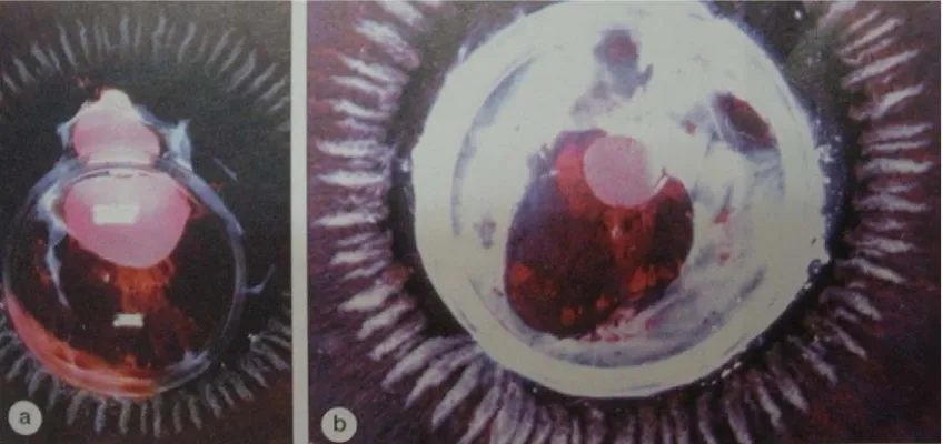
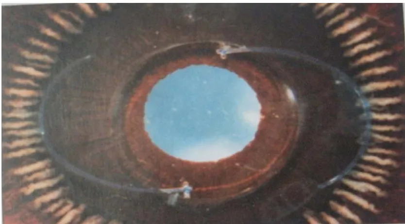
[image:34.612.117.544.598.694.2]Figure 18- An autopsy eye with capsular fixation of an Alcon
acrysof foldable lense
[image:35.612.116.543.491.703.2]OCULAR INJURIES AND TRAUMATIC CATARACT
CLASSIFICATION OF OCULAR INJURIES
Ocular injuries can be anatomically classified as intraocular and extraocular injuries. Extraocular injuries include lid lacerations, orbital
fractures, orbital hemorrhage and traumatic optic neuropathy. Ocular
Trauma classification group has classified intraocular mechanical injuries
into closed globe and open globe injuries18. The classification is based on
the type of injury:
1.CLOSED GLOBE INJURIES - Ocular injury without full thickness defect
of the coats. They are contusion or lamellar laceration(partial thickness
injury of the coats).
2.OPEN GLOBE INJURIES - Full thickness defects in the corneoscleral
coat of the eye. They are full thickness laceration outside to inside break in
ocular coats (can be penetrating injury if the object traverses the coats only
once or perforating injury if both an entry and exit wounds are present) and
rupture globe (full thickness inside to outside break in ocular coats).
Injuries can be:
1.Mechanical injuries : i. Blunt injuries ii. Penetrating injuries
iii. Radiational cataract iv. Chemical injuries induced cataract
BLUNT INJURIES
Blunt trauma of eyeball produces damage by different forces which
include:
1) Direct impact on the globe : It produces maximal effect at the point
where the blow is received (COUP)
2) Compression wave force : Transmitted force appearing as a wave
of pressure when the eye is suddenly compressed, travelling throughout its
fluid contents in all directions and the maximum damage may be at a point
distant from the actual place of impact (CONTRE COUP).
3) Reflected compression wave force : After striking the outer coats,
the compression waves are reflected towards the posterior pole and cause
foveal damage.
4) Rebound compression wave force : After striking the posterior wall
of the globe, the compression waves round back interiorly. This force
damages the retina and the choroid by forward pull and lens iris diaphragm
by forward thrust from the back.
5) Indirect force : Globe is suddenly hurled against the elastic contents
PATHOPHYSIOLOGY
1.When the anterior surface of the eye is struck bluntly, there is a
rapid anterior posterior shortening accompanied by equatorial expansion.
This equatorial stretching can disrupt the lens capsule, zonules or both.
Combination of coup, contrecoup and equatorial expansion is responsible
for formation of traumatic cataract following blunt injury.
2. Even when the capsule is not torn, the slightly traumatised lens
imbibes twice as much water as controls. This has been attributed to a
separation of the normal integrity of lens fibres.
3. Damage to capsule due to concussion may impair its
semipermiability, allowing the imbibation of aqueous by the lens substance
and distributing the active transport of metabolites.
The various forms of cataract following blunt trauma include:1.
Discrete subepithelial opacities 2.Traumatic rosette cataract 3. Traumatic
zonular cataract 4.Early maturation of senile cataract 5.Diffuse concussion
cataract.
Blunt trauma causes traumatic cataract, anterior subluxation or
dislocation and posterior dislocation or subluxation of lens19.
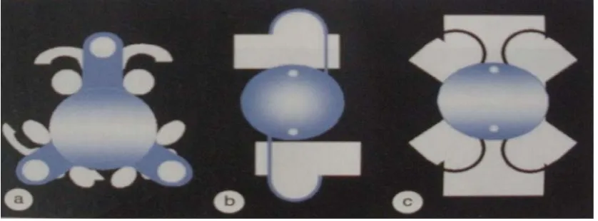
Figure 20- MECHANISMS OF BLUNT TRAUMA TO
EYE BALL
A. Direct impact
B. Compression wave form
C. Reflected compression wave form
POST SURGICAL APHAKIA
Cataract surgery is by far the most common intraocular surgery
performed world wide. It may not be possible to implant PCIOL in the
posterior chamber in all cases due to unforeseen intraoperative
complications during surgery. The incidence of posterior rupture is higher in
ICCE followed by ECCE,SICS and phacoemulsification.
POSTERIOR CAPSULAR TEAR
Posterior capsule rupture is the most common serious intraoperative
complication of cataract surgery. A posterior capsular rent is more likely to
occur in eyes with small pupils, hard nucleus, or pseudoexfoliation
syndrome. Recent reports suggest that the visual prognosis of patients who
have broken posterior capsules is excellent. The key factors are to minimize
ocular trauma, meticulously clean prolapsed vitreous from the anterior
segment, if present, and ensure secure fixation of the IOL.
BEFORE NUCLEUS REMOVAL.
A capsular break noted before nucleus extraction is a potential
disaster. The first objective is to prevent the nucleus from being dislodged
into the vitreous cavity by using an OVD injected posterior and anterior to
the nucleus to prevent its posterior displacement and to cushion the corneal
plana incision 3 mm posterior to the limbus into the vitreous, which Kelman
has described as “posterior assisted levitation”. The nucleus is pushed gently
anteriorly, so that it can be captured in front of the iris and safely removed
from the eye.
In most circumstances, the nucleus should be managed by sufficiently
enlarging the wound to facilitate easy extraction of the nucleus on a lens
loop. However, in the case of a small break or when only a small amount of
nucleus is left, it may be possible to cover the posterior capsular opening
with a retentive OVD and complete the phacoemulsification. One can also
use a Sheets glide as a “pseudo–posterior capsule” to facilitate completion
of phacoemulsification.
Vitreous loss almost always accompanies posterior capsular rupture
and vitrectomy should be performed before the nuclear pieces are removed.
Clearly, one should not do this if it makes loss of the nucleus into the
vitreous more likely.
DURING CORTICAL IRRIGATION-ASPIRATION.
When capsular rupture occurs during aspiration of the cortex ( the
most common cause), a key factor is the status of the vitreous. If no vitreous
is present in the anterior segment, vitreous loss often can be averted. An
posteriorly. Cortical removal can be completed using low-flow irrigation.
Options include using a manual system; a dry approach, aspirating with a
cannula in the chamber filled with OVD; a bimanual approach through two
paracentesis openings; and automated irrigation-aspiration with all settings
reduced. Cortex should be stripped first in the region farthest from the rent,
and the direction of stripping should be toward the rent. Because it can be
hazardous to remove cortex in the region of the rent, the cortex is sometimes
better left in the eye, to avoid the possibility of enlarging the rent and
precipitating vitreous loss. One option to prevent extension of the rent is to
convert the tear into a small posterior capsulorrhexis, which eliminates any
radially orientated tears that could extend with further surgical
manipulation.
If vitreous is present in the anterior segment, vitrectomy should be
performed first, with the necessary caution being taken to prevent extension
of the rent. Depending on the type of capsular tear, the vitrectomy is
performed through either the limbal incision or the pars plana. In either
case, irrigation is provided with an infusion cannula in the paracentesis
opening. After a thorough anterior vitrectomy, the remaining cortical
material can be removed using one of the techniques described earlier or
OPTICS OF APHAKIA AND PSEUDOPHAKIA
The normal 72 yrs old man has total dioptric power of 58 D , with
nearly 75% of the power from the cornea and 25% of the power from the
crystalline lens. Removal of the crystalline lens leaves the eye extremely
deficient in dioptric power, which must be replaced to restore vision.
The replacement of the dioptric power can be in the form of
spectacles, contact lenses or intraocular lenses. Although each modality can
restore the patient's vision the optical consequences are dramatically
different.
OPTICS OF APHAKIA:
Replacement of crystalline lens power with spectacle lenses causes
the image that is formed on the patients retina to be roughly 25% larger than
that of the image formed by the crystalline lens. There is approximately
2% magnification for each dioptre of power in the spectacles. The average
aphakic spectacle is therefore 12.5 dioptre21.
The magnification from aphakic spectacle causes other optical
aberrations such as a ring scotoma. The magnification from other
spectacle causes other optical aberrations such as a ring scotoma, jack in the
Magnification of about 25% by aphakic spectacles reduces field of
vision by 25%, a loss of vision of peripheral fields or a ring scotoma.
When an object moves from the peripheral field of vision towards the
centre of fixation it disappears through the ring scotoma till it appears in the
central island of vision.
The jumping in to and out of the patients vision has been referred to
as the jack in box phenomenon. Pin cushion effect is the property of all plus
lenses and is directly proportional to their power . The square looks like a
pin cushion with sides pushed in and the corners have a stretched out
appearance. Every object viewed through aphakic spectacles appear this
way. This would be a handicap for professional like Draftsman or an
architect.
OPTICS OF CORRECTION OF APHAKIC WITH
CONTACT LENSES
To correct aphakia at the corneal plane involves the use of contact
lenses or surgery that adds refractive power to the cornea.
The power at the corneal plane that is equivalent to 12.5D at a vertex
of 12mm is 14.7D; a patient who needs a 12.5D in aphakic spectacles would
At the corneal plane the magnification is 6-8%.This value is near the
limit of aniseikonia, (image size disparity between the eyes), so that
unilateral aphakic patients can have binocular vision, with the aphakic eye
corrected using a contact lens and the other eye aphakic. Binocular vision is
not the possible with one aphakic spectacle and a normal phakic lens.
OPTICS OF PSEUDOPHAKIA
A posterior chamber lens in the bag following cataract extraction just
as the average spectacle power for aphakia is 12.5D , the average power for
an equiconvex lens is 21.0D.The average magnification of an IOL in this
position is 1.5% compared with the original crystalline lens.
For an anterior chamber IOL the average power would be less,
approximately 18.0D, and the magnification would be approximately
2.0%.Almost every one can achieve binocular vision with one eye
pseudophakia and other eye phakic.
IOLs available are either biconvex, convexoplano or meniscus. As a
result of clinical performance and optical analysis the majority of lenses
implanted today are biconvex. The quality of the optical designs of an IOL
70% is measured based on its performance with respect to tilting,
decentration and spherical aberration. The optimal mechanical and optical
functions of an IOL in the human eye is that of biconvex lenses.They reduce
SECONDARY INRAOCULAR LENS IMPLANTATION
Secondary intraocular lens implantation (IOL) is defined as insertion
of lens into an eye which is rendered aphakic by prior cataract extraction by
any methods, or by an exchange IOL which is a special case of secondary
IOL implantation. In Sanjeev Kumar et al and Lee et al study, it is found
that secondary SFIOL implantation has better visual outcome compared to
primary SFIOL implantation23, 24.
IOL exchange or explantation is a special case of secondary IOL
implantation where the original AC or PC lens may have to be removed due
to excessive corneal decompensation, UGH syndrome and CME.
INTRAOCULAR LENS INSERTION.
Careful inspection of the anatomy of the capsule and zonules is required to
determine the appropriate site for IOL implantation. There are five choices:
capsular bag, ciliary sulcus, sutured posterior chamber, and anterior
chamber.
1.CAPSULAR BAG
If the rent is small and relatively central, and if the anterior capsular
margins are well defined, the posterior chamber IOL can be implanted into
the capsular bag. If possible, conversion of posterior capsule tears to
This technique is applied to avoid an anticipated extension of the
inadvertent linear or triangular tear during manoeuvers such as a required
vitrectomy or lens placement. The surgeon should ensure that the haptics are
orientated away from the rent (to avoid haptic placement or subsequent
migration into the vitreous) and that the lens is inserted gently to avoid
enlargement of the rent.
2.CILIARY SULCUS
If the rent exceeds 4–5mm in length or there is extensive zonular loss,
the capsular bag probably is not adequate for IOL support. In such cases, the
ciliary sulcus is opened with an OVD, and the iris is retracted in all
quadrants to assess the status of the peripheral capsule and zonules. The IOL
is inserted with its haptics oriented away from the area of the rent and
positioned in areas of intact zonules and capsule. Another alternative, if the
anterior capsulorrhexis is intact, is sulcus placement of the IOL, with
capture of the optic through the capsulorrhexis25.
3.IRIS FIXATION
Some surgeons advocate iris suture fixation of one or both haptics to
prevent IOL decentration. After the IOL optic is captured through the pupil,
McCannel sutures are used to secure the haptics to the iris, and then the
optic is repositioned through the pupil. The complications include iris chafe
(motility of iris over the lens), chronic inflammation or intraocular
4.SUTURED SCLERAL FIXATED INTRAOCULAR LENS
If loss of more than 4–5 clock hours of capsule or zonules occurs, the
ciliary sulcus may be inadequate for lens stability. The lens can be fixated to
the sclera using single or dual 10–0 polypropylene sutures. If one region of
solid peripheral capsule and zonules exists, one haptic can be inserted into
the sulcus in this area, and the opposite haptic can be sutured to the sclera27.
5.ANTERIOR CHAMBER
A Kelman-type multiflex anterior chamber IOL design is a good
option for patients who do not have glaucoma, peripheral anterior
synechiae, or chronic uveitis28. A peripheral iridectomy should be
performed in these patients to prevent pupillary block29.
SCLERAL FIXATED INTRAOCULAR LENSES
The posterior chamber is the normal anatomic position of the human
lens. Here the lens reduces the risk of bullous keratopathy, injury to angle
structures, pseudophacodonesis and the risk of pupillary block glaucoma as
it is closer to the rotational centre of the eye. The centrifugal forces acting
on the lens is reduced and ocular contents are stabilised thus reducing the
risk of iritis, CME & retinal detachment. It improves the optical properties
of the eye. In the eye without an intact posterior capsule, a PCIOL can be
inserted only if it is sutured to either the sclera or the iris. Another
advantage of positioning the lens closer to the nodal point and centre of
rotation of the eye is the superior optical properties acquired by the lens in
this position30. The indications for placement of a PCIOL fixated to the
sclera include the following:
1) Patients with fibrosed anterior-posterior capsule, with extensive
posterior synechiae or zonular or posterior capsular tears.
2) An eye with inadequate capsular support or zonular support(more
than three clock hours).
3) An aphakic patient who is contact lens intolerant.
4) An eye that has undergone an ICCE.
5) For secondary IOL used in combination with penetrating
6) In young patients to avoid the risk of corneal decompensation and
other ACIOL complications.
7) In cases of inadequate intact iris diaphragm where iris fixated IOLs
cannot be used.
Suture fixated IOLs were first introduced by Parry in the
1950s.PCIOLs designed for suturing to the sclera have eyelets on both
haptics as well as a large diameter (6.5-7mm) optic to decrease the risk of
decentration. Although suture fixated lenses are technically difficult to
insert, they often provide good results when implanted as secondary IOLs.
There are many methods for scleral fixation of posterior chamber lenses.
SURGICAL TECHNIQUES
1. CLASSIC AB EXTERNO TECHNIQUE FOR CILIARY SULCUS FIXATION
In 1991,Lewis described a technically facile technique for ab externo
sulcus fixation of PCIOL. By definition, the ab externo technique avoids the
passage of a needle from the inside of the eye to the outside through the
sclera. The surgeon can thus reduce the risk of hemorrhage, retinal
detachment and lens malposition by avoiding the potential inaccuracies of
suture placement that are inherent to the ab externo technique. One
disadvantage of the Lewis method is that the one - point fixation of the
suture to the sclera creates a less stable fixation than would a two - point
After creating a conjunctival peritomy from 4 o'clock to 10 0'clock
position a partial thickness limbal based triangular scleral flap that is 3mm
high and 2mm wide is made. Complete anterior vitrectomy is made through
a 7mm corneal scleral wound. A straight needle carrying a 10-0
polypropylene suture is passed through the 10 o'clock scleral bed 1mm
posterior to surgical limbus. When the needle tip is visualised through the
pupil, insert the straight needle into the barrel of a 28 - gauge needle on a
standard insulin syringe at 4 o'clock position and withdraw the syringe from
the eye. Deliver a loop of suture through the corneal scleral incision. Cut the
loop and tie the free ends to the haptics of the lens. Insert the lens into the
ciliary sulcus. Dial the lens while removing slack from the sutures. Use a
second 10-0 polypropylene suture on a half- circle needle to take a short bite
in the 4 o'clock scleral bed just anterior to the first suture's exist. Suture
together the short end of this suture to the IOL fixated suture and consider
this as a hybrid suture. Tie the long end of the second suture to the hybrid
suture in a square knot with four throws. The same steps are followed in the
10 o'clock position. Close the scleral flaps and reapproximate the
conjunctiva.
2. CLASSIC AB INTERNO TECHNIQUE FOR CILIARY SULCUS FIXATION
1n 1990,Smiddy described a straight forward technique that produces
disadvantage of this method is the hemorrhage risk and decreased stabilty of
the one-point fixation. After preparing a shelved limbal incision pass the
needle transsclerally 1 mm posterior to the limbus in the 3 o'clock meridian.
Apply gentle counter pressure with the forceps externally while passing the
suture through the sclera. Pass the second needle in a similar manner at 9
o'clock meridian. A properly positioned scleral fixated lens with the haptics
oriented in the 3 and 9 o'clock meridians is obtained after making a mid -
thickness scleral pass with the needle and tying the suture to itself.
3.SMALL INCISION AB EXTERNO TECHNIQUE FOR CILIARY SULCUS FIXATION
Regillo and Tidwell published a modified version of the Lewis
technique. The straight needle end of the 10-0 polypropylene suture is
inserted through the scleral bed temporally and retrieved within the barrel of
a 28 gauge needle. The single transscleral suture is externalised through the
temporal incision and the loop is cut. The two free ends are tied to the
haptics of the silicone lens. The folded silicone lens is inserted through the
4mm corneal incision. The lens is secured in the ciliary sulcus with the
haptics at 3 and 9 o'clock position.
4.KNOTLESS AB EXTERNO TECHNIQUE FOR CILIARY SULCUS FIXATION
In 1995,Erylidirim introduced the knotless ab externo ciliary sulcus
method leaves the surgeon with two lines of suture at each scleral clock
hour at the end of the operation, these sutures cannot be used to perform a
more stable two- point fixation because the two strands exit through the
same port.
After preparing the flaps, a needle is inserted in reverse position. The
suture is captured with the lens dialer and pulled through the corneal
incision. The procedure is repeated on the other port. The suture is passed
through the passing the lens through the loop and is locked in place. The
procedure is repeated in the other haptic. The haptic is passed through the
loop and the suture is tightened with an equal pulling force on either side.
One part of the suture is passed to the other side of the haptic. A 8-0 suture
can be used as a guide to 10-0 suture through the eyelet31.
5.AB INTERNO TECHNIQUE WITH TWO POINT CILIARY SULCUS FIXATION
This technique is a stable two point fixation because two sutures exit
the sclera at two different spots. After dissecting two limbal based, partial
thickness scleral flaps 180˚ apart, a double armed suture is passed through
the ciliary sulcus and out through the sclera 1 mm posterior to limbus. The
other needle of the double armed suture is passed in a similar manner,
haptics to the double armed suture. Once the lens is centred in the ciliary
sulcus, a 3-1-1 surgeon's knot is used to tie the superior and inferior loops.
6.AB INTERNO TECHNIQUE WITH PARS PLANA FIXATION
In 1981,Girard introduced the pars plana fixation technique. The
advantage of this technique is that hemorrhagic complications, retinal
detachments and iris pigment dispersion can be reduced. The diameter of
the lens is increased to 17mm, the diameter of the biconvex optic to 7mm
and the haptics have to be angled backward at 10˚ or 20˚.Teichman advised
that to enter the pars plana safely, the sclera should be entered 3-5 mm
TECHNIQUES OF SFIOL IMPLANTAION
Figure 21- Classic ab externo technique
[image:55.612.115.543.356.671.2]
Figure 23- Small incision ab externo technique
Figure 25- Ab interno two point technique
COMPLICATIONS OF SFIOL IMPLANTATION
There is a 12% risk of serious complications with SFIOL
implantation which include :
1. CYSTOID MACULAR EDEMA - This is one of the most common
complications seen in 9% to 36% of the patients after SFIOL implantation.
2.GLAUCOMA - Glaucoma after SFIOL implantation is seen in 30.3% of
the patients when the surgery is performed at the same time as penetrating
keratoplasty.
3.LENS DECENTRATION - Lens tilt or decentration is found in
5%-10% of patients. Proper polypropylene suture placement and tension are
more important in avoiding this complication.
4.RETINAL DETACHMENT - The risk of retinal detachment is 2.3% in
patients with SFIOL implantation33.
5.UVEITIS - There is increased risk of uveitis in diabetic patients and in
patients with recurrent anterior uveitis.
6.CHOROIDAL DETACHMENT - The risk of choroidal detachment is
3.6% in patients with transscleral sutures.
7. SUTURE RELATED COMPLICATIONS - Suture related complications
AIMS/OBJECTIVES OF THE STUDY
PRIMARY OBJECTIVE
To determine the factors affecting the visual outcome following
vitrectomy & scleral fixated intraocular lens implantation in:
1. Post surgical aphakia
2. Post traumatic cataract , subluxation of lens and anterior lens dislocation
3. Spontaneous subluxation or anterior lens dislocation in collagen vascular
diseases or pseudoexfoliation.
SECONDARY OBJECTIVE
To study about the various factors during SFIOL implantation like:
1. Preoperative corneal opacity, striate keratopathy, chronic uveitis, cystoid macular edema, type of cataract surgery done, mode of injury, duration between trauma/cataract surgery and SFIOL implantation, associated systemic diseases.
2.Intraoperative factors like duration of surgery, complications during SFIOL implantation.
3.Postoperative complications like striate keratopathy, iritis, corneal decompensation(<500 cells/cu.mm),IOL malposition, cystoid macular edema (OCTthickness>250µ),retinal detachment, rise of IOP, endophthalmitis, vitritis.
MATERIALS AND METHODS
This prospective study was conducted in Regional Institute of
Ophthalmology and Government Ophthalmic Hospital, Egmore, Chennai
from September 2010 to August 2012 for a period of 24 months.
INCLUSION CRITERIA
All patients with postsurgical aphakia, posttraumatic cataract,
posttraumatic subluxation/ anterior dislocation, spontaneous subluxated or
anteriorly dislocated lenses, patients with endothelial cell count >1200
cells/cu.mm.
EXCLUSION CRITERIA
Patients with endothelial cell count less than 1200 cells/cu.mm
,retinal detachment, vision not improving with aphakic correction, macular
disorder, previous intraocular surgery other than cataract surgery and
corneal tear suturing.
METHODOLOGY:
In this study totally 50 patients were included based on the inclusion
criteria. Pre-operative evaluation in these patients include visual acuity by
Snellen’s chart, applanation tonometry ,Slit lamp biomicroscopy, B-Scan
Ultrasonography, A-Scan Biometry, Keratometry, Indirect ophthalmoscopy,
1. Anterior vitrectomy with ab externo two point scleral fixated
intraocular lens implantation is done with Aurolab single piece SFIOL (with
overall diameter of 13 mm and optic diameter of 6 mm with single eyelet at
each haptic) and 10-0 polypropylene suture.
2. Postoperatively patients are treated with topical eyedrops like
1%prednisolone acetate & 0.5% moxifloxacin 6 times/day tapered over 6
weeks,0.5%ketorolac tromethamine 4 times/day for 1 week and
Prednisolone tablets 1mg/kg in cases of vitritis which is tapered later.
3. Postoperative evaluation includes-visual acuity by Snellen’s chart,
applanation tonometry, Slit lamp biomicroscopy, indirect ophthalmoscopy
and specular microscopy, OCT,UBM.
4. During the follow up period of 1,4,6 weeks and late follow up at 12
months complications if any are treated, retinoscopic refraction is done and
best glasses are prescribed. Success rate of the surgery was defined as vision
≥6/18.
SCREENING PROCEDURES
PROCEDURES
Best corrected Snellen’s visual acuity, IOP, B-scan ultrasonography,
indirect ophthalmoscopic examination, slit lamp biomicroscopy, A-Scan
Biometry, keratometry, specular microscopy, OCT for macular thickness,
FOLLOW UP PROCEDURES/VISITS
Best corrected Snellen’s visual acuity, IOP, slit lamp
biomicroscopy, indirect ophthalmoscopic examination, specular
microscopy, OCT, retinoscopic refraction, UBM at 1,2,4, 6 weeks and 12
months.
ASSESSMENT OF PARAMETERS
a) Age, sex, Visual acuity better in which group.
b) Pre-operative factors corneal opacity, striate keratopathy, chronic uveitis,
cystoid macular edema, type of cataract surgery done, mode of injury,
duration between trauma/cataract surgery and SFIOL implantation,
associated systemic diseases.
c) Intraoperative factors like duration of surgery, complications during
SFIOL implantation.
d) Postoperative complications like striate keratopathy, iritis, corneal
decompensation(<500 cells),IOL malpositon, cystoid macular
edema(OCT thickness>250µ),retinal detachment, rise of IOP,
endophthalmitis, vitritis.
1.AG Tabl S.N 1 2 3 4 5 6 Cha year 0 2 4 6 8 10 12 14 16 18
GE AT PR e no 1 : A
NO A
P 25 35 45 55 65 75
art no. 1: A
The
rs. 58% of
2
25‐35 3
RESENTA ge distribu AGE OF T
ATIENTS 5-35 5-45 5-55 5-65 5-75 5-85 Age distri mean age
f patients w
1
12
35‐45 45‐55
AG
RES
TION ution of p THE S bution of of present were betwe 17
55‐65 6
E
DISTRI
SULTS
atients NO 02 01 12 17 12 06 patientstation in ou
en 45-65 y
12
6
65‐75 75‐85
IBUTION
O. OF PAT
ur study is
yrs
N
AGE D
TIENTS
61.54(SD-ISTRIBUTION
2. SE
Tabl
M
Fe
Cha
1
EX DIST
e No 2 : SEX
Male
emale
art No 2
.
In
8(36%) we
18
TRIBUTI
Sex distri
: SEX D
n this study
ere female
ION
ibution NO. OF
ISTRIBU
y out of 50
s.
SEX
F PATIEN
32
18
UTION
0 patients 3
32
NTS
32(64%) w
MALE FEMA
were males
ALE
3.LA
Tab
Rig
Lef
Cha
In th
ATERAL
le No 3 :
E
ght
ft
art No 3
his study b
24
LITY
Latera
EYE
: Lateral
both eyes w
ality
lity
were equall
EYE
NO. OF
ly involved
26
F PATIEN
26
24
d.
RIG LEF
TS
4. D
Tab
S.NO
1 2 3 4. 5 6Cha
MajIAGNOS
le.No 4 :
DIAG
Post sur Post tra Post tra Post tra lens Spontan or sublu vascula Spontan or subluart no 4 :
jority of th
35 0 5 10 15 20 25 30 35 40
SIS AT P
: Diagnos
GNOSIS
rgical apha aumatic cat aumatic sub aumatic an neous ante uxation in ar disease neous ante uxation in: Diagnos
he patients 3DIA
PRESEN
sis at pre
akia taract bluxated le nt. disloca erior disloc collagen erior disloc PXF
sis at pre
were in the
6 2
AGNOSIS
TATION
esentation
N
P
3 3 ens 6ation of 2
cation 2
cation 2
esentation
e post surg
2
S AT PRES
N
n
NO.OF
PATIENT
5n
gical aphak 2SENTATIO
TS
PER
70 6 12 4 4 4
kia group (
ON
DIAGNOSIS AT
RCENTA
%
70%).
T PRESENTATIO
AGE
5. P
Tab
Ch
The spon S.NO 1 2 3 4 5 6 7 8 9 10PRE - OP
ble no 5 :
art No 5:
e most com ntaneous d 0 2 4 6 8 10 12 14 16 18 20 19 O PRE FAC 2 3 4
5 C
6 sub 7 sub 8 9 0
PERATIV
: Pre-op
: Preope
mmon facto dislocation 3 0
PREOPE
EOPERAT CTORS No com Corne striate k Chron Cystoid ma Spon bluxation/d Tra bluxation/d Diabete Hype IschaemicVE FAC
erative f
erative fa
ors were ch / subluxat 18 0
ERATIVE
TIVE mplications eal opacity keratopathy nic uveitis acular oed ntaneous dislocation aumatic dislocation es mellitus ertension c heart diseCTORS
factors
actors
hronic uvei tion of lens
4
2 3
E
FACTOR
N P s y ema of lens of lens s ease
itis (38%) f s (8%) and
3 4 0
RS
NO. OF PATIENT 19 3 0 18 0 4 2 3 4 0 followed b d hypertens pr TS PERC by sion (8%).reoperative fac
6. TY
Tab
S.N 1 2 3 4
Ch
apha
SIC
YPE OF
le No 6 :
NO C S 1 2 3 4 PVP R
hart No.6
akics
Majori
CS (58%)
CATAR
Type of
TYPE OF CATARAC SURGERY ECCE SICS PHACO P WITH L REMOVA
: Type o
ity of the
.
1
TYPE
O
S
RACT SU
cataract
F CT Y N LENS AL
of catarac
post surg
3 29 2OF
CATAR
SURGICA
URGERY
t surgery
patient
NO OF PA
03 29 01 02
ct surger
patien
gical apha
RACT
SU
AL
APHA
Y
in post s
ts
TIENTS
ry in post
nts
akia patie
URGERY
AKIC
CAS
surgical a
PERCE
t surgical
ents had u
IN
POST
SES
ECCE SICS PHACO PPV W
aphakics
ENTAGE % 06 58 02 04l
undergone
T
O
WITH LENS REM
s
7.MO
Tab
Cha
13(2 hadODE OF
le No 7: M
MODE
Blunt
Sharp
art no 7 :
Total n
26%) and a
sharp inju
MOD
F INJURY
Mode of
OF INJU t trauma p injuryMode of
number of among the ury. 3E
OF
INJ
Y IN PO
injury in
URY
f injury in
f patients
em 10(77%
JURY
IN
APHAK
OST TRA
n post tra
NO.O
n post tr
presenting
%) had blun
10
POST
TR
KIA
AUMATIC
aumatic a
OF PATIEN 10 03aumatic
g with his
nt trauma t
RAUMAT
C APHA
aphakia
NTS
aphakia
story of tr
to the eye
8.D
SUR
Ch
vari pati 0 2 4 6 8 10 12 14 16 18
DURATIO
RGERY
art no 8:
Duratio ied betwee ents presen 14 0 2 4 6 8 0 2 4 6 8
0‐2 month
Du
ON BE
Y AND SF
Duratio
on of trau
n minimum
nted within
16
hs 2‐4 mon
ration
b
and
SF
ETWEEN
FIOL IM
on of trau
i
uma or ca
m of 1 mon
n 2 to 6 mo
13
nths 4‐6 mo
between
FIOL
imp
N TRAU
MPLANTA
uma/cata
implanta
ataract sur
nth to max
onths durat
3
7
onths 6 mo
n
trauma
plantatio
UMA O
ATION
aract surg
tion
rgery and
ximum of 2
tion.
7
onths
a/catarac
on
in
mo
D t S
OR CAT
gery and
SFIOL im25 years. B
ct
surge
onths
Duration betw trauma/catara SFIOL implanta
TARACT
SFIOL
mplantatio
But most th
ry
een
ct surgery and ation in month
9..P
50 p patiTab
Cha
0 5 10 15 20 25 30 35 40PRE OPE
Preoper patients 11ents had v
le No 8 :
S.NO
1
2
3
4
art No. 9:
0 0 0 0 0
6/6‐6/12 0
ERATIV
rative visu 1(22%) pat isual acuityPreoper
Preoper
6/18‐6/36 0
PREOP
VE VISUA
al acuity w
tients had v
y of ≤3/60
rative visu
VISUAL 6/6‐ 6/18 6/60 ≤3rative vis
6 6/60‐4/60 11
PERATIV
AL ACU
was measu visual acui 0.ual acuit
ACUITY ‐6/12 ‐6/36 ‐4/60 360sual acuit
≤360 39VE VISU
UITY
ured in all
ity of 6/60
ty
Y N
ty
UAL ACU
patients. O
0 to 4/60 an
NO. OF PA
0
0
11
39
UITY
preoperative v
Out of thes
nd 39(78% ATIENTS 0 0 1 9
visual acquity
se
%)
S
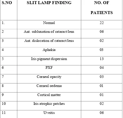
10. SLIT LAMP FINDINGS
Table N o 9 :Preoperative slit lamp examination
S.NO
SLIT LAMP FINDING
NO. OF
PATIENTS
1. Normal 22
2 Ant. subluxation of cataract/lens 06
3 Ant. dislocation of cataract/lens 02
4 Aphakia 03
5 Iris pigment dispersion 13
6 PXF 04
7 Corneal opacity 03
8 Corneal oedema 01
9 Cortical matter 01
10 Iris atrophic patches 02
11 Uveitis 06
The most common slit lamp examination findings in preoperative
evaluation was found to be iris pigment dispersion in 13(26%) patients,
followed by uveitis in 6 patients(12%).
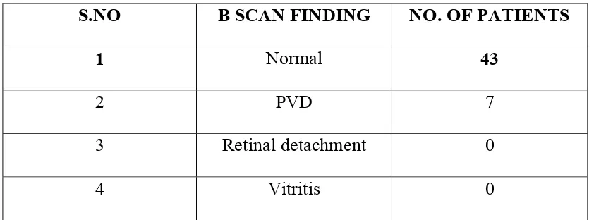
11. B SCAN FINDINGS
Table No 10: Preoperative B scan findings
S.NO B SCAN FINDING NO. OF PATIENTS
1 Normal 43
2 PVD 7
3 Retina