Copyright © 2003, American Society for Microbiology. All Rights Reserved.
The Stable 2.0-Kilobase Intron of the Herpes Simplex Virus Type 1
Latency-Associated Transcript Does Not Function as an Antisense
Repressor of ICP0 in Nonneuronal Cells
Edward A. Burton,† Chang-Sook Hong, and Joseph C. Glorioso*
Department of Molecular Genetics and Biochemistry, School of Medicine, University of Pittsburgh, Pittsburgh, Pennsylvania
Received 2 August 2002/Accepted 23 December 2002
During latency, herpes simplex virus expresses a unique set of latency-associated transcripts (LATs). As the 2.0-kb LAT intron is complementary to, and overlaps, the 3ⴕend of the ICP0 transcript, it has been suggested that the stable LAT intron might function as an antisense repressor of ICP0 expression. We tested this hypothesis in cell culture by dissociating cis- and trans-acting effects of the 2.0-kb LAT, using a series of complementary strategies. Initially, we constructed 293T cell lines that stably express the nuclear 2.0-kb LAT intron to determine whether LAT accumulation in transaffects ICP0 expression. ICP0 mRNA and protein expression profiles were studied (i) following infections with a viral mutant containing wild-type LAT and ICP0 sequences but having deletions of other immediate-early (IE) genes, thus preventing the progression of viral early gene expression, (ii) at early time points after infection with wild-type virus, before viral LAT expression, and (iii) by plasmid transfections. Northern and Western blot analysis showed thattransexpression of the 2.0-kb LAT intron does not affect ICP0 mRNA expression, stability, accumulation, splicing, or translation. In addition, suppression of viral replication by overexpression of the 2.0-kb LAT, which has been detected previously in neuronal cell lines, was not found in these nonneuronal cell lines. However, deletion of the latency-active promoter (LAP) region of the virus resulted in overexpression of IE genes, which occurred soon after infection, before viral LAT expression had commenced. This was not complemented by the expression of LAT intrans, suggesting that the LAP deletion affected transcriptional regulation of the IE genes incis. We conclude that the function of the highly conserved LAT intron is unlikely to involve a direct-acting anti-ICP0 antisense mechanism but that the LAT region could affect ICP0 mRNA expression from the viral genome.
Herpes simplex virus (HSV) is a common human pathogen that is responsible for oral and genital infection, severe en-cephalitis, and blindness. Following primary mucosal or cuta-neous infection, the viral nucleocapsid is transported to the sensory ganglia associated with the afferent nerve supply of the affected region. Within the nuclei of ganglionic sensory neu-rons, the virus is able to establish a latent infection; the viral genome may persist in this state for the lifetime of the host. Periodic reactivation triggers the formation of infectious viri-ons that are transported by anterograde axonal transport to the periphery, where they are shed to allow infection of a new host (37).
The processes regulating the establishment and mainte-nance of latency are incompletely understood. During latent infection, only the latency-associated transcript (LAT) gene is expressed abundantly from the viral genome (11, 35, 42, 46). A primary 8.3-kb LAT of low steady-state abundance (56) un-dergoes posttranscriptional processing to form other LAT spe-cies. The 2.0-kb LAT is a stable lariat intron (36, 54) that is spliced from the primary 8.3-kb transcript (2, 17, 55), located within the nuclei of infected cells, and can be readily detected during either lytic or latent infection (13, 46). The 1.5-kb LAT
is also a stable lariat intron (54) formed by the removal of a second intron from within the 2.0-kb LAT sequence (2, 44, 52). However, the 1.5-kb LAT intron has been detected only during viral latency (43, 44, 51), implying that the relevant splicing event is specific to both neurons and latent infection. The unusual stability of the LATs has been ascribed to peculiarities in the lariat structure adjacent to the branch point and splice acceptor region (25, 53, 55). The presumed 6.3-kb spliced exon product of LAT is very difficult to detect and appears to be highly unstable (12).
Despite the extensive characterization of the origin and na-ture of the LATs, relatively little is known about their function. Difficulty is encountered in producing viruses that are pure LAT null mutants; the genome of HSV is extremely compact, and the immediate-early (IE) gene encoding ICP0 is situated on the complementary strand of the viral genome, with the 3⬘
ends of ICP0 and LAT overlapping (Fig. 1). Thus, only the LAT promoters and the 5⬘ region of LAT may be deleted without affecting the ICP0 gene. The remainder of the LAT region has been inactivated by other means, including the use of synthetic transcriptional stop signals which do not disrupt the ICP0 reading frame on the opposite strand (4). The most striking conclusion from studies on LAT-null strains is that LAT deletion does not completely prevent the establishment of, or reactivation from, latency (22, 24, 41, 45). Various more subtle deficits have been reported, however. First, there are reports that LAT-null mutants are less efficient at establishing latent infection than wild-type or rescuant virus (40, 50). Sec-ond, latent LAT-null mutants show different reactivation
ki-* Corresponding author. Mailing address: Department of Molecular Genetics and Biochemistry, University of Pittsburgh, School of Med-icine, E1240 Biomedical Science Tower, Pittsburgh, PA 15261. Phone: (412) 648-8105. Fax: (412) 624-8997. E-mail: [email protected].
† Present address: University of Oxford, Department of Clinical Neurology, Radcliffe Infirmary, Oxford OX2 6HE, United Kingdom.
3516
on November 8, 2019 by guest
http://jvi.asm.org/
netics than controls (3, 15, 21, 31, 32, 45), although this may be attributable in some instances to the reduced efficiency with which latent infection is established in the first place. Finally, LAT-null mutants appear to be more toxic to neurons than LAT-expressing viruses (33, 49), resulting in more cell death following infection, which may be apoptotic (33). Correspond-ingly, LAT expression seems to enhance cellular resistance to toxic stimuli in tissue culture (1, 33). Enhanced cytotoxicity may account for the reduced efficiency with which LAT-null viruses establish latency.
The molecular events underlying these phenotypes are poorly characterized. LAT-null mutants show alterations in the regulation of other viral genes. Thus, the steady-state levels of the IE gene products ICP0, ICP4, and ICP27 are increased following infection with LAT-null mutants (7, 30). Conse-quently, the early gene encoding thymidine kinase is also over-expressed in the absence of LAT (7). The higher levels of IE gene products might account for the greater cell toxicity and diminished ability to establish latent infection exhibited by LAT-null mutants. However, viral deletion experiments do not distinguish between effects of LAT locus deletions occurring in
cisand effects of the products of the LAT gene occurring in
trans. Thus, the mechanisms responsible for dysregulation of
gene expression in LAT-null mutants remain unclear. ICP0 has profound effects on viral gene expression levels during the HSV infectious cycle (6). The ICP0 gene lies in close proximity to the LAT gene, and various mechanisms could be envisaged whereby LAT expression could affect ICP0 expression. Thus, at a cis-acting level, the LAT locus might contain regulatory elements that affect transcription of ICP0, or promoter interference might occur through two transcrip-tionally active genes being in opposite orientation and in close proximity. At a trans-acting level, LAT RNA might interact
with the viral genome or trigger secondary effects on the chro-matin structure and accessibility of viral gene promoters to transcription factors, or the LAT RNA might have direct an-tisense effects on ICP0 mRNA levels. Finally, LAT might en-code a protein that has a regulatory function.
The antisense overlap between the abundant 2.0-kb LAT intron and the 3⬘end of the ICP0 transcript has given rise to speculation that the 2.0-kb LAT intron might function as a
trans-acting antisense repressor of ICP0 mRNA stability or
translation (18, 30, 46). This notion is attractive, because it attributes an RNA-level function to the evolutionarily con-served stable structure of the abundant 2.0-kb LAT. The fol-lowing data have provided limited evidence in support of this idea. (i) Triple transient-transfection experiments, using simul-taneous transfection with three different plasmids, showed that the ability of an ICP0 expression plasmid totransactivate the thymidine kinase promoter was suppressed by the presence of a LAT-expressing plasmid (17). Unfortunately, these experi-ments are difficult to interpret because multiple variables, which are hard to control, are involved in simultaneous plas-mid transfection. (ii) Overexpression of LAT in neuronal cell lines was able to suppress replication of wild-type and LAT-null mutant viruses, in addition to suppressing the expression of ICP0 (30). However, the replication of LAT-expressing ruses was suppressed to a greater degree than LAT-null vi-ruses, implying that LAT may exert additional cis-acting ef-fects. Furthermore, the reduction in IE gene steady-state RNA levels was not restricted to ICP0; the transcripts corresponding to ICP4 and ICP27 were also reduced, implying that a simple antisense mechanism might not be the full explanation.
[image:2.603.112.476.67.299.2]As an initial effort to elucidate the regulatory events under-lying the establishment of latency and the enhanced cytotoxic-ity of LAT-null viruses, we decided to study putative LAT
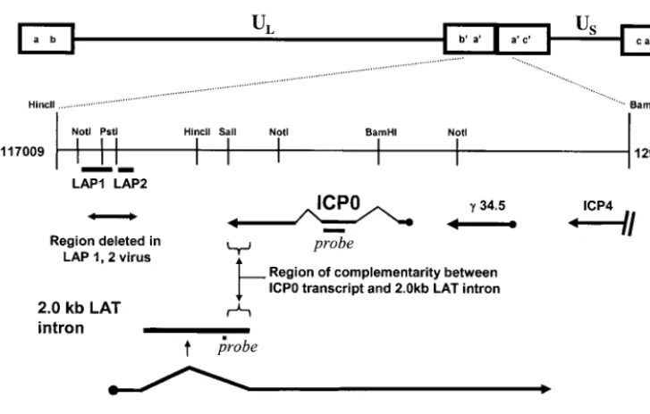
FIG. 1. Schematic depiction of the HSV-1 genome, with the repeat region flanking the unique long (UL) region of the genome expanded to illustrate the positions of probes used in this study, the positions of LAP1 and LAP2 promoters deleted in the⌬LAP1-2 virus, and the region of antisense overlap between the 2.0-kb LAT intron and the ICP0 transcript.
on November 8, 2019 by guest
http://jvi.asm.org/
antisense mechanisms. We reasoned that if the 2.0-kb LAT intron had antisense activity against the ICP0 mRNA, which was simply dependent on interstrand duplex formation be-tween the LAT and the ICP0 RNA, this should be detectable
in trans in cultured nonneuronal cells. If, however, the LAT
had a complex neuron-specific function involving a cell-type-dependent mechanism, then thetrans-acting effect previously demonstrated in neuronal cells should not be detected in non-neuronal cells.
We took the following approaches to dissociating thetrans -acting properties of the 2.0-kb LAT intron from thecis-acting properties of the LAT locus. First, we generated multiple cul-tured 293T cell lines and populations stably expressing the nuclear 2.0-kb LAT intron at various levels, similar to those found during lytic infection. Then,trans-acting effects of cell-encoded LAT on ICP0 expression in these cells were examined by using three different strategies: (i) infection with viruses in which the wild-type LAT loci are intact but which do not express LAT because of derangements in the viral transcrip-tional program induced by null mutations at the ICP4 and ICP27 loci, (ii) infection with wild-type virus and examination of ICP0 expression at time points before expression of viral LAT, and (iii) transfection with an ICP0 expression plasmid. Using these complementary techniques, we were able to ex-clude anytrans-acting effect of the 2.0-kb LAT intron on the stability or accumulation of ICP0 mRNA or protein in non-neuronal cells. We then examined thecis-acting effects induced by a deletion in the latency-active promoters LAP1 and LAP2 (9). We found that the expression of viral IE genes following infection of wild-type 293T cells with this mutant virus was enhanced with respect to expression in wild-type virus, even at time points before the expression of viral LAT. The delivery of 2.0-kb LAT intron RNA intransdid not alter this phenotype, implying that acis-acting, non-LAT RNA-dependent effect on viral gene expression was responsible. We conclude that an antisense mechanism involving the 2.0-kb LAT intron is un-likely to account for the changes in viral gene expression, and associated phenotypic alterations, that accompany the deletion of the LAT loci in the context of the viral genome in neuronal cells.
MATERIALS AND METHODS
Plasmids and probes.pIRES-Bleo-17 was made by inserting theEcoRV-MluI restriction fragment of strain 17 LAT into theEcoRV-BstXI sites of pIRES-Bleo (Clontech). pIRES-Bleo-SD was identical except that theEcoRV-NotI restric-tion fragment derived from strain 17 LAT contained a mutarestric-tion within the 2.0-kb consensus splice donor site (AGgtaggt to AGagtaga) (lowercase letters indicate introns, uppercase letters indicate exons, and bold letters indicate bases that were mutated). This mutation was generated as described in a previous study, in which the mutation was shown to abolish 2.0-kb LAT splicing (2). The ICP0 expression plasmid, pEG110, was a kind gift from Patrick Lomonte, Universite´ Claude Bernard Lyon 1, Villeurbanne Cedex, France; it was made by inserting the ICP0 cDNA into the pEGFP-C1 vector (Clontech) (29). Templates for the generation of riboprobes were constructed with pDP18 (Ambion). The ICP0 riboprobe template, pDP18-ICP0, was made by inserting a 255-bpXhoI-KpnI fragment of ICP0 exon 2 into theKpnI-SalI sites of pDP18. The LacZ riboprobe template, PDP18-LacZ, was made by inserting a 365-bpPvuII-EcoRI fragment of theEscherichia coliLacZ gene into theEcoRI (PstI/Klenow blunt-ended) sites of pDP18. The ICP22 riboprobe template was a kind gift from S. Wendell, Pittsburgh, Pa.; it was made by inserting a 210-bp PCR fragment spanning nucleotides 133056 to 133265 of the HSV-1 strain KOS ICP22 gene into pGEM-T (Promega). All constructs were characterized by restriction digestion and prepared at high concentration and purity by using a commercially available
maxiprep kit (Qiagen). Sequences of oligonucleotide probes were as follows: LAT-probe, 5⬘-CCGGACTGACCTGGCCTCTGG-3⬘; 18S-probe, 5⬘-GCTATC AATCTGTCAATCCTGTCCGTGTCC-3⬘.
Viruses.Wild-type HSV infections were carried out using an HSV-1 labora-tory strain, KOS. The LAT mutant,⌬LAP1-2, contains a deletion encompassing LAP1 and LAP2; the construction and characterization of this vector were previously described (9). The construction and characterization of QOZ.HG (genotype ICP4⫺, ICP27⫺:HCMV IEp-GFP, -ICP22, -ICP47, UL41⫺:
ICP0placZ) was previously described (8); the vector is based on thed106 back-bone, which was generated by N. DeLuca (University of Pittsburgh). The genome of QOZ.HG is depicted schematically in Fig. 7A. KOS and ⌬LAP1-2 were propagated in Vero cells. QOZ.HG was propagated in complementing 7B cells, which stably express ICP4 and ICP27. All vectors were purified by three rounds of limiting dilution and verified by Southern blot analysis. High-titer and high-purity vector stocks were prepared as described previously (5) and titrated in quadruplicate at each dilution using Vero or 7B cells as described above.
Cell culture and transfection.293T cells were maintained in Dulbecco’s mod-ified Eagle medium (Gibco-BRL) supplemented with 10% fetal bovine serum, 100 U of penicillin per ml, and 100g of streptomycin per ml. Cells were transfected with 1 g of plasmid expression construct at 80% confluence in six-well plates with Lipofectamine Plus (Gibco-BRL), following the manufactur-er’s protocol. Plasmid-expressing cell populations were selected by the addition of 200g of bleomycin per ml to the media, which killed 100% of control nontransfected cells. Selected clones were combined and cultured in the ongoing presence of bleomycin. Clonal isolation was carried out using limiting dilution in 96-well plates. Cultured monolayers at 80% confluence were subjected to HSV-1 infection for 1 h. Time points postinfection were counted from the time of addition of HSV-1 to the cell culture medium.
Northern blots.Total RNA was extracted from cultured cells by using a kit (RNAqueous; Ambion, Austin, Tex.), according to the manufacturer’s protocol. RNA was quantified by spectrophotometry at 260 nm. Denaturing electrophore-sis was carried out with a 1.2% agarose gel made with proprietary formaldehyde gel buffer (NorthernMax; Ambion) according to the manufacturer’s protocol, and electrophoresis was carried out in 1⫻MOPS (morpholinepropanesulfonic acid) running buffer. Total RNA (5g) was dissolved in 5l of H2O and 10l
of loading buffer (NorthernMax) and heated to 65°C for 15 min prior to gel loading. Transfer to a Nytran positively charged nylon membrane (Schleicher & Schuell) was carried out by using proprietary transfer buffer (NorthernMax) and a TurboBlotter apparatus (Schleicher & Schuell). Membranes were stored at ⫺20°C prior to hybridization, which was carried out in Ultrahyb (Ambion). Prehybridization for 2 h was followed by overnight hybridization at 68°C (ribo-probes) or 42°C (oligonucleotide (ribo-probes). Membranes were washed five times in 2⫻SSC (1⫻SSC is 0.15 M NaCl plus 0.015 M sodium citrate)–0.1% sodium dodecyl sulfate (SDS) at room temperature over 30 min and then three times in 0.1⫻SSC–0.1% SDS at 68°C (riboprobes) or 42°C (oligonucleotide probes) over 45 min. Membranes were exposed to film at room temperature for 5 to 120 min (riboprobes and the 18S oligonucleotide probe) or at⫺80°C overnight with intensifying screens (LAT probe). Band quantification was carried out with a Storm 840 PhosphorImager and ImageQuant software (Molecular Dynamics). Riboprobes were generated by in vitro transcription. Probe templates were linearized by digestion withHindIII (pDP18-LacZ and pDP18-ICP0) orNotI (pGEM-T-ICP22), phenol-chloroform extracted, ethanol salt precipitated, and resuspended at a concentration of 1g/l. The 20-l transcription reaction mixture contained 1g of linearized probe template, 10 U of T3 polymerase, 1⫻ transcription buffer (40 mM Tris-HCl [pH 7.9] at 25°C, 10 mM NaCl, 6 mM MgCl2, 2 mM spermidine, 0.05% Tween-20) and 10 mM dithiothreitol
(Ribo-probe; Promega), 20 U of recombinant RNase inhibitor (RNasin; Promega), high-purity unlabeled riboguanosine triphosphate (rGTP), rUTP, and rATP (Pharmacia-Amersham) at final concentrations of 500M each, and 3,000 Ci of [␣-32P]rCTP (NEN Life Science) per mmol at a final concentration of
approxi-mately 1.15M. After incubation at 37°C for 2 h, the DNA probe template was digested by the addition of 20l of DNase I solution (2 U in 1⫻transcription buffer). Following a further incubation of 1 h, the unincorporated labeled nu-cleotide was removed by minicolumn chromatography purification (G-25 Micro-Spin; Amersham). Oligonucleotide probes were end-labeled with T4 polynucle-otide kinase (PNK; New England Biolabs). The 20-l reaction mixture contained 10 U of PNK, 1⫻PNK buffer (70 mM Tris-HCl [pH 7.6], 10 mM MgCl2, 5 mM
dithiothreitol; New England Biolabs), 20 nM oligonucleotide, 5l of [␥-32P]ATP
at 6,000 Ci/mmol. After incubation for 1 h, unincorporated label was removed by using a MicroSpin column as described above.
Western blots and antibodies.Protein samples were harvested directly from cells in six-well plates. Monolayer samples were washed three times in phos-phate-buffered saline (PBS) at room temperature and then lysed in 100l of 1⫻
on November 8, 2019 by guest
http://jvi.asm.org/
lysis buffer (50 mM Tris [pH 8.0], 150 mM NaCl, 0.02% sodium azide, and 1% NP-40). Lysates were centrifuged to remove insoluble debris, and the protein concentration of the supernatant was quantified by a Bradford assay (Bio-Rad). Samples of 20g of protein were heated to 95°C for 5 min in 1⫻SDS loading buffer (50 mM Tris [pH 6.8], 5% 2-mercaptoethanol, 10% glycerol, 1% SDS, and 5g of bromphenol blue per ml) prior to electrophoresis, which was carried out with a 4% stacking gel and an 8% resolving gel. Transfer to a nitrocellulose membrane (Schleicher & Schuell) was carried out at 150 V, in 1⫻transfer buffer (25 mM Tris, 192 mM glycine, and 10% methanol) at 4°C for 2 h. Membranes were blocked in 1⫻TBST (Tris-buffered saline–Tween)–5% milk overnight at 4°C. Primary antibody was incubated for 2 h in 1⫻TBST–5% milk at room temperature, followed by four washes in 1⫻TBST over 40 min. Peroxidase-conjugated secondary antibody was incubated in 1⫻TBST–5% milk at room temperature for 1 h, followed by four washes in 1⫻TBST over 1 h. Hybridizing bands were visualized by using photographic film after incubation of the mem-brane with chemiluminescent peroxidase substrate (ECL Western blotting de-tection reagents; Amersham). The primary antibodies were ICP0 monoclonal antibody (Virusys), LacZ monoclonal antibody (reference G6282; Sigma), and fibronectin polyclonal antibody (reference F3648; Sigma). Secondary antibodies were peroxidase-conjugated rabbit anti-mouse antibody (reference A9044; Sigma) and horseradish peroxidase-conjugated goat anti-rabbit antibody (refer-ence A0545; Sigma).
In situ hybridization.The riboprobe template for LAT RNA in situ hybrid-ization was generated by inserting theHpaI-SalI fragment of the HSV-1 strain 17 2.0-kb LAT intron into theEcoRV-XhoI sites of the pcDNA3 vector (Invitro-gen). The resulting plasmid was linearized by digestion withEcoRI, and LAT-antisense riboprobe was made by in vitro transcription using SP6 RNA polymer-ase. The probe was labeled by incorporation of digoxigenin-UTP (Roche Boehringer Mannheim) into the transcription reaction mixture, as described previously (8). Cells were plated on one eight-well Lab-Tek chamber slide (Nalge Nunc), such that each slide contained the test cell population and all of the controls in adjacent wells, to ensure that all samples were processed identically. In situ hybridization was performed as described in a previous study (8). Cells were fixed in 4% paraformaldehyde for 10 min, treated with 1% HCl in PBS for 5 min, and washed with PBS. After equilibration in 5⫻SSC for 5 min, hybrid-ization with digoxigenin-labeled riboprobe was carried out overnight at 68°C in 15l of hybridization solution (50% formamide, 5⫻SSC, 40g salmon sperm DNA per ml, 0.5g of riboprobe per ml) per well. Following hybridization, the slide was rinsed with 2⫻SSC and 0.1⫻SSC at 68°C for 1 h. After blocking with
5% normal goat serum for 1 h, the LAT probe was localized with alkaline phosphatase-conjugated antidigoxigenin antibody (1:5,000; Roche Boehringer Mannheim) and detected with nitroblue tetrazolium–5-bromo-4-chloro-3-in-dolylphosphate substrate solution (Roche Boehringer Mannheim).
RESULTS
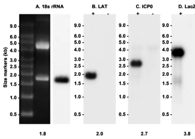
Probes. The positions of probes to the 2.0-kb LAT intron and ICP0 are depicted schematically in Fig. 1. All probes were single stranded to avoid problems of interpretation inherent in RNA strand-specific experiments. Figure 2 shows that each of the probes for ICP0, LacZ, LAT, and 18S rRNA hybridized with a single transcript of the expected size in Northern blot analyses. It was thus possible and admissible to quantify rela-tive transcript abundance by measuring the intensities of spe-cific hybridizing bands with a PhosphorImager and correcting them for gel loading and transfer by using the 18S rRNA signal as a control.
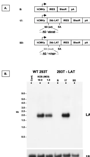
[image:4.603.126.460.67.305.2]Generation of cell populations and clonal lines stably ex-pressing the 2.0-kb LAT intron. We decided to isolate any direct trans-acting effect of the 2.0-kb LAT intron on ICP0 mRNA by generating nonneuronal cell populations that ex-press the 2.0-kb LAT intron. The EcoRV-MluI fragment of strain 17 LAT, encompassing the 2.0-kb intron, was cloned into an expression vector, pIRES-Bleo (Clontech). Three con-structs were made (Fig. 3A). The17construct contained the wild-type sequence. The Bleo construct comprised unmodified vector and served as a negative control. The SD construct contained a LAT sequence similar to that of the17construct, except that the conserved 2.0-kb LAT splice donor signal was mutated. The mutation, which consisted of two 3-bp deletions, was based on that described in a previous study, in which it was
FIG. 2. Northern blot controls showing that the probes used in this study hybridized specifically with the intended target RNA. (A) 18S RNA oligonucleotide. The RNA sample was from 293T cells. (B) LAT oligonucleotide. RNA was derived from Vero cells infected with HSV-1 strain KOS (⫹) and uninfected controls (⫺). (C) ICP0 antisense riboprobe. RNA was from 293T cells infected with QOZ.HG (⫹) and uninfected controls (⫺). (D) LacZ antisense riboprobe. RNA samples were as in panel C.
on November 8, 2019 by guest
http://jvi.asm.org/
FIG. 3. Construction of cell lines and populations that stably express the 2.0-kb LAT intron. (A) Schematic depiction of transgene expression cassettes introduced into cells by transfection prior to selection. Abbreviations: hCMVp, major IE promoter of human cytomegalovirus; IRES, internal ribosome entry site from encephalomyocarditis virus; BleoR, bleomycin resistance gene. (B) Northern blot hybridization analysis of control 293T cells (cont.), 293T cells 18 h after infection with HSV-1 strain KOS, and cell populations produced with the expression cassettes shown in panel A (293T-LAT). The blot was sequentially hybridized to probes for LAT and 18S rRNA. The 17 cell population expresses the 2.0-kb LAT intron at levels approximating those seen after wild-type infection at an MOI of 10. B, Bleo.
on November 8, 2019 by guest
http://jvi.asm.org/
shown to abolish 2.0-kb LAT splicing in the context of the entire LAT locus during viral infection (2). The SD plasmid thus contains almost the same sequences as the 17 plasmid but does not give rise to 2.0-kb intron splicing. The pIRES-Bleo vector contains an internal ribosome entry site 3⬘of the cloning site containing the LAT sequence, followed by an expression cassette for the bleomycin resistance gene. The 2.0-kb LAT would thus be spliced as an intron from the mRNA encoding the selectable marker. Following transfection of 293T cells with the three LAT constructs, cultures were selected for bleo-mycin resistance. This revealed many hundreds of resistant colonies in each sample. Some experiments used mixed se-lected cell populations, avoiding the problems associated with the idiosyncratic properties of single clonal cell lines. For other experiments, we selected, characterized, and expanded multi-ple clonal cell lines in order to assess the effects of LAT expression level.
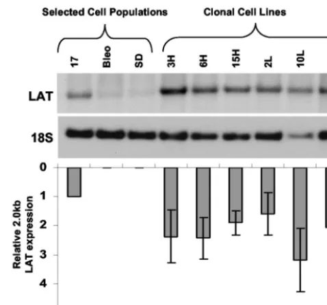
Characterization of cell populations and clonal lines stably expressing the 2.0-kb LAT intron. Total RNA was harvested from the cell populations and subjected to Northern blot anal-ysis (Fig. 3B). A 2.0-kb transcript hybridized with the single-stranded LAT oligonucleotide probe shown in Fig. 2. The electrophoretic mobility of the band was identical to that seen when RNA from cells infected with wild-type HSV-1 was hy-bridized with the same probe. This transcript was not present in cells expressing a LAT sequence carrying a mutation in the 2.0-kb splice donor. These features identified the hybridizing transcript as the 2.0-kb LAT intron. The expression level of the LAT 2.0-kb intron in this selected cell population was similar to that seen after infection of 293T cells with wild-type HSV strain KOS. A series of eight different clonal cell lines was isolated from the17population by limiting dilution culture and expanded under continued selective pressure. The Northern blot hybridization profile of these cell lines is shown in Fig. 4. Each of the clonal lines used in subsequent experiments ex-pressed LAT at higher levels than the selected17population from which it was derived.
The subcellular localization of the 2.0-kb LAT intron was examined by using RNA in situ hybridization (Fig. 5). Cells from the selected Bleo and SD populations were cultured on four-well slides, alongside cells from the selected17population and wild-type 293T cells that had been infected with wild-type HSV strain KOS. The slides were processed for in situ hybrid-ization, such that all of the cell samples were exposed to iden-tical reagents under ideniden-tical conditions. As predicted from Northern hybridization experiments, no detectable signal was observed in the Bleo or SD cell populations (Fig. 5A and C). The17cell population showed expression of LAT in the nuclei of⬎90% of the selected cell population (Fig. 5B). The appear-ances were identical to those arising from infection with wild-type HSV strain KOS (Fig. 5D). Elimination of the nuclear permeabilization step from the protocol also revealed a smaller amount of LAT within the cytoplasm of the selected cell population (data not shown).
These analyses show that the selected cell populations ex-press the 2.0-kb LAT intron at levels similar to those arising during wild-type infection and that the resulting stable intron is located in a similar subcellular compartment. These cell pop-ulations and the derived clonal lines should therefore be useful
for investigation of the possible trans-acting effects of the 2.0-kb LAT intron.
Measurement of wild-type HSV-1 replication in the pres-ence of LAT. We tested the hypothesis that the 2.0-kb LAT intron acts intransto repress viral replication. Parallel samples of the 293T cell populations described above were infected at a multiplicity of infection (MOI) of 0.1, 1.0, and 10.0 with wild-type HSV-1 strain KOS. Virus was harvested from the cultures at 18 h postinfection and titrated on permissive Vero cells. The number of infectious particles was determined in each sample at different dilutions. There was no difference in the titer of virus passaged in any of the three cell populations, indicating that the 2.0-kb LAT intron does not repress viral replication intransin these nonneuronal cells (Fig. 6).
Measurement of ICP0 accumulation during infection of LAT-expressing cells.The generation of cell lines allowed us to supply the LAT 2.0-kb intron in trans, but to examinetrans -acting effects of the LAT on ICP0 expression, it was necessary to eliminate cis-derived expression of LAT, without altering the genetic context of the ICP0 gene. We achieved this in three ways: (i) using a virus in which the LAT locus and ICP0 gene are present in their entirety but in which the viral transcrip-tional program is disrupted by mutations in the genes encoding other IE genes, such that the LAT genes are not expressed; (ii) using wild-type virus but examining IE gene expression at time points before LAT is expressed, and (iii) using an ICP0 ex-pression plasmid which does not contain any other HSV-1 sequences.
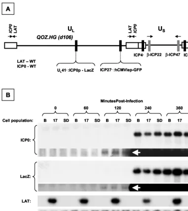
[image:6.603.303.542.68.290.2](i) QOZ.HG (Fig. 7A) is a derivative of the previously de-scribed mutantd106 (38).d106 has deletions of both copies of the gene encoding ICP4, the major transcriptional regulator of HSV-1. In addition, d106 has a deletion of the ICP27 gene,
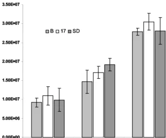
FIG. 4. Northern hybridization analysis of clonal cell lines derived from the 17 cell population. The blot was sequentially hybridized with LAT and 18S rRNA loading control probes. The graph shows the level of LAT expression in each of the clonal cell lines relative to controls and the parent cell population. Error bars indicate standard error of the mean for three measurements.
on November 8, 2019 by guest
http://jvi.asm.org/
which is replaced by a green fluorescent protein expression cassette and has mutations in the promoters of the ICP22 and ICP47 genes that render them unresponsive to VP16. The mutations of ICP4 and ICP27 prevent progression of viral gene expression to the E and L phases and thus prevent LAT ex-pression from the virus in nonneuronal cells. QOZ.HG does express LAT in neuronal cells, in keeping with its neuron-specific promoter activity (unpublished observation). The QOZ.HG derivative ofd106 has a deletion of the UL41 gene,
which is replaced with the bacterial-galactosidase gene under transcriptional regulation of the ICP0 promoter (8). As tran-scription of both ICP0 and LacZ is regulated similarly in this virus, any difference in the ratio of the two transcripts from sample to sample must arise from differences in the stability or splicing and transport of one transcript or the other. This serves as a control for small differences in the percentage of cells infected in each sample and in the viral copy number in each cell. The ICP0/LacZ ratio thus provides a surrogate marker of ICP0 mRNA stability.
The LAT-expressing cell populations and controls were in-fected with QOZ.HG at an MOI of 3. Samples were harvested before infection and at 60, 120, 240, and 360 min postinfection. RNA was subjected to Northern blot analysis with probes to ICP0, LacZ, and LAT and with 18S rRNA as a loading control (Fig. 7B). LAT was not expressed from QOZ.HG during this experiment, as evidenced by the absence of a hybridizing
2.0-kb LAT band in either of the non-LAT-expressing (Bleo or SD) cell populations at any time point. The first appearance of both LacZ and ICP0 mRNA was noted at 120 min postinfec-tion in all three of the cell populapostinfec-tions, by prolonged exposure of the blots. mRNA encoding both ICP0 and LacZ was present in abundance in all three cell populations by 240 min postin-fection. Minor differences apparent in the levels of ICP0 and LacZ mRNA between samples in Fig. 7B were caused by loading artifacts, as shown by examination of the loading con-trol. The experiment was repeated four times, and quantitative analysis was undertaken by measuring the relative levels of LacZ and ICP0 mRNA in each of the samples at 240 and 360 min (Fig. 7C). This showed no significant difference in the ratio of ICP0 to LacZ between the different cell populations and time points, indicating that ICP0 mRNA stability was not af-fected by the presence or absence of the 2.0-kb LAT intron. Furthermore, we did not see an accumulation of ICP0 primary transcripts, excluding the possibility that the 2.0-kb LAT intron interferes with splicing of the ICP0 pre-mRNA or the forma-tion of mature mRNA. We considered the possibility that LAT prevented translation of ICP0 mRNA without affecting the steady-state levels of the mRNA. Western blot hybridization analysis excluded this possibility; ICP0 accumulation at 6 and 12 h postinfection was identical in all three selected cell pop-ulations (Fig. 7D).
[image:7.603.119.470.71.364.2](ii) We next examined the accumulation of ICP0 protein in
FIG. 5. RNA in situ hybridization analysis of LAT expression in 293T cell populations. (A) Negative control cells expressing the bleomycin resistance gene only (Bleo); (B) cells from the 17 population expressing the 2-kb LAT intron; (C) cells from the SD population, in which 2.0-kb LAT splicing is prevented by a mutation in the splice donor consensus; (D) positive control 293T cells, 18 h after infection with HSV-1 strain KOS at an MOI of 1. Dark staining in panels B and D shows that the LAT probe specifically hybridizes to the nuclei of infected cells and those expressing the 2.0-kb LAT intron.
on November 8, 2019 by guest
http://jvi.asm.org/
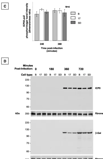
a series of LAT-expressing clonal cell populations derived from the selected 293T cell population (Fig. 8A). The cells were infected alongside control cells with wild-type HSV-1 strain KOS at an MOI of 3. Protein was extracted at 5 h postinfection, before the viral LAT gene was expressed. The samples were subjected to electrophoresis and transfer and probed simultaneously with antibodies to ICP0 and a protein loading control, fibronectin. No systematic or appreciable dif-ferences in the expression of ICP0 between the different cell lines and controls following infection with wild-type virus were identified.
(iii) To confirm the results obtained by methods 1 and 2, we performed plasmid transfections, which completely dissociate the presence of LAT intransfrom any possiblecis-acting effect in the context of the viral genome (Fig. 8B). An ICP0 expres-sion plasmid in which the ICP0 cDNA is under the transcrip-tional control of the cytomegalovirus IE promoter, was used to transfect the 2.0-kb LAT-expressing cell lines and controls. ICP0 protein levels were determined by Western blot hybrid-ization analysis at various time points after the transfections. ICP0 expression in the LAT-expressing clonal cell lines and controls was similar at early time points following the trans-fection. At later time points, there was more variability, as might be expected in a transient-transfection assay. However, there was no relationship between the ICP0 expression level and the LAT expression level at any time point.
IE gene expression following infection of 293T cells with a LAP1-2 deletion virus.In order to study thecis-acting effects of deletions of the LAT locus on the expression of IE genes
following viral infection in culture, we utilized a previously described virus bearing deletions for both LAP1 and LAP2 (9). This virus contains a deletion encompassing thePstI sites at positions⫺800 and⫺3 with respect to the 5⬘end of the 2.0-kb LAT intron. Although the deletion greatly reduces expression of LAT, the entire sequences encoding the 2.0-kb LAT intron and ICP0 gene are spared.
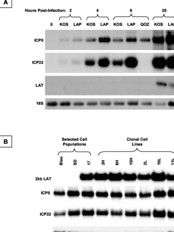
Figure 9A shows a representative Northern blot analysis of gene expression following infection of 293T cells with wild-type HSV strain KOS and the LAP deletion derivative. The viral preparations were done in parallel, and the resulting stocks were titrated in parallel in Vero cells in culture. Identical numbers of PFU of virus were added to 293T cells, and the samples were lysed at 2, 4, 6, and 20 h postinfection. Total RNA was extracted and subjected to Northern blot hybridiza-tion analysis. No LAT expression occurs from either virus at early time points following infection. At 20 h, the ⌬LAP1-2 virus expresses substantially less 2.0-kb LAT than the wild-type virus. At all time points postinfection, the ICP0 and ICP22 mRNAs are clearly more abundant in cell samples infected with LAP deletion viruses. Thus, the LAP deletion results in overexpression of viral IE genes before any expression of LAT occurs.
LAT intransdoes not complement the absence of LAT incis.
[image:8.603.129.457.71.343.2]Finally, we examined the effect of the 2.0-kb LAT intron, delivered in trans, on the expression of IE genes following infection with ⌬LAP1-2, to test the hypothesis that comple-mentation would occur and that IE gene expression would be reduced by the presence of the 2.0-kb LAT intron in the cells
FIG. 6. Replication of HSV-1 strain KOS in LAT-expressing cell populations. Multiple samples of each cell population were infected at three different multiplicities. The resulting virus-containing medium was titrated on permissive Vero cells, and the number of PFU per milliliter of medium was determined. HSV KOS replicated equally well in all three cell populations at all three MOI. B, Bleo.
on November 8, 2019 by guest
http://jvi.asm.org/
(Fig. 9B). Cells were infected, and total RNA was extracted at 6 h postinfection, before the endogenous LAT genes are ex-pressed. The samples were subjected to Northern blot hybrid-ization analysis using probes to ICP0, ICP22, LAT, and a loading control, 18S rRNA. After adjustment for loading arti-facts, no differences in ICP0 and ICP22 expression between LAT-expressing and control cells were detected.
DISCUSSION
Taken together, these experiments suggest that the 2.0-kb LAT intron does not exert atrans-acting effect on the
expres-sion of ICP0 in nonneuronal cells in culture. Our observations do not concur with earlier reported evidence that LAT may function intransto inhibit IE gene expression in neuronal cells (30). The difference between this report and our work may relate to our use of nonneuronal cells. If antisense regulation of ICP0 expression occurred through hybridization of the nu-clear LAT intron with ICP0 mRNA, it might be expected that this would occur in any cell type. The absence of atrans-acting effect of the 2.0-kb LAT intron RNA on ICP0 expression in 293T cells might result from one of several putative mecha-nisms.
First, a cell-type specific antisense mechanism could be
in-FIG. 7. Dynamic expression profile of ICP0 and LacZ in LAT-expressing cell populations after infection with QOZ.HG. (A) Schematic depiction of the QOZ.HG genome. The virus contains wild-type LAT and ICP0 loci but does not express LAT, as the viral transcriptional program is disrupted by mutations in the ICP4, -22, and -27 loci. (B) Northern blot hybridization analysis of ICP0, LacZ, and LAT expression in LAT-expressing and control cell populations before and at four time points after infection with QOZ.HG. The second and fourth panels show prolonged exposures of the blots depicted in the first and third panels. ICP0 and LacZ mRNAs appear at 120 min postinfection (arrows) in all cell populations and peak at similar levels. (C) Quantitative analysis of the ICP0/LacZ ratio provides a surrogate marker of ICP0 stability (see the text). This is not altered by the presence or absence of the 2.0-kb LAT at any time point. (D) Western blot hybridization analysis of ICP0 and LacZ protein expression in LAT-expressing cell populations and controls before and after infection with QOZ.HG. The upper two and lower two panels represent duplicate blots that were probed for ICP0 or LacZ and then stripped and reprobed for the fibronectin loading control. B, Bleo.
on November 8, 2019 by guest
http://jvi.asm.org/
[image:9.603.104.486.70.496.2]voked. It is well established that the effect of an RNA targeting reagent in suppressing gene expression may be critically de-pendent on the cellular location of the reagent with respect to the targeted transcript. For example, targeting a ribozyme to the nucleolus greatly enhances cleavage of a nucleolar RNA
[image:10.603.110.473.71.630.2](39), whereas cytoplasmic RNAs may be targeted by a suitably localized ribozyme (27). Antisense regulation is more compli-cated; the cellular location of the resulting RNA duplex seems to determine both the catabolic pathway taken by the duplex and the response of the cell to the presence of dsRNA (26). It
FIG. 7—Continued.
on November 8, 2019 by guest
http://jvi.asm.org/
FIG. 8. Western blot hybridization analysis of ICP0 expression in LAT-expressing clonal cell lines. The blots were simultaneously probed with antibodies for ICP0 (0) and fibronectin (F) as a loading control. Quantification of the densitometric ICP0/fibronectin ratio is shown below each lane of the gel. (A) Cells were infected with HSV-1 strain KOS and harvested 5 h postinfection, before viral LAT expression. (B) Cells were transfected with an ICP0 expression plasmid and harvested at the time points shown. There is no systematic difference in ICP0 expression between the different cell lines and controls at any time point following either infection or transfection. As might be expected from a transient-transfection assay, there is more variability at later time points in panel B.
on November 8, 2019 by guest
http://jvi.asm.org/
has recently been shown that the posttranscriptional regulation of ICP0 is different in neuronal and nonneuronal cell types (8). This raises the possibility that some aspect of posttranscrip-tional processing or transport of the ICP0 transcript, which is unique to neurons, makes it susceptible to antisense repression by LAT in a cell-specific manner. Alternatively, the cell-type-dependent splicing of the 1.5-kb LAT intron might account for
differences between previous studies and ours. If the 1.5-kb intron were an effective antisense repressor of ICP0, then the detection oftrans-acting LAT-dependent suppression of ICP0 mRNA would depend on the neuron-specific splicing event that generates the smaller lariat.
[image:12.603.119.472.84.553.2]The second possible type of mechanism to explain the dis-crepancy between our results and those obtained in neuronal
FIG. 9. Analysis of IE gene expression from the HSV-1 LAT-null mutant⌬LAP1-2. (A) Northern blot analysis of viral gene expression from wild-type HSV-1 (strain KOS),⌬LAP1-2, and QOZ.HG at different time points following infection of 293T cells. The expression of ICP0, ICP22, and LAT is shown by sequential hybridization of a Northern blot with different probes; 18S rRNA acts as a loading control. (B) Northern blot hybridization analysis of ICP0 and ICP22 mRNA expression in LAT-expressing cell populations, clonal LAT-expressing cell lines, and controls after infection with⌬LAP1-2 at an MOI of 1. There is no evidence that ICP0 mRNA accumulation is suppressed in the presence of LAT intrans, even though the LAT-null deletion was shown to increase ICP0 mRNA expression (A), presumably by acting incis.
on November 8, 2019 by guest
http://jvi.asm.org/
cell lines invokes a cell-type-specific, nonantisense mechanism. One possibility would be that the 2.0-kb LAT sequence is translated in neurons, resulting in a protein that functioned, directly or indirectly, as a viral transcriptional regulator. Re-cent work has provided limited evidence that the 2.0-kb LAT intron may encode a protein. The LAT is associated with the polyribosome fraction of lytically and latently infected cells (20), and an open reading frame (ORF) within the 2.0-kb LAT intron appears to give rise to a protein that enhances viral growth when overexpressed outside the context of the 2.0-kb lariat (47). Stable overexpression of this LAT ORF in cultured cell lines appears to allow sustained expression of a reporter gene driven by the cytomegalovirus IE promoter in the context of a HSV genome, which is null for ICP0, ICP4, and ICP27 (48). Examination of the properties of the epitope-tagged pro-tein provides preliminary evidence that this LAT ORF-en-coded protein may be nuclear, phosphorylated, and localized to viral replication compartments. The protein does not appear to directlytransactivate the promoters of IE genes (10), and its action on viral replication is directly opposite to that reported from expression of the 2.0-kb intron in cultured neurons (30). In fact, it is not at all clear that this ORF can be translated in the context of the lariat 2.0-kb LAT intron. Although earlier work described polyclonal antisera directed against a LAT-encoded peptide which detected a protein in latently infected cells (14), this finding has never been reproduced (28). Fur-thermore, insertion of marker genes into the intron does not allow their efficient translation unless aberrant splicing events are induced by the foreign sequence (10, 28). Finally, selective deletions of the LAT ORFs do not appear to induce an ab-normal phenotype in the virus (16). The role of 2.0-kb-LAT-encoded proteins thus remains uncertain, as does whatever putative mechanism dictates its selective translation in neu-rons. Other possible non-antisense-dependent trans-acting mechanisms whereby LAT might affect IE gene expression include functions at an RNA level, which do not depend on hybridization between the LAT intron and the ICP0 mRNA. Specific LAT-binding RNA or protein species within neurons, whose activity is modulated in some way by the LAT intron, could be possible.
Recent work has focused on the role of LAT in protecting neurons from apoptosis following viral infection (33). The re-gions of the LAT implicated in these effects map to the first 1.5 kb of the LAT and do not include any sequences that are complementary to the ICP0 transcript or any conserved ORFs (1, 23). This strongly suggests that the antiapoptotic mecha-nism of LAT does not depend on an anti-ICP0 antisense mech-anism. Furthermore, it has recently been demonstrated that the reactivation phenotype of a LAT-null virus can be restored to wild-type levels by the expression of a heterologous anti-apoptotic gene (34). This suggests that the IE gene dysregula-tion occurring after LAT deledysregula-tion may be dissociable from the antiapoptotic function of LAT and that the latter is associated with the impaired latency establishment and reactivation phe-notype seen in LAT-null viruses. These data provide limited insight into the function of the stable 2.0-kb LAT intron, only part of which appears to contribute to the noncoding se-quences necessary and sufficient for the putative antiapoptotic function of the LAT gene.
Finally, we studied the effects of deletion of the latency
promoters to examine whether the effects of LAT expression in
cis were different from those in trans. Although ICP0 and ICP22 expression from the LAP deletion mutant⌬LAP1-2 was enhanced with respect to control virus, this effect was seen at time points well before the detectable expression of LAT. It is possible that the LAP1-2 deletion affects elements within the virus that result in IE gene transcriptional dysregulation via a mechanism that does not depend on expression of the LAT RNA, for example by deleting cis-acting negative transcrip-tional regulatory sequences. This hypothesis is consistent with previously reported data, in which LAP deletion had a signif-icant cis-acting effect on IE gene expression (7, 19, 30). The results dictate caution in the interpretation of data obtained with LAP deletion mutants.
The present study has not elucidated the function of the conserved 2.0-kb LAT intron or the mechanism whereby LAT affects the expression of viral IE genes intransin neuronal cells (30). Our studies ontrans-acting mechanisms do not demon-strate involvement of the 2.0-kb LAT intron in the regulation of ICP0 mRNA accumulation in nonneuronal cells. We con-clude that the mechanisms for the previously described
trans-acting effects of LAT are cell type specific and are unlikely to be mediated by an antisense mechanism involving the 2.0-kb intron.
ACKNOWLEDGMENTS
E.A.B. and C.-S.H. contributed equally to this work.
We thank Steve Wendell and Darren Wolfe for generously sharing plasmid resources and Xiao-Ping Chen for technical help for in situ hybridization.
This work was supported by the following grants: 5R01 GM34534-17 from the NIH/NIGMS, DAMD17-98-1-8626 from the U.S. Army, 5P01 DK44935 from the NIH, 1PO1 NS40923 from the NIH, and 1R01 NS44323 from the NIH/NINDS.
REFERENCES
1. Ahmed, M., M. Lock, C. G. Miller, and N. W. Fraser.2002. Regions of the herpes simplex virus type 1 latency-associated transcript that protect cells from apoptosis in vitro and protect neuronal cells in vivo. J. Virol.76:717– 729.
2. Alvira, M. R., W. F. Goins, J. B. Cohen, and J. C. Glorioso.1999. Genetic studies exposing the splicing events involved in herpes simplex virus type 1 latency-associated transcript production during lytic and latent infection. J. Virol.73:3866–3876.
3. Block, T. M., S. Deshmane, J. Masonis, J. Maggioncalda, T. Valyi Nagi, and N. W. Fraser.1993. An HSV LAT null mutant reactivates slowly from latent infection and makes small plaques on CV-1 monolayers. Virology192:618– 630.
4. Bloom, D. C., J. M. Hill, G. Devi Rao, E. K. Wagner, L. T. Feldman, and J. G. Stevens.1996. A 348-base-pair region in the latency-associated transcript facilitates herpes simplex virus type 1 reactivation. J. Virol.70:2449–2459. 5. Burton, E. A., S. Huang, W. F. Goins, and J. C. Glorioso.2003. Use of the
herpes simplex viral genome to construct gene therapy vectors. Methods Mol. Med.76:1–31.
6. Cai, W., and P. A. Schaffer.1992. Herpes simplex virus type 1 ICP0 regulates expression of immediate-early, early, and late genes in productively infected cells. J. Virol.66:2904–2915.
7. Chen, S. H., M. F. Kramer, P. A. Schaffer, and D. M. Coen.1997. A viral function represses accumulation of transcripts from productive-cycle genes in mouse ganglia latently infected with herpes simplex virus. J. Virol.71: 5878–5884.
8. Chen, X., J. Li, M. Mata, J. Goss, D. Wolfe, J. C. Glorioso, and D. J. Fink. 2000. Herpes simplex virus type 1 ICP0 protein does not accumulate in the nucleus of primary neurons in culture. J. Virol.74:10132–10141. 9. Chen, X., M. C. Schmidt, W. F. Goins, and J. C. Glorioso.1995. Two herpes
simplex virus type 1 latency-active promoters differ in their contributions to latency-associated transcript expression during lytic and latent infections. J. Virol.69:7899–7908.
10. Coffin, R. S., S. K. Thomas, D. P. Thomas, and D. S. Latchman.1998. The herpes simplex virus 2 kb latency associated transcript (LAT) leader
on November 8, 2019 by guest
http://jvi.asm.org/
quence allows efficient expression of downstream proteins which is enhanced in neuronal cells: possible function of LAT ORFs. J. Gen. Virol.79:3019– 3026.
11. Deatly, A. M., J. G. Spivack, E. Lavi, and N. W. Fraser.1987. RNA from an immediate early region of the type 1 herpes simplex virus genome is present in the trigeminal ganglia of latently infected mice. Proc. Natl. Acad. Sci. USA 84:3204–3208.
12. Devi Rao, G. B., S. A. Goodart, L. M. Hecht, R. Rochford, M. K. Rice, and E. K. Wagner.1991. Relationship between polyadenylated and nonpolyade-nylated herpes simplex virus type 1 latency-associated transcripts. J. Virol. 65:2179–2190.
13. Dobson, A. T., F. Sederati, G. Devi Rao, W. M. Flanagan, M. J. Farrell, J. G. Stevens, E. K. Wagner, and L. T. Feldman.1989. Identification of the latency-associated transcript promoter by expression of rabbit beta-globin mRNA in mouse sensory nerve ganglia latently infected with a recombinant herpes simplex virus. J. Virol.63:3844–3851.
14. Doerig, C., L. I. Pizer, and C. L. Wilcox.1991. An antigen encoded by the latency-associated transcript in neuronal cell cultures latently infected with herpes simplex virus type 1. J. Virol.65:2724–2727.
15. Drolet, B. S., G. C. Perng, R. J. Villosis, S. M. Slanina, A. B. Nesburn, and S. L. Wechsler.1999. Expression of the first 811 nucleotides of the herpes simplex virus type 1 latency-associated transcript (LAT) partially restores wild-type spontaneous reactivation to a LAT-null mutant. Virology253:96– 106.
16. Fareed, M. U., and J. G. Spivack.1994. Two open reading frames (ORF1 and ORF2) within the 2.0-kilobase latency-associated transcript of herpes simplex virus type 1 are not essential for reactivation from latency. J. Virol. 68:8071–8081.
17. Farrell, M. J., A. T. Dobson, and L. T. Feldman.1991. Herpes simplex virus latency-associated transcript is a stable intron. Proc. Natl. Acad. Sci. USA 88:790–794.
18. Farrell, M. J., T. P. Margolis, W. A. Gomes, and L. T. Feldman.1994. Effect of the transcription start region of the herpes simplex virus type 1 latency-associated transcript promoter on expression of productively infected neu-rons in vivo. J. Virol.68:5337–5343.
19. Garber, D. A., P. A. Schaffer, and D. M. Knipe.1997. A LAT-associated function reduces productive-cycle gene expression during acute infection of murine sensory neurons with herpes simplex virus type 1. J. Virol.71:5885– 5893.
20. Goldenberg, D., N. Mador, M. J. Ball, A. Panet, and I. Steiner.1997. The abundant latency-associated transcripts of herpes simplex virus type 1 are bound to polyribosomes in cultured neuronal cells and during latent infec-tion in mouse trigeminal ganglia. J. Virol.71:2897–2904.
21. Hill, J. M., F. Sedarati, R. T. Javier, E. K. Wagner, and J. G. Stevens.1990. Herpes simplex virus latent phase transcription facilitates in vivo reactiva-tion. Virology174:117–125.
22. Ho, D. Y., and E. S. Mocarski.1989. Herpes simplex virus latent RNA (LAT) is not required for latent infection in the mouse. Proc. Natl. Acad. Sci. USA 86:7596–7600.
23. Inman, M., G. C. Perng, G. Henderson, H. Ghiasi, A. B. Nesburn, S. L. Wechsler, and C. Jones.2001. Region of herpes simplex virus type 1 latency-associated transcript sufficient for wild-type spontaneous reactivation pro-motes cell survival in tissue culture. J. Virol.75:3636–3646.
24. Javier, R. T., J. G. Stevens, V. B. Dissette, and E. K. Wagner.1988. A herpes simplex virus transcript abundant in latently infected neurons is dispensable for establishment of the latent state. Virology166:254–257.
25. Krummenacher, C., J. M. Zabolotny, and N. W. Fraser.1997. Selection of a nonconsensus branch point is influenced by an RNA stem-loop structure and is important to confer stability to the herpes simplex virus 2-kilobase latency-associated transcript. J. Virol.71:5849–5860.
26. Kumar, M., and G. G. Carmichael.1998. Antisense RNA: function and fate of duplex RNA in cells of higher eukaryotes. Microbiol. Mol. Biol. Rev. 62:1415–1434.
27. Kuwabara, T., M. Warashina, S. Koseki, M. Sano, J. Ohkawa, K. Nakayama, and K. Taira.2001. Significantly higher activity of a cytoplasmic hammer-head ribozyme than a corresponding nuclear counterpart: engineered tRNAs with an extended 3⬘end can be exported efficiently and specifically to the cytoplasm in mammalian cells. Nucleic Acids Res.29:2780–2788. 28. Lock, M., C. Miller, and N. W. Fraser.2001. Analysis of protein expression
from within the region encoding the 2.0-kilobase latency-associated tran-script of herpes simplex virus type 1. J. Virol.75:3413–3426.
29. Lomonte, P., and R. D. Everett.1999. Herpes simplex virus type 1 immedi-ate-early protein Vmw110 inhibits progression of cells through mitosis and from G1into S phase of the cell cycle. J. Virol.73:9456–9467.
30. Mador, N., D. Goldenberg, O. Cohen, A. Panet, and I. Steiner.1998. Herpes simplex virus type 1 latency-associated transcripts suppress viral replication and reduce immediate-early gene mRNA levels in a neuronal cell line. J. Virol.72:5067–5075.
31. Perng, G. C., E. C. Dunkel, P. A. Geary, S. M. Slanina, H. Ghiasi, R. Kaiwar, A. B. Nesburn, and S. L. Wechsler.1994. The latency-associated transcript gene of herpes simplex virus type 1 (HSV-1) is required for efficient in vivo spontaneous reactivation of HSV-1 from latency. J. Virol.68:8045–8055.
32. Perng, G. C., H. Ghiasi, S. M. Slanina, A. B. Nesburn, and S. L. Wechsler. 1996. The spontaneous reactivation function of the herpes simplex virus type 1 LAT gene resides completely within the first 1.5 kilobases of the 8.3-kilobase primary transcript. J. Virol.70:976–984.
33. Perng, G. C., C. Jones, J. Ciacci Zanella, M. Stone, G. Henderson, A. Yukht, S. M. Slanina, F. M. Hofman, H. Ghiasi, A. B. Nesburn, and S. L. Wechsler. 2000. Virus-induced neuronal apoptosis blocked by the herpes simplex virus latency-associated transcript. Science287:1500–1503.
34. Perng, G. C., B. Maguen, L. Jin, K. R. Mott, N. Osorio, S. M. Slanina, A. Yukht, H. Ghiasi, A. B. Nesburn, M. Inman, G. Henderson, C. Jones, and S. L. Wechsler.2002. A gene capable of blocking apoptosis can substitute for the herpes simplex virus type 1 latency-associated transcript gene and restore wild-type reactivation levels. J. Virol.76:1224–1235.
35. Rock, D. L., A. B. Nesburn, H. Ghiasi, J. Ong, T. L. Lewis, J. R. Lokensgard, and S. L. Wechsler.1987. Detection of latency-related viral RNAs in tri-geminal ganglia of rabbits latently infected with herpes simplex virus type 1. J. Virol.61:3820–3826.
36. Rodahl, E., and L. Haarr.1997. Analysis of the 2-kilobase latency-associated transcript expressed in PC12 cells productively infected with herpes simplex virus type 1: evidence for a stable, nonlinear structure. J. Virol.71:1703– 1707.
37. Roizman, B., and A. E. Sears.1996. Herpes simplex viruses and their repli-cation, p. 2231–2295.InB. N. Fields, D. M. Knipe, and P. M. Howley (ed.), Fields virology, 3rd ed., vol. 2. Lippincott-Raven, Philadelphia, Pa. 38. Samaniego, L. A., L. Neiderhiser, and N. A. DeLuca.1998. Persistence and
expression of the herpes simplex virus genome in the absence of immediate-early proteins. J. Virol.72:3307–3320.
39. Samarsky, D. A., G. Ferbeyre, E. Bertrand, R. H. Singer, R. Cedergren, and M. J. Fournier.1999. A small nucleolar RNA:ribozyme hybrid cleaves a nucleolar RNA target in vivo with near-perfect efficiency. Proc. Natl. Acad. Sci. USA96:6609–6614.
40. Sawtell, N. M., and R. L. Thompson.1992. Herpes simplex virus type 1 latency-associated transcription unit promotes anatomical site-dependent establishment and reactivation from latency. J. Virol.66:2157–2169. 41. Sedarati, F., K. M. Izumi, E. K. Wagner, and J. G. Stevens.1989. Herpes
simplex virus type 1 latency-associated transcription plays no role in estab-lishment or maintenance of a latent infection in murine sensory neurons. J. Virol.63:4455–4458.
42. Spivack, J. G., and N. W. Fraser.1987. Detection of herpes simplex virus type 1 transcripts during latent infection in mice. J. Virol.61:3841–3847. 43. Spivack, J. G., and N. W. Fraser.1988. Expression of herpes simplex virus
type 1 latency-associated transcripts in the trigeminal ganglia of mice during acute infection and reactivation of latent infection. J. Virol. 62:1479– 1485.
44. Spivack, J. G., G. M. Woods, and N. W. Fraser.1991. Identification of a novel latency-specific splice donor signal within the herpes simplex virus type 1 2.0-kilobase latency-associated transcript (LAT): translation inhibition of LAT open reading frames by the intron within the 2.0-kilobase LAT. J. Virol. 65:6800–6810.
45. Steiner, I., J. G. Spivack, R. P. Lirette, S. M. Brown, A. R. MacLean, J. H. Subak Sharpe, and N. W. Fraser.1989. Herpes simplex virus type 1 latency-associated transcripts are evidently not essential for latent infection. EMBO J.8:505–511.
46. Stevens, J. G., E. K. Wagner, G. B. Devi Rao, M. L. Cook, and L. T. Feldman. 1987. RNA complementary to a herpesvirus alpha gene mRNA is prominent in latently infected neurons. Science235:1056–1059.
47. Thomas, S. K., G. Gough, D. S. Latchman, and R. S. Coffin.1999. Herpes simplex virus latency-associated transcript encodes a protein which greatly enhances virus growth, can compensate for deficiencies in immediate-early gene expression, and is likely to function during reactivation from virus latency. J. Virol.73:6618–6625.
48. Thomas, S. K., C. E. Lilley, D. S. Latchman, and R. S. Coffin.2002. A protein encoded by the herpes simplex virus (HSV) type 1 2-kilobase latency-asso-ciated transcript is phosphorylated, localized to the nucleus, and overcomes the repression of expression from exogenous promoters when inserted into the quiescent HSV genome. J. Virol.76:4056–4067.
49. Thompson, R. L., and N. M. Sawtell.2001. Herpes simplex virus type 1 latency-associated transcript gene promotes neuronal survival. J. Virol.75: 6660–6675.
50. Thompson, R. L., and N. M. Sawtell.1997. The herpes simplex virus type 1 latency-associated transcript gene regulates the establishment of latency. J. Virol.71:5432–5440.
51. Wagner, E. K., W. M. Flanagan, G. Devi Rao, Y. F. Zhang, J. M. Hill, K. P. Anderson, and J. G. Stevens.1988. The herpes simplex virus latency-associ-ated transcript is spliced during the latent phase of infection. J. Virol. 62:4577–4585.
52. Wechsler, S. L., A. B. Nesburn, R. Watson, S. M. Slanina, and H. Ghiasi. 1988. Fine mapping of the latency-related gene of herpes simplex virus type 1: alternative splicing produces distinct latency-related RNAs containing open reading frames. J. Virol.62:4051–4058.
on November 8, 2019 by guest
http://jvi.asm.org/
53. Wu, T. T., Y. H. Su, T. M. Block, and J. M. Taylor.1998. Atypical splicing of the latency-associated transcripts of herpes simplex type 1. Virology243: 140–149.
54. Wu, T. T., Y. H. Su, T. M. Block, and J. M. Taylor.1996. Evidence that two latency-associated transcripts of herpes simplex virus type 1 are nonlinear. J. Virol.70:5962–5967.
55. Zabolotny, J. M., C. Krummenacher, and N. W. Fraser.1997. The herpes
simplex virus type 1 2.0-kilobase latency-associated transcript is a stable intron which branches at a guanosine. J. Virol.71:4199–4208.
56. Zwaagstra, J. C., H. Ghiasi, S. M. Slanina, A. B. Nesburn, S. C. Wheatley, K. Lillycrop, J. Wood, D. S. Latchman, K. Patel, and S. L. Wechsler.1990. Activity of herpes simplex virus type 1 latency-associated transcript (LAT) promoter in neuron-derived cells: evidence for neuron specificity and for a large LAT transcript. J. Virol.64:5019–5028.